Article

Towards a Tissue-Engineered Ligament: Design and Preliminary Evaluation of a Dedicated Multi-Chamber Tension-Torsion Bioreactor

1
CNRS, LEMTA, UMR 7563, Université de Lorraine, 2 Avenue de la Forêt de Haye, 54504 Vandeuvre-lès-Nancy, France
2
Institute of Health and Biomedical Innovation, Queensland University of Technology, Cnr Blamey Street & Musk Avenue, Kelvin Grove Brisbane, Queensland 4059, Australia
3
CNRS, LRGP, UMR 7274, Université de Lorraine, 2 Avenue de la Forêt de Haye, 54504 Vandeuvre-lès-Nancy, France
4
Department of Electromechanical Engineering, School of Mechanical Engineering, Yangtze University, 1 Xueyuan Road, Jingzhou 434023, China
5
CNRS, IMoPA, UMR 7365, Biopôle, Université de Lorraine, 9 Avenue de la Forêt de Haye, 54504 Vandeuvre-lès-Nancy, France
6
PTIBC-IBISA, CNRS, FR3209, Biopôle, Université de Lorraine, 9 Avenue de la Forêt de Haye, 54504 Vandeuvre-lès-Nancy, France
*
Author to whom correspondence should be addressed.
These authors contributed equally to this work.
Processes 2014, 2(1), 167-179; https://doi.org/10.3390/pr2010167
Submission received: 3 January 2014 / Revised: 7 February 2014 / Accepted: 10 February 2014 / Published: 19 February 2014
(This article belongs to the Special Issue Design of Bioreactor Systems for Tissue Engineering)

Abstract

:
Tissue engineering may constitute a promising alternative to current strategies in ligament repair, providing that suitable scaffolds and culture conditions are proposed. The objective of the present contribution is to present the design and instrumentation of a novel multi-chamber tension-torsion bioreactor dedicated to ligament tissue engineering. A preliminary biological evaluation of a new braided scaffold within this bioreactor under dynamic loading is reported, starting with the development of a dedicated seeding protocol validated from static cultures. The results of these preliminary biological characterizations confirm that the present combination of scaffold, seeding protocol and bioreactor may enable us to head towards a suitable ligament tissue-engineered construct.

1. Introduction

The principle of tissue engineering offers large horizons in the field of reconstructive surgery of the musculoskeletal system, providing that: (1) the native biomechanical function of the tissue to repair is well defined [1,2]; (2) a biodegradable scaffold able to restore this function and to encourage tissue formation is proposed [3,4,5,6,7,8,9,10]; (3) suitable cells are selected [11] and protocols are proposed to scale-up and seed them within this scaffold [12,13,14,15]; (4) an adapted bioreactor is used both to simulate in vitro the tissue implantation and to provide cells with suitable mechanical stimuli [16,17,18,19,20,21]. However, there is still no clear consensus concerning the best solution for each of these steps, thus requiring additional intensive and pluridisciplinary research. For some ligaments such as the Anterior Cruciate Ligament (ACL) associated with frequent injuries, the range of physiological loadings and the associated strains have been well documented [22,23], as well as the properties of the native tissue [24,25]. We have recently proposed a novel multilayer braided scaffold made of a copoly(lactic acid-co-(ε-caprolactone)) (PLCL) for ligament tissue engineering, and we have brought some evidences about its suitability for ACL tissue engineering in terms of morphology [26] and tensile response [27]. The next natural step of the approach is to propose protocols enabling a homogeneous seeding of such a scaffold, and to imagine a bioreactor able to simulate its implantation and to provide seeded cells with suitable stimuli.
Although ligaments of the musculoskeletal system are obviously mainly subject to cyclic tensile loading, it is now acknowledged that the ACL is also subject to twisting that obviously plays a role in the resulting stresses [28,29]. It has been indeed suggested that a knee flexion of 90° could result in ACL torsion of more than 35°. The role of torsion in the mechanisms involved in ACL failure has been also emphasized [30]. Consequently, it seems that a bioreactor able to simulate in vitro the implantation of a scaffold for ligaments should reproduce the physiological tension-torsion cycles. Moreover, it is well reported that cell respond both to the strain of their substrate [31,32] and the wall shear stress due to the circulation of physiological fluid [33]. Cyclic loading enable to provide the cells with both types of stimuli, as long as (1) external loads are transferred to the cells through the scaffold deformation; (2) stretching a fluid-saturated porous media such as the scaffold obviously result in fluid flow, which may be adjusted by playing on the frequency of cyclic loads [34]. Particularly, we have previously shown [35] that the braided scaffold was compatible with a computer-aided approach that may enable to quantify these local signals that are not measurable experimentally. The dynamic culture of such a scaffold under tension-torsion cycles, combined with these computer simulations, could therefore bring crucial information regarding the evaluation of the effects of these local stimuli and their magnitudes on cellular activity. However, one of the major difficulties associated with the required experimental evaluations lies in the large variability inherently related to biological material. Indeed, characterizations of tissue engineering constructs must be largely repeated in order to draw conclusions about the suitability of a scaffold or a culture protocol, and a large amount of experiments are therefore necessary. The multi-chamber bioreactor described in the following sections enables to address this variability by simultaneously co-culturing several samples under identical conditions (mechanical and biochemical). Alternatively, the multi-chamber bioreactor may be used to compare different protocols (e.g., in terms of culture medium composition or scaffold coating) under similar mechanical loading.
The evaluation of such a bioreactor must obviously involves the culture of a large sample of scaffolds over a long period, which firstly requires that adapted seeding protocols are proposed in order to get an initial homogeneous cell distribution. As far as such seeding procedure is concerned, it has been emphasized in the literature that cellular penetration was preferable when a rationally-designed scaffold was used rather than a random porous structure [36], and that morphological gradients from core to periphery were suitable as far as nutrients supply is concerned [37]. These particular statements have strongly motivated the choice of the multilayer braided structure that will be used in the present study, which clearly offers non-homogeneous pore sizes from the braid axis to the braid periphery, as emphasized previously [35]. It is then our hypothesis that this structure could enable good cellular penetration and cell colonization, even without the perfusion of a cell suspension.
The objective of the present contribution is to present the novel multi-chamber tension-torsion bioreactor that has been designed in our team in order to meet the requirements for ligament tissue engineering. We will then present a preliminary biological evaluation of the bioreactor: firstly, the seeding procedure enabling a homogeneous cell distribution within the multilayer braided scaffold will be described and evaluated through static culture; secondly, this procedure will be applied to perform a preliminary dynamic culture of the scaffold within the bioreactor.

2. Materials and Methods

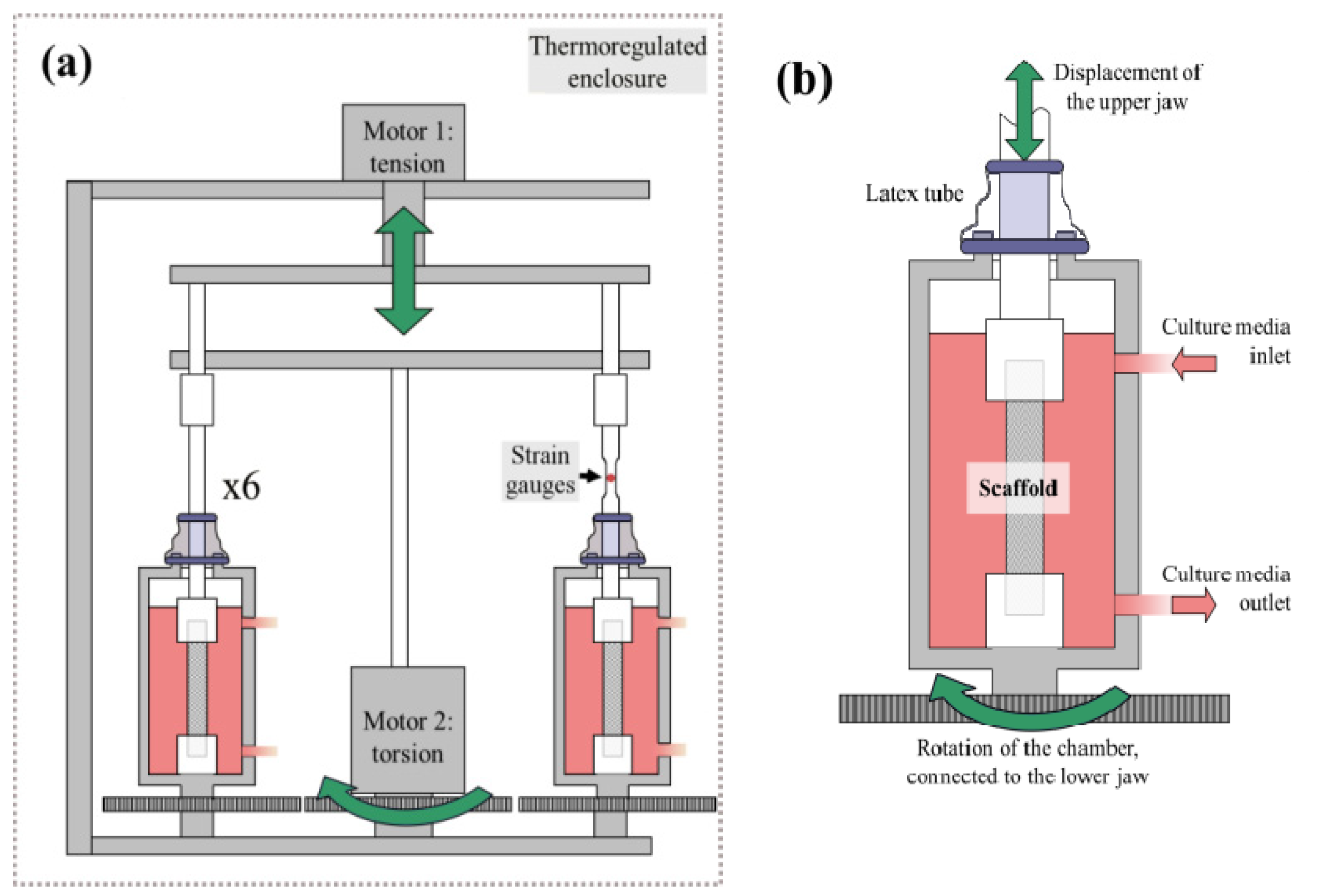
2.1. Bioreactor Design and Instrumentation

Our initial goal was to design and build a bioreactor able to (1) co-culture parallel six scaffold samples; (2) impose identical, programmable and adjustable loading cycles to the six chambers; (3) impose tension, torsion or tension-torsion cycles; (4) co-culture these six samples under identical or different biochemical conditions, depending on the objective (i.e., reproducibility study or optimization of the culture conditions); (5) provide the samples with a thermo-regulated and sterile environment; (6) impose a flow circulation of regulated culture medium in order to provide the samples with nutrients and to evacuate wastes; (7) record the evolution of reaction force within the scaffold in response to the prescribed displacement. We retained a technical solution in which samples are cultured within six separated polycarbonate chambers (see [38]), disposed circularly around a central axis, the whole system being placed in a thermoregulated enclosure. The temperature regulation is realized through a circulation of hot air, a thermocouple (Prosensor, Amanvillers, France) and a PID controller (BMS France, Pringy, France). Sterilized scaffolds are clamped in jaws through a frontal opening in each chamber. Chamber impermeability at the level of the translating upper jaw was performed using sterile latex tubes, in order not to restrict the relative translation and rotation between the upper jaw and the chambers.
Tension and torsion loading take advantage of two electric servomotors (Transtechnik, Ahuy, France, Figure 1a), driven in an uncoupled way by a servodriver (Delta Electronics, Taipei, Taiwan). Translation is performed using an endless screw, controlled by the first motor, activating a tray connected to the upper jaws of each chamber (Figure 1b). Rotation of the chambers is performed using a gear transmission: the lower jaw of each chamber is connected to a gear driven by the central gear, activated by the second motor. The current position in translation and rotation are controlled using a magnetic scale and an incremental magnetic encoder ring respectively (ASM, Munich, Germany). A feedback on the prescribed displacement and rotation is ensured using these encoders and can be recorded throughout the dynamic culture. As far as the reaction force in the scaffolds is concerned, one shaft connecting the upper jaw to the translating tray is instrumented using strain gauges (linear patterns strain gauges, Vishay Precision Group, Malvern, PA, USA). These strain gauges record the deformation within a specific part of the shaft, directly and linearly linked to the forces in the scaffold in reaction to the prescribed displacement. This feedback on applied force enables (1) to evaluate mechanical properties of the scaffold, such as dynamic creep or fatigue properties, when loaded physiologically within a fluid environment; (2) to measure the evolution of scaffold properties due to both the fibers degradation and neo-tissue formation. A high-definition camera (Lumenera LM135, Ottawa, Canada) is disposed in the enclosure and is synchronized to the loading cycles, in such a way that the evolution of the scaffold can be visualized and recorded in time.
Figure 1. (a) Global scheme of the bioreactor functioning (b) Zoom on one of the six bioreactor chambers.
Processes 02 00167 g001
A dedicated acquisition interface has been developed and enables (1) to specify the type, amplitude and frequency of the prescribed displacement and their scheduling over several weeks; (2) to acquire high resolution pictures synchronized with the loading cycles; (3) to record the prescribed displacement and rotation throughout the dynamic culture; (4) to record the evolution of force applied to one scaffold; (5) to manually control the servomotors in order to be compatible with different scaffold lengths.

2.2. Culture Medium Circulation

As far as the circulation of culture medium is concerned, chambers are connected to buffered medium tanks (0.4 liter working volume) and aerated with air containing 5% CO2 through versilic silicone tubing, and culture medium circulation is ensured via an external peristaltic pump. The buffered medium tank is regulated in pH and pO2 using appropriate sensors (Mettler Toledo, Viroflay, France). The six chambers can be connected to identical or different buffered medium tanks, allowing performing reproducible or independent studies. In these conditions, a 7-days long evaluation of the biochemical regulation has been performed: chambers without scaffolds were mounted in the bioreactor regulated at 37 °C and a flow circulation rate of 5 mL per minute was insured. The components in contact with the culture medium (chambers, jaws, keys, tanks, tubes and hydraulic connections, BioExpert Applikon as well as the culture medium tank equipped with pO2 and pH sensors) were firstly sterilized by autoclaving (121 °C, 30 min). Chambers were then mounted in the bioreactor and progressively filled with culture medium made of DMEM-F12 and supplemented with 10% calf fetal serum and 1% (v/v) antibiotics cocktail (Merckmillipore, 10,000 U/mL penicillin G, 10 mg/mL streptomycin, 25 µg/mL amphotericin B). pO2 and pH were set and controlled at 25% air saturation and 7.3 respectively using a controller (Applikon ADI 1030, Schiedam, The Netherlands) and were recorded over the 7-days period (BioExpert, Applikon, Schiedam, The Netherlands).

2.3. Scaffold Fabrication and Preparation

The bioreactor presented above aims at culturing novel scaffolds for ligament tissue engineering recently proposed and evaluated in our group [26]. Briefly, the scaffold is made of concentric braided layers of PLCL fibers. We used a commercial copolymer (Purac Biomaterials, Gorinchem, The Netherlands) with a lactic acid/ε-caprolactone proportion of 85/15. This copolymer has been selected because of its biocompatibility and slow biodegradability, and allows both the brittle behavior of polylactic acid and the low stiffness of polycaprolactone to be compensated. PLCL fibers have been processed from raw material using a custom plastic extruder enabling the processing of adjustable and homogeneous fibers from a small amount of polymer. Fibers have been then arranged into concentric circular braids using a custom maypole braider (Composite and Wire Machinery, North Kingstown, RI, USA) enabling the processing of braided scaffolds from small lengths of fiber. This architecture has been chosen for the following reason: (1) it is deformable in the low strain range but exhibits high stiffness for large strains; (2) it offers a network of interconnected pores with a pore size gradient which is thought suitable for the migration of cells, the supply in biochemical factors, the evacuation of wastes and the formation of ligamentous tissue; (3) it is largely tailorable in terms of morphology and mechanics by playing with the number of layers of the structure, the fiber diameters and the braiding angle of each layer; (4) it is adapted to computer-aided tissue engineering because of its predictable geometry. For the experiments reported below, scaffolds were made of 4 layers of 16 fibers, with a fiber diameter of 170 µm. For static cultures, a long braid was processed and then cut into 7 mm long samples using a heat sealer. For dynamic culture, scaffolds were approximately 45 mm long in order to allow their fixing, but only the central 30 mm was braided. As far as the sterilization of both types of scaffolds is concerned, there were placed in 70% ethanol for 30 min and UV-irradiated for another 30 min. They were left to dry in the sterile laminar hood for 12 h and kept sterile until use.

2.4. Seeding Procedure

In order to culture such a scaffold within the bioreactor, a suitable seeding procedure enabling a homogeneous cell distribution and cellular adhesion within the scaffold was firstly required. We therefore selected bone marrow mesenchymal stem cells (bMSC) from Merino sheep and further expanded until P3. A cell suspension (200,000 cells in 15 µL of culture medium of the same composition as above) was deposited within the periphery of the scaffolds. Due to the pore size gradient offered by the scaffold architecture (see [26]), the cell suspension immediately penetrated the scaffold center and spread along the scaffold. The seeded scaffolds were placed in the incubator for 30min after which small amounts of medium were progressively added. The medium was supplemented with 100 µg/mL ascorbate-2-phosphate.
Static culture was then performed during four weeks, the medium being changed 3 times a week. Scaffolds (n = 3) were harvested at day 3, 7, 14, 21 and 28 for DNA quantification by using a PicoGreen working solution (P11496, Invitrogen, Carlsbad, CA, USA) prepared according to the manufacturer’s instructions so as to assess the cell proliferation. As far as the SEM analysis is concerned, the samples were fixed in a 2.5% glutaraldehyde solution and then dehydrated in a concentration gradient of ethanol and finally carbon coated. For confocal imaging, the constructs were fixed in a 4% paraformaldehyde solution in PBS. The cell membrane was permeabilized for 5 min in a 0.2% triton X solution, cytoskeleton stained for 45 min with a mixture of 0.8 U/mL TRITC-conjugated phalloidin and nuclear stained with 5 µg/mL DAPI solution. The biphasic scaffolds were rinsed another three times and imaged with a Leica SP5 microscope (TCS SP5 II, Leica, Solms, Germany) with a 20× dry objective. 4 µm thick Z-stacks were acquired over a total height of 400–600 µm.

2.5. A Preliminary Dynamic Culture within the Bioreactor

Once the seeding procedure validated, similar protocol were applied to seed the scaffolds prior to dynamic culture within the bioreactor. The components of the bioreactor were sterilized as described previously, and a cell suspension was deposited within two scaffolds. The chambers were then closed and placed in an incubator for 30 min in order to enhance cellular adhesion. An additional part fixed to both the upper jaw and the chamber was used to restrict their relative movement throughout this phase. The whole hydraulic system was then connected to chambers under the laminar air flow hood. Chambers were mounted in the bioreactor, and the part used to restrict the jaw translation was removed.
Two scaffolds were seeded according to the presented protocol with 100 µL of suspended human bMSC, using respectively 200,000 and 600,000 cells/100 µL. The scaffolds were cultured 48 h within the bioreactor under a circulation of culture medium regulated as described previously. The cultures were kept static for the first 24 h, and a planned cyclic loading was then applied (2.5% strain and 10° rotation at 1 Hz) during 2 min every 30 min for 10 h. The constructs were then fixed after 48 h as previously described and stained with EthD-1 at 4 mM. Scaffold’s images were acquired with a macromultiphoton [38] developed at the PTIBC-IBISA Imaging Core Facility in Nancy, used in confocal macroscope mode. This modality uses a Leica Z16 APO A coupled to a Leica SP5-II CFS confocal microscope with a 5× objective (NA 0.5). Fifteen micrometer thick Z-stacks were acquired over a total height of 2 mm and superposed with a 514 nm excitation wavelength, which permitted to visualize both the autofluorescence of polymer fibers and the cellular staining nuclei.

3. Results and Discussion

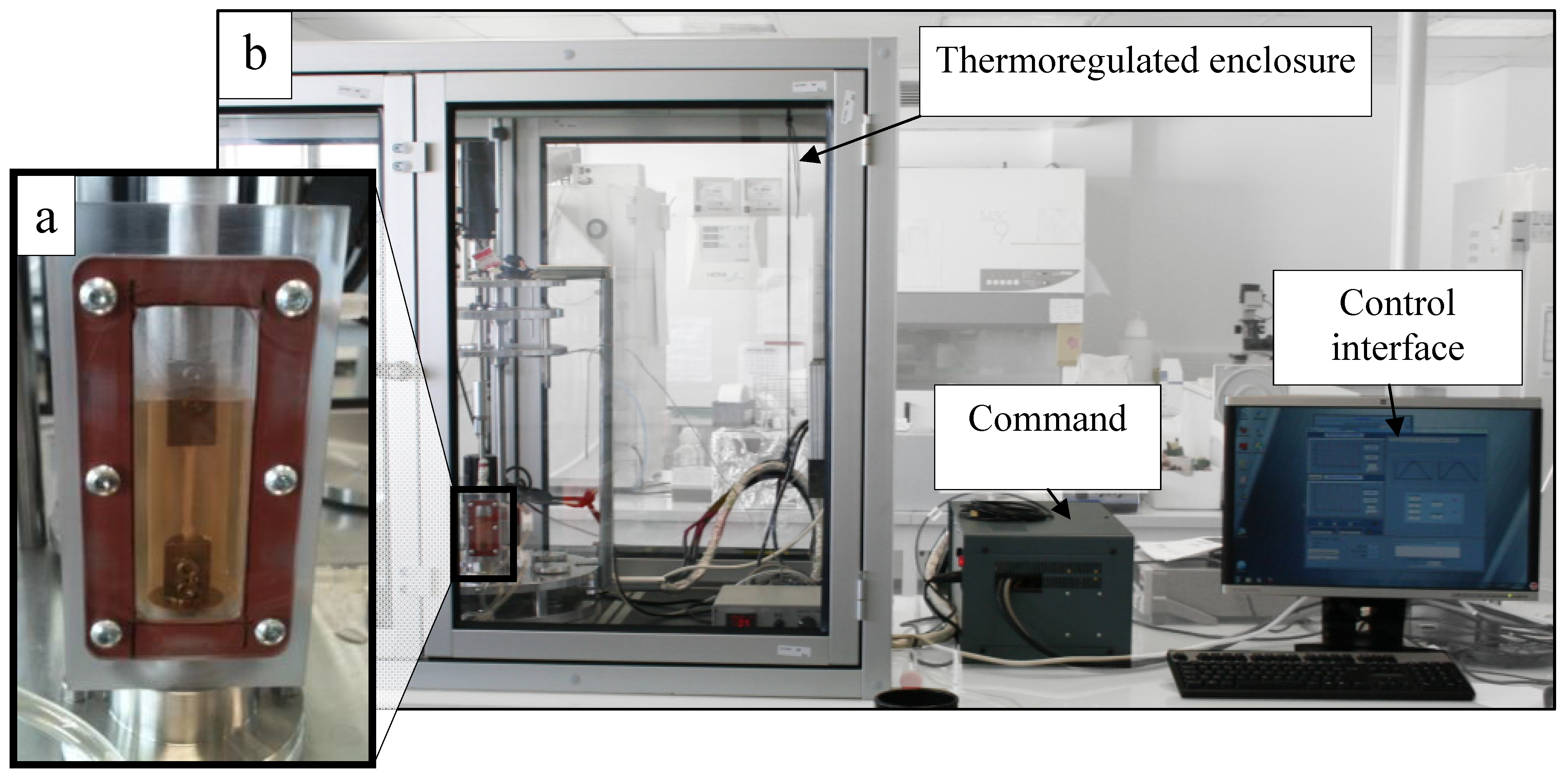
3.1. Bioreactor System
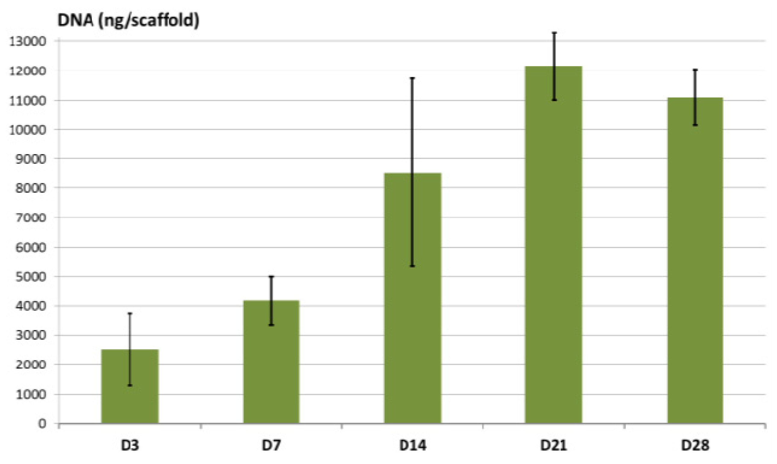
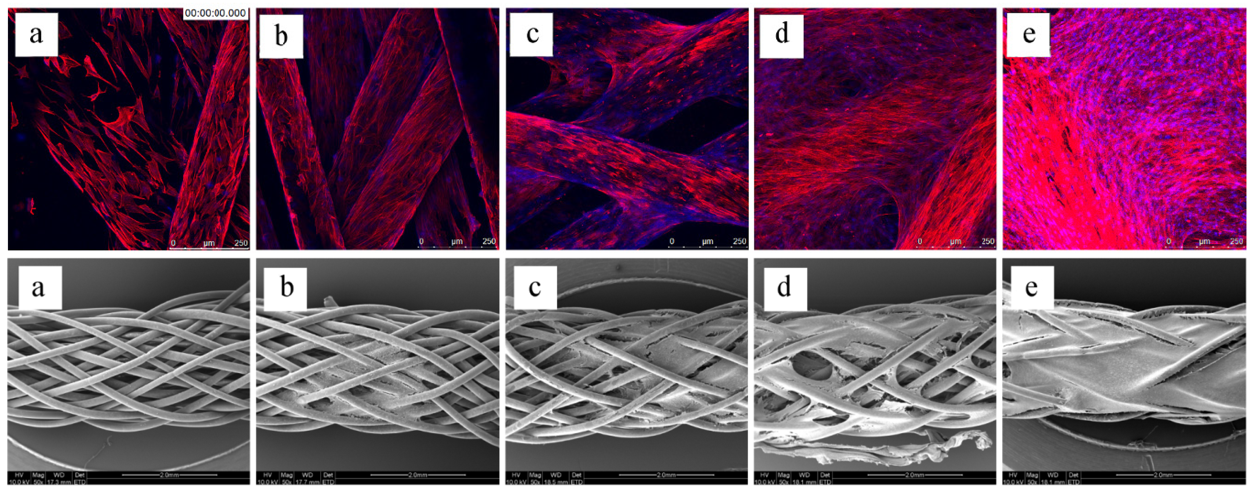
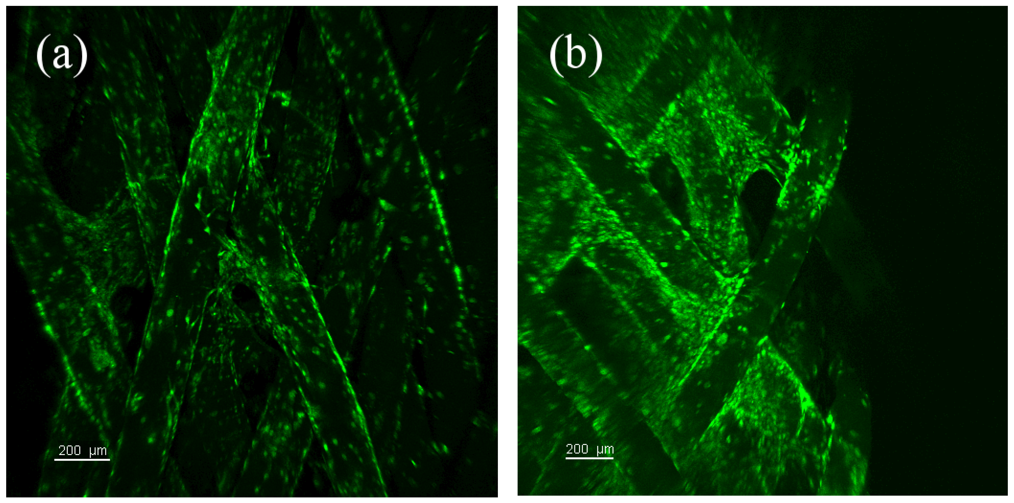
Our novel multichamber tension-torsion bioreactor has been manufactured in the mechanical department of our university. The management of electronic devices and motors and a dedicated user-friendly control interface have been also realized in our common services. Sensors (microswitches Honeywell, Morristown, NJ, USA) have been added in order to prevent the user to impose displacements that would lead bioreactor parts to enter into contact. In comparison to previously reported bioreactor [19,39], the system proposed in the present contribution enables the co-culture of six scaffolds under tension-torsion (synchronized or not) cyclic loads scheduled over several weeks. The final bioreactor configuration is showed below (Figure 2).
Figure 2. Multi-chamber tension-torsion bioreactor in its final configuration. (a) scaffold mounted in a bioreactor chamber (b) entire set-up including command and interface.
Processes 02 00167 g002
Results of the biochemical regulation test indicated that pO2 and pH parameters of the culture medium were maintained stable over this 7-days period and no signs of biological contamination were noticed. One must however emphasize that the culture medium contained antibiotics which may have delayed any potent contamination, without strictly demonstrating the impermeability of the system to external contamination. Nevertheless, an identical culture medium composition will be used for forthcoming long-term biological cultures. Therefore, we concluded to the ability of the bioreactor to prevent contamination.

3.2. Static Culture

The results from the DNA quantification (Figure 3) and the confocal and SEM images at different time points (Figure 4) clearly show that the cells are able to adhere and proliferate within the scaffold when seeded as described above. Cells slightly clustered in the first days, and then spread along the whole scaffold. Interestingly, the cells have a tendency to spontaneously orientate in the fiber direction. Cellular bridges between fibers appear from day 14, and it is likely that cyclic stretching of the scaffold and the corresponding sliding between fibers will impede the phenomenon. Noteworthy, there is a substantial variability concerning the DNA values at day 14, indicating that proliferation rate differs from one scaffold to another. This variability is largely reduced at day 21. At day 28, the decrease in cell number is thought to be due to the obstruction of external pores by ECM, which may cause deficiencies in mass transport. Firstly, as seen above, a different scaffold configuration may result in bigger pore sizes at the scaffold periphery, reducing the risk of obstruction. Moreover, this should not be the case with another scaffold configuration (with higher fiber diameter and/or number of layers) under dynamic culture. Indeed, cyclic stretching of the scaffold-even with a small frequency-will involve fluid flow (essentially in the radial direction) due to the deformation of the saturated porous scaffold. Noteworthy, Computed Fluid Dynamics (CFD) may help to determinate such effects, which is currently addressed by our team (see [35]).
Figure 3. Evolution of DNA from 3 (D3) to 28 (D28) days.
Processes 02 00167 g003

3.3. Preliminary Dynamic Culture in the Bioreactor

The qualitative results issued from the culture of constructs within our novel bioreactor indicated that the cells satisfyingly adhered to the scaffold fibers as a result of the dedicated seeding protocol, and that they were still present on a large extent after the prescribing of cyclic loads and the circulation of culture medium (Figure 5). No contamination was observed during this period. We observed that cells were present in a larger amount when the cell suspension was more concentrated, but were not more homogeneously distributed. We also observed that cells were gathered into clusters, which may indicate that (1) the seeding procedure did not perfectly enable to distribute homogeneously the cells within the whole scaffold, and/or (2) the cells have clustered under dynamic loading. The second hypothesis is unlikely, as long as this effect has been also observed in the first days of static culture.
Figure 4. Images issued from confocal microscopy (up) and scanning electron microscopy (down) at (a) Day 3 (b) Day 7 (c) Day 14 (d) Day 21 (e) Day 28.
Processes 02 00167 g004
Figure 5. Macroconfocal images of scaffolds cultured three days in the bioreactor and seeded with (a) 200,000 cells (b) 600,000 cells.
Processes 02 00167 g005
This may be due to the well-known hydrophobic properties of PLCL [40] that tends to repulse the cell suspension. This effect could be counterbalanced in the future by initially coating the scaffolds with fibronectin [4,41], RGD peptides [42] or collagen [43,44], which is currently tested in our team.
These preliminary results should be corroborated in the future by performing repeated in vitro dynamic culture, which will be precisely made possible by the use of the multi-chamber bioreactor. Some future enhancements will be envisaged starting from the present study. Firstly, numerical studies that we recently reported on the braided scaffold [26,27] will be used to determine the best-suited design for the specific application that we target here. We have indeed observed that, for instance, the pore size distribution of the scaffolds used in the present study may not perfectly prevent the obstruction of external pores after 28 days. Particularly, we should integrate in these studies the effect of sterilizing protocol on the mechanical properties of PLCL fibers, in order to take into account this effect during the numerical optimization of the scaffold properties.

4. Conclusions

The present contribution reported recent advances towards the development of a tissue-engineered ligament, by presenting a novel dedicated multi-chamber tension-torsion bioreactor and its first preliminary evaluation using a recently reported multilayer braided scaffold. Results of static cultures have enabled to conclude that the proposed seeding protocols led to a high cellular adhesion and proliferation, and to the whole scaffold colonization in 28 days. This protocol has been applied to perform a preliminary dynamic culture, which results in the adhesion of a large amount of cells after 48 days of dynamic culture. The bioreactor will be used in the future in order to optimize and evaluate scaffold properties, and presents large possibilities to be extended to the dynamic culture of other tissues such as cartilage or skin. It is the conviction of the authors that these preliminary results could benefit the tissue engineering community in the research of the most suited approach towards the design of a tissue-engineered construct.

Acknowledgments

The authors would like to thank the French Agence Nationale pour la Recherche for the support of their research activities concerning tendon and ligament tissue engineering (ANR Blanc TELiTeR 2007-2012). Authors also thank Jean-Yves Morel (mechanical design), Timothée Baudequin (imaging) and M Ghyslaine Cauchois (cell culture) for their precious assistance.

Conflict of Interest

The authors declare no conflict of interest.

References

  1. Butler, D.L.; Goldstein, S.A.; Guilak, F. Functional tissue engineering: The role of biomechanics. J. Biomech. Eng. 2000, 122, 570–575. [Google Scholar] [CrossRef]
  2. Vieira, A.C.; Guedes, R.M.; Marques, A.T. Development of ligament tissue biodegradable devices: A review. J. Biomech. 2009, 42, 2421–2430. [Google Scholar] [CrossRef]
  3. Cooper, J.A.; Lu, H.H.; Ko, F.K.; Freeman, J.W.; Laurencin, C.T. Fiber-based tissue-engineered scaffold for ligament replacement: design considerations and in vitro evaluation. Biomaterials 2005, 26, 1523–1532. [Google Scholar] [CrossRef]
  4. Lu, H.H.; Cooper, J.A., Jr.; Manuel, S.; Freeman, J.W.; Attawia, M.A.; Ko, F.K.; Laurencin, C.T. Anterior cruciate ligament regeneration using braided biodegradable scaffolds: In vitro optimization studies. Biomaterials 2005, 26, 4805–4816. [Google Scholar] [CrossRef]
  5. Laurencin, C.T.; Freeman, J.W. Ligament tissue engineering: An evolutionary materials science approach. Biomaterials 2005, 26, 7530–7536. [Google Scholar] [CrossRef]
  6. Nair, L.; Laurencin, C. Biodegradable polymers as biomaterials. Prog. Polym. Sci. 2007, 32, 762–798. [Google Scholar] [CrossRef]
  7. Hollister, S.J. Scaffold design and manufacturing: From concept to clinic. Adv. Mater. 2009, 21, 3330–3342. [Google Scholar] [CrossRef]
  8. Chen, X.; Qi, Y.-Y.; Wang, L.-L.; Yin, Z.; Yin, G.-L.; Zou, X.-H.; Ouyang, H.-W. Ligament regeneration using a knitted silk scaffold combined with collagen matrix. Biomaterials 2008, 29, 3683–3692. [Google Scholar] [CrossRef]
  9. Liu, Y.; Ramanath, H.S.; Wang, D.-A. Tendon tissue engineering using scaffold enhancing strategies. Trends Biotechnol. 2008, 26, 201–209. [Google Scholar] [CrossRef]
  10. Liu, C.; Xia, Z.; Czernuszka, J.T. Design and development of three-dimensional scaffolds for tissue engineering. Chem. Eng. Res. Des. 2007, 85, 1051–1064. [Google Scholar] [CrossRef]
  11. Ge, Z.; Goh, J.; Lee, E. Selection of cell source for ligament tissue engineering. Cell Transplant. 2005, 14, 573–583. [Google Scholar] [CrossRef]
  12. Holy, C.E.; Shoichet, M.S.; Davies, J.E. Engineering three-dimensional bone tissue in vitro using biodegradable scaffolds: Investigating initial cell-seeding density and culture period. J. Biomed. Mater. Res. 2000, 51, 376–382. [Google Scholar] [CrossRef]
  13. Wendt, D.; Marsano, A.; Jakob, M.; Heberer, M.; Martin, I. Oscillating perfusion of cell suspensions through three-dimensional scaffolds enhances cell seeding efficiency and uniformity. Biotechnol. Bioeng. 2003, 84, 205–214. [Google Scholar] [CrossRef]
  14. Awad, H.A.; Butler, D.L.; Harris, M.T.; Ibrahim, R.E.; Wu, Y.; Young, R.G.; Kadiyala, S.; Boivin, G.P. In vitro characterization of mesenchymal stem cell-seeded collagen scaffolds for tendon repair: Effects of initial seeding density on contraction kinetics. J. Biomed. Mater. Res. 2000, 51, 233–240. [Google Scholar] [CrossRef]
  15. Burg, K.J.L.; Holder, W.D.; Culberson, C.R.; Beiler, R.J.; Greene, K.G.; Loebsack, A.B.; Roland, W.D.; Eiselt, P.; Mooney, D.J.; Halberstadt, C.R. Comparative study of seeding methods for three-dimensional polymeric scaffolds. J. Biomed. Mater. Res. 2000, 51, 642–649. [Google Scholar] [CrossRef]
  16. Martin, I.; Wendt, D.; Heberer, M. The role of bioreactors in tissue engineering. Trends Biotechnol. 2004, 22, 80–86. [Google Scholar] [CrossRef]
  17. Butler, D.; Hunter, S.; Chokalingam, K.; Cordray, M.J.; Shearn, J.; Juncosa-Melvin, N.; Nirmalanandhan, S.; Jain, A. Using functional tissue engineering and bioreactors to mechanically stimulate tissue-engineered constructs. Tissue Eng. Part A 2009, 15, 741–749. [Google Scholar] [CrossRef]
  18. Shearn, J.T.; Juncosa-Melvin, N.; Boivin, G.P.; Galloway, M.T.; Goodwin, W.; Gooch, C.; Dunn, M.G.; Butler, D.L. Mechanical stimulation of tendon tissue engineered constructs: Effects on construct stiffness, repair biomechanics, and their correlation. J. Biomech. Eng. 2007, 129, 848. [Google Scholar] [CrossRef]
  19. Jeong, S.I.; Lee, Y.M.; Shin, H. Tissue engineering using a cyclic strain bioreactor and gelatin/PLCL scaffolds. Macromol. Res. 2008, 16, 567–569. [Google Scholar] [CrossRef]
  20. Benhardt, H.A.; Cosgriff-Hernandez, E.M. The role of mechanical loading in ligament tissue engineering. Tissue Eng. Part B 2009, 15, 467–475. [Google Scholar] [CrossRef]
  21. Wang, T.; Lin, Z.; Day, R.E.; Gardiner, B.; Landao-Bassonga, E.; Rubenson, J.; Kirk, T.B.; Smith, D.W.; Lloyd, D.G.; Hardisty, G.; et al. Programmable mechanical stimulation influences tendon homeostasis in a bioreactor system. Biotechnol. Bioeng. 2013, 110, 1495–1507. [Google Scholar]
  22. Beynnon, B.D.; Fleming, B.C. Anterior cruciate ligament strain in-vivo: A review of previous work. J. Biomech. 1998, 31, 519–525. [Google Scholar] [CrossRef]
  23. Toutoungi, D.E.; Lu, T.W.; Leardini, A.; Catani, F.; O’Connor, J.J. Cruciate ligament forces in the human knee during rehabilitation exercises. Clin. Biomech. 2000, 15, 176–187. [Google Scholar] [CrossRef]
  24. Karmani, S.; Ember, T. The anterior cruciate ligament—1. Curr. Orthop. 2003, 17, 369–377. [Google Scholar] [CrossRef]
  25. Jones, R.S.; Nawana, N.S.; Pearcy, M.J.; Learmonth, D.J.A.; Bickerstaff, D.R.; Costi, J.J.; Paterson, R.S. Mechanical properties of the human anterior cruciate ligament. Clin. Biomech. 1995, 10, 339–344. [Google Scholar] [CrossRef]
  26. Laurent, C.P.; Ganghoffer, J.-F.; Babin, J.; Six, J.-L.; Wang, X.; Rahouadj, R. Morphological characterization of a novel scaffold for anterior cruciate ligament tissue engineering. J. Biomech. Eng. 2011, 133, 065001. [Google Scholar] [CrossRef]
  27. Laurent, C.P.; Durville, D.; Mainard, D.; Ganghoffer, J.-F.; Rahouadj, R. A multilayer braided scaffold for Anterior Cruciate Ligament: Mechanical modeling at the fiber scale. J. Mech. Behav. Biomed. Mater. 2012, 12, 184–196. [Google Scholar] [CrossRef]
  28. Dargel, J.; Gotter, M.; Mader, K.; Pennig, D.; Koebke, J.; Schmidt-Wiethoff, R. Biomechanics of the anterior cruciate ligament and implications for surgical reconstruction. Strateg. Trauma Limb Reconstr. 2007, 2, 1–12. [Google Scholar] [CrossRef]
  29. Li, G.; DeFrate, L.E.; Rubash, H.E.; Gill, T.J. In vivo kinematics of the ACL during weight-bearing knee flexion. J. Orthop. Res. 2005, 23, 340–344. [Google Scholar] [CrossRef]
  30. Azangwe, G.; Mathias, K.J.; Marshall, D. The effect of torsion on the appearance of the rupture surface of the ACL of rabbits. Knee 2002, 9, 31–39. [Google Scholar] [CrossRef]
  31. Wang, J.H.-C.; Yang, G.; Li, Z. Controlling cell responses to cyclic mechanical stretching. Ann. Biomed. Eng. 2005, 33, 337–342. [Google Scholar] [CrossRef]
  32. Webb, K.; Hitchcock, R.W.; Smeal, R.M.; Li, W.; Gray, S.D.; Tresco, P.A. Cyclic strain increases fibroblast proliferation, matrix accumulation, and elastic modulus of fibroblast-seeded polyurethane constructs. J. Biomech. 2006, 39, 1136–1144. [Google Scholar] [CrossRef]
  33. McGarry, J.G.; Klein-Nulend, J.; Mullender, M.G.; Prendergast, P.J. A comparison of strain and fluid shear stress in stimulating bone cell responses—A computational and experimental study. FASEB J. 2004, 18, 482–484. [Google Scholar]
  34. Milan, J.-L.; Planell, J.A.; Lacroix, D. Computational modelling of the mechanical environment of osteogenesis within a polylactic acid-calcium phosphate glass scaffold. Biomaterials 2009, 30, 4219–4226. [Google Scholar] [CrossRef]
  35. Laurent, C.; Durville, D.; Vaquette, C.; Rahouadj, R.; Ganghoffer, J.-F. Computer-aided tissue engineering: Application to the case of anterior cruciate ligament repair. Biomech. Cells Tissues 2013, 9, 1–44. [Google Scholar] [CrossRef]
  36. Melchels, F.P.W.; Barradas, A.M.C.; van Blitterswijk, C.A.; de Boer, J.; Feijen, J.; Grijpma, D.W. Effects of the architecture of tissue engineering scaffolds on cell seeding and culturing. Acta Biomater. 2010, 6, 4208–4217. [Google Scholar] [CrossRef] [Green Version]
  37. Ahn, G.; Park, J.; Kang, T.; Lee, J.W.; Kang, H.-W.; Cho, D.-W. Effect of pore architecture on oxygen diffusion in 3D scaffolds for tissue engineering. J. Biomech. Eng. 2010, 132, 104506. [Google Scholar]
  38. Dumas, D.; Hupont, S. Improved Biomedical Imaging with Multiphoton Excitation. Patent WO 2012160312 A3, 2013. [Google Scholar]
  39. Kahn, C.; Vaquette, C.; Rahouadj, R.; Wang, X. A novel bioreactor for ligament tissue engineering. Biomed. Mater. Eng. 2008, 18, 283–287. [Google Scholar]
  40. Sailynoja, E.; Koskinen, M.; Salonen, J.; Holmlund, P.; Sodergard, A. Immobilization of a biologically active coating on a hydrophobic L-lactide-epsilon-caprolactone copolymer. J. Mater. Sci. Mater. Med. 1999, 10, 703–705. [Google Scholar] [CrossRef]
  41. Puk, C.K.; Miller, D.J.; Gamradt, S.; Wu, B.M.; McAllister, D.R. The effects of short-term stimulation on fibroblast spreading in an in vitro 3D system. J. Biomed. Mater. Res. A 2006, 76A, 665–673. [Google Scholar] [CrossRef]
  42. Hersel, U.; Dahmen, C.; Kessler, H. RGD modified polymers: Biomaterials for stimulated cell adhesion and beyond. Biomaterials 2003, 24, 4385–4415. [Google Scholar] [CrossRef]
  43. Ragetly, G.; Griffon, D.J.; Chung, Y.S. The effect of type II collagen coating of chitosan fibrous scaffolds on mesenchymal stem cell adhesion and chondrogenesis. Acta Biomater. 2010, 6, 3988–3997. [Google Scholar] [CrossRef]
  44. Kawase, T.; Yamanaka, K.; Suda, Y.; Kaneko, T.; Okuda, K.; Kogami, H.; Nakayama, H.; Nagata, M.; Wolff, L.F.; Yosie, H. Collagen-coated poly(L-lactide-co-epsilon-caprolactone) film: A promising scaffold for cultured periosteal sheets. J. Periodontol. 2010, 81, 1653–1662. [Google Scholar] [CrossRef]

