Article

Design and Validation of a Cyclic Strain Bioreactor to Condition Spatially-Selective Scaffolds in Dual Strain Regimes

Joint Biomedical Engineering Graduate Program, University of Memphis-University of Tennessee Health Science Center, University of Memphis, ET 330, Memphis, TN 38152, USA
*
Author to whom correspondence should be addressed.
Processes 2014, 2(2), 345-360; https://doi.org/10.3390/pr2020345
Submission received: 22 November 2013 / Revised: 31 January 2014 / Accepted: 21 February 2014 / Published: 31 March 2014
(This article belongs to the Special Issue Design of Bioreactor Systems for Tissue Engineering)

Abstract

:
The objective of this study was to design and validate a unique bioreactor design for applying spatially selective, linear, cyclic strain to degradable and non-degradable polymeric fabric scaffolds. This system uses a novel three-clamp design to apply cyclic strain via a computer controlled linear actuator to a specified zone of a scaffold while isolating the remainder of the scaffold from strain. Image analysis of polyethylene terephthalate (PET) woven scaffolds subjected to a 3% mechanical stretch demonstrated that the stretched portion of the scaffold experienced 2.97% ± 0.13% strain (mean ± standard deviation) while the unstretched portion experienced 0.02% ± 0.18% strain. NIH-3T3 fibroblast cells were cultured on the PET scaffolds and half of each scaffold was stretched 5% at 0.5 Hz for one hour per day for 14 days in the bioreactor. Cells were checked for viability and proliferation at the end of the 14 day period and levels of glycosaminoglycan (GAG) and collagen (hydroxyproline) were measured as indicators of extracellular matrix production. Scaffolds in the bioreactor showed a seven-fold increase in cell number over scaffolds cultured statically in tissue culture plastic petri dishes (control). Bioreactor scaffolds showed a lower concentration of GAG deposition per cell as compared to the control scaffolds largely due to the great increase in cell number. A 75% increase in hydroxyproline concentration per cell was seen in the bioreactor stretched scaffolds as compared to the control scaffolds. Surprisingly, little differences were experienced between the stretched and unstretched portions of the scaffolds for this study. This was largely attributed to the conditioned and shared media effect. Results indicate that the bioreactor system is capable of applying spatially-selective, linear, cyclic strain to cells growing on polymeric fabric scaffolds and evaluating the cellular and matrix responses to the applied strains.

1. Introduction

Tissue engineering strategies have shown promise in aiding the repair of damaged tendons [1,2,3,4]. Studies have shown that the addition of cells on scaffold-based constructs has greatly improved the constructs’ elastic moduli, tensile strength, and biological response of native tissue as compared to synthetic/non-cell materials [5,6,7].
Because tendons are mechanically responsive, studies have examined the effects of mechanical stretch on engineered tendon tissues by a variety of mechanisms such as linear actuators, perfusion chambers, electric motors, vacuum systems, and audio speakers [5,8,9,10]. In general, cell-scaffold constructs subjected to mechanical strain showed increased stiffness and tensile strength over statically cultured constructs [8,9]. The addition of cyclic strain showed increased collagen type I production as well as cytoskeletal rearrangement [8,9]. Intermittent cyclic strain caused increased cell proliferation while constant cyclic strain led to cell-mediated apoptosis [3,9].
Due to the complexity of the bone-tendon enthesis, one challenge that still remains is the integration of engineered tendon into bone because these constructs lack the normal transition zones in the bone-tendon enthesis [5,8,9,10]. To address this problem, researchers have co-cultured tenocytes or fibroblasts, and osteoblasts [11,12,13,14]. Wang et al. showed the ability to create an unstrained construct divided into three zones: primarily fibroblastic, primarily osteoblastic, and a third zone that was a mix of fibroblasts and osteoblasts [14].
While bone is also a mechanically responsive tissue, if subjected to the same large strains as tendon, it tends to form fibrocartilagenous tissue instead of bone [14]. To engineer tendon that mimics the bone-tendon enthesis, it would be ideal to be able to use large mechanical stretch to stimulate tendon formation on one end of the scaffold while minimizing tensile stretch on the other end to favor bone formation. Therefore, this study aimed to design a bioreactor capable of providing spatially differential strain conditions to be able to support the engineering of scaffolds with differentiated osseous and fibroblastic attachment zones.
This design was initially validated using fibroblast culture to investigate the difference in cellular and matrix response to the different strain zones on the scaffold. Responses were characterized by cell proliferation, and deposition of glycosaminoglycan (GAG), and collagen, key extracellular matrix components, onto the scaffold [14,15].

2. Materials and Methods

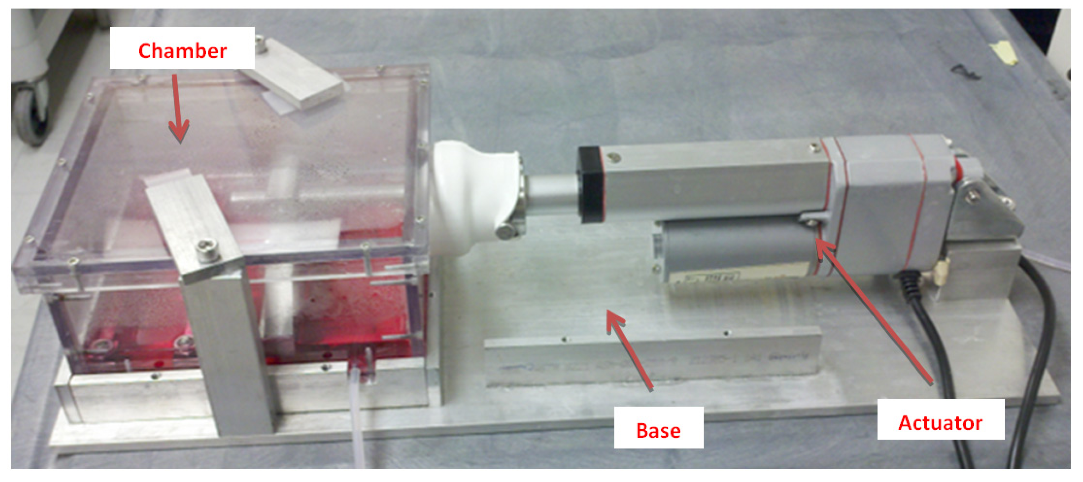
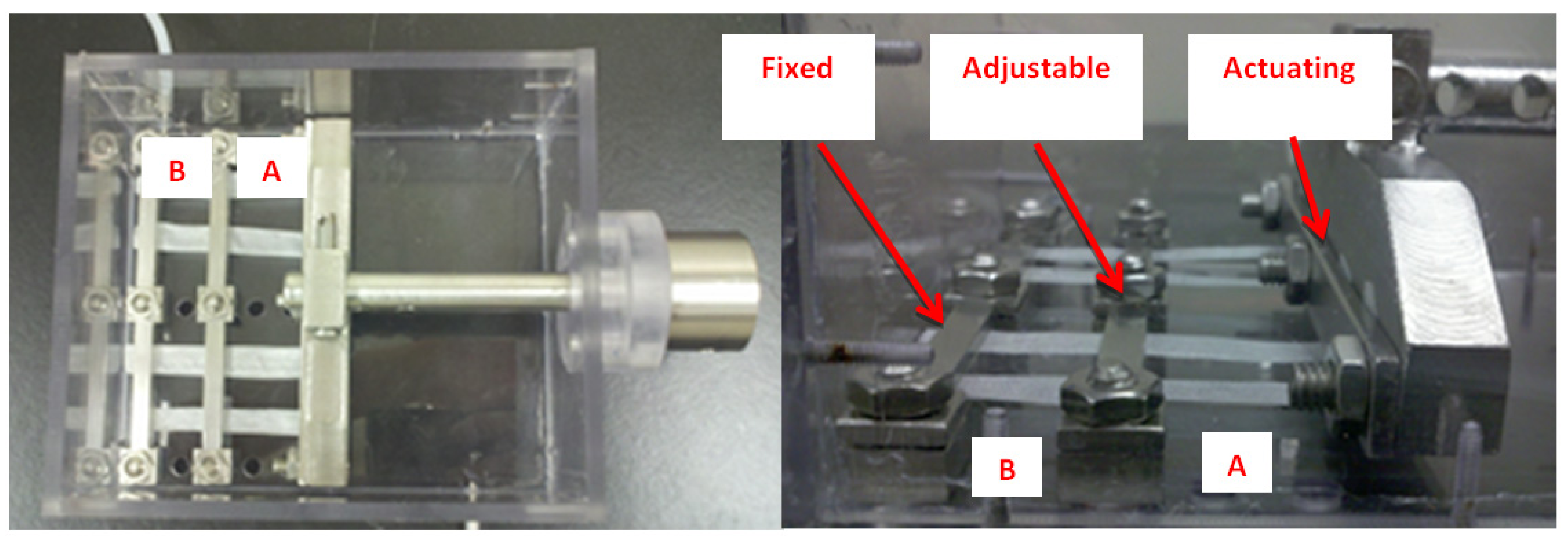
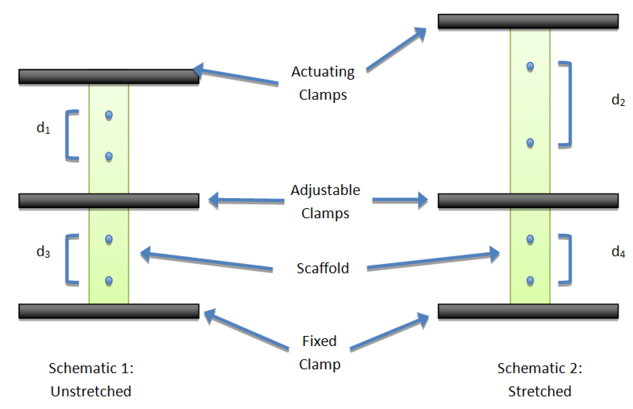
2.1. Bioreactor System

The bioreactor system design was modified from a commercially available bioreactor (BOSE Electroforce 3200; Eden Prairie, MN, USA) to provide differential stretch to portions of the scaffolds. The overall system is composed of two major sections: a baseplate (composed of 6061 aluminum alloy) and a removable polycarbonate chamber (Figure 1). The chamber is removable from the base and replaced with another chamber so concurrent tests may be performed. This system uses a linear actuator to apply linear, uniaxial strain to a group of parallel scaffolds fixed inside the chamber. The overall dimensions of the system are 17.8 cm × 44.5 cm. which allows it to fit inside a standard cell incubator.
Figure 1. Bioreactor chamber in stretching base.
Processes 02 00345 g001
The bioreactor chamber is constructed of 0.64 cm plates of polycarbonate fitted together using 316L stainless steel socket head cap screws and then sealed with silicone (100% Clear Aquarium Silicone, Marineland, Blacksburg, VA, USA) along both the outside and inside edges to make the chamber watertight. The clamps, screws, and actuator inside the chamber are also made of 316L stainless steel. A single compartment chamber was used in this initial reactor design to explore enthesis generation in vitro based on the idea that bone and tendon tissue interfaces share similar extracellular fluids in vivo and may participate in paracrine signaling that is important to tissue development.
The novel three-clamp design involves three clamps: an actuating, fixed, and adjustable clamp (Figure 2). This design allows scaffolds of different materials and sizes, as well as portions of scaffold subjected to mechanical perturbation. The actuating clamp is connected to the linear actuator and is used to mechanically stretch the scaffold by pulling on one end of the scaffold. The fixed clamp is used to hold the opposite end of the scaffold in a fixed position. The adjustable clamp may be used to create two distinct zones of mechanical stretch (a non-stretched zone (A) and a stretched zone (B)) by creating a fixed section in the central region of the scaffold. The adjustable clamp may be moved to other positions designated by the extra holes visible in Figure 2. This adjustability allows for varying scaffold lengths and larger/smaller percentages of the scaffold to be stretched. In addition, different cell types may be seeded in zones A and B allowing for spatially segregated co-cultures as well as intermixed co-cultures. After stretching begins, the fixed clamp and actuating clamp are intended to remain in place; but, the adjustable clamp may be removed for non-segregated co-culture when two cell types are used.
Figure 2. Bioreactor chamber clamp design. (A) Portion of the scaffold experiencing strain; (B) Portion of the scaffold that remains under 0% strain conditions.
Processes 02 00345 g002
Medium is pumped with a Masterflex pump through the bioreactor chamber at 60–70 mL/min through an inlet to the side of the scaffolds and an outlet at the opposite corner. Gas permeable tubing (Cole-Parmer, Vernon Hills, IL, USA) allows for CO2 and O2 gas exchange as the medium circulates. The bioreactor chamber and tubing are sterilized using steam autoclave at 121 °C and 15 psi for 20 min.
A LabVIEW program (v. 8.0 National Instruments, Austin, TX, USA) was created to control the 5 cm stroke 667 N force linear actuator, with potentiometer feedback (FA-PO-150-12-2, Firgelli Automations, Surrey, BC, Canada), that is used to mechanically stretch the scaffolds. The program is used to control frequency, magnitude, and duration of mechanical stretch to meet experimental conditions.

2.2. Mechanical Characterization

To characterize the mechanical performance of the bioreactor, four Polyethylene Terephthalate (PET) (Biomedical Structures, Warwick, RI, USA) fabric samples (10 mm × 80 mm) were clamped in the bioreactor and marked with small graphite dots (Figure 3). The LabVIEW program was set up to apply a 3% stretch chosen as an expected applied stretch to be used with this bioreactor. Three percent is also large enough to measure a noticeable strain but small enough that the accuracy/repeatability of the system can be tested. The LabVIEW program was run through 24 strain cycles and pictures were taken at each step in the cycle. Observed strain was recorded from the measurements taken.
Figure 3. Two schematics of the strain characterization. Schematic 1 represents the photo of an unstretched scaffold. Schematic 2 represents a picture of the same scaffold under a specified stretch. Comparing to Figure 4, d1 and d2 are markers inside of zone A while d3 and d4 are markers inside of zone B.
Processes 02 00345 g003
Figure 4. Relative position of each section of scaffold for assays. Positions 1, 2, and 3 refer to position on the scaffold and proximity to clamps. L/D refers to sections of the scaffolds designated for Live/Dead imaging.
Processes 02 00345 g004

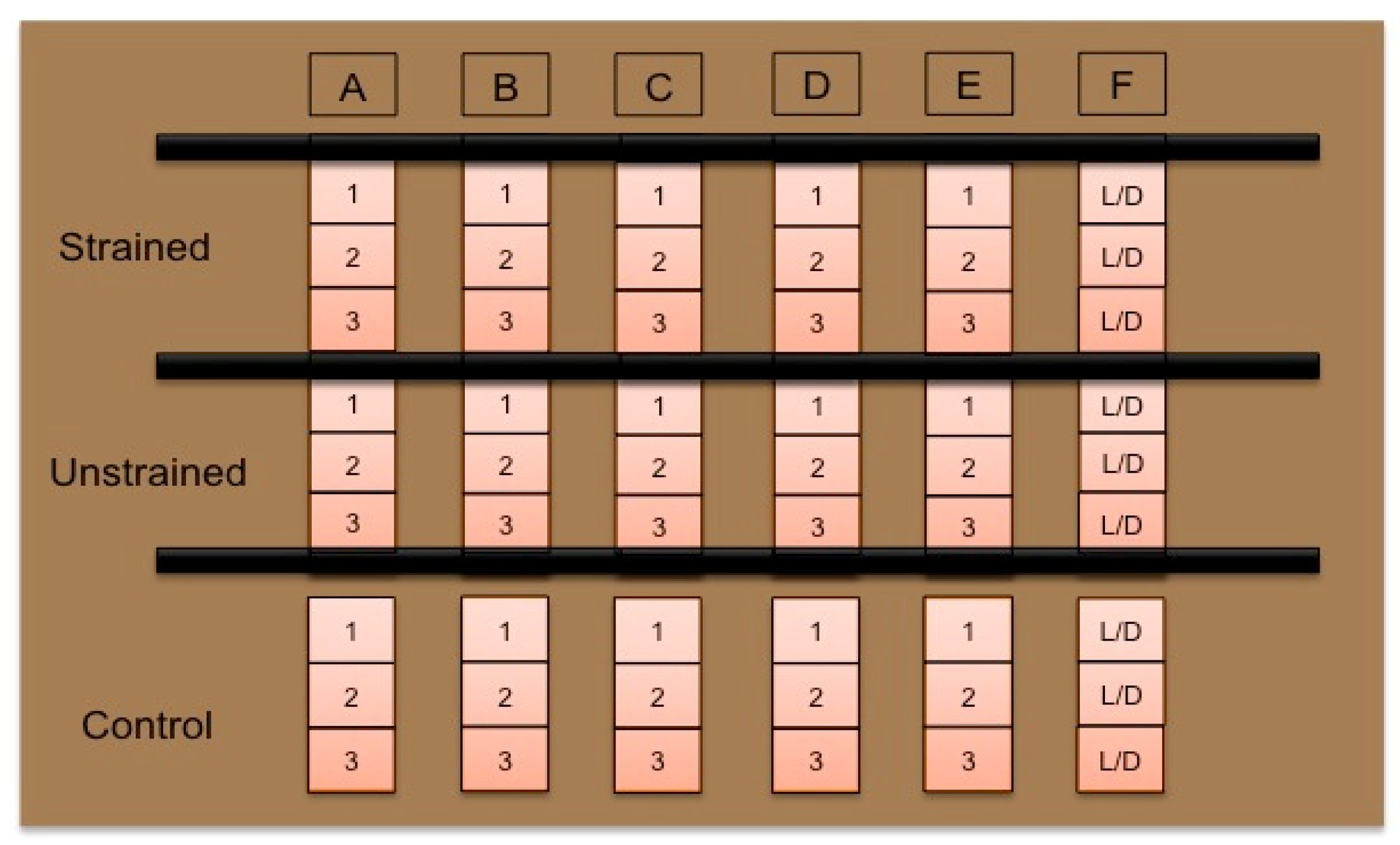
2.3. Cellular Characterization
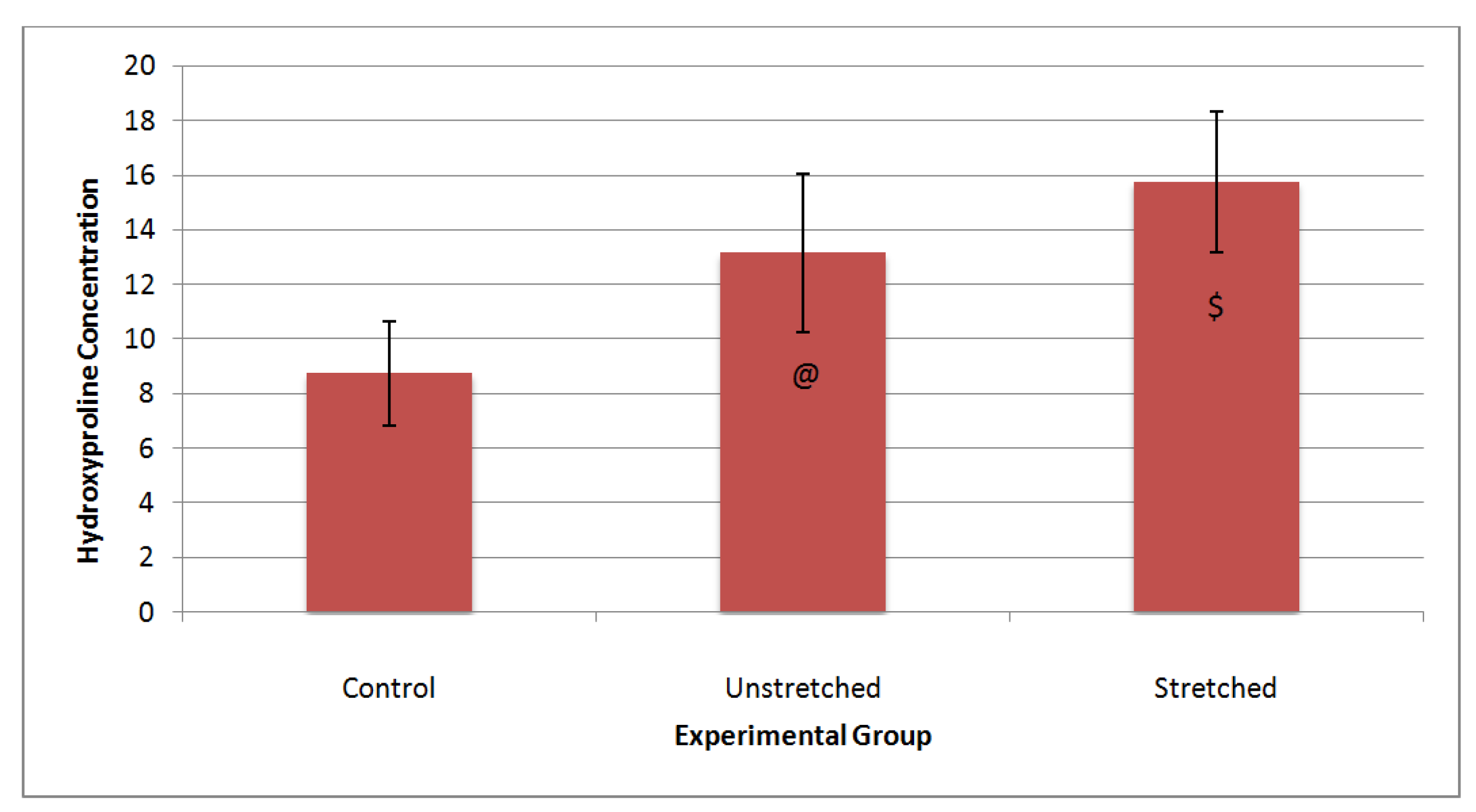
Six large PET scaffolds (8 mm × 87.5 mm) and seven small PET scaffolds (8 mm × 25 mm) were cut from a large spool and their cut ends lightly melted by touching them to a heated steel bar to prevent fraying. Scaffolds were sterilized by sonicating in each of the following solutions for 30 min: 1% Ivory dish soap solution, deionized (DI) water, and a second time in DI water. Samples were placed in 70% ethanol for 30 min and finally sterilized by exposure to UV light for at least 30 min per side.
After sterilization, but before seeding, scaffolds were rinsed with sterile 1× PBS and then pre-exposed to culture medium, Dulbecco’s Modified Eagle Medium High Glucose (HyQ® DMEM, HyClone, Logan, UT, USA) supplemented with 10% fetal bovine serum (FBS, Cat. No. 30-2020, American Type Culture Collections (ATCC), Manassas, VA, USA) and 1% antibiotic/antimycotic (AB/AM, 100 U/mL penicillin, 100 μg/mL streptomycin, 0.25 μg/mL amphotericin B) by soaking the scaffolds in sterile 50 mL centrifuge tubes for at least 30 min to allow the medium to penetrate the scaffold and proteins to start attaching.
NIH-3T3 mouse fibroblast cells (ATCC® CRL-1658TM, Manassas, VA, USA) were cultured in DMEM-High Glucose + 10% FBS + 1% AB/AM at 37 °C with 5% CO2. Cells were seeded at a density of 1 × 10⁵ cells/cm2 in four spots along the length of six large (8 mm × 87.5 mm) and seven small (8 mm × 25 mm) scaffolds, and kept in three 8.9 cm diameter TCP petri dishes and seven individual 3.8 cm TCP petri dishes, respectively, overnight to allow attachment.
Two groups were tested in this experiment: a control group (static culture) and an experimental group (bioreactor). The experimental group was divided into two subgroups. Subgroup 1 of the experimental group was cultured in the bioreactor, but experienced no stretch. Subgroup 2 of the experimental group was also cultured in the bioreactor, but was subjected to 5% mechanical stretch at 0.5 Hz for one hour per day for 14 consecutive days. Table 1 lists the characteristics for each experimental group.
Table 1. Test and control group experimental conditions.
Group Location Stretch Schedule
Control TCP Petri Dish No stretch
Unstretched Bioreactor No stretch
Stretched Bioreactor 5%, 0.5 Hz, 1 h daily, 14 days
After seeding and incubation overnight, scaffolds were divided into their respective groups. On day 1, the six scaffolds were clamped in the bioreactor and 250 mL of DMEM-High Glucose medium (Fisher Scientific, St. Louis, MO, USA) supplemented with 1 mM beta-glycerophosphate (β-GP) and 10 μg/mL of ascorbic acid [16] was added. The six smaller scaffolds, sized such to represent the same surface area as the other groups, remained in their petri dishes and 2 mL of medium was added to each. The remaining small scaffold selected for determining initial cell attachment was removed, rinsed with sterile 1× PBS, and then stored at −20 °C. Stretching on day 1 began 2 h after clamping to allow any cells that may have become dislodged or perturbed to reattach to the scaffold.
Over the 14-day period, the reservoir of medium for the bioreactor was changed every 6 days. On day 3 between reservoir changes, fresh ascorbic acid was added sterilely to a final 10 μg/mL concentration to account for loss due to decomposition. The medium of the control scaffolds was changed every 3 days with fresh medium.
Experimental parameters were chosen based on their use in the literature. The stretching protocol is similar to that used by other groups such as Garvin et al. and Riboh et al. [6,9] Daily intermittent straining was chosen as it has been shown that intermittent straining increases collagen I production and cell proliferation [9]. Fourteen days is also a common timeframe for tendon tissue engineering bioreactors [9,10]. Concentrations of β-GP and ascorbic acid were chosen as having the most positive effect on cell proliferation and collagen I production in tests performed by Wang et al. [14]. Mechanical stretching of the scaffolds took place in a cell incubator (37 °C, 5% CO2, 95% humidity).
Scaffolds were removed from the bioreactor and petri dishes 2 h following the final stretch of day 14. All scaffolds were cut into three approximately equal sized sections (8 mm × 8 mm) and their surface area recorded (Figure 4). By sectioning the scaffolds, we are able to determine if the matrix on the scaffolds is uniform over the whole scaffold as well as across all of the scaffolds. If any non-uniformity is noted, it may be possible to determine if effects are caused by proximity of the clamps (section 1, 2, or 3) or by position of the scaffold (scaffold A–F).
To remove the cells for analysis, each scaffold section was rinsed with sterile 1× PBS and placed into labeled Erlenmeyer tubes with 1 mL of 50 μg/mL Proteinase K (Pro-K, Promega Corporation, Madison, WI, USA) solution (50 μg/mL Pro-K, 50 mM Tris-HCL, 50 mM CaCl2 buffer, pH 7.5) and placed overnight in an oven at 60 °C. The following day, 50 μL of proteinase inhibitor (phenylmethylsulfonyl fluoride (PMSF), MP Biomedicals, Solon, OH, USA) was added and a sonic dismembraner (Fisher Scientific Sonic Dismembraner 550, Fisher Scientific, Pittsburg, PA, USA, Setting 2) was used to disrupt any remaining cells and matrix. This cell lysate was used to estimate cell number. One scaffold was not digested in the Pro-K solution and used instead to view cells on scaffold using a Live/Dead® assay (Invitrogen, Grand Island, NY, USA). From the assay protocol, viable cells will fluoresce green and dead cells will fluoresce red.
The PicoGreen assay (Invitrogen, Grand Island, NY, USA) was to measure DNA as an indicator of the number of cells on scaffolds. For the assay, the 100 μL of PicoGreen reagent (Life Technologies Corp, Grand Island, NY, USA) was added to 20 μL of the cell/matrix lysate in 80 μL of tris-HCL-EDTA buffer in a 96 well opaque microplate. The plates were read at 485 nm excitation and 528 nm emission using a spectrophotometer (Molecular Devices, LLC, Sunnyvale, CA, USA). A linear standard curve (2 μg/mL serial dilution) was prepared from a DNA standard (100 μg/mL). DNA was normalized to surface area, ngDNA/mm2, for each scaffold section.
Glycosaminoglycans are polysaccharides that are a major component of tendon extracellular matrix [17]. To quantify the concentration of GAG, the Alcian blue assay (Kamiya Biomedical Co., Seattle, WA, USA) was used. 250 μL of each prepared sample (cell lysate with Alcian blue) and 100 μL of each prepared chondroitin sulfate standard (400 μg/mL serial dilution) was transferred to a 96-well plate and the absorbance read at 620 nm. Sample concentrations of GAG were then normalized by DNA to ngGAG/ngDNA.
Elaboration of collagen by cells is an indicator of cellular matrix production. The hydroxyproline assay was used to measure collagen. According to Stegemann and Salter, hydroxyproline can be linearly translated to collagen by multiplying by a factor of 7.46, i.e., 1 μg/mL hydroxyproline is equal to 7.46 μg/mL collagen [18]. First, 25 μL of prepared hydroxyproline (HP) standard (Sigma-Aldrich, St. Louis, MO, USA) (100 μg/mL serial dilution) and 25 μL of 16× concentrated prepared sample were added to each well of a 96-well plate. Prepared chloramine-T solution (112.5 μL) was added and incubated at room temperature for 20 min. Immediately following, 125 μL of prepared Ehrlich’s reagent were added to each well, incubated for 20 min at 65 °C in an oven, and the absorbance read at 550 nm. Sample concentrations of collagen were then normalized by DNA to ngHP/ngDNA.

2.4. Statistical Analysis

Means among section position and scaffold location were calculated and a two-factor ANOVA was performed; one factor was the individual scaffolds (A–E) and the other factor was scaffold section (1–3) to one another. This was done for each of the three groups. Differences were considered significant at p < 0.05. Differences were determined using Tukey’s post-hoc testing. When no significant differences were determined, results were pooled and one-way ANOVA was performed to test differences between test groups (n = 5) for the GAG and hydroxyproline data. Differences were determined using Tukey’s post hoc testing and were considered significant at p < 0.05.

3. Results

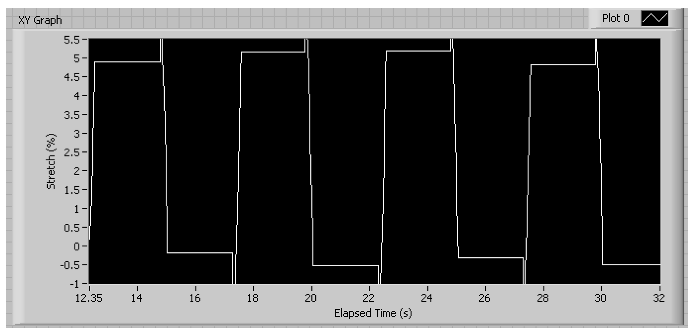
3.1. Mechanical Characterization

Figure 5 shows the LabVIEW waveform for the actuator to apply mechanical stretch to test scaffolds in the bioreactor. The large spikes at the end of each plateau are an artifact caused by the sampling and recording and are not representative of stretched applied to the scaffolds.
Figure 5. Sample waveform for controlling actuator to apply mechanical stretch to scaffolds in bioreactor. The parameters set were 5% stretch at 0.2 Hz.
Processes 02 00345 g005
Results of the video analysis of the reference dots on the four PET scaffolds subjected to the programmed 3% stretch at 0.1 Hz for 24 cycles are shown in Figure 6. The figure shows the mechanical stretch of the stretched portion and the non-stretched portion of the scaffolds. The average strain experienced by the stretched scaffolds was 2.97% ± 0.13%. The average strain experienced by the unstretched scaffolds was 0.02% ± 0.18%.
Figure 6. Stretch cycles of the PET scaffolds in the bioreactor. Each bar represents the average strain across all four scaffolds for that stretch cycle. Error bars represent one standard deviation. n = 4.
Processes 02 00345 g006
Due to camera resolution limitations, visual representation of 0% strain was very difficult to obtain. However, the resolution of the pictures taken was 1 pixel ~0.5% strain, so values less than 0.5% were assumed to be 0%. Because the LabVIEW program was designed as an open loop control system, errors from one stretch cycle do not translate to the following stretch cycle as evidenced by the positive and negative differences of cycles 10–15.

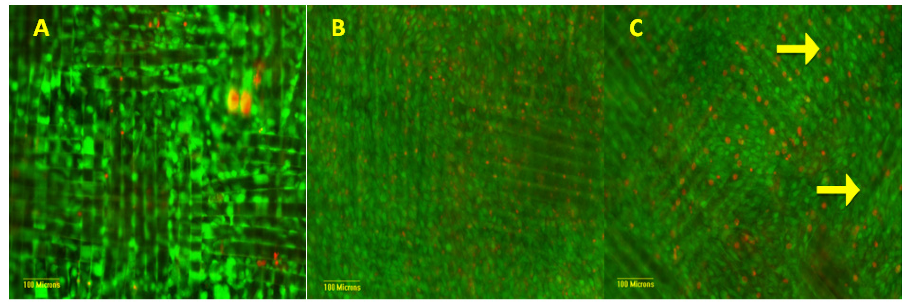
3.2. Cellular Characterization

Images of the experimental and control scaffolds stained with the Live/Dead reagent at the end of the 14-day culture period are shown in Figure 7. Images were taken of each of the three assayed sections in each group at magnifications of 10× and 20×. The images showed that there were many live/viable cells on all scaffold groups and scaffold sections. The cells uniformly covered the majority of the surface area of the scaffolds and cells were observed growing on fabric fibers within the scaffold.
Using measured cell DNA data, two-way ANOVA was performed on test samples to determine if there were any effects based on proximity of cells on scaffolds to the clamps or on position of scaffolds in the bioreactor. No statistical differences were found between zones or proximity of cells to clamps with in each experimental group (p > 0.18) nor were differences detected for position of scaffolds in bioreactor for each experimental group. Since there were no effects of position on the scaffold or position within the bioreactor based on DNA data, scaffolds were then treated as a single test unit in subsequent analyses.
Figure 7. Live/Dead images of section 2 of each scaffold magnified at 10×. Green represents those cells that are alive/viable and red represents those cells that are dead/non-viable. (A) Control; (B) Unstretched; (C) Stretched. Arrows signify cells aligning along scaffold fibers.
Processes 02 00345 g007
Figure 8 shows the results of total DNA on scaffolds in each experimental group after 14 days. One-way ANOVA revealed that the DNA concentration in all groups was not equal (p < 1 × 10⁻⁶). A large increase was found in the DNA concentration of the bioreactor unstretched group as compared to the control group (p < 1 × 10⁻⁶) as well as the bioreactor stretched group as compared to the control group (p < 1 × 10⁻⁶). However, no difference was found between the DNA concentration of the bioreactor unstretched group and bioreactor stretched group (p > 0.90).
Figure 8. DNA concentration of each experimental group. Data from all three sections of each scaffold were combined to compare the effect had over the whole scaffold. n = 5. Error bars represent one standard deviation. # Statistically different than control (p < 1 × 10⁻⁶).
Processes 02 00345 g008

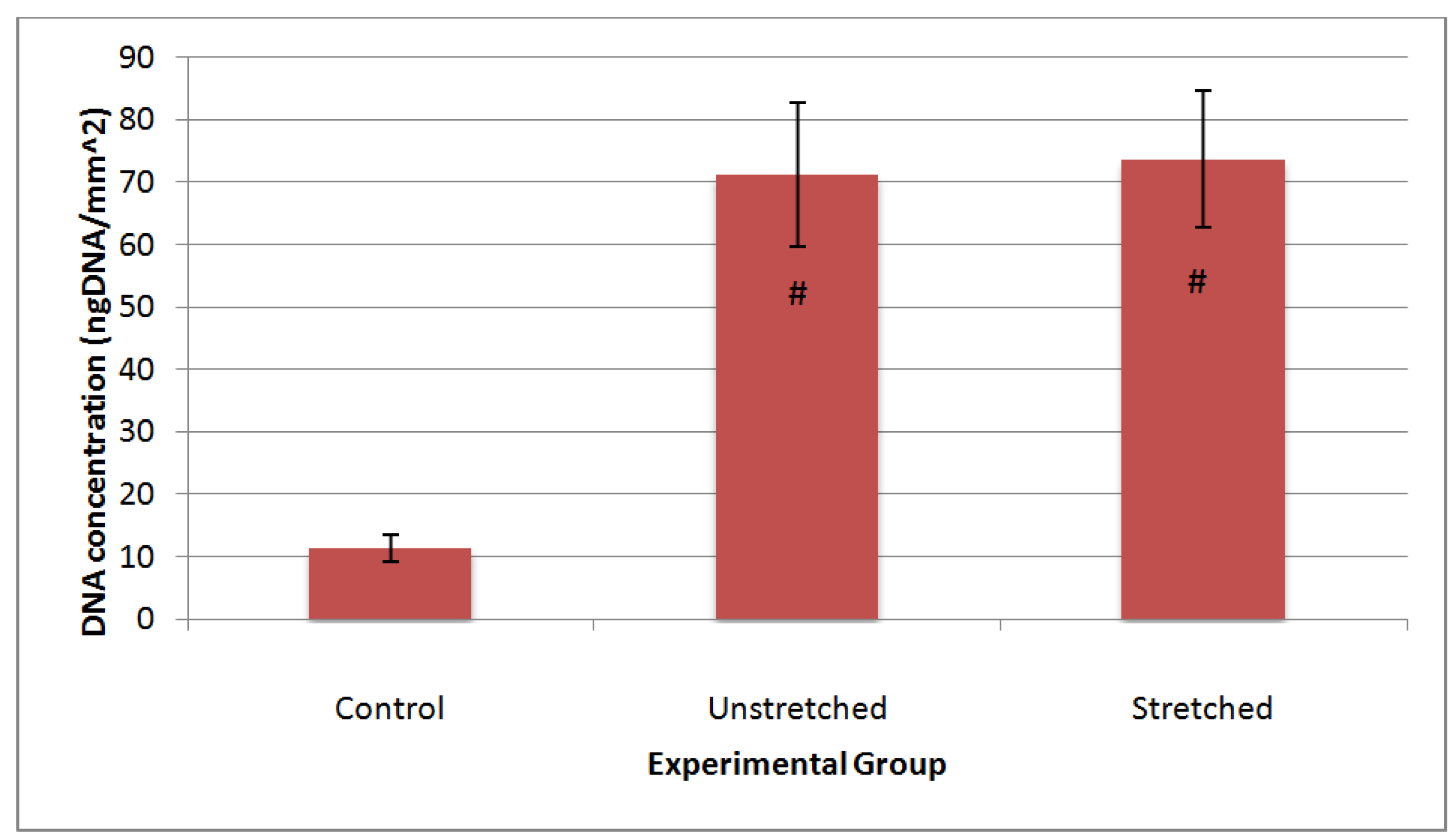
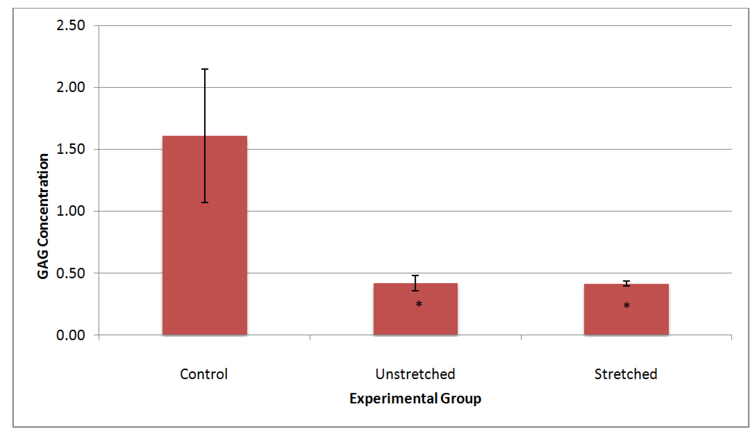
3.3. Matrix Characterization

To investigate the effect of mechanical stretch on levels of extracellular matrix, GAG was measured via the Alcian Blue assay and then normalized to DNA. One-way ANOVA with post-hoc Tukey tests showed that the amount of GAG per DNA was statistically lower in the bioreactor as compared to the static culture (p < 0.001), but there was no difference between strained and unstrained sections of scaffolds in the bioreactor (Figure 9) (p > 0.99).
Figure 9. GAG deposition normalized to DNA (ngGAG/ngDNA). n = 5. * Statistically different than control (p < 0.001).
Processes 02 00345 g009
Hydroxyproline (HYP), like GAG, was normalized to DNA and showed a difference between experimental groups (Figure 10) (p < 0.01). The bioreactor unstretched group showed an increase in hydroxyproline per DNA as compared to the control group (p < 0.05) as did the bioreactor stretched group (p < 0.01). The bioreactor unstretched and stretched groups still showed no significant differences in hydroxyproline deposition when normalized to DNA (p > 0.25).
Figure 10. Hydroxyproline deposition normalized to DNA (ngHYP/ngDNA). n = 5. @, Statistically different than control (p < 0.05); $, Statistically different than control (p < 0.01).
Processes 02 00345 g010

4. Discussion

This study presented an enhanced bioreactor design that is able to cyclically strain preferential zones of multiple, fabric, cell-seeded scaffolds in a controlled in vitro setting. This system has potential to develop degradable and non-degradable fabric scaffolds with improved matrix deposition over basic statically cultured scaffolds. Analysis with photographic video demonstrated that the system is able to deliver accurate and repeatable cyclic stretch to a designated portion of each scaffold while isolating the remainder of the scaffold from stretch. In addition, the system may accommodate numerous variables including, scaffold length, width, and material as well as stretch zone, magnitude, frequency, and schedule. Furthermore, each bioreactor chamber could interchange with a second identical bioreactor chamber for concurrent studies using similar or differing experimental conditions.
Minor variations of less than 10% of the desired strain magnitude were noticed between stretch cycles, which were attributed mostly to the process of the marking for the video analysis. The method used to mark the scaffolds for analysis was sufficient for initial validation purposes, but lacked fine resolution. Many methods were used to mark the scaffolds and measure the distance between marks—graphite providing the most accurate method. The camera used also had a limited resolution. For more exact values, a camera or microscope with higher resolution would give decreased error to the measurements taken. However, the system did prove that it could consistently stretch as low as 1% and at least as high as 10% for a 1-inch scaffold (data not shown). Unfortunately, over a 14-day period, there was some laxity noticed in the scaffolds likely due to viscoelastic properties of the PET material. This laxity may have decreased strain on the cells and thereby altered the cellular response over the time period studied. Based on a study on single PET fibers with a creep rate at 0.15% strain per decade of time at 50% relative humidity and 20 °C, an approximate 0.7% strain may be anticipated over the 14 days of culture at 1 hour per day loading [19]. Since the woven membranes are not single fibers and the membranes were not subjected to constant loads and were submerged in a saline environment at 37 °C, we cannot say more than that we estimate a possible decrease of peak strain in the scaffolds from 5% to 4%. It is unclear at this time if cells in these types of studies are able to distinguish and, hence, respond differently to a 1% change in strain. Additional studies will be needed to determine the effects, if any, of this laxity of the scaffold on the cells.
Investigating the effect of location within the bioreactor and the proximity to a clamp revealed that the scaffolds were statistically equivalent and could be treated as uniform. Live/Dead staining demonstrated that cells remained viable on the scaffolds in both the bioreactor and in control cultures over the 14-day experimental timeframe. The viable cells on the scaffolds in the bioreactor were still at a much greater density based on visual examination than the viable cells on the control scaffolds. Also, the cells in the bioreactor, especially the stretched cells, began to show alignment along the scaffold fibers. Riboh et al. also noticed this phenomenon with cytoskeletal and nuclear rearrangement due to uniaxial cyclic stress [9].
Quantitative analysis of the DNA concentration on the scaffolds supported our qualitative findings. Intermittent cyclic strain increased DNA concentration seven times on the scaffolds in the bioreactor as compared to the control scaffolds. This agreed with other studies investigating the effects of varying schedules of intermittent cyclic strain over static culture [9,20]. Application of mechanical strain to these cells activates the mitogen-activated protein kinase pathway as well as mechanosensitive calcium channels initiating the proliferative response [9]. However, our findings did not agree with Screen et al. who did not see any change in cell proliferation on a cyclically strained tendon [15]. However, their studies were only performed for 24 h, and grafted tendon was used as their scaffold material [10,15].
In addition to the mechanical perturbations, fluid flow of the medium may have also contributed to increased cell proliferation in the bioreactor as compared to the controls. Fluid shear forces are known to effect fibroblast morphology, proliferation, migration, and matrix remodeling [21]. For example, studies have shown that at low flow rates, 1.67–3.34 µL/min, proliferation of fibroblasts increased significantly as compared to static conditions [12,22]. In a microfluidic device, Park et al. also reported that fibroblastic cell proliferation was significantly increased at low flow rates (0.14–0.52 µL/min) with the maximal increase observed at flow rates approximating interstitial fluid flow (0.14 µL/min) [23]. Wang et al., using a spinner flask, noted increased fibroblast matrix production and proliferation at 13–40 rpm stirring speeds [24]. These fluid flow effects are attributed to increased nutrient exchange, conformational changes in membrane proteins (e.g., integrins) that affect binding properties to extracellular matrix, membrane deformations activating cytoskeletal elements, and or activating membrane signaling proteins (e.g., G proteins) [21]. In this study, the pump flow rate was set at 60–70 mL/min, but the actual flow rate in the chamber is likely to be substantially lower due to its larger volume than the pump tubing. The actual flow rate in the chamber was not determined in this initial evaluation, due to the complexity of the chamber components, and limits direct comparison to previous studies. Nevertheless, it may be speculated that fluid flow in the chamber may also have had an effect on cell growth in addition to the cyclic mechanical effects. Characterization of fluid flow in the chamber will be a focus of future studies.
Initially, it was surprising that there were no differences detected in DNA and elaboration of extracellular matrix components between strained and non-strained scaffold sections since cyclic strain has been shown to have a large effect over the static control petri dish scaffolds. It was expected that the proliferation increase of the strained scaffolds over the unstrained scaffolds would be much greater than the unstrained scaffolds over the control scaffolds. We attributed the lack of difference in part to a concept known as conditioned media since the media was recycled by the pump system. Multiple groups have shown that instead of directly or mechanically stimulating cells, replacing the culture medium with a conditioned culture medium can induce a response from the cells based on chemical signals present in the medium [25,26,27]. For example, by using medium that was used to stretch osteoblast cells, Soma et al. was able to induce osteoclast proliferation by using the discarded medium as a conditioned medium [27]. Their results suggest that cellular signals, generated from the mechanically perturbed and growing osteoblasts, in the culture medium caused the osteoclast cells to initiate homeostatic tendencies by proliferating [27]. This same phenomenon may be occurring inside the bioreactor as the unstretched scaffolds shared medium with the strained scaffolds. The lack of difference in cell proliferation on strained vs. non-strained scaffolds may also have been be due in part to the effects of fluid flow in the chamber as already discussed. Studies have suggested that cells may be more responsive to fluid shear flow forces than mechanical forces, though some questions still remain [28,29,30]. Thus, both the shared conditioned medium and fluid flow effects may have confounded effects of mechanical stretch on cell proliferation.
The tendon reconstruction schedule, proposed by Rust et al., shows that cell proliferation peaks around day 5, followed by a large increase in unorganized collagen deposition (days 5–28) [31]. The collagen deposition overlaps slightly with an increase in GAG deposition beginning around day 21 and increasing through day 28 and beyond [31]. Our analysis occurred in the middle of the collagen growth peak based on data from Rust et al., which would ideally show us if cyclic strain increased the concentration of collagen as well as the rate of production. We found a 44% increase in hydroxyproline concentration on the unstrained scaffolds in the bioreactor as well as a 75% increase in hydroxyproline concentration on the strained scaffolds in the bioreactor as compared to the control scaffolds. A similar trend has been previously reported by Riboh et al. as intermittent cyclic strain was shown to increase type I collagen production using fibroblastic cells [9]. The strained scaffolds cultured by Screen et al. showed no significant increase in hydroxyproline concentration over the unstrained control scaffolds [15]. Interestingly though, there was a large increase in hydroxyproline concentration present in the medium of the strained scaffolds over the unstrained scaffolds [15].
Analysis of GAG concentration on the scaffolds in the bioreactor shows an interesting trend. Overall, there was a greater mass of GAG on the bioreactor scaffolds than on the control scaffolds, but the static control petri dish scaffolds deposited four times more GAG per cell on the scaffolds as the bioreactor scaffolds. It would appear that cyclic strain had a negative effect on GAG deposition; but the trend is misleading because of the drastically large increase in DNA concentration on the bioreactor scaffolds. At day 14, when these scaffolds were assayed, the large increase in GAG deposition proposed by Rust et al. would not have begun [31]. Therefore, it is not surprising to have a lower concentration of GAG per cell on the bioreactor scaffolds due to the large increase in bioreactor cell proliferation. Sampling around day 28 may provide a better investigative time point to analyze the bioreactor’s effect on the elaboration of GAG. While these results seem surprising, our findings do agree with Screen et al. where they saw a statistically significant decrease in GAG concentration on their strained scaffolds as compared to their unstrained scaffolds [15].
In this study, medium was supplemented with ascorbic acid, an important factor for both stimulating proliferation and extracellular matrix production [16,32]. Since ascorbic acid degrades overtime, the medium used in the bioreactor was supplemented in-between medium exchanges in an attempt to keep ascorbic acid levels similar to the control cultures. It has been reported that ascorbic acid in conjunction with cyclic strain had a synergistic effect on both fibroblast proliferation and gene expression of extracellular matrix proteins such as elastin and collagen as compared to static conditions [33]. Additional studies are warranted to further explore the synergistic effects of ascorbic acid and mechanical forces.
In this bioreactor design, the effects of paracrine signaling between mechanically strained and unstrained sections of the constructs were not explored separately from mechanical factors. This is because in vivo, the bone-tendon interface shares similar environments and may participate in paracrine signaling that is important to tissue development. However, a simple barrier and a second medium reservoir-pump system may be added to the chamber to separate the strained and unstrained compartments to prevent medium exchange and to enable exploration of applied mechanical forces separate from signaling phenomena. The bioreactor design also did not separate out the effects of fluid flow in the chamber from that of scaffold mechanical strain. This is a challenge for most bioreactor designs intended for load bearing tissues like bones and tendons [29]. Nevertheless, study designs involving comparing scaffolds subjected only to medium flow in the chamber to those subjected to both medium flow and mechanical strain are planned in order to better explore their contributions to the engineered tissue.

5. Conclusions

A unique bioreactor system was designed, developed, and shown to apply a known, user-specified, cyclic strain to cells growing on polymeric fabric scaffolds. The strain environment experienced by the cells was quantified and reproducible. Strain was able to be isolated from a specified portion of each scaffold while stretching the remainder. Differences in cellular proliferation, GAG concentration, and hydroxyproline concentration were measured based on static and dynamic culture conditions experienced by the cells. The bioreactor system may be used to evaluate the effects of mechanical stretch on degradable and non-degradable fabric scaffolds.

Acknowledgments

This project was funded by the USAMRMC W81XWH-10-1-0768 grant and by the Biomaterials Applications of Memphis (BAM) Laboratories in the Biomedical Engineering Department at the University of Memphis. Special thanks to Richard Voyles and Dan Kohn for their assistance in the design and technical help of this project.

Conflicts of Interest

The authors declare no conflict of interest.

References

  1. Caliari, S.R.; Harley, B.A.C. The effect of anisotropic collagen-GAG scaffolds and growth factor supplementation on tendon cell recruitment, alignment, and metabolic activity. Biomaterials 2011, 32, 5330–5340. [Google Scholar] [CrossRef]
  2. Caliari, S.R.; Ramirez, M.A.; Harley, B.A.C. The development of collagen-GAG scaffold-membrane composites for tendon tissue engineering. Biomaterials 2011, 32, 8990–8998. [Google Scholar] [CrossRef]
  3. Sahoo, S.; Toh, S.L.; Goh, J.C.H. A bFGF-releasing silk/PLGA-based biohybrid scaffold for ligament/tendon tissue engineering using mesenchymal progenitor cells. Biomaterials 2010, 31, 2990–2998. [Google Scholar] [CrossRef]
  4. Whitlock, P.W.; Smith, T.L.; Poehling, G.G.; Shilt, J.S.; van Dyke, M. A naturally derived, cytocompatible, and architecturally optimized scaffold for tendon and ligament regeneration. Biomaterials 2007, 28, 4321–4329. [Google Scholar] [CrossRef]
  5. Deng, D.; Liu, W.; Xu, F.; Yang, Y.; Zhou, G.; Zhang, W.J.; Cui, L.; Cao, Y. Engineering human neo-tendon tissue in vitro with human dermal fibroblasts under static mechanical strain. Biomaterials 2009, 30, 6724–6730. [Google Scholar] [CrossRef]
  6. Garvin, J.; Qi, J.; Maloney, M.; Banes, A.J. Novel system for engineering bioartificial tendons and application of mechanical load. Tissue Eng. 2003, 9, 967–979. [Google Scholar] [CrossRef]
  7. Joshi, S.D.; Webb, K. Variation of cyclic strain parameters regulates development of elastic modulus in fibroblast/substrate constructs. J. Orthop. Res. 2008, 26, 1105–1113. [Google Scholar] [CrossRef]
  8. Butler, D.L.; Hunter, S.A.; Chokalingam, K.; Cordray, M.J.; Shearn, J.; Juncosa-Melvin, N.; Nirmalanandhan, S.; Jain, A. Using functional tissue engineering and bioreactors to mechanically stimulate tissue-engineered constructs. Tissue Eng. 2009, 15, 741–749. [Google Scholar] [CrossRef]
  9. Riboh, J.; Chong, A.K.S.; Pham, H.; Longaker, M.; Jacobs, C.; Chang, J. Optimization of flexor tendon tissue engineering with a cyclic strain bioreactor. J. Hand Surg. 2008, 33A, 1388–1396. [Google Scholar]
  10. Wolchok, J.C.; Brokopp, C.; Underwood, C.J.; Tresco, P.A. The effect of bioreactor induced vibrational stimulation on extracellular matrix production from human derived fibroblasts. Biomaterials 2009, 30, 327–335. [Google Scholar] [CrossRef]
  11. Abousleiman, R.I.; Reyes, Y.; McFetridge, P.; Sikavitsas, V. Tendon tissue engineering using cell-seeded umbilical veins cultured in a mechanical stimulator. Tissue Eng. 2009, 15, 787–795. [Google Scholar] [CrossRef]
  12. Korin, N.; Bransky, A.; Dinnar, U.; Levenberg, S. A parametric study of human fibroblasts culture in a microchannel bioreactor. Lab Chi 2007, 7, 611–617. [Google Scholar] [CrossRef]
  13. Titze, I.R.; Hitchcock, R.W.; Broadhead, K.; Webb, K.; Li, W.; Gray, S.D.; Tresco, P.A. Design and validation of a bioreactor for engineering vocal fold tissues under combined tensile and vibrational stresses. J. Biomech. 2004, 37, 1521–1529. [Google Scholar] [CrossRef]
  14. Wang, N.E.; Shan, J.; Choi, R.; Oh, S.; Kepler, C.K.; Chen, F.H.; Lu, H.H. Role of osteoblast-fibroblast interactions in the formation of the ligament-to-bone interface. J. Orthop. Res. 2007, 25, 1609–1620. [Google Scholar] [CrossRef]
  15. Screen, H.R.C.; Shelton, J.C.; Bader, D.L.; Lee, D.A. Cyclic tensile strain upregulates collagen synthesis in isolated tendon fascicles. Biochem. Biophys. Res. Commun. 2005, 336, 424–429. [Google Scholar] [CrossRef]
  16. Geesin, J.C.; Darr, D.; Kaufman, R.; Murad, S.; Pinnell, S.R. Ascorbic acid specifically increases type I and type III procollagen messenger RNA levels in human skin fibroblasts. J. Invest. Dermatol. 1988, 90, 420–424. [Google Scholar]
  17. Redaelli, A.; Vesentini, S.; Soncini, M.; Vena, P.; Mantero, S.; Montevecchi, F.M. Possible role of decorin glycosaminoglycans in fibril to fibril force transfer in relative mature tendons—A computational study from molecular to microstructural level. J. Biomech. 2003, 36, 1555–1569. [Google Scholar] [CrossRef]
  18. Stegemann, H.; Stalder, K. Determination of hydroxyproline. Clin. Chim. Acta 1967, 18, 267–273. [Google Scholar] [CrossRef]
  19. Lechat, C.; Bunsell, A.R.; Davies, P. Tensile and creep behaviour of polyethylene terephthalate and polyethylene naphthalate fibres. J. Mater. Sci. 2011, 46, 528–533. [Google Scholar] [CrossRef]
  20. Blair, B.; Ricci, A.J.L.; Casar, R.S. The effect of mechanical strain on rat tendon fibroblasts grown on a three-dimensional scaffold. In Proceedings of Transactions of the Annual Meeting—Orthopaedic Research Society, New Orleans, LA, USA, 21–24 February 1994.
  21. Dan, L.; Chua, C.K.; Leong, K.F. Fibroblast response to interstitial flow: A state-of-the-art review. Biotechnol. Bioeng. 2010, 107, 1–10. [Google Scholar] [CrossRef]
  22. Jarman-Smith, M.; Bodamyali, T.; Stevens, C.; Howell, J.A.; Horrocks, M.; Chaudhuri, J.B. Human fibroblast culture on a crosslinked dermal porcine collagen matrix. Biochem. Eng. J. 2004, 20, 217–222. [Google Scholar] [CrossRef]
  23. Park, J.Y.; Yoo, S.J.; Patel, L.; Lee, S.H.; Lee, S.H. Cell morphological response to low shear stress in a two-dimensional culture microsystem with magnitudes comparable to interstitial shear stress. Biorheology 2010, 47, 165–178. [Google Scholar]
  24. Wang, H.J.; Bertrand-De Haas, M.; Riesle, J.; Lamme, E.; van Blitterswijk, C.A. Tissue engineering of dermal substitutes based on porous PEGT/PBT copolymer scaffolds: Comparison of culture conditions. J. Mater. Sci. 2003, 14, 235–240. [Google Scholar]
  25. GanjiBakhsh, M.; Nejati, V.; Delirezh, N.; Asadi, M.; Gholami, K. Mixture of fibroblast, epithelial and endothelial cells conditioned media induce monocyte-derived dendritic cell maturation. Cell. Immunol. 2001, 272, 18–24. [Google Scholar]
  26. Lee, S.R.; Kim, M.O.; Kim, S.H.; Kim, B.S.; Yoo, D.H.; Par, Y.S.; Park, Y.B.; Ha, J.H.; Ryoo, Z.Y. Effect of conditioned medium of mouse embryonic fibroblasts produced form EC-SOD transgenic mice in nuclear maturation of canine oocytes in vitro. Anim. Reprod. Sci. 2007, 99, 106–116. [Google Scholar] [CrossRef]
  27. Soma, S.; Matsumoto, S.; Takano-Yamamoto, T. Enhancement by Conditioned medium of stretched calvarial bone cells of the osteoclast-like cell formation induced by parathyroid hormone in mouse bone marrow cultures. Arch. Oral Biol. 1997, 42, 205–211. [Google Scholar] [CrossRef]
  28. Owan, I.; Burr, D.B.; Turner, C.H.; Qiu, J.Y.; Tu, Y.; Onyia, J.E.; Duncan, R.L. Mechanotransduction in bone: Osteoblasts are more responsive to fluid forces than mechanical strain. Amer. J. Physiol.-Cell Physiol. 1997, 273, C810–C815. [Google Scholar]
  29. Van Dyke, W.S.; Sun, X.; Richard, A.B.; Nauman, E.A.; Akkus, O. Novel mechanical bioreactor for concomitant fluid shear stress and substrate strain. J. Biomech. 2012, 45, 1323–1327. [Google Scholar] [CrossRef]
  30. You, J.; Yellowley, C.E.; Donahue, H.J.; Zhang, Y.; Chen, Q.; Jacobs, C.R. Substrate deformation levels associated with routine physical activity are less stimulatory to bone cells relative to loading-induced oscillatory fluid flow. J. Biomech. Eng. 2000, 122, 387–393. [Google Scholar] [CrossRef]
  31. Rust, P.A.; Eckersley, R. Twenty questions on tendon injuries in the hand. Curr. Orthop. 2008, 22, 17–24. [Google Scholar] [CrossRef]
  32. Sommer, F.; Kobuch, K.; Brandl, F.; Wild, B.; Framme, C.; Weiser, B.; Tessmar, J.; Gabel, V.P.; Blunk, T.; Goepferich, A. Ascorbic acid modulates proliferation and extracellular matrix accumulation of hyalocytes. Tissue Eng. 2007, 13, 1281–1289. [Google Scholar] [CrossRef]
  33. Webb, K.; Hitchcock, R.W.; Smeal, R.M.; Li, W.; Gray, S.D.; Tresco, P.A. Cyclic strain increases fibroblast proliferation, matrix accumulation, and elastic modulus of fibroblast-seeded polyurethane constructs. J. Biomech. 2006, 39, 136–144. [Google Scholar]

