Bioreactor Systems for Human Bone Tissue Engineering

The New York Stem Cell Foundation Research Institute, 1995 Broadway, New York, NY 10023, USA
*
Author to whom correspondence should be addressed.
Processes 2014, 2(2), 494-525; https://doi.org/10.3390/pr2020494
Submission received: 24 February 2014
/
Revised: 19 April 2014
/
Accepted: 24 April 2014
/
Published: 11 June 2014
(This article belongs to the Special Issue Design of Bioreactor Systems for Tissue Engineering)
Abstract
:Critical size skeletal defects resulting from trauma and pathological disorders still remain a major clinical problem worldwide. Bone engineering aims at generating unlimited amounts of viable tissue substitutes by interfacing osteocompetent cells of different origin and developmental stage with compliant biomaterial scaffolds, and culture the cell/scaffold constructs under proper culture conditions in bioreactor systems. Bioreactors help supporting efficient nutrition of cultured cells and allow the controlled provision of biochemical and biophysical stimuli required for functional regeneration and production of clinically relevant bone grafts. In this review, the authors report the advances in the development of bone tissue substitutes using human cells and bioreactor systems. Principal types of bioreactors are reviewed, including rotating wall vessels, spinner flasks, direct and indirect flow perfusion bioreactors, as well as compression systems. Specifically, the review deals with: (i) key elements of bioreactor design; (ii) range of values of stress imparted to cells and physiological relevance; (iii) maximal volume of engineered bone substitutes cultured in different bioreactors; and (iv) experimental outcomes and perspectives for future clinical translation.
1. Introduction
The human skeleton consists of 206 distinct bones, which support and protect the body, and plays a role in metabolism, calcium storage and blood cell production [1]. Despite its ability to remodel throughout lifetime and self-healing property, reconstructive therapies are needed to restore functionality in clinical conditions characterized by large skeletal defects resulting from congenital disorders, degenerative diseases and trauma [2]. The economic burden of skeletal defects is massive and expected to rapidly increase over the next decades due to the rapid global population growth and extension of life expectancy [3], with a combined annual US market for bone repair and regeneration therapies projected to reach 3.5 billion by 2017 [4].
A large number of bone substitute materials are today available for skeletal reconstructions, with the transplantation of autologous bone grafts still remaining the gold standard treatment [5]. Nevertheless, current treatments for patients in need of complex skeletal reconstructions have never reached full clinical potential and can be associated with life-threatening complications. The engineering of viable bone substitutes using a combination of patient-specific cells and compliant biomaterial scaffolds therefore represents a promising therapeutic solution. Attempts to grow bone tissue grafts by interfacing human stem cells of different origin and development stage with a large spectrum of three-dimensional biomaterial scaffolds (with different composition, topography, geometry and mechanical properties) under proper osteogenic conditions have recently been reported (for a review see [6]). Human mesenchymal stem cells (hMSCs) derived from a set of different adult tissues have extensively been used in bone engineering studies and showed encouraging results in preclinical models of bone healing [7] and clinical case report series [8]. In addition, hMSCs are recognized to display strong immunomodulatory activity and paracrine regulatory effects [9,10], which could contribute to promote healing, neovascularization and graft integration following in vivo implantation. However, hMSCs display limited proliferation potential and inability to differentiate toward other lineages constituting the mature bone tissue, in addition to exhibiting strong decrease in functionality with protracted in vitro expansion and donor age [11,12,13,14], which might hinder their use for the effective reconstruction of skeletal defects in aged patients. As opposite, human embryonic stem cells (hESC) and human induced pluripotent stem cells (hiPSC) possess virtually unlimited expansion potential and ability to differentiate toward all specialized cell types constituting the human body [15,16,17] including cells of the vascular systems, with an increasing number of scientific reports lately demonstrating the potential of pluripotent stem cells and their mesenchymal derivatives for bone engineering in vitro and in vivo [18,19,20,21,22,23,24,25]. Nevertheless, the field of pluripotent stem cells is still in its infancy, and the use of hESCs and hiPSCs for regenerative medicine applications subjected to more stringent technical guidelines and regulatory policies than hMSCs.
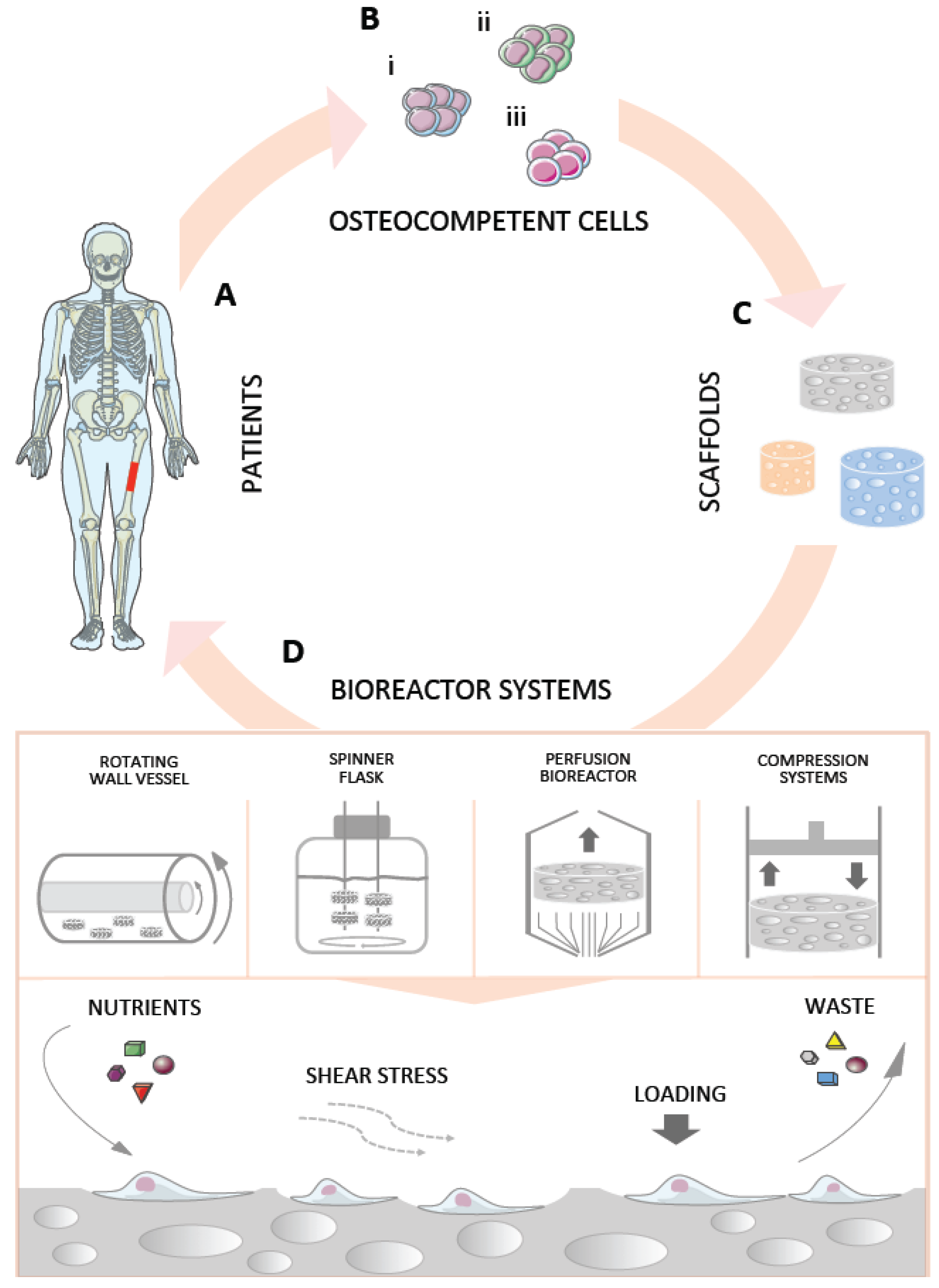
Traditional attempts to grow bone grafts in the laboratory were based on culturing cell/scaffold constructs under static conditions in the presence of osteogenesis-inducing factors. However, static cultures are not optimal to grow centimeter-sized bone grafts for clinical translation due to poor nutrient supply and removal of metabolic waste. Under these conditions, in fact, mass transport occurs only via diffusion, which is not sufficient to support cell survival and proliferation inside the core of large cell/scaffold constructs, resulting in necrosis and poor tissue formation. In addition, cell proliferation and matrix synthesis at the construct periphery over the culture period further impede medium diffusion and contribute to the formation of a nutrient gradient that drive cell migration towards the substitute borders [26]. On top of this, culture in static conditions does not allow provision of those biophysical stimuli that are critical for functional regeneration [27,28]. In fact, bone cells are sensitive to mechanical stimuli, whose integration and conversion into intracellular signals play an important role in driving bone remodeling throughout lifetime and regeneration during fracture healing [29,30]. Human bone is principally subjected to two types of mechanical stimuli, i.e., strain caused by deformation (bending and compression) resulting from physical activity (estimated values <2000 µε), and fluid shear stress resulting from interstitial fluid movement through the lacunae as a consequence of loading (estimated values 0.8–3 Pa) [31]. Conversion of physical stimuli into molecular signals and biological responses is termed mechanotransduction, which principally relies on the regulation of stretch-activated ion channels and integrin-initiated cytoskeleton deformations and organelle displacement (for a review see [32]) that ultimately triggers the initiation of a cascade of events culminating in the activation of genes involved in osteogenic pathways [31]. Based on this knowledge, it is clear that the recapitulation of these mechanisms in vitro is essential for fostering the regenerative properties of human osteocompetent cells seeded onto biomaterial scaffolds, thus enabling the formation of mature tissue substitutes for enhanced skeletal reconstructions. Advances in bioreactor systems over the last two decades have opened new possibilities in the field of bone engineering as they allow to nurture the development of bone tissue by providing an appropriate physiological environment with stimulatory biochemical and biophysical signals [33] (Figure 1). The combination of stem cells, biomaterials, osteogenesis-inducing factors and bioreactor systems has recently been defined as the “Tissue Engineering Quadriad” [34], with the culture under dynamic conditions representing a paradigm shift for the ex vivo construction of viable tissue substitutes for replacement and reconstructive therapies. Not least, the construction of viable bone substitutes using bioreactor systems opens new opportunities for the generation of valid experimental models to study bone development and pathologies, screen new drugs and test biomaterials within a context that better reflects the native tissue environment.
2. Bioreactors in Bone Tissue Engineering
Bioreactors were initially developed to allow the high-mass culture of cells used for applications in diverse areas, including fermentation, wastewater treatment and purification, food processing and drug production [35]. Many of the principles established by these applications have recently been adapted for tissue engineering purposes. A bioreactor for tissue engineering applications should: (i) facilitate uniform cell distribution; (ii) provide and maintain the physiological requirements of the cell (e.g., nutrients, oxygen, growth factors); (iii) increase mass transport both by diffusion and convection using mixing systems of culture medium; (iv) expose cells to physical stimuli; and (v) enable reproducibility, control, monitoring and automation. The ultimate design of a tissue engineering bioreactor is application specific, but basic characteristics are required when engineering tissue substitutes for potential clinical applications, such as the use of materials that do not release toxic products and can withstand numerous cycle of high temperature and pressure for repeated steam sterilization in autoclaves. Furthermore, bioreactors should present a simple design in order to prevent contamination and allow quick access to the engineered tissue if any problem arises in the system during the operational period (e.g., fluid leakage and flow obstruction). Despite the fact that several design solutions and range of stress values imparted to the cells have been explored to date, bioreactors for bone engineering applications are broadly classified in few main categories, including rotating wall vessels, spinner flasks, perfusion bioreactors and compression systems. In addition to these, combinations of different types of bioreactors have been explored in order to better mimic the bone physiological environment in vitro, such as for example compression bioreactors with added perfusion [36]. A comprehensive list of studies exploring the effects of dynamic conditions on human bone engineering and regeneration is examined in the following sections. Given the large amount of work reported, the authors apologize if not all studies hitherto published in the field have been cited in this review.
Figure 1.
Engineering bone tissue grafts using bioreactor systems. (A) Patients affected by skeletal defects (in red) resulting from congenital malformations, disease or trauma are examined for bone reconstructions. (B) Patient-specific osteocompetent cells derived from: (i) adult tissues; (ii) induced pluripotent stem cells; or (iii) blastocysts generated via somatic cell nuclear transfer are interfaced to (C) three-dimensional porous biomaterial scaffolds of different nature and architecture, and cultured under dynamic conditions in (D) bioreactors systems, which nurture the development of bone tissue by supporting efficient nutrition of cultured cells and applying mechanical stimuli that are critical for functional regeneration.

2.1. Rotating Wall Vessel Bioreactors
The rotating wall vessel bioreactor was originally designed for applications in simulated microgravity conditions [37] (Figure 2). This bioreactor is a horizontally tissue culture vessel composed of rotating concentric cylinders filled with culture medium, where media oxygenation is provided via a coaxial tubular silicon membrane. When the velocity of the rotating fluid evens out the sedimentation rate of the cell/scaffold constructs, these are maintained in a “free-fall” state and subjected to dynamic laminar flow [38]. Alternative configurations with cell/scaffold constructs attached to the rotating vessel have also been reported such as the rotating bed bioreactor. Rotating wall vessel bioreactors have largely been used to explore the effects of spaceflight conditions on cells and tissues [39], but only occasionally for tissue engineering applications, with a few examples of studies exploring their potential to support bone tissue formation using human osteocompetent cells.
Figure 2.
Rotating wall vessel. Schematic representation of a rotating wall vessel showing the outer (i) and inner (ii) cylinders, the cell/scaffold constructs (iii) and the rotator base (iv). Rotating wall vessels are systems completely filled with culture medium (without a gas-liquid interface), where medium oxygenation is provided via a silicone-rubber gas-transfer membrane. Constructs are cultured in a “free-fall” state when the velocity of the rotating fluid is equal and opposite to the sedimentation rate of the constructs.

A list of studies using rotating wall vessels and human cells is reported in Table 1. Botchwey et al. cultured SaOS-2 bone cells onto porous, three-dimensional microcarriers of degradable poly(lactic-co-glycolic acid) in a rotating wall vessel bioreactor at 25 rotation per minute (rpm) for seven days, with a maximum shear stress imparted to the cells of 3.9 dynes/cm2 (0.39 Pa), and demonstrated that cells maintained an osteoblastic phenotype and showed significant increase in alkaline phosphatase (ALP) activity and matrix mineralization compared to cells cultured under static conditions [40].
In a different study, Facer et al. cultured aggregates of a human preosteoblast cell line for 28 days, and found that dynamic culture (15 rpm) under osteogenic conditions resulted in increased cell aggregation and calcium and phosphate deposition [41].
Using bone marrow (BM)-derived hMSCs interfaced to films of gelatin-hyaluronic acid (1.5 × 1.5 mm), Wang et al. demonstrated that culture under dynamic osteogenic conditions (30 rpm) for 21 days resulted in increased cell proliferation but decreased expression of bone specific genes [42].
Another study by Diederichs et al. demonstrated that adipose tissue-derived hMSCs cultured for 47 days under osteogenic conditions onto macroporous zirconium dioxide-based ceramic discs with a diameter of 65 mm (natural or hydroxyaptatite (HA)-coated) in rotating wall vessels (1 rpm) displayed increased cell proliferation and bone matrix synthesis and mineralization, as evidenced by glucose consumption and positive staining for osteocalcein, Alizarin red and von Kossa respectively [43]. However, no significant improvements were observed on the HA-coated scaffolds, indicating a role played by the biomaterial properties in bone tissue development under the investigated conditions.
Araujo et al. cultured nano-meshes of polycaprolactone (PCL) and bicalcium phosphate-coated PCL (10 × 10 × 0.06 mm) seeded with MG63 cells for seven days under static conditions, then additional seven days at 16 rpm [44]. Culture in bioreactors did not support either cell proliferation or osteogenic differentiation.
In 2010, Ben-David et al. explored the bone formation ability of grafts engineered using rotating wall vessels in vivo [45] (Table 2). The authors cultured constructs of BM-derived hMSCs and a mixture of gelatin-based hydrogel and CaCO3/HA particles (particle diameter: 0.5–1 mm) for 21 days before subcutaneous implantation in nude mice for eight weeks. Following the implantation period new bone formation was observed in the cell/scaffold constructs, whereas no bone formation was observed in transplants containing no cells. However, the lack of control samples cultured under static conditions does not allow drawing conclusions on the effects of dynamic culture on bone formation. Taken together, the above studies demonstrate large variability in the results, and it is not clear whether this variability arises from the different cell source, scaffolds and dynamic culture regimes investigated, or the specific combination thereof.
Rotating wall vessels are limited to the cultures of small cell/scaffold constructs since they do not support optimal mass transport inside the construct core. In addition, due to the low range of values of shear stress imparted to the cells, these systems may not be efficient in promoting robust osteogenic differentiation. On the other hand, rotating wall vessels allow the concomitant culture of several cell/scaffold constructs, resulting in high volume of engineered bone tissue substitutes per vessel (Figure 3). Nevertheless, it remains to be seen whether cell/scaffold constructs cultured in rotating wall vessels exhibit improved bone regenerative capacity in vivo compared to static cultures. In that case, especially considering that cell/scaffold constructs are culture in a “free-fall” state under specific rotating conditions, these systems could be adopted to engineer thin bone substitutes for the reconstruction of flat bones or as bone patches for restorative applications of the skeletal system.

| Reference | Cell type and density | Scaffold type | Max V of constructs/bioreactor | Rotation rate | Shear stress | Culture period and media | Results (vs. static culture) |
|---|---|---|---|---|---|---|---|
| Botchwey et al. 2001 [40] | SaOS-2 (ATCC) ~222 × 103/scaffold | PLGA disc (d 4 × h 2.5 mm) | 1.116 cm3 | 25 rpm | 0.39 Pa | 7 days OS+ medium | Proliferation: ↓ Differentiation: ↑ ALP, Ca |
| Facer et al. 2005 [41] | HEPM (ATCC) 10 × 106/suspension | No scaffold | na | 15 ** rpm | na | 28 days OS− medium OS+ medium | ↑ cell aggregation; ↑ Ca and P deposition |
| Wang et al. 2009 [42] | BM-hMSCs na/scaffold | Gelatin-hyaluronic acid film (1.5 × 1.5 cm) | na | 30 rpm | na | 21 days OS+ medium | Proliferation: ↑ Differentiation: ↓ ALP, OC, COL1 |
| Diederichs et al. 2009 [43] | AT-hMSCs 14 × 106/scaffold | ZrO2 based ceramic (Sponceram®) disc natural HA-coated (d 65 × h 3 mm) | 9.96 cm3 | 1 rpm | na | 47 days OS+ medium | Only in natural scaffolds Proliferation: ↑ Differentiation: ↑ bone specific ECM; ↑ mineralization |
| Araujo et al. 2010 [44] | MG63 (ATCC) 1.5 × 10⁵/cm2 | BCP-PCL nano-mesh PCL nano-mesh (10 × 10 × 0.06 mm) | na | 16 rpm | na | 14 * days OS− medium | Proliferation: ↔ Differentiation: ↑ protein amount |
| Ben-David et al. 2010 [45] | BM-hMSCs 0.5 × 10⁶/scaffold | Gelatin-Coral/HA (ProOsteon™) particles (0.5–1 mm) | na | 7.5 rpm | na | 21 days OS+ medium | na |
Abbreviations: ↑ = increase; ↓ = decrease; ↔ = similar; ALP = alkaline phosphatase; AT-hMSCs = adipose tissue-derived hMSCs; BCP = bicalcium phosphate; BM-hMSCs = bone marrow-derived hMSCs; Ca = calcium deposition; COL1 = collagen 1; d = diameter; h = height; HA = hydroxyapatite; HEPM = human embryonic palatal mesenchymal preosteoblast cell line; hMSCs = human mesenchymal stem cells; MG63 = human osteosarcoma-derived cell line; na = not available; OC = osteocalcin; OS− = proliferative culture medium; OS+ = osteogenic culture medium; P = phosphate deposition; PCL = poly(caprolactone); PLGA = poly(lactic-co-glycolic acid); rpm = rotation per minute; SaOS-2 = human osteosarcoma cell line; V = volume; ZrO2 = zirconium dioxide; * = cells were cultured in static conditions the first half of the culture period; ** because of the increasing size of cell aggregates over time the rotational speed was later increased to maintain them in suspension.
| Reference | Cell type | Scaffold type | Bioreactor Pre-culture period | Animal model | Implantation period | Bone formation |
|---|---|---|---|---|---|---|
| Braccini et al. 2005 [46] | BM-hMSCs | HA (d 8 × h 4 mm) | Direct perfusion bioreactor 19 days | Nude mice sc | 8 weeks | Dynamic > Static 52% vs. 9.6% of available pore space |
| Karageorgiou et al. 2006 [47] | BM-hMSCs (Clonetics) | Silk loaded with BMP-2 (d 4 × h 2 mm) | Spinner flask 28 days | Mice Cranial 4 mm defect | 5 weeks | Dynamic ↔ Static 0.28 vs. 0.11 mm2 |
| Schliephake et al. 2009 [48] | hTBCs | CaCO3 (Biocoral®) (d 5 × h 3 mm) | Direct perfusion bioreactor 14 days | Nude rat im | 6 weeks | Dynamic ↔ Static 22% vs. 16% of area |
| Schliephake et al. 2009 [49] | hTBCs | CaCO3 (Biocoral®) Mineralized collagen TCP (Cerasorb®) (d 5 × h 3 mm) | Direct perfusion bioreactor 14 days | Nude rats Mandibular 5 mm defect | 6 weeks | Dynamic ↔ Static CaCO3: 7.2 vs. 8.7% of area Mineralized collagen: 10.3% vs. 12.5% of area TCP: 14.6% vs. 22.8% of area |
| Zhang et al. 2009 [50] | hfMSCs | PCL-TCP (6 × 6 × 4 mm) | Biaxial rotating bioreactor 14 days | NOD/SCID mice sc | 12 weeks | Dynamic > Static Dynamic: 3.2x fold increase |
| Ben-David et al. 2010 [45] | BM-hMSCs | Gelatin-Coral/HA (ProOsteon®) particles (0.5–1 mm) | Rotating wall vessel 21 days | Nude mouse sc | 8 weeks | Bone formation No static group |
| Janssen et al. 2010 [51] | BM-hMSCs | BCP particles (OsSatura™) (2–6 mm) | Indirect perfusion bioreactor 7; 20; 40 days | Nude mice sc | 6 weeks | Dynamic ↔ Static |
| Marolt et al. 2012 [24] | hESC-MPs | Decellularized cow bone (d 4 × h 4 mm) | Direct perfusion bioreactor 35 days | SCID-beige mice sc | 8 weeks | Dynamic > CTL 7% vs. 2% of area |
| de Peppo et al. 2013 [25] | hiPSC-MPs | Decellularized cow bone (d 4 × h 4 mm) | Direct perfusion bioreactor 35 days | SCID-beige mice sc | 12 weeks | Bone-like tissue formation No static group |
| Yeatts et al. 2014 [52] | BM-hMSCs (Lonza) | PLGA/PCL (d 3 × h 3 mm) | Indirect perfusion bioreactor 10 days | Nude rats Femoral condyle 2.5 mm defect | 3 and 6 weeks | Dynamic > Static (6 weeks only) 1.72 vs. 1.26 mm2 |
Abbreviations: > = superior; < = inferior; ↔ = similar; BCP = bicalcium phosphate; BM-hMSCs = bone marrow-derived hMSCs; BMP-2 = bone morphogenetic protein 2; CaCO3 = calcium carbonate; CTL = scaffolds seeded with cells prior implantation without pre-culturing; d = diameter; h = height; HA = hydroxyapatite; hESC-MPs = human embryonic stem cell-derived mesenchymal progenitors; hfMSCs = human fetal mesenchymal stem cells; hiPSC-MPs = human induced pluripotent stem cell-derived mesenchymal progenitors; hMSCs = human mesenchymal stem cells; hTBCs = human trabecular bone cells; im = intramuscular site; NOD/SCID = nonobese diabetic/severe combined immunodeficiency; PCL = polycaprolactone; PLGA = poly(lactic-co-glycolic acid; sc = subcutaneous site; TCP = tricalcium phosphate.
Figure 3.
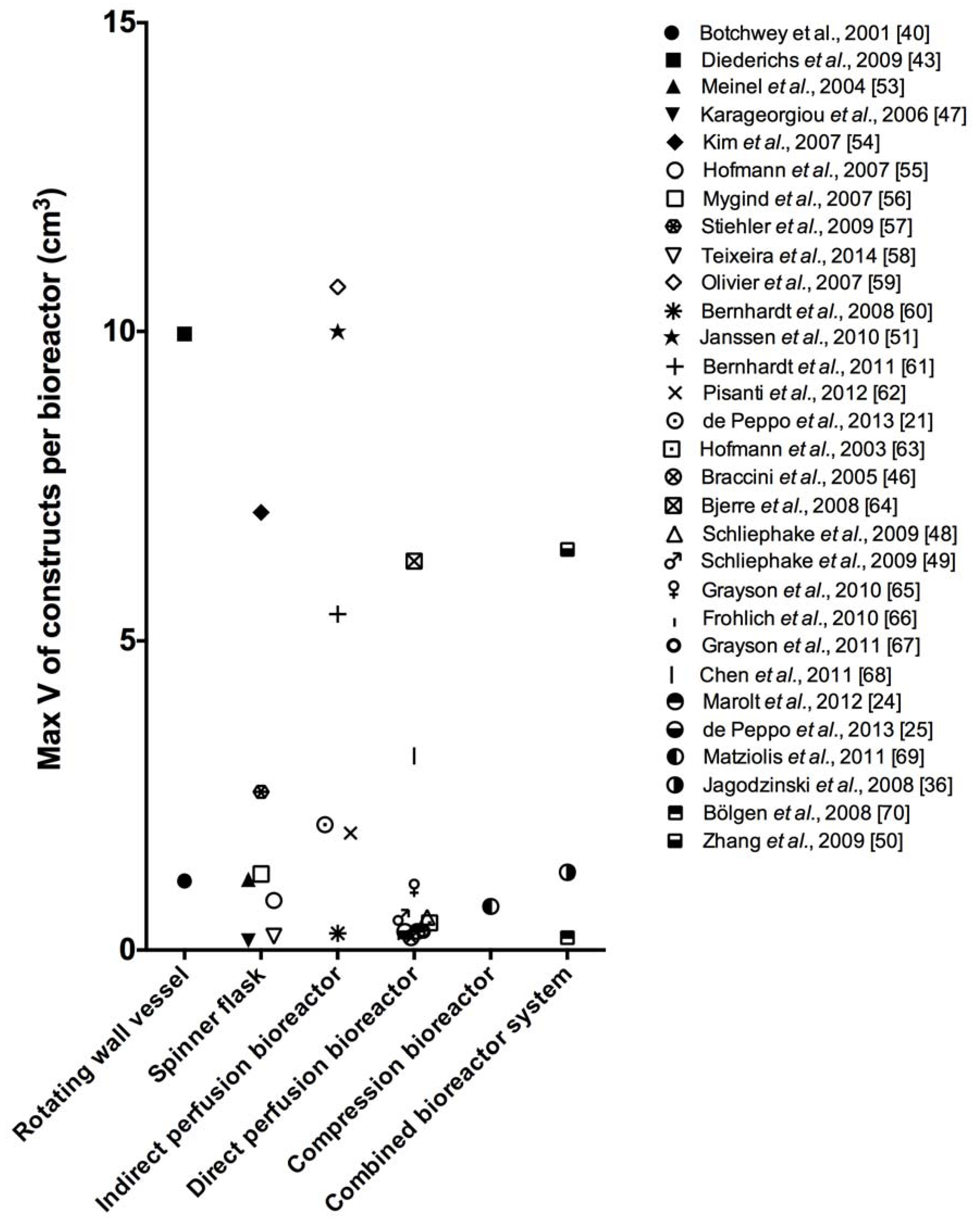
Maximal volume of engineered bone. Scatter dot plot graph showing the maximal volume of engineered bone substitutes per culture chamber for all different bioreactor systems examined in this review.
Figure 3.
Maximal volume of engineered bone. Scatter dot plot graph showing the maximal volume of engineered bone substitutes per culture chamber for all different bioreactor systems examined in this review.

2.2. Spinner Flasks
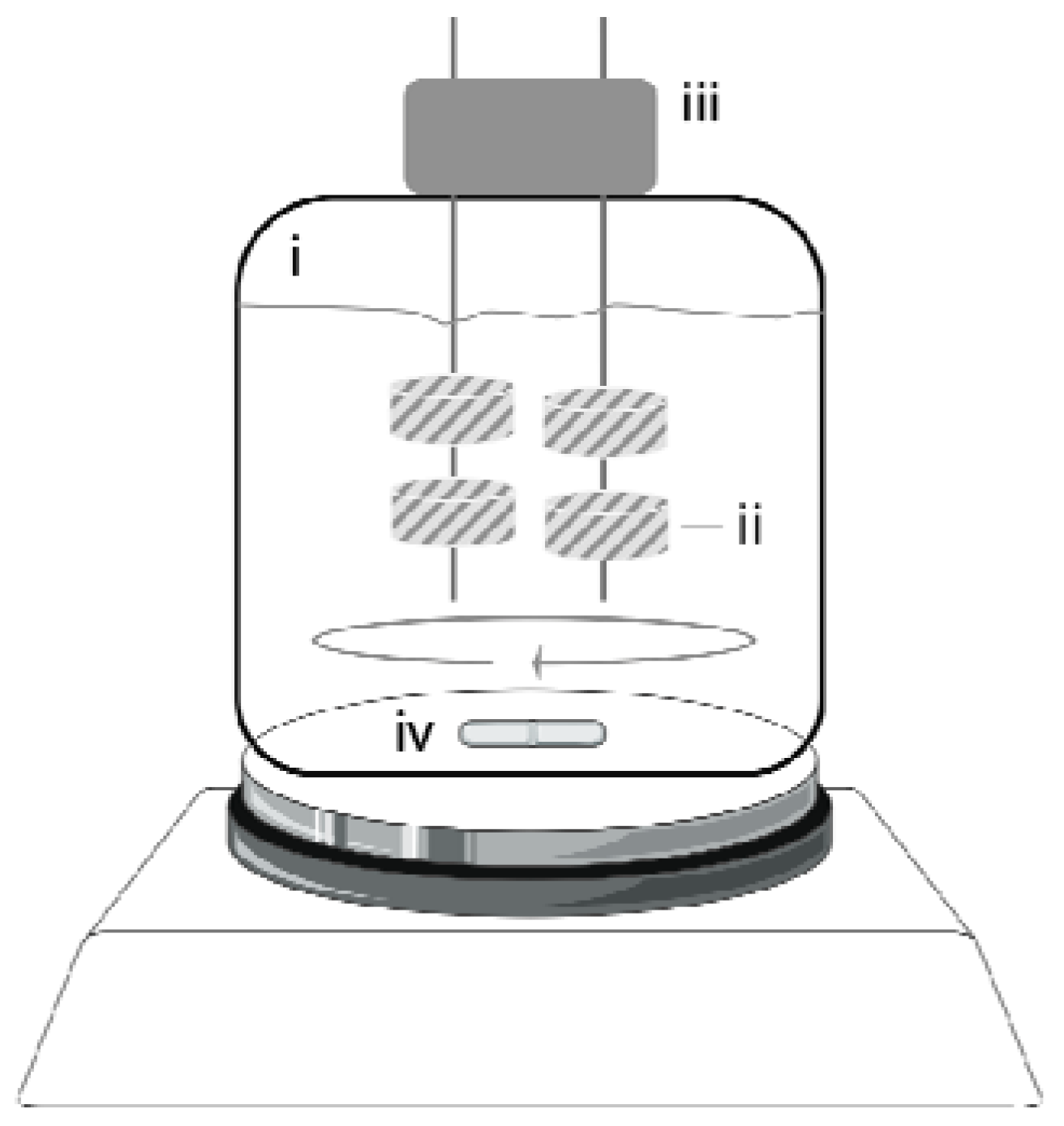
Spinner flasks are simple bioreactor systems composed of a glass or plastic vessel in which cell/scaffold constructs are attached to vertical needles hanging from the top of the vessel and immersed in the culture medium (Figure 4). The top of the vessel is usually used for gas exchange and medium oxygenation. Mixing of the medium is maintained with a stir bar at the bottom of the vessel or other mixing mechanisms. The convective forces generated during stirring mitigate the nutrient concentration gradients at the surface of the cell/scaffold constructs and produce turbulences that enhance mass transport toward the center of the samples [71]. Spinner flasks were traditionally used to support large biomass growth [72] and have recently been exploited for tissue engineering applications, including the culture and maturation of bone tissue substitutes using human osteocompetent cells derived from adult tissues.
Figure 4.
Spinner flask. Schematic representation of a spinner flask showing the glass or plastic vessel (i), the cell/scaffold constructs (ii) immersed in the culture medium and attached to vertical needles hanging from the top of the vessel (iii), and the magnetic stir bar (iv) enabling mixing of the culture medium. In these systems the top of the vessel is usually used for gas exchange and medium oxygenation.

A summary of these studies is reported in Table 3. Yasuda et al. interfaced adipose tissue-derived hMSCs to polyethylene terephthalate discs (6 mm in diameter), and cultured the cell/disc constructs at 50 rpm in osteogenic conditions for 21 days, which resulted in increased cell proliferation but decreased ALP activity compared to static conditions [73].
As opposite, Meinel et al. reported that bone marrow-derived hMSCs seeded onto collagen scaffolds (11 mm diameter and 1.5 mm thickness) and cultured in spinner flasks (50 rpm) for five weeks displayed increased ALP activity and calcium deposition compared to static cultures [53]. Interestingly, the authors observed that the orientation and distribution of deposited calcium rods where affected by the fluid dynamics, highlighting the critical role played by specific hydrodynamic conditions in the maturation of bone tissues in bioreactors.
A study by Karageorgiou et al. investigated the effects of dynamic conditions on constructs of human trabecular bone cells and BMP2-loaded silk discs (4 mm in diameter and 2 mm in thickness) in vitro after 28 days and in mouse calvaria defects after a 5-week implantation period [47] (Table 2). Although the dynamic conditions seemed to promote the formation of bone-like tissue in vitro, no significant differences in bone formation ability in vivo were observed between constructs cultured under dynamic and static conditions.
| Reference | Cell type and density | Scaffold type | Max V of constructs/bioreactor | Rotation rate | Shear stress | Culture period and media | Results (vs. static culture) |
|---|---|---|---|---|---|---|---|
| Yasuda et al. 2004 [73] | AT-hMSCs 1 × 10⁶/scaffold | PET fiber disc (d 6 mm × h na mm) | na | 50 rpm | na | 21 days OS+ medium | Proliferation: ↑ Differentiation: ↓ ALP |
| Meinel et al. 2004 [53] | BM-hMSCs (Clonetics) 5 × 10⁶/scaffold | Collagen film (Ultrafoam®) (d 11 × h 1.5 mm) | 1.144 cm3 | 50 rpm | na | 35 days OS+ medium | Differentiation: ↑ ALP, Ca |
| Karageorgiou et al. 2006 [47] | BM-hMSCs (Clonetics) 5 × 10⁶/scaffold | Silk loaded with BMP-2 (d 4 × h 2 mm) | 0.151 cm3 | 50 rpm | na | 28 days OS+ medium | na |
| Kim et al. 2007 [54] | BM-hMSCs (Cambrex) 1 × 10⁶/scaffold | Aqueous-derived silk disc (d 15 × h 5 mm) | 7.072 cm3 | 50 rpm | na | 84 days OS+ medium | Proliferation: ↑ Differentiation: ↑ ALP, Ca; ↑ ALP, BSP, COL1, OP; ↑ compressive modulus ↑ compressive strength |
| Hofmann et al. 2007 [55] | BM-hMSCs (Clonetix) 5 × 10⁶/scaffold | Silk fibroin disc (d 8 × h 2 mm) small pores (112–224 µm) large pores (400–500 µm) mixed pores | 0.804 cm3 | 60 rpm | na | 35 days OS− medium; OS+ medium | na |
| Mygind et al. 2007 [56] | BM-hMSCs-TERT 2 × 10⁶/scaffold | Coralline HA disc (ProOsteonTM) (d 10 × h 2 mm) small pores (200 µm) large pores (500 µm) | 1.232 cm3 | 30 rpm | na | 21 days OS− medium | Proliferation: P200 ↔; P500 5 Differentiation: P200 ↑ ALP ↑ ALP, COL1, RUNX2, BMP-2, BSP, Osterix, ON P500 ↔ ALP ↑ ALP, COL1, RUNX2, OP, ON |
| Stiehler et al. 2009 [57] | BM-hMSCs-TERT 2 × 10⁶/scaffold | PLGA block (8 × 8 × 5 mm) | 2.56 cm3 | 30 rpm | na | 21 days OS+ medium | Proliferation: ↔ Differentiation: ↑ ALP, Ca ↑ COL1, BMP-2, RUNX2, ON |
| Wang et al. 2009 [42] | BM-hMSCs
na/scaffold | Gelatin- hyaluronic acid film (1.5 × 1.5 mm) | na | 60 rpm | na | 21 days OS+ medium | Proliferation: ↑ Differentiation: ↑ ALP, OC, COL1 |
| Teixeira et al. 2014 [58] | hMSCs; (Lonza) 0.5 × 10⁶/scaffold | Chitosan disc (d 4 × h 3 mm) | 0.226 cm3 | 50 rpm | na | 14 days; OS− medium; OS+ medium | Proliferation: ↑ Differentiation: ↑ ALP, von Kossa |
Abbreviations: ↑ = increase; ↓ = decrease; ↔ = similar; ALP = alkaline phosphatase; AT-hMSCs = adipose tissue-derived hMSCs; BM-hMSCs = bone marrow-derived hMSCs; BMP-2 = bone morphogenetic protein 2; BSP = bone sialoprotein; Ca = calcium deposition; COL1 = collagen 1; d = diameter; h = height; HA = hydroxyapatite; hMSCs = human mesenchymal stem cells; na = not available; OC = osteocalcin; ON = osteonectin; OP = osteopontin; OS− = proliferative culture medium; OS+ = osteogenic culture medium; P = pore size (µm); PET = polyethylene terephthalate; PLGA = poly(lactic-co-glycolic acid); rpm = rotation per minute; RUNX2 = runt-related transcription factor 2; TERT = telomerase reverse transcriptase; V = volume.
Later on, Kim et al. reported the beneficial effects of dynamic culture in spinner flasks (50 rpm) on BM-derived hMSCs seeded onto large aqueous-derived macroporous silk scaffolds (15 mm diameter and 5 mm thickness) for 84 days [54]. Compared to static controls, constructs cultured under dynamic conditions exhibited enhanced cell proliferation, osteogenic differentiation and mechanical properties, as evidenced by increased expression of bone-specific genes, ALP activity, deposition of mineralized matrix and formation of organized bone-like structures. The authors attributed the good bone formation outcomes obtained for large-sized scaffolds in spinner flask systems to the large pore size (~900–100 µm) of the scaffold materials used in the study.
Independent studies by Hofmann et al. investigated the effects of dynamic conditions (60 rpm) on BM-derived hMSCs seeded onto silk fibroin discs (8 mm diameter and 5 mm thickness) with different pore size (small: 112–224 µm, large: 400–500 and mixed: 112–500 µm) for five weeks [55]. Cell differentiation in osteogenic medium resulted in increased ALP activity and calcium deposition when compared to control medium. However, the lack of control samples cultured under static conditions does not allow estimating the contribution of the dynamic conditions in supporting the formation of bone-like tissue.
In a different study, BM-derived hMSCs modified to express telomerase reverse transcriptase (hMSC-TERT) were cultured for 21 days on coralline HA scaffolds (10 mm diameter and 2 mm thickness) with 200 or 500 µm pore size in spinner flasks (30 rpm) [56]. Results showed that dynamic cultures were associated with increased proliferation, homogenous cell distribution and osteogenic differentiation. Interestingly, the results demonstrated that scaffolds with smaller pore size exhibited a faster rate of osteogenic differentiation as evidenced by increased ALP activity and expression of several osteogenic markers, while scaffolds with 500 µm pore size exhibited higher cellularity, indicating that scaffold porosity plays a role when cell/scaffold constructs are cultured under specific conditions in bioreactors. Under similar dynamic culture conditions, cells seeded onto scaffolds with smaller pore size are subjected to higher wall shear stresses [74], which could support differentiation in place of proliferation.
Independent studies using BM-derived hMSC-TERT seeded onto porous poly(lactic-co-glycolic acid) scaffolds (8 × 8 × 5 mm) and cultured in spinner flasks (30 rpm) up to 21 days showed similar results [57], characterized by a positive effect provided by the dynamic conditions on cell distribution and differentiation toward the osteogenic lineage, as evidenced by increased ALP activity and calcium deposition.
In another study, Wang et al. demonstrated that culture of BM-derived hMSCs seeded onto films of gelatin-hyaluronic acid (1.5 × 1.5 mm) under dynamic osteogenic conditions (60 rpm) for 21 days resulted in increased proliferation and expression of bone specific genes [42]. Following comparison with culture of the same constructs under dynamic conditions in rotating wall vessels, the authors concluded that the increased expression of bone specific genes resulted from the increased shear stress associated with culture in spinner flasks, although no real values of stresses imparted to the cells were reported.
Teixeira et al. recently reported the beneficial effects of dynamic culture conditions (50 rpm) on the proliferation and osteogenic differentiation of commercially available hMSCs seeded onto chitosan disks (4 mm diameter and 3 mm thickness) for 14 days [58], as evidenced by improved cell distribution within the scaffolds, and increased matrix deposition and mineralization. Taken together, these results show that the dynamic conditions provided by spinner flask systems can support osteogenic differentiation and bone-like tissue formation, although these effects are also highly dependent on operation regimes and scaffold properties.
Spinner flasks display higher ability to enhance mass transport compared to rotating wall vessels, and can be used to impart higher values of stress to the cells, although no estimations about these values have been reported in the studies examined in this review. However, these effects are still limited and concerns exist regarding their ability to support functional bone tissue maturation in large cell/scaffold constructs. On the other hand, as for rotating wall vessels, spinner flasks can accommodate a large amount of small constructs (Figure 3), and therefore allow the culture of clinically relevant volumes of engineered tissues for the reconstructions of segmental skeletal defects. In addition, like for rotating wall vessels, spinner flasks could be adopted to engineer thin bone substitutes for the reconstruction of flat bones or as bone patches for restorative applications. Again, the low amount of studies reported, as well as the lack of control groups cultured under static conditions, does not allow drawing definite conclusions regarding the real potential of spinner flask-engineered substitutes in supporting bone tissue formation in vivo.
2.3. Perfusion Bioreactors
Perfusion bioreactors for bone engineering applications are culture systems composed of several key elements, including one or more perfusion chambers where the cell/scaffold constructs are placed, a medium reservoir, a tubing circuit and a pump enabling mass transport of nutrients and oxygen throughout the perfusion chamber (Figure 5). Perfusion bioreactors are broadly classified into indirect or direct systems, depending on whether the culture medium is perfused around or throughout the cell/scaffold constructs.
Figure 5.
Perfusion bioreactors. Schematic representation of an indirect (A) and a direct (B) perfusion bioreactor showing the culture chambers (i), the cell/scaffold constructs (ii), the culture medium reservoirs (iii), the peristaltic pumps (iv) and the tubing systems (v). In indirect perfusion bioreactors the culture medium follows the path of less resistance around the constructs. In direct perfusion bioreactors the cell/scaffold constructs are placed in a press-fitted fashion in the culture chamber and the medium is perfused throughout the constructs.

2.3.1. Indirect Perfusion Bioreactors
In this type of perfusion bioreactors the cell/scaffold constructs are loosely placed in the perfusion chamber, and the culture medium preferentially follows the path of least resistance around the constructs (Figure 5A), resulting in reduced mass transfer throughout the core of the samples. Therefore, the convective forces generated by the perfusion pump mitigate the nutrient concentration gradients principally at the surface of the cell/scaffold constructs, thus limiting the size of bone substitutes that can be engineered using these systems. On the other hand, indirect perfusion bioreactors may represent valuable systems for the collective culture of a large number of small particulate cell/scaffold constructs [21,75] that could be then assembled to repair large and geometrically complex skeletal defects.
A list of works using indirect perfusion bioreactors is summarized in Table 4. Using indirect perfusion bioreactors, human MG63 cells were cultured onto beta tricalcium phosphate cylinders (14 mm diameter and 33 mm length), with 65% porosity and 500–630 μm range of pore size, in bioreactors under convergent or divergent perfusion (flow rate: 3 mL/min) for up to 28 days [59]. Compared to controls, perfusion cultures resulted in increased cell proliferation and distribution as evidenced by glucose consumption, DNA content and new tissue formation. Interestingly, the authors observed better outcomes when constructs were cultured under convergent perfusion, suggesting that operation configurations are crucial for optimal bone tissue regeneration.
In another study, Bernhardt et al. showed that cultures of BM-derived hMSCs under dynamic conditions (flow rate: 1 mL/min) for 35 days on tapes composed of collagen type I with nanocrystalline HA (12 mm diameter) did not result in increased proliferation and osteogenic differentiation compared to static conditions. Same findings were observed regardless the cell/scaffold constructs where cultured in presence or absence of osteogenic supplements [60].
In 2010, Janssen et al. published a study reporting the use of BM-derived hMSCs for the production of clinically relevant amount of tissue-engineered bone substitutes [51] (Table 2). Cells were seeded onto macroporous biphasic calcium phosphate particles (2–6 mm diameter) and constructs cultured in a large-volume perfusion bioreactor system for 20 days with a medium recirculation speed of 4 mL/min. Following the experimental period, the particles were covered with a homogeneous layer of viable cells and interconnected. However, no significant differences were found in the expression of bone-specific markers and bone formation in vivo compared to control groups cultured under static conditions.
In 2011, Bernhardt et al. showed that dynamic culture conditions (flow rate: 1.5 mL/min) had an effect on proliferation and osteoblastic differentiation (evidenced by ALP activity) of BM-derived hMSCs seeded onto βTCP scaffolds (12 mm diameter and 6 mm thickness) [61]. However, the observed effects were dependent on the serum concentration in the medium, suggesting a synergistic effect of chemical and physical stimulation on cell response.
| Reference | Cell type and density | Scaffold type | Max V of constructs/bioreactor | Perfusion rate | Shear stress | Culture period and media | Results (vs. static culture) |
|---|---|---|---|---|---|---|---|
| Olivier et al. 2007 [59] | MG63 10 × 106/scaffold | ßTCP cylinder (d 14 mm × h 33 mm) | 10.721 cm3 | 3 mL/min
convergent and divergent flow set-up | na | 28 days OS− medium | Proliferation: ↑; Homogeneous cell distribution: Convergent > divergent > static |
| Bernhardt et al. 2008 [60] | BM-hMSCs 0.015 × 10⁶/scaffold | COL1-nanocrystalline HA tape (d 12 × h 0.2 mm) | 0.271 cm3 | 1 mL/min | na | 35 days OS− medium OS+ medium | Proliferation: ↔ OS−; ↔ Os+; Differentiation: ↔ ALP in OS−; ↓ ALP in Os+ |
| Janssen et al. 2010 [51] | BM-hMSCs (1–12) × 106/all scaffolds | BCP particles (OsSatura™) (2–6 mm) | 10 cm3 | 4 mL/min | na | 20 days OS+ medium | Differentiation: ↔ ALP, CBFA1, COL1, OC, ON, S100A4, BMP-2 |
| Bernhardt et al. 2011 [61] | BM-hMSCs 0.2 × 10⁶/scaffold | βTCP (Cerasorb®) (d 12 × h 6 mm)
750 and 1400 µm pore size | 5.43 cm3 | 1.5 mL/min | na | 21 days OS+ medium with 10% serum or 2% serum | Proliferation: ↑ OS+ medium with 10% serum; ↔ OS+ medium with 2% serum; Differentiation: ↔ OS+ medium with 10% serum; ↑ OS+ medium with 2% serum |
| Pisanti et al. 2012 [62] | BM-hMSCs (Lonza) 0.12 × 10⁶/scaffold | PLLA disc (d 4 × h 5 mm)
100, 250, and 500 µm pore size | 1.89 cm3 | 0.3 mL/min | na | 24 days Os+ medium | Differentiation: ↑ ALP, BMP-2 |
| de Peppo et al. 2013 [21] | BM-hMSCs hESC-MPs 0.1 × 10⁶/scaffold | CaCO3 cube (Biocoral®) (3 mm) | 2.025 cm3 | 10 mL/min | 0.001 Pa | 35 days OS+ medium | Proliferation: ↑; hESC-MPs > BM-hMSCs; Differentiation: ↑ RUNX2, COL1, ALP, ON, OP; hESC-MPs > BM-hMSCs |
| Yeatts et al. 2014 [52] | BM-hMSCs (Lonza) 0.25 × 10⁶/scaffold | PLGA-PCL (d 3 × h 3 mm) | na | 1 mL/min | na | 10 days OS+ medium | na |
Abbreviations: ↑ = increase; ↓ = decrease; ↔ = similar; ALP = alkaline phosphatase; BCP = bicalcium phosphate; BM-hMSCs = bone marrow-derived hMSCs; BMP-2 = bone morphogenetic protein; ßTCP = beta tricalcium phosphate; CaCO3 = calcium carbonate; CBFA1 = core-binding factor subunit alpha-1; COL1 = collagen 1; convergent = flow direction where culture medium infiltrates the scaffold from scaffold’s exterior; d = diameter; divergent = flow direction where culture medium infiltrates the scaffold from scaffold’s central inlet; h = height; HA = hydroxyapatite; hESC-MPs = human embryonic stem cell-derived mesenchymal progenitors; hMSCs = human mesenchymal stem cells; MG63 = human osteosarcoma-derived cell line; na = not applicable; OC = osteocalcin; ON = osteonectin; OP = osteopontin; OS− = proliferative culture medium; OS+ = osteogenic culture medium; PCL = poly(caprolactone); PLGA = poly(lactic-co-glycolic acid); PLLA = poly-l-lactic acid; RUNX2 = runt-related transcription factor 2; S100A4 = negative regulator of mineralization; V = volume.
In another study, Pisanti et al. explored the influence of scaffold microstructure and dynamic culture in perfusion bioreactors (flow rate: 0.3 mL/min) on commercially available hMSCs [62]. Cells were seeded onto poly-l-lactic acid scaffolds with average pore size of 100, 250, and 500 µm and cultured under dynamic or static conditions for up to 24 days. Results indicated that proliferation and osteogenic differentiation were enhanced when cells where cultured under dynamic conditions in the 250 µm and 500 µm pore size scaffolds, indicating a synergistic effect of scaffold microstructure and culture in bioreactors on hMSC behavior.
A recent study by de Peppo et al. demonstrated that culture of BM-derived hMSCs and hESC-derived mesenchymal progenitors seeded onto biocoral scaffolds (3 × 3 × 3 mm) in a packed bed/column bioreactor (flow rate: 10 mL/min; average shear stress estimated to be 0.001 Pa) over a period of five weeks resulted in increased cell proliferation as shown by increased DNA and protein content, and improved osteogenesis as evidenced by increased gene expression, tissue formation and matrix deposition and mineralization [21]. Interestingly, the authors found that the beneficial effects of dynamic culture were significantly higher for constructs of hESC-derived mesenchymal progenitors, suggesting that different operation regimes may result optimal when cells of different origin and developmental stage are used for bone engineering applications using bioreactors.
In a different study, Yeatts et al. interfaced BM-derived hMSC with poly(lactic-co-glycolic acid)/PCL cylinders (3 mm diameter and 3 mm height), and cultured the constructs under dynamic conditions for 10 days before implantation in femoral condyle defects in nude rats [52] (Table 2). Results showed that dynamic cultures of these constructs resulted in increased bone formation compared to static culture after six weeks in vivo, highlighting the positive effects of the perfusion culture for bone engineering applications.
Using a version of a perfusion bioreactor combined with a biaxial rotating system, Zhang et al. similarly reported the beneficial effects of dynamic conditions (5 biaxial rpm; flow perfusion: 3.8 mL/min) on human fetal MSCs seeded onto PCL/tricalcium phospahate blocks (6 × 6 × 4 mm) both in vitro, as evidenced by increased proliferation and calcium deposition, and in subcutaneous pockets in mice [50] (Table 2), resulting in 3.2 fold increase of ectopic bone formation compared to statically cultured constructs.
As yet, indirect perfusion bioreactors have allowed engineering the highest volume of bone substitutes per culture chamber (Figure 3), therefore representing valuable systems for the construction of large amount of bone substitutes for extensive skeletal reconstructions. However, although indirect flow perfusion bioreactors have shown the potential to support maturation of bone substitutes both in vitro and in vivo, outcomes seems to depend on experimental protocol, type of cells and scaffolds used, and bioreactor configuration and operation regimes. Additionally studies are therefore recommended to better explore the potential of these systems for the culture of bone substitutes that can be used to treat large skeletal defects.
2.3.2. Direct Perfusion Bioreactors
In direct perfusion bioreactors the cell/scaffold constructs are placed in the perfusion chamber in a press-fit fashion so that the culture medium is forced to pass through the center of the samples (Figure 5B). In view of this advantage, direct perfusion bioreactors have extensively been used to engineer bone substitutes using a combination of different human osteocompetent cells and biomaterial scaffolds, as summarized in Table 5.
| Reference | Cell type and density | Scaffold type | Max V of constructs/bioreactor | Perfusion rate | Shear stress | Culture period and media | Results (vs. static culture) |
|---|---|---|---|---|---|---|---|
| Hofmann et al. 2003 [63] | Human osteoblasts 12,500 cells/cm2 | Cancelous human bone HA (Endobon®) (d 7.5 × h 10 mm) | 0.442 cm3 | 1 mL/min | na | 10 days OS− medium | na |
| Braccini et al. 2005 [46] | hBMNCs; ~18 × 106/scaffold | HA (Engipore®) (d 8 × h 4 mm) | 0.201 cm3 | 100 μm/s | na | 19 days; OS+ medium | na |
| Bjerre et al. 2008 [64] | BM-hMSC-TERT 2 × 10⁶/scaffold | 67% Si-TCP/33% HA/ßTCP (SkeliteTM) (d 10 × h 5 mm) | 6.288 cm3 | 0.1 mL/min | na | 21 days OS− medium | Proliferation: ↑; Differentiation: ↑ ALP; ↑ OP, BSP, BMP-2 |
| Schliephake et al. 2009 [48] | hTBCs 5 × 10⁶ cells/cm3 | CaCO3 (Biocoral®) (d 5 × h 3 mm) | 0.530 cm3 | na | na | 14 days OS− medium | na |
| Schliephake et al. 2009 [49] | hTBCs 5 × 10⁶ cells/cm3 | CaCO3 (Biocoral®) Mineralized collagen TCP (Cerasorb®) (d 5 × h 3 mm) | 0.530 cm3 | na | na | 14 days OS− medium | na |
| Grayson et al. 2010 [65] | BM-hMSCs (Cambrex) 3.4 × 10⁶/scaffold | Decellularized cow bone hTMJ-shaped (~15 × 15 × 5 mm) | ~1 cm3 | 1.8 mL/min | na | 35 days OS+ medium | Proliferation: ↑; Differentiation: ↑ bone volume |
| Fröhlich et al. 2010 [66] | AT-hMSCs 1.5 × 10⁶/scaffold | Decellularized cow bone plugs (d 4 × h 4 mm) | 0.302 cm3 | 1.8 mL/min | ~0.01 Pa | 35 days OS− medium; OS+ medium | Proliferation: ↔ OS− ↔ OS+; Differentiation: ↑ bone specific ECM |
| Grayson et al. 2011 [67] | BM-hMSCs (Cambrex) 1.2 × 10⁶/scaffold | Decellularized cow bone plugs (d 4 × h 4 mm) | 0.302 cm3 | 80, 400, 800, 1200 or 1800 µm/s | 0.0006–0.02 Pa | 35 days OS+ medium | na |
| Chen et al. 2011 [68] | AM-hMSCs 5 × 10⁴ cells/mg microcarriers | Porcine gelatin microcarrier (CultiSpher S) (d 20 × h 10 mm) | 3.142 cm3 | 2 mL/min | na | 28 + 7 days; OS− medium; OS+ medium | na |
| Marolt et al. 2012 [24] | hESC-MPs 1.5 × 10⁶/scaffold | Decellularized cow bone plugs (d 4 × h 4 mm) | 0.302 cm3 | 3.6 mL/min | na | 35 days OS+ medium | Proliferation: ↑; Differentiation: ↑ ALP, OP; ↑ bone specific ECM |
| de Peppo et al. 2013 [25] | hiPSC-MPs 1.5 × 10⁶/scaffold | Decellularized cow bone plugs (d 4 × h 4 mm) | 0.302 cm3 | 3.6 mL/min | na | 35 days OS+ medium | Differentiation: ↑ OP; ↑ bone specific ECM |
Abbreviations: ↑ = increase; ↓ = decrease; ↔ = similar; ALP = alkaline phosphatase; AM-hMSCs = amniotic membrane hMSCs; AT-hMSCs = adipose tissue-derived hMSCs; BM-hMSCs = bone marrow-derived hMSCs; BMP-2 = bone morphogenetic protein 2; BSP = bone sialoprotein; ßTCP = beta tricalcium phosphate; CaCO3 = calcium carbonate; d = diameter; ECM = extracellular matrix; h = height; HA = hydroxyapatite; hBMNCs = human bone marrow nuclear cells; hESC-MPs = human embryonic stem cell-derived mesenchymal progenitors; hiPSC-MPs = human induced pluripotent stem cell-derived mesenchymal progenitors; hMSCs = human mesenchymal stem cells; hTBCs = human trabecular bone cells; hTMJ = human temporomandibular joint; na = not available; OP = osteopontin; OS− = proliferative culture medium; OS+ = osteogenic culture medium; Si-TCP = silicate-substituted tricalcium phosphate; TCP = tricalcium phosphate; TERT = telomerase reverse transcriptase; V = volume.
A relatively large amount of these studies have also explored the bone formation potential of these substitutes in vivo using ectopic and orthotopic animal models (Table 2). In a study by Hofmann et al., human osteoblasts were cultured in a perfusion bioreactor (flow rate: 1 mL/h) for ten days after seeding onto demineralized and non-demineralized human spongiosa, and HA cylinder scaffolds (7.5 mm diameter and 10 mm height) [63]. The results demonstrated significant increased expression of bone-specific genes when cells where seeded onto demineralized matrices compared to the other scaffolds. However, the lack of static groups does not allow evaluating the real effects of dynamic conditions on cell proliferation and osteogenic differentiation.
In another study, Braccini et al. seeded BM-derived hMSCs onto HA scaffolds (8 mm diameter and 4 mm thickness) and cultured the constructs under static or dynamic conditions (100 µm/s) before subcutaneous implantation in nude mice for eight weeks. Results demonstrated a superior ability of the constructs cultured in perfusion bioreactors in promoting bone formation in vivo [46] (Table 2).
Bjerre and colleagues later demonstrated that dynamic cultures (flow rate: 0.1 mL/min) of BM-derived hMSC-TERT seeded onto silicate-substituted tricalcium phosphate scaffolds (10 mm diameter and 5 mm thickness) for 21 days were associated with higher construct cellularity, improved cell distribution and increased osteogenic differentiation as evidenced by ALP and expression of other bone-specific markers [64].
In 2009, Schliephake et al. cultured constructs of human trabecular bone cells and CaCO3 scaffolds (pore size: 250–750 µm) or mineralized collagen scaffolds (pore size: 100–200 µm) in vitro for 14 days, and then implanted the constructs in intramuscular pockets or 5-mm mandibular defects in nude rats for six weeks [48,49]. As opposite to the study by Braccini et al., the authors did not found significant differences in bone formation between the constructs cultured under static or dynamic conditions, suggesting that other factors couls play a role in enhancing the bone formation ability of these constructs.
Using a biomimetic approach of bone engineering, Grayson et al. demonstrated the effect of perfusion rate on BM-derived hMSCs seeded onto decellularized bone plugs (4 mm diameter and 4 mm height) during a five-week perfusion culture [67]. The authors found that increasing the flow velocity of perfusion medium from 80 to 1800 μm/s (corresponding to estimated initial shear stresses ranging from 0.0006 to 0.02 Pa) significantly affected cell morphology, cell-cell interactions, matrix production and composition, and the expression of osteogenic genes and that intermediate flow velocities (400 to 800 μm/s) yielded the best osteogenic outcomes. As proof of principle for potential clinical translation to treat complex skeletal defects, they also reported the ability to engineer anatomically-shaped human bone substitutes using a combination of decellularized bone scaffolds (in the shape of a temporomandibular joint condylar bone), BM-derived hMSCs and a customized direct perfusion bioreactor [65]. In a different study, the same group reported that an interstitial velocity of 400 µm/s (corresponding to shear stress values of the order of 0.01 Pa) imparted to adipose tissue-derived hMSCs seeded onto decellularized bone plugs (4 mm diameter and 4 mm height) resulted in more uniform distributions of cells and bone matrix (collagen, bone sialoprotein, and bone osteopontin) compared to control groups cultured under static conditions. The presence of osteogenic supplements significantly increased construct cellularity, and the perfusion markedly improved the amounts and distributions of cells and bone matrix [66].
Using the same biomimetic approach of bone development, Marolt and de Peppo recently demonstrated that viable bone substitutes could be engineered from hESC- [24] and hiPSC-derived mesenchymal progenitors [25] respectively. Culture in bioreactors resulted in common alterations in the expression of several important genes involved in molecular pathways controlling cell proliferation and tissue maturation, indicating a strong phenotypic transition toward the osteogenic lineage driven by the investigated conditions. Interestingly, both studies demonstrated phenotypic stability and further bone tissue maturation following ectopic implantation of engineered bone substitutes in nude mice, opening the possibility to engineer unlimited amounts of functional bone substitutes for personalized clinical applications.
A new strategy of bone engineering was reported by Chen et al. using a two-stage culture protocol [68]. First, amniotic hMSCs were cultured on porcine gelatin microcarriers in a spinner flask for 28 days (3 min agitation at 30 rpm followed by 30 min in static conditions). During this cultivation process, cell-laden microcarriers aggregated into 700–800 µm clusters that displayed increased expression of bone markers and matrix deposition over time, as evidenced by alizarin red staining and calcium quantification. In the second step, microtissues were transferred into cylindrical perfusion culture chamber (2 cm in diameter) and assembled through applying pulsatile flow (flow rate: 2 mL/min) of osteogenic medium for seven days, resulting into a centimeter-sized construct (2 cm diameter and 1 cm height), which displayed high cell viability, homogenous cell distribution and deposition of bone-specific extracellular matrix.
Altogether, these studies demonstrate that direct perfusion bioreactors, irrespective of the specific cell source and properties of the biomaterial scaffolds used, display high potential in supporting tissue maturation and bone formation both in vitro and in vivo, suggesting they could represent suitable culture systems for the construction of viable bone substitutes for future applications in clinical settings. The press-fit setup that characterizes the direct perfusion systems on one hand allows the production of anatomically shaped bone substitutes but on the other limits the size of the cell/scaffold constructs that can be cultured using these systems due to the resistance built in the systems upon tissue maturation (Figure 3), and it is therefore crucial to develop innovative engineering solutions to account for this limitation. Again, a lot of work remains to be done before optimal protocols of dynamic culture can be developed for the reproducible production of bone tissue substitutes for effective reconstruction of the skeletal system using direct perfusion systems.
2.4. Compression Bioreactors and Combined Systems
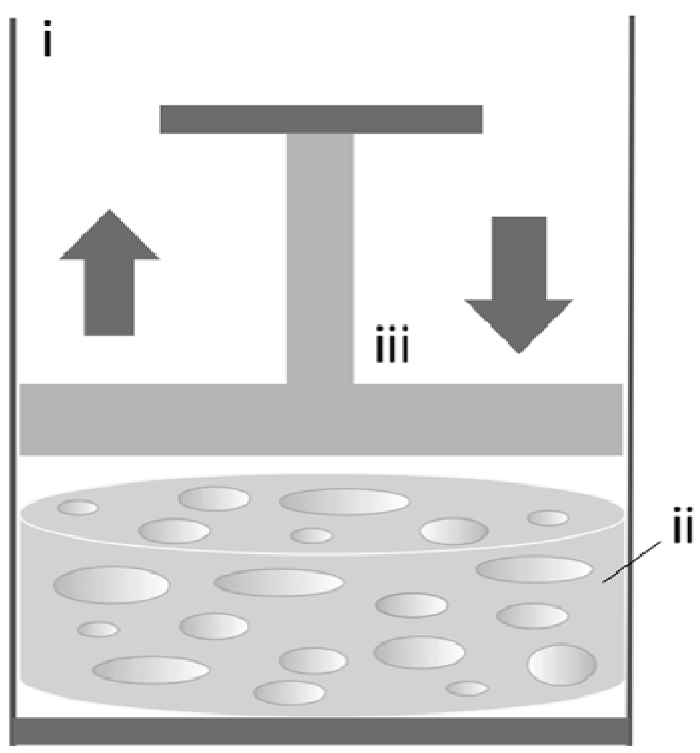
Compression bioreactors were intended to mimic the bone physiological environment in vitro, characterized by repeated mechanical stimulation required for functional bone regeneration. These culture systems consist of a motor, a system providing linear motion and a compression chamber in which one or more pistons apply static or dynamic compressive loads directly to the cell/scaffold constructs (Figure 6).
In a study by Matziolis et al., BM-derived hMSCs were cultured on human cancellous bone-fibrin composites (15 mm diameter and 4 mm thickness) in a compression bioreactor, and the effect of short term mechanical stimulation (24-hour cyclic loading of 4 kPa, 25% strain at 0.05 Hz) on osteogenic differentiation was investigated [69] (Table 6). Short-term mechanical stimulation enhanced the expression of several genes encoding for factors involved in osteogenesis, including RUNX2, osteopontin, integrin-β1, TGFßR1, SMAD5, annexin-V and PDGFα. These results demonstrate that even short mechanical stimuli can be sufficient to activate the osteogenic differentiation pathways in hMSCs.
Figure 6.
Compression bioreactor. Schematic representation of a compression bioreactor showing the compression chamber (i), the cell/scaffold construct (ii) and the piston (iii), which applies static or dynamic compressive loads directly to the constructs. In these systems the compression is controlled by a motor and a system that provides linear motion.

| Reference | Cell type and density | Scaffold type | Max V of constructs/bioreactor | Compression rate | Culture period and media | Results (vs. static culture) |
|---|---|---|---|---|---|---|
| Matziolis et al. 2011 [69] | BM-hMSCs 1 × 10⁶/scaffold | Human cancellous bone-fibrin composite (d 15 × h 4 mm) | 0.707 cm3 | 4 kPa 25% strain 0.05 Hz | 24 h OS−medium | ↑ OP, integrin-ß-1, TGFßR1, SMAD5, PDGFα, annexin-V |
Abbreviations: ↑ = increase; ↓ = decrease; ↔ = similar; BM-hMSCs = bone marrow-derived hMSCs; d = diameter; h = height; hMSCs = human mesenchymal stem cells; OP = osteopontin; PDGFα = platelet-derived growth factor alpha; TGFßR1 = transforming growth factor beta-receptor type 1; V = volume.
In order to enhance mass transport and allow long-term culture of large cell/scaffold constructs, combined configurations of compression systems coupled with perfusion loops have been developed, as summarized in Table 7.
Jagodzinski et al. reported a study aimed at investigating the effect of mechanical stimuli on BM-derived hMSCs. Cells were seeded on bovine spongiosa discs (20 mm diameter and 4 mm thickness) and incubated in a bioreactor system under either perfusion (flow rate: 10 mL/min) or a combination of perfusion and cyclic loading (10% cyclic compression at 0.5 Hz) for three weeks [36]. Both conditions demonstrated enhanced proliferation compared to static cultures. However, hMSCs stimulated by cyclic compression clearly demonstrated an enhanced secretion of osteocalcin along the entire experimental period.
| Reference | Mechanical stimulation | Cell type and density | Scaffold type | Max V of constructs/bioreactor | Rotation rate | Perfusion rate | Compression rate | Culture period and media | Results (vs. static culture) |
|---|---|---|---|---|---|---|---|---|---|
| Jagodzinski et al. 2008 [36] | Perfusion | BM-hMSCs 1 × 10⁶/scaffold | Decellularized cow bone (Tutobone®) (d 20 × h 4 mm) | 1.257 cm3 | Not done | 10 mL/min | Not done | 21 days OS+ medium | Proliferation: ↑ Differentiation: ↔ OC |
| Compression-perfusion | Not done | 10 mL/min | 10% strain, 0.5 Hz | Proliferation: ↑ Differentiation: ↑ OC; also > to perfusion only | |||||
| Bölgen et al. 2008 [70] | Compression | MG63 (ATTC) 1 × 10⁶/scaffold | HEMA–lactate–dextran (d 8 × h 4 mm) | 0.201 cm3 | Not done | 0.1 mL/min | Not done | 10 days OS− medium | Differentiation: ↔ ALP |
| Compression-perfusion | Not done | 0.1 mL/min | 1.5% strain, 1 Hz, 1 h/day | Differentiation: ↑ ALP | |||||
| Zhang et al. 2009 [50] | Perfusion-rotation | hfMSCs 0.5 × 10⁶/scaffold | PCL-TCP (6 × 6 × 4 mm) | 6.48 cm3 | 5 rpm | 3.8 mL/min | Not done | 28 days OS+ medium | Proliferation: ↑ Differentiation: ↑ ALP, Ca |
| Liu et al. 2012 [76] | Perfusion | BM-hMSCs 6 × 10⁶/scaffold | PU-BDI shape of human meniscus (size na) | na | Not done | 10 mL/min | Not done | 14 days na medium | Proliferation: ↑ Differentiation: ↔ ALP; ↔ mechanical strength |
| Compression-perfusion | Not done | 10 mL/min | 10% strain, 0.5 Hz, 1 time/day, 8 h/time | Proliferation: ↔; Differentiation: ↔ ALP; ↔ mechanical strength | |||||
| Not done | 10 mL/min | 10% strain, 0.5 Hz, 4 times/day, 2 h/time, 4 h of rest | Proliferation: ↑; also > to perfusion only; Differentiation: ↑ ALP; ↑ mechanical strength` |
Abbreviations: ↑ = increase; ↓ = decrease; ↔ = similar; ALP = alkaline phosphatase; BM-hMSCs = bone marrow-derived hMSCs; Ca = calcium deposition; d = diameter; h = height; HEMA = 2-hydroxyethyl methacrylate; hfMSCs = human fetal mesenchymal stem cells; hMSCs = human mesenchymal stem cells; MG63 = human osteosarcoma-derived cell line; na = not available; OC = osteocalcin; OS− = proliferative culture medium; OS+ = osteogenic culture medium; PCL = poly(caprolactone); PU-BDI = polyurethane-based 1,4-butanediisocyanate; rpm = rotation per minute; TCP = tricalcium phosphate; V = volume.
Similarly, Bölgen et al. cultured a human osteoblast-like cell line seeded on biodegradable cryogels made of L-lactide and dextran with 2-hydroxyethyl methacrylate end groups (8 mm diameter and 4 mm thickness) in compression bioreactor (1.5% strain level at 1 Hz for 1 h/day) with or without perfusion (flow rate: 0.1 mL/min) for two weeks [70]. Culture in both conditions resulted in increased expression of markers of osteoblastic differentiation and deposition of bone extracellular matrix compared to control. However, as above, combination of compression and perfusion appeared to be more beneficial in supporting osteogenic differentiation as evidenced by increased ALP activity.
In a different study, Liu et al. cultured constructs of BM-derived hMSCs and meniscal-shaped polyurethane-based 1,4-butanediisocyanate scaffolds under either perfusion (flow rate: 10 mL/min) or a combination of perfusion and compression for 14 days [76]. Among the different conditions, only the combination of perfusion and cyclic loading of 2-hour compression followed by 4-hour rest resulted in increased proliferation and bone-like tissue maturation compared to static cultures, as evidenced by increased ALP activity and mechanical strength.
In conclusion, these studies provide evidence that mechanical loading, especially when combined with flow perfusion, can play a pivotal role in promoting survival and functional ostegenic differentiation of the cells within the cultured constructs. However, no in vivo studies have been reported so far and much work remains to be done to assess the potential of these systems for clinically relevant bone engineering applications.
3. Simulation Techniques for Improved Bioreactor Design
Traditionally bioreactor designs and operation regimes were chosen by trial-and-error approaches, which are time consuming and often result in suboptimal performance and poor reproducibility [77]. To overcome such limitations and allow future translation of bone-engineered products, over the last years mathematical models and computer simulation techniques have been applied to an increasing extent to bioreactor systems in order to determine a priori the most favorable parameters for functional tissue regeneration from a set of available alternatives (for a review see [78]). Simulations in tissue engineering are usually divided into simulation of the biophysical environments (distribution of all physical forces) [71,79] or biological environments (including dynamics of nutrient transport, tissue growth, matrix deposition and morphological evolution) [80], or both environments [81].
As opposite to cumbersome optical measuring techniques, such as for examples laser Doppler anemometry, particle image velocimetry and planar laser-induced fluorescence, computational fluid dynamics (CFD) softwares are now extensively used to provide simulations of the hydrodynamic environment of bioreactor systems and the factors influencing it. Distribution of mechanical forces, nutrient consumption and kinetics of tissue growth highly depend on mass transport, scaffold architecture and properties, as well as bioreactor design and operation regimes. In this view, given specific models of scaffolds filled with biological material, which can be developed using finite element methods, CFD simulations can be used to give recommendations about key elements of bioreactor design including shape, inlet and outlet location and size, and operation regimes, with the aim of providing adequate hydrodynamic environments supporting optimal bone tissue regeneration.
However, in the case of compression bioreactors, the dynamic loading of cell/scaffold constructs results in cyclic deformation of the scaffold and cellular material, which in turn affects the hydrodynamic behavior of the system, the distribution and intensity of the physical forces throughout the constructs over time, and the cellular response. Therefore, rigorous models must be developed using different numerical methods to specifically account for these fluid-structure interactions [82], and better predict formation and differentiation of tissues under specific loading conditions, which could also help in the development of more appropriate scaffold and bioreactor designs. The application of CFD simulations therefore plays an important role in the design of perfusion bioreactor configurations to engineer bone substitutes of clinical relevance, therefore reducing time and cost of product development for therapeutic translation, especially in clinical situations where customized bioreactors must be developed for functional regeneration of anatomically-shaped bone substitutes.
4. Future Directions for Clinical Translation
As discussed in previous sections, bioreactor systems have shown great utility in supporting de novo growth of bone tissue substitutes in vitro, with the perfusion bioreactors representing the optimal systems to support cell viability, increase construct cellularity and deposition of bone matrix, and guide maturation of functional tissue.
Nevertheless, despite these promising results, no examples of successful clinical applications of bioreactor-engineered bone substitutes has as yet been reported, and several challenges remain to be addressed before these products can be used as treatment of choice for reconstructive therapies of the skeletal system. Bioreactor systems have been used to support functional tissue regeneration for about two decades, but relatively few studies exist thus far and it is important to further explore the effects of dynamic conditions on different sources of human progenitor cells (reviewed in this manuscript), the role of biomaterials properties in tissue regenerations, and the safety and efficacy of bioreactor-engineered bone grafts in valid animal models of skeletal defects. First of all, the relative potential of hMSC, hESCs, hiPSCs for the construction of large bone substitutes must be investigated, as well as their genetic and phenotypic stability following protracted in vitro expansion and osteogenic differentiation onto biomaterial scaffolds in bioreactor systems. Similarly important, the effect of biomaterial properties and scaffold design in promoting bone tissue regeneration in bioreactor systems must also been assessed. For example, it is essential to understand how the hydrodynamic environment in bioreactors affects the surface energy of the scaffolds and their interaction with cells, as well as the resorption rate and release of by-products, and how these changes alter the stress imparted to the cells over time and influence cell behavior and differentiation toward the bone lineage. Systematic studies aimed at exploring these relations will certainly contribute to the development of more optimal biomaterial scaffolds for application in bone engineering using bioreactor systems.
In addition, the regenerative potential of bioreactor-engineered bone substitutes must be explored in preclinical studies using valid animal models of skeletal defects as a guide to safety and efficacy, and to establish and quantify their relative benefit compared to bone substitutes engineered under static conditions and currently available treatments. In fact, dynamic conditions in bioreactors support maturation of bone-like tissue in vitro and in isolated cases of skeletal defects in animal models, but do not guarantee graft survival following in vivo implantation due to the lack of a vascular system that provides adequate exchange of nutrients and gases, and removal of waste products. As a result, cell death often occurs in the center of the implanted grafts prior to blood vessel ingrowth from the surrounding host tissues [83].
One possibility to overcome this problem is to engineer vascularized bone substitutes that support rapid integration and survival of the cells inside the graft. Considering the pivotal role of the vascular compartment in development and regeneration [84], pre-vascularization strategies of bone engineering could also lead to the generation of tissues of higher physiological complexity and enhanced regenerative potential. In this context, the broad regenerative potential of hiPSCs make these cells of great interest for the generation of unlimited amount of multicellular bone tissue substitutes for personalized applications. Nevertheless, it remains to be clarified whether bioreactor conditions supporting functional maturation of bone-like tissues are also favorable in sustaining the formation of a stable and well-organized network of vascular structures inside the construct. Another important aspect is to understand if the proposed experimental conditions in bioreactors result in highly reproducible outcomes, so that suitable bone engineering protocols can be developed for translational applications. The use of miniature multi-chamber bioreactor systems has been proposed to address the problem of the large variability associated with the use of biological material, resulting for example from genetic and developmental differences of the cell source used to engineering tissues [85,86]. Bioreactor systems support dynamic maturation of tissue substitutes by providing physical stimulation to the cells in the three-dimensional context within biomaterial scaffolds. As seen in this review, several different operational regimes of dynamic culture have been tested, but only a few studies have reported the corresponding values of stress imparted to the cells. It is therefore crucial, with the help of CFD simulations, to estimate these values in order to develop better protocols for functional and reproducible maturation of tissues for future clinical translations. Other reasons behind the unsuccessful clinical translation of bioreactor-engineered bone substitutes have recently been reviewed by Salter et al., and regard cost, time and ease of application [33].
A few bioreactors for bone engineering are available on the market and many concerns arise whether the proposed configurations are optimal for translation in specific clinical situations [87]. On the other hand, development of customized bioreactors that meet specific clinical needs is expensive, time consuming and often do not comply with international regulatory requirements for translation into the clinics. In this view, the development of proper manufacturing and clinical procedures that meet international regulatory requirements is paramount for future clinical translation. The prevention of microbial contamination using environmentally controlled areas, process standardization and validation and quality control testing are among some of the most important challenges that must be addressed before bioreactor-engineered bone substitutes can be used in clinical settings [88]. Production time and cost of customized bioreactor systems will also play a role in enabling the production of replacement bone substitutes for the treatment of complex skeletal defects. It is likely that advances and decreasing cost of rapid prototyping technologies, and the ability to automate each manufacturing step using closed-system bioreactors for real-time monitoring and controlling of tissue maturation would increase product consistency, and facilitate the transition from a research scale to a clinically applicable mass production of bioreactor-engineered bone substitutes, in a reproducible, GMP-compliant and economically affordable fashion [35,89,90].
5. Conclusions
An increasing number of studies have demonstrated the feasibility of using bioreactor systems to engineer bone tissue substitutes from human osteocompetent cells interfaced with different biomaterial scaffolds. Using bioreactor systems, biophysical and biochemical stimuli can be integrated to enhance cell survival and differentiation, and control functional maturation of three-dimensional bone tissue substitutes. However, it is still crucial to understand the independent role of the bioreactor environment, scaffold properties and cell origin and phenotype in the formation of bone substitutes before standardized protocols of bone engineering can be developed for safe and effective reconstructive therapies. In conclusion, it is only via the use of state-of-the-art cell culture technologies, process automation under GMP-compliant conditions and adherence to regulatory policies that lab-made bone grafts engineered using bioreactor systems can start being used to treat human patients.
Acknowledgments
This work was supported by the New York Stem Cell Foundation’s Helmsley Investigator award, the Leona M. and Harry B. Helmsley Charitable Trust, Goldman Sachs Gives (with the recommendations of Alan and Deborah Cohen), New York State Stem Cell Science (Shared Facility Grant C024179), and the New York Stem Cell Foundation.
Conflicts of Interest
The authors declare no conflict of interest.
References
- de Peppo, G.M. Human Embryonic Stem Cells for Bone Engineering Applications. Ph.D. Thesis, University of Gothenburg, Gothenburg, Sweden, 7 June 2011. [Google Scholar]
- Braddock, M.; Houston, P.; Campbell, C.; Ashcroft, P. Born again bone: Tissue engineering for bone repair. News Physiol. Sci. 2001, 16, 208–213. [Google Scholar]
- Hollinger, J.O.; Winn, S.; Bonadio, J. Options for tissue engineering to address challenges of the aging skeleton. Tissue Eng. 2000, 6, 341–350. [Google Scholar] [CrossRef]
- U.S. Markets for Orthopedic Biomaterials for Bone Repair and Regeneration; MedTech Insight: Bedminster, NJ, USA, 2013.
- Albert, A.; Leemrijse, T.; Druez, V.; Delloye, C.; Cornu, O. Are bone autografts still necessary in 2006? A three-year retrospective study of bone grafting. Acta Orthop. Belg. 2006, 72, 734–740. [Google Scholar]
- de Peppo, G.M.; Marolt, D. Modulating the biochemical and biophysical culture environment to enhance osteogenic differentiation and maturation of human pluripotent stem cell-derived mesenchymal progenitors. Stem Cell Res. Ther. 2013, 4, 106. [Google Scholar]
- Frohlich, M.; Grayson, W.L.; Wan, L.Q.; Marolt, D.; Drobnic, M.; Vunjak-Novakovic, G. Tissue engineered bone grafts: Biological requirements, tissue culture and clinical relevance. Curr. Stem Cell Res. Ther. 2008, 3, 254–264. [Google Scholar] [CrossRef]
- Marolt, D.; Knezevic, M.; Vunjak-Novakovic, G. Bone tissue engineering with human stem cells. Stem Cell Res. Ther. 2010, 1, 10. [Google Scholar] [CrossRef]
- Murphy, M.B.; Moncivais, K.; Caplan, A.I. Mesenchymal stem cells: Environmentally responsive therapeutics for regenerative medicine. Exp. Mol. Med. 2013, 45, e54. [Google Scholar]
- Duffy, G.P.; Ahsan, T.; O’Brien, T.; Barry, F.; Nerem, R.M. Bone marrow-derived mesenchymal stem cells promote angiogenic processes in a time- and dose-dependent manner in vitro. Tissue Eng. Part A 2009, 15, 2459–2470. [Google Scholar] [CrossRef]
- de Peppo, G.M.; Svensson, S.; Lennerås, M.; Synnergren, J.; Stenberg, J.; Strehl, R.; Hyllner, J.; Thomsen, P.; Karlsson, C. Human embryonic mesodermal progenitors highly resemble human mesenchymal stem cells and display high potential for tissue engineering applications. Tissue Eng. Part A 2010, 16, 2161–2182. [Google Scholar] [CrossRef]
- Wagner, W.; Horn, P.; Castoldi, M.; Diehlmann, A.; Bork, S.; Saffrich, R.; Benes, V.; Blake, J.; Pfister, S.; Eckstein, V.; et al. Replicative senescence of mesenchymal stem cells: A continuous and organized process. PLoS One 2008, 3, e2213. [Google Scholar] [CrossRef]
- Zhou, S.; Greenberger, J.S.; Epperly, M.W.; Goff, J.P.; Adler, C.; Leboff, M.S.; Glowacki, J. Age-related intrinsic changes in human bone-marrow-derived mesenchymal stem cells and their differentiation to osteoblasts. Aging Cell 2008, 7, 335–343. [Google Scholar] [CrossRef]
- Bertram, H.; Mayer, H.; Schliephake, H. Effect of donor characteristics, technique of harvesting and in vitro processing on culturing of human marrow stroma cells for tissue engineered growth of bone. Clin. Oral Implants Res. 2005, 16, 524–531. [Google Scholar] [CrossRef]
- Thomson, J.A.; Itskovitz-Eldor, J.; Shapiro, S.S.; Waknitz, M.A.; Swiergiel, J.J.; Marshall, V.S.; Jones, J.M. Embryonic stem cell lines derived from human blastocysts. Science 1998, 282, 1145–1147. [Google Scholar] [CrossRef]
- Tachibana, M.; Amato, P.; Sparman, M.; Gutierrez, N.M.; Tippner-Hedges, R.; Ma, H.; Kang, E.; Fulati, A.; Lee, H.S.; Sritanaudomchai, H.; et al. Human Embryonic Stem Cells Derived by Somatic Cell Nuclear Transfer. Cell 2013, 153, 1228–1238. [Google Scholar] [CrossRef]
- Takahashi, K.; Tanabe, K.; Ohnuki, M.; Narita, M.; Ichisaka, T.; Tomoda, K.; Yamanaka, S. Induction of pluripotent stem cells from adult human fibroblasts by defined factors. Cell 2007, 131, 861–872. [Google Scholar] [CrossRef] [Green Version]
- Kim, S.; Kim, S.S.; Lee, S.H.; Eun Ahn, S.; Gwak, S.J.; Song, J.H.; Kim, B.S.; Chung, H.M. In vivo bone formation from human embryonic stem cell-derived osteogenic cells in poly(d,l-lactic-co-glycolic acid)/hydroxyapatite composite scaffolds. Biomaterials 2008, 29, 1043–1053. [Google Scholar] [CrossRef]
- Bigdeli, N.; de Peppo, G.M.; Lennerås, M.; Sjövall, P.; Lindahl, A.; Hyllner, J.; Karlsson, C. Superior osteogenic capacity of human embryonic stem cells adapted to matrix-free growth compared to human mesenchymal stem cells. Tissue Eng. Part A 2010, 16, 3427–3440. [Google Scholar] [CrossRef]
- de Peppo, G.M.; Sjovall, P.; Lennerås, M.; Strehl, R.; Hyllner, J.; Thomsen, P.; Karlsson, C. Osteogenic potential of human mesenchymal stem cells and human embryonic stem cell-derived mesodermal progenitors: A tissue engineering perspective. Tissue Eng. Part A 2010, 16, 3413–3426. [Google Scholar] [CrossRef]
- de Peppo, G.M.; Sladkova, M.; Sjövall, P.; Palmquist, A.; Oudina, K.; Hyllner, J.; Thomsen, P.; Petite, H.; Karlsson, C. Human embryonic stem cell-derived mesodermal progenitors display substantially increased tissue formation compared to human mesenchymal stem cells under dynamic culture conditions in a packed bed/column bioreactor. Tissue Eng. Part A 2013, 19, 175–187. [Google Scholar] [CrossRef]
- Kuznetsov, S.A.; Cherman, N.; Robey, P.G. In vivo bone formation by progeny of human embryonic stem cells. Stem Cells Dev. 2011, 20, 269–287. [Google Scholar] [CrossRef]
- Levi, B.; Hyun, J.S.; Montoro, D.T.; Lo, D.D.; Chan, C.K.; Hu, S.; Sun, N.; Lee, M.; Grova, M.; Connolly, A.J.; et al. In vivo directed differentiation of pluripotent stem cells for skeletal regeneration. Proc. Natl. Acad. Sci. USA 2012, 109, 20379–20384. [Google Scholar] [CrossRef]
- Marolt, D.; Campos, I.M.; Bhumiratana, S.; Koren, A.; Petridis, P.; Zhang, G.; Spitalnik, P.F.; Grayson, W.L.; Vunjak-Novakovic, G. Engineering bone tissue from human embryonic stem cells. Proc. Natl. Acad. Sci. USA 2012, 109, 8705–8709. [Google Scholar] [CrossRef]
- de Peppo, G.M.; Marcos-Campos, I.; Kahler, D.J.; Alsalman, D.; Shang, L.; Vunjak-Novakovic, G.; Marolt, D. Engineering bone tissue substitutes from human induced pluripotent stem cells. Proc. Natl. Acad. Sci. USA 2013, 110, 8680–8685. [Google Scholar] [CrossRef]
- Goldstein, A.S.; Juarez, T.M.; Helmke, C.D.; Gustin, M.C.; Mikos, A.G. Effect of convection on osteoblastic cell growth and function in biodegradable polymer foam scaffolds. Biomaterials 2001, 22, 1279–1288. [Google Scholar] [CrossRef]
- Yeatts, A.B.; Fisher, J.P. Bone tissue engineering bioreactors: Dynamic culture and the influence of shear stress. Bone 2011, 48, 171–181. [Google Scholar] [CrossRef]
- Klein-Nulend, J.; Bakker, A.D.; Bacabac, R.G.; Vatsa, A.; Weinbaum, S. Mechanosensation and transduction in osteocytes. Bone 2013, 54, 182–190. [Google Scholar] [CrossRef]
- Klein-Nulend, J.; Bacabac, R.G.; Mullender, M.G. Mechanobiology of bone tissue. Pathol. Biol. 2005, 53, 576–580. [Google Scholar] [CrossRef]
- Huang, C.; Ogawa, R. Mechanotransduction in bone repair and regeneration. FASEB J. 2010, 24, 3625–3632. [Google Scholar] [CrossRef]
- McCoy, R.J.; O’Brien, F.J. Influence of shear stress in perfusion bioreactor cultures for the development of three-dimensional bone tissue constructs: A review. Tissue Eng. Part B Rev. 2010, 16, 587–601. [Google Scholar] [CrossRef]
- Hoffman, B.D.; Crocker, J.C. Cell mechanics: Dissecting the physical responses of cells to force. Annu. Rev. Biomed. Eng. 2009, 11, 259–288. [Google Scholar] [CrossRef]
- Salter, E.; Goh, B.; Hung, B.; Hutton, D.; Ghone, N.; Grayson, W.L. Bone tissue engineering bioreactors: A role in the clinic? Tissue Eng. Part B Rev. 2012, 18, 62–75. [Google Scholar]
- de Peppo, G.M.; Thomsen, P.; Karlsson, C.; Strehl, R.; Lindahl, A.; Hyllner, J. Human progenitor cells for bone engineering applications. Curr. Mol. Med. 2013, 13, 723–734. [Google Scholar] [CrossRef]
- Martin, I.; Wendt, D.; Heberer, M. The role of bioreactors in tissue engineering. Trends Biotechnol. 2004, 22, 80–86. [Google Scholar] [CrossRef]
- Jagodzinski, M.; Breitbart, A.; Wehmeier, M.; Hesse, E.; Haasper, C.; Krettek, C.; Zeichen, J.; Hankemeier, S. Influence of perfusion and cyclic compression on proliferation and differentiation of bone marrow stromal cells in 3-dimensional culture. J. Biomech. 2008, 41, 1885–1891. [Google Scholar] [CrossRef]
- Schwarz, R.P.; Thomas, J.; Wolf, D.A. Cell culture for three-dimensional modeling in rotating-wall vessels: An application of simulated microgravity. J. Tissue Cult. Methods 1992, 14, 51–55. [Google Scholar] [CrossRef]
- Vunjak-Novakovic, G.; Martin, I.; Obradovic, B.; Treppo, S.; Grodzinsky, A.J.; Langer, R.; Freed, L.E. Bioreactor cultivation conditions modulate the composition and mechanical properties of tissue-engineered cartilage. J. Orthop. Res. 1999, 17, 130–138. [Google Scholar] [CrossRef]
- Freed, L.E.; Vunjak-Novakovic, G. Spaceflight bioreactor studies of cells and tissues. Adv. Space Biol. Med. 2002, 8, 177–195. [Google Scholar] [CrossRef]
- Botchwey, E.A.; Pollack, S.R.; Levine, E.M.; Laurencin, C.T. Bone tissue engineering in a rotating bioreactor using a microcarrier matrix system. J. Biomed. Mater. Res. 2001, 55, 242–253. [Google Scholar] [CrossRef]
- Facer, S.R.; Zaharias, R.S.; Andracki, M.E.; Lafoon, J.; Hunter, S.K.; Schneider, G.B. Rotary culture enhances pre-osteoblast aggregation and mineralization. J. Dent. Res. 2005, 84, 542–547. [Google Scholar] [CrossRef]
- Wang, T.W.; Wu, H.C.; Wang, H.Y.; Lin, F.H.; Sun, J.S. Regulation of adult human mesenchymal stem cells into osteogenic and chondrogenic lineages by different bioreactor systems. J. Biomed. Mater. Res. A 2009, 88, 935–946. [Google Scholar]
- Diederichs, S.; Röker, S.; Marten, D.; Peterbauer, A.; Scheper, T.; van Griensven, M.; Kasper, C. Dynamic cultivation of human mesenchymal stem cells in a rotating bed bioreactor system based on the Z RP platform. Biotechnol. Prog. 2009, 25, 1762–1771. [Google Scholar]
- Araujo, J.V.; Cunha-Reis, C.; Rada, T.; da Silva, M.A.; Gomes, M.E.; Yang, Y.; Ashammakhi, N.; Reis, R.L.; El-Haj, A.J.; Neves, N.M. Dynamic culture of osteogenic cells in biomimetically coated poly(caprolactone) nanofibre mesh constructs. Tissue Eng. Part A 2010, 16, 557–563. [Google Scholar] [CrossRef] [Green Version]
- Ben-David, D.; Kizhner, T.; Livne, E.; Srouji, S. A tissue-like construct of human bone marrow MSCs composite scaffold support in vivo ectopic bone formation. J. Tissue Eng. Regen. Med. 2010, 4, 30–37. [Google Scholar]
- Braccini, A.; Wendt, D.; Jaquiery, C.; Jakob, M.; Heberer, M.; Kenins, L.; Wodnar-Filipowicz, A.; Quarto, R.; Martin, I. Three-dimensional perfusion culture of human bone marrow cells and generation of osteoinductive grafts. Stem Cells 2005, 23, 1066–1072. [Google Scholar] [CrossRef]
- Karageorgiou, V.; Tomkins, M.; Fajardo, R.; Meinel, L.; Snyder, B.; Wade, K.; Chen, J.; Vunjak-Novakovic, G.; Kaplan, D.L. Porous silk fibroin 3-D scaffolds for delivery of bone morphogenetic protein-2 in vitro and in vivo. J. Biomed. Mater. Res. A 2006, 78, 324–334. [Google Scholar]
- Schliephake, H.; Zghoul, N.; Jäger, V.; van Griensven, M.; Zeichen, J.; Gelinsky, M.; Wülfing, T. Effect of seeding technique and scaffold material on bone formation in tissue-engineered constructs. J. Biomed. Mater. Res. A 2009, 90, 429–437. [Google Scholar]
- Schliephake, H.; Zghoul, N.; Jäger, V.; van Griensven, M.; Zeichen, J.; Gelinsky, M.; Szubtarsky, N. Bone formation in trabecular bone cell seeded scaffolds used for reconstruction of the rat mandible. Int. J. Oral Maxillofac. Surg. 2009, 38, 166–172. [Google Scholar] [CrossRef]
- Zhang, Z.; Teoh, S.H.; Chong, W.S.; Foo, T.T.; Chng, Y.C.; Choolani, M.; Chan, J. A biaxial rotating bioreactor for the culture of fetal mesenchymal stem cells for bone tissue engineering. Biomaterials 2009, 30, 2694–2704. [Google Scholar] [CrossRef]
- Janssen, F.W.; van Dijkhuizen-Radersma, R.; van Oorschot, A.; Oostra, J.; de Bruijn, J.D.; van Blitterswijk, C.A. Human tissue-engineered bone produced in clinically relevant amounts using a semi-automated perfusion bioreactor system: A preliminary study. J. Tissue Eng. Regen. Med. 2010, 4, 12–24. [Google Scholar]
- Yeatts, A.B.; Both, S.K.; Yang, W.; Alghamdi, H.S.; Yang, F.; Fisher, J.P.; Jansen, J.A. In vivo bone regeneration using tubular perfusion system bioreactor cultured nanofibrous scaffolds. Tissue Eng. Part A 2014, 20, 139–146. [Google Scholar] [CrossRef]
- Meinel, L.; Karageorgiou, V.; Fajardo, R.; Snyder, B.; Shinde-Patil, V.; Zichner, L.; Kaplan, D.; Langer, R.; Vunjak-Novakovic, G. Bone tissue engineering using human mesenchymal stem cells: Effects of scaffold material and medium flow. Ann. Biomed. Eng. 2004, 32, 112–122. [Google Scholar] [CrossRef]
- Kim, H.J.; Kim, U.J.; Leisk, G.G.; Bayan, C.; Georgakoudi, I.; Kaplan, D.L. Bone regeneration on macroporous aqueous-derived silk 3-D scaffolds. Macromol. Biosci. 2007, 7, 643–655. [Google Scholar] [CrossRef]
- Hofmann, S.; Hagenmüller, H.; Koch, A.M.; Müller, R.; Vunjak-Novakovic, G.; Kaplan, D.L.; Merkle, H.P.; Meinel, L. Control of in vitro tissue-engineered bone-like structures using human mesenchymal stem cells and porous silk scaffolds. Biomaterials 2007, 28, 1152–1162. [Google Scholar] [CrossRef]
- Mygind, T.; Stiehler, M.; Baatrup, A.; Li, H.; Zou, X.; Flyvbjerg, A.; Kassem, M.; Bünger, C. Mesenchymal stem cell ingrowth and differentiation on coralline hydroxyapatite scaffolds. Biomaterials 2007, 28, 1036–1047. [Google Scholar] [CrossRef]
- Stiehler, M.; Bünger, C.; Baatrup, A.; Lind, M.; Kassem, M.; Mygind, T. Effect of dynamic 3-D culture on proliferation, distribution, and osteogenic differentiation of human mesenchymal stem cells. J. Biomed. Mater. Res. A 2009, 89, 96–107. [Google Scholar]
- Teixeira, G.Q.; Barrias, C.C.; Lourenço, A.H.; Gonçalves, R.M. A multi-compartment holder for spinner flasks improves expansion and osteogenic differentiation of mesenchymal stem cells in 3D scaffolds. Tissue Eng. Part C Methods 2014. [Google Scholar] [CrossRef]
- Olivier, V.; Hivart, P.; Descamps, M.; Hardouin, P. In vitro culture of large bone substitutes in a new bioreactor: Importance of the flow direction. Biomed. Mater. 2007, 2, 174–180. [Google Scholar] [CrossRef]
- Bernhardt, A.; Lode, A.; Boxberger, S.; Pompe, W.; Gelinsky, M. Mineralised collagen—An artificial, extracellular bone matrix—Improves osteogenic differentiation of bone marrow stromal cells. J. Mater. Sci. Mater. Med. 2008, 19, 269–275. [Google Scholar]
- Bernhardt, A.; Lode, A.; Peters, F.; Gelinsky, M. Optimization of culture conditions for osteogenically-induced mesenchymal stem cells in β-tricalcium phosphate ceramics with large interconnected channels. J. Tissue Eng. Regen. Med. 2011, 5, 444–453. [Google Scholar] [CrossRef]
- Pisanti, P.; Yeatts, A.B.; Cardea, S.; Fisher, J.P.; Reverchon, E. Tubular perfusion system culture of human mesenchymal stem cells on poly-L-lactic acid scaffolds produced using a supercritical carbon dioxide-assisted process. J. Biomed. Mater. Res. A 2012, 100, 2563–2572. [Google Scholar]
- Hofmann, A.; Konrad, L.; Gotzen, L.; Printz, H.; Ramaswamy, A.; Hofmann, C. Bioengineered human bone tissue using autogenous osteoblasts cultured on different biomatrices. J. Biomed. Mater. Res. A 2003, 67, 191–199. [Google Scholar]
- Bjerre, L.; Bünger, C.E.; Kassem, M.; Mygind, T. Flow perfusion culture of human mesenchymal stem cells on silicate-substituted tricalcium phosphate scaffolds. Biomaterials 2008, 29, 2616–2627. [Google Scholar] [CrossRef]
- Grayson, W.L.; Fröhlich, M.; Yeager, K.; Bhumiratana, S.; Chan, M.E.; Cannizzaro, C.; Wan, L.Q.; Liu, X.S.; Guo, X.E.; Vunjak-Novakovic, G. Engineering anatomically shaped human bone grafts. Proc. Natl. Acad. Sci. USA 2010, 107, 3299–3304. [Google Scholar] [CrossRef]
- Fröhlich, M.; Grayson, W.L.; Marolt, D.; Gimble, J.M.; Kregar-Velikonja, N.; Vunjak-Novakovic, G. Bone grafts engineered from human adipose-derived stem cells in perfusion bioreactor culture. Tissue Eng. Part A 2010, 16, 179–189. [Google Scholar] [CrossRef]
- Grayson, W.L.; Marolt, D.; Bhumiratana, S.; Fröhlich, M.; Guo, X.E.; Vunjak-Novakovic, G. Optimizing the medium perfusion rate in bone tissue engineering bioreactors. Biotechnol. Bioeng. 2011, 108, 1159–1170. [Google Scholar] [CrossRef]
- Chen, M.; Wang, X.; Ye, Z.; Zhang, Y.; Zhou, Y.; Tan, W.S. A modular approach to the engineering of a centimeter-sized bone tissue construct with human amniotic mesenchymal stem cells-laden microcarriers. Biomaterials 2011, 32, 7532–7542. [Google Scholar] [CrossRef]
- Matziolis, D.; Tuischer, J.; Matziolis, G.; Kasper, G.; Duda, G.; Perka, C. Osteogenic Predifferentiation of Human Bone Marrow-Derived Stem Cells by Short-Term Mechanical Stimulation. Open Orthop. J. 2011, 5, 1–6. [Google Scholar] [CrossRef]
- Bölgen, N.; Yang, Y.; Korkusuz, P.; Güzel, E.; El Haj, A.J.; Pişkin, E. Three-Dimensional Ingrowth of Bone Cells within Biodegradable Cryogel Scaffolds in Bioreactors at Different Regimes. Tissue Eng. Part A 2008, 14, 1743–1750. [Google Scholar] [CrossRef]
- Sucosky, P.; Osorio, D.F.; Brown, J.B.; Neitzel, G.P. Fluid mechanics of a spinner-flask bioreactor. Biotechnol. Bioeng. 2004, 85, 34–46. [Google Scholar] [CrossRef]
- Martin, Y.; Vermette, P. Bioreactors for tissue mass culture: Design, characterization, and recent advances. Biomaterials 2005, 26, 7481–7503. [Google Scholar] [CrossRef]
- Yasuda, K.; Inoue, S.; Tabata, Y. Influence of culture method on the proliferation and osteogenic differentiation of human adipo-stromal cells in nonwoven fabrics. Tissue Eng. 2004, 10, 1587–1596. [Google Scholar] [CrossRef]
- Boschetti, F.; Raimondi, M.T.; Migliavacca, F.; Dubini, G. Prediction of the micro-fluid dynamic environment imposed to three-dimensional engineered cell systems in bioreactors. J. Biomech. 2006, 39, 418–425. [Google Scholar]
- David, B.; Bonnefont-Rousselot, D.; Oudina, K.; Degat, M.C.; Deschepper, M.; Viateau, V.; Bensidhoum, M.; Oddou, C.; Petite, H. A Perfusion Bioreactor for Engineering Bone Constructs: An in vitro and in vivo Study. Tissue Eng. Part C Methods 2011, 17, 505–516. [Google Scholar] [CrossRef]
- Liu, C.; Abedian, R.; Meister, R.; Haasper, C.; Hurschler, C.; Krettek, C.; von Lewinski, G.; Jagodzinski, M. Influence of perfusion and compression on the proliferation and differentiation of bone mesenchymal stromal cells seeded on polyurethane scaffolds. Biomaterials 2012, 33, 1052–1064. [Google Scholar] [CrossRef]
- Hutmacher, D.W.; Singh, H. Computational fluid dynamics for improved bioreactor design and 3D culture. Trends Biotechnol. 2008, 26, 166–172. [Google Scholar] [CrossRef]
- Vetsch, J.R.; Müller, R.; Hofmann, S. The evolution of simulation techniques for dynamic bone tissue engineering in bioreactors. J. Tissue Eng. Regen. Med. 2013. [Google Scholar] [CrossRef]
- Milan, J.L.; Planell, J.A.; Lacroix, D. Simulation of bone tissue formation within a porous scaffold under dynamic compression. Biomech. Model. Mech. 2010, 9, 583–596. [Google Scholar] [CrossRef]
- Galbusera, F.; Cioffi, M.; Raimondi, M.T.; Pietrabissa, R. Computational modeling of combined cell population dynamics and oxygen transport in engineered tissue subject to interstitial perfusion. Comput. Methods Biomech. Biomed. Eng. 2007, 10, 279–287. [Google Scholar] [CrossRef]
- Lappa, M. Organic tissues in rotating bioreactors: Fluid-mechanical aspects, dynamic growth models, and morphological evolution. Biotechnol. Bioeng. 2003, 84, 518–532. [Google Scholar] [CrossRef]
- Verbruggen, S.W.; Vaughan, T.J.; McNamara, L.M. Fluid flow in the osteocyte mechanical environment: A fluid-structure interaction approach. Biomech. Model. Mechanobiol. 2014, 13, 85–97. [Google Scholar] [CrossRef]
- Brennan, M.A.; Jean-Michel Davaine, J.M.; Layrolle, P. Pre-vascularization of bone tissue-engineered constructs. Stem Cell Res. Ther. 2013, 4, 96. [Google Scholar]
- Rivron, N.C.; Liu, J.J.; Rouwkema, J.; de Boer, J.; van Blitterswijk, C.A. Engineering vascularised tissues in vitro. Eur. Cells Mater. 2008, 21, 27–40. [Google Scholar]
- Laurent, C.P.; Vaquette, C.; Martin, C.; Guedon, E.; Wu, X.; Delconte, A.; Dumas, D.; Hupont, S.; Isla, N.D.; Rahouadj, R.; Wang, X. Towards a tissue-engineered ligament: Design and preliminary evaluation of a dedicated multi-chamber tension-torsion bioreactor. Processes 2014, 2, 167–179. [Google Scholar] [CrossRef]
- Figallo, E.; Cannizzaro, C.; Gerecht, S.; Burdick, J.A.; Langer, R.; Elvassore, N.; Vunjak-Novakovic, G. Micro-bioreactor array for controlling cellular microenvironments. Lab Chip 2007, 7, 710–719. [Google Scholar] [CrossRef]
- Rauh, J.; Milan, F.; Günther, K.P.; Stiehler, M. Bioreactor systems for bone tissue engineering. Tissue Eng. Part B Rev. 2011, 17, 263–280. [Google Scholar] [CrossRef]
- de Peppo, G.M.; Marolt, D. Make no bones about it: Cells could soon be reprogrammed to grow replacement bones? Expert Opin. Biol. Ther. 2014, 14, 1–5. [Google Scholar] [CrossRef]
- Archer, R.; Williams, D.J. Why tissue engineering needs process engineering. Nat. Biotechnol. 2005, 23, 1353–1355. [Google Scholar] [CrossRef]
- Martin, I.; Smith, T.; Wendt, D. Bioreactor-based roadmap for the translation of tissue engineering strategies into clinical products. Trends Biotechnol. 2009, 27, 495–502. [Google Scholar] [CrossRef] [Green Version]
© 2014 by the authors; licensee MDPI, Basel, Switzerland. This article is an open access article distributed under the terms and conditions of the Creative Commons Attribution license (http://creativecommons.org/licenses/by/3.0/).
Share and Cite
MDPI and ACS Style
Sladkova, M.; De Peppo, G.M. Bioreactor Systems for Human Bone Tissue Engineering. Processes 2014, 2, 494-525. https://doi.org/10.3390/pr2020494
AMA Style
Sladkova M, De Peppo GM. Bioreactor Systems for Human Bone Tissue Engineering. Processes. 2014; 2(2):494-525. https://doi.org/10.3390/pr2020494
Chicago/Turabian StyleSladkova, Martina, and Giuseppe Maria De Peppo. 2014. "Bioreactor Systems for Human Bone Tissue Engineering" Processes 2, no. 2: 494-525. https://doi.org/10.3390/pr2020494