Article

Light-Induced Production of An Antibody Fragment and Malaria Vaccine Antigen from Chlamydomonas reinhardtii

by
Neera Munjal
1,
Andrea Juliana Garzon-Sanabria
1,
Katelyn Wilson Quinones
1,
James Gregory
2 and
Zivko L. Nikolov
1,*
1
Biological and Agricultural Engineering Department, Texas A & M University, 2117 TAMU, College Station, TX 77843, USA
2
Pediatric Allergy and Immunology and the Jaffe Food Allergy Institute, Ichan School of Medicine at Mt. Sinai, New York, NY 10029, USA
*
Author to whom correspondence should be addressed.
Processes 2014, 2(3), 625-638; https://doi.org/10.3390/pr2030625
Submission received: 31 May 2014 / Revised: 19 July 2014 / Accepted: 21 July 2014 / Published: 7 August 2014
(This article belongs to the Special Issue Advances in Bioseparation Engineering)

Abstract

:
The eukaryotic green alga, Chlamydomonas reinhardtii, is a unique expression platform that can efficiently express complex therapeutic proteins. However, demonstrating that therapeutic molecules can be produced in quantifiable levels is essential to establish the potential of the C. reinhardtii expression system. Thus, the objective of this investigation was to determine the process conditions that could maximize C. reinhardtii biomass accumulation and induced-production of the two recombinant proteins, a single chain fragment antibody molecule (αCD22 scFv) and malaria vaccine antigen (Pfs25), produced in the chloroplast of C. reinhardtii. To achieve a higher production of recombinant proteins, cultivation variables of C. reinhardtii, such as mixing, light-induction time and intensity, nutrient depletion and culture age, were investigated and optimized. The optimal light-induction time was 24 h at a light intensity of 300 μmol m−2 s−1. Replacement of the culture media in the late exponential growth with fresh media was beneficial to the accumulation of recombinant proteins. Optimization led to increases in the accumulation of recombinant proteins by six-fold and the recombinant protein fraction in the extracted soluble protein by two-fold.

1. Introduction

The possibility of producing complex and diverse therapeutic proteins, such as monoclonal antibodies, antibody conjugates and vaccine antigens, in the chloroplast of Chlamydomonas reinhardtii has been reported [1,2,3,4,5,6]. The potential of microalgae to produce recombinant protein was previously reported [1,2,3,4,5]. Simple growth requirements, rapid growth and scalable production makes the unicellular eukaryotic green algal, Chlamydomonas reinhardtii, an attractive alternative, especially for complex therapeutic proteins that are not efficiently expressed in other heterologous systems. The expression of molecules that are not glycosylated or that do not require glycosylation for function are best suited for chloroplast expression in C. reinhardtii. Examples include antibody fragments, anthrax toxin blocking IgG [7], immunotoxins [3] and transmission-blocking malaria vaccines (TBV) [8]. Although the expression, authenticity and activity of complex recombinant proteins in microalgae chloroplast are important prerequisites, one has to establish that these molecules can be produced at a competitive cost [9]. The aim of this study was to identify and evaluate variables that affect the accumulation and extraction of recombinant proteins produced in algal chloroplast.
Plasmodium falciparum surface protein 25 (Pfs25TBV/Pfs25) [4] and single-chain antibody fragment (αCD22 scFv) [3] were chosen as representative therapeutic protein molecules that have been produced in the chloroplast of C. reinhardtii. Pfs25 is a structurally complex, aglycosylated outer membrane protein and a leading subunit TBV candidate for malaria. The chloroplast of C. reinhardtii is a particularly attractive location for the production of aglycosylated therapeutic molecules, like Pfs25, that require eukaryotic-like machinery for proper folding and disulfide bond formation. The antibody fragment (αCD22 scFv) recognizes the CD22 B-cell surface epitope and has been used to generate fully-functional antibody-toxin chimeric proteins [3,10]. Expression of these genes is regulated by the psbA promoter in psbA-deficient C. reinhardtii strains, which are non-photosynthetic, because the psbA gene product D1 is required for photosynthesis. This specific promoter and strain combination was made because it resulted in the highest levels of recombinant protein accumulation in C. reinhardtii [11,12] and allows for the decoupling of heterotrophic growth and light-induced gene expression.
While many studies in C. reinhardtii have demonstrated light-induced gene expression [13,14,15], few have demonstrated the profound effect it can have on recombinant protein accumulation. Previous studies [16] established that a high cell density reduces light transmission in growing cultures and negatively affected recombinant protein accumulation. Recent reports indicate that, in addition to the cell shading effect (reduced light transmission), the size of the synthesized molecule is also an important light induction variable. For example, a 5–8-h light induction time was used for Pfs25 using a photon irradiance of 5000 lux (68 μmol m−2 s−1) [4], whereas the CtBx-Pfs25 fusion protein required a 24-h induction time at the same irradiance level [17]. The larger of the two proteins, a monoclonal antibody-toxin conjugate, required a 96-h light exposure at 10,000 lux (135 μmol m−2 s−1) [3]. Therefore, the effect of light exposure conditions (time and irradiance levels) on recombinant protein accumulation is a critical issue.
The objective of this study is to provide a quantifiable account of light-induced recombinant protein accumulation in transgenic C. reinhardtii. Specifically, we wanted to determine the process conditions that could maximize C. reinhardtii biomass concentration and recombinant protein accumulation. Pfs25 (25 kDa) and αCD22 scFv (30 kDa) have similar molecular weights and, thus, reduce the potential effect of molecular size on light-induced accumulation.

2. Experimental Section

2.1. Gene Constructs for αCD22 scFv and Pfs25

In both constructs, the endogenous psbA locus was replaced by Pfs25 or αCD22 scFv via direct homologous recombination. Thus, transgene expression in these strains is regulated by the psbA promoter and the 5' and 3' untranslated regions (UTRs) and, therefore, is light inducible. A kanamycin resistance cassette was incorporated for selection. In the case of αCD22 scFv, the variable domains of a human antibody against the B-cell surface antigen CD22 were separated by a linker consisting of four glycines and a serine repeated four times (4× G4S) to create an scFv [3]. Both of the gene cassettes (αCD22 scFv and Pfs25) were ligated with a sequence coding for a 1× FLAG peptide (DYKDDDDKS) and separated by a sequence that encodes a tobacco etch virus (TEV) protease cleavage site (ENLYFQG) [4].

2.2. Cultivation of Recombinant Pfs25 and αCD22 scFv Chlamydomonas reinhardtii Strains

Algal biomass from a single agar plate (Tris-Acetate-Phosphate TAP agar with 150 μg/mL kanamycin) was transferred to 100 mL of TAP media without kanamycin and grown for 3 days. A subsequent volumetric culture scale up was performed using 10% inoculum in the exponential phase (100 mL) in 1 L of fresh TAP media containing 25 μg/mL kanamycin. One-liter cultures were grown heterotrophically (in the dark) for 5 days, reaching ~4 to 5 × 10⁵ cells/mL. For resuspension experiments at the end of the fifth day, the biomass from 1-L cultures was resuspended in 1-L of fresh TAP media containing 25 μg/mL kanamycin and grown for 1 day, reaching about 106 cells/mL. The latter cultures were then exposed to light to induce recombinant protein synthesis. Control experiments without resuspension were performed under the same time and light regimes. For each recombinant protein, three replicate batches were grown in different conditions (resuspension vs. non-resuspension) followed by light induction. Cell growth and cell concentration were monitored daily by counting cells using a hemocytometer (Bright Line, Hausser Scientific, Horsham, PA, USA) and by measuring the optical density at a 750-nm wavelength using a DU640 spectrophotometer (Beckman Coulter, Brea, CA, USA).

2.3. Effect of Light Duration and Light Intensity on Light-Induced Production of αCD22 scFv

The effect of photon irradiance flux on the accumulation of αCD22 scFv was evaluated in cultures grown under heterotrophic conditions followed by resuspension to a final cell concentration of ~106 cells/mL, as described above. Cultures were exposed to light for 12 h and 24 h consecutively at a photon irradiance of 101 μmol m−2 s−1 and 300 μmol m−2 s−1. Recombinant protein was extracted and quantified by anti-FLAG affinity purification, as described below.

2.4. Protein Extraction

C. reinhardtii cultures producing recombinant proteins were grown in liquid media until they reached the desired cell concentration of ~106 cells/mL. At the end of the light exposure period, cells were harvested by centrifugation at 10,000× g for 15 min at 4 °C. Pelleted algal biomass was washed with fresh TAP media, weighed and then resuspended at a 1:5 biomass-to-lysis buffer ratio (50 mM Tris-HCl, 400 mM NaCl and 0.5% Tween, pH 8.0). The buffer contained a complete protease inhibitor cocktail (Roche, Mannheim, Germany) dissolved in 200 mL of the buffer. Algal cells were lysed by sonication for 8 min with 30 s on/off intervals at 4 °C using a sonicator (Sonifier 250, Branson, Danbury, CT, USA) at 30% output control and 30% duty cycle with a micro probe (1/8” microtip A3-561 Branson, Danbury, CT, USA). Cell lysates were centrifuged (10,000× g for 10 min) to produce clarified crude extracts.

2.5. Protein Analysis

Filtered algal crude extract and purified samples were analyzed by SDS-PAGE and western blot, and the total eluted protein was determined by the Bradford assay [18]. Total soluble protein from crude extracts and purified samples were quantified using the microplate protocol (working range from 1 to 25 μg/mL and 25 to 1,500 μg/mL) Coomassie plus (Bradford) assay kit (Thermo Scientific, Waltham, MA, USA). Absorption at 595 nm was measured using the VERSA max microplate reader (Molecular Devices, Sunnyvale, CA, USA).
NuPAGE Novex Bis-Tris pre-cast gradient gels (4%–12%) from Invitrogen™ (Carlsbad, CA, USA) (1.5 mm × 10 wells), (Cat No. NP0335BOX) were used for SDS-PAGE electrophoresis. Reducing buffer was prepared using LDS sample buffer (4×) (NuPAGE NP0007) containing 10% of reducing agent (Cat No. NP0004). Reduced samples were prepared using a 1:4 ratio reducing-buffer:sample and heated at 70 °C for 10 min. MES SDS Running Buffer (20×) (Cat No. NP0002) stock solution was used to prepare 1× running buffer in reverse osmosis water. Antioxidant (NuPAGE NP0005, Invitrogen™, Carlsbad, CA, USA) was added to ensure reduced samples during electrophoresis. Gels were run for 35 min at a constant voltage (200 V). For SDS analysis, the gels were stained in Coomassie™ (Thermo Fisher Scientific, Waltham, MA, USA) G-250 stain (Cat No. LC6065) for 3 h, followed by destaining in RO water. For western blot analysis, the gel was transferred to nitrocellulose membranes using the iBlot® 7-Minute Blotting System, Life Technologies Corporation (Carlsbad, CA, USA).
After protein transfer to a nitrocellulose membrane, the membrane (free sites) was blocked with 2.5% non-fat milk in TBS containing 0.05% Tween 20 at pH 7.5 buffer for 1 h to prevent nonspecific binding of the detection antibodies. FLAG-tagged recombinant proteins (αCD22 scFv and Pfs25) were detected by using anti-FLAG M2-AP (alkaline phosphatase conjugated) antibody from Sigma Aldrich (Cat No. A9469, St. Louis, MO, USA) at a concentration of 1:1000. After incubation with the antibody for 1 h, the membrane was washed with TBS containing 0.05% Tween 20 at pH 7.5; buffer and blots were visualized (developed) with nitro-blue tetrazolium (NBT) and 5-bromo-4-chloro-3’-indolyphosphate p-toluidine salt (BCIP) (Sigma FAST B5655, St. Louis, MO, USA) dissolved in 10 mL of filtered RO water.

2.6. FLAG Affinity Purification

Crude extracts were filtered using a polyethersulfone (PES) 0.45-μm filter and mixed with anti-FLAG affinity resin (Sigma Aldrich A4596, St. Louis, MO, USA) equilibrated in the same lysis buffer used for protein extraction. Approximately 1 mL of resin was used per every 4 g of wet algal biomass. Binding of the recombinant protein to the affinity resin was performed for 2 h at 4 °C by continuous end-over-end mixing in a Glas-Col rotor (Glas-Col LLC, Terre Haute, IN, USA) at ~33 rpm (40% speed control). Affinity resin was washed with 10 column volumes (CV) of lysis buffer followed by 3 column volumes of lysis buffer without Tween. The washed FLAG resin was transferred into Bio Spin disposable chromatography columns (Bio Rad, Cat No. 732-6008, Hercules, CA, USA) for protein elution at room temperature. Recombinant protein was eluted at pH 3.5 using 5 CV of 100 mM glycine buffer, pH 3.5, that contained 400 mM NaCl. Eluted protein fractions were collected in 5 tubes containing a predetermined amount of 1M Tris-HCl, pH 8.0, to immediately increase the pH of the eluted protein and avoid protein denaturation. Typically, three elution fractions (E2 to E4) were used for the estimation of purity and yield, although some losses occurred by not taking into account E1 (Elution Fraction 1). By pooling these three fractions, more than 80% of extracted FLAG-tagged proteins were recovered. Extraction buffer and all of the materials used, including the sonication probe (1/8” microtip A3-561 Branson, Danbury, CT, USA), were cooled in advance.
The FLAG affinity purification method was used as a convenient analytical tool to determine the recombinant extraction yield. The resin was added in sufficient amounts to bind all available FLAG fusion protein present in clarified extracts. Cell debris and supernatants at the end of the batch adsorption period were regularly analyzed by western blotting to assure complete extraction and adsorption, respectively. Although minor recombinant protein losses have occurred during resin washing and pH 3.5 elution from the anti-FLAG resin, this determination of recombinant protein concentration was considered appropriate for estimating recombinant protein in crude extracts.

2.7. Statistical Analysis

Design Expert software (Version 9, Stat-Ease, Inc., Minneapolis, MN, USA, 2014) was used for the experimental design and analysis. The statistical significance of the models was evaluated by the analysis of variance (ANOVA). Effects with more than 95% of significance (95% confidence interval), that is effects with a p-value lower than 0.05, were significant. Significantly different means (p < 0.05) were separated by Tukey’s test.

3. Results and Discussion

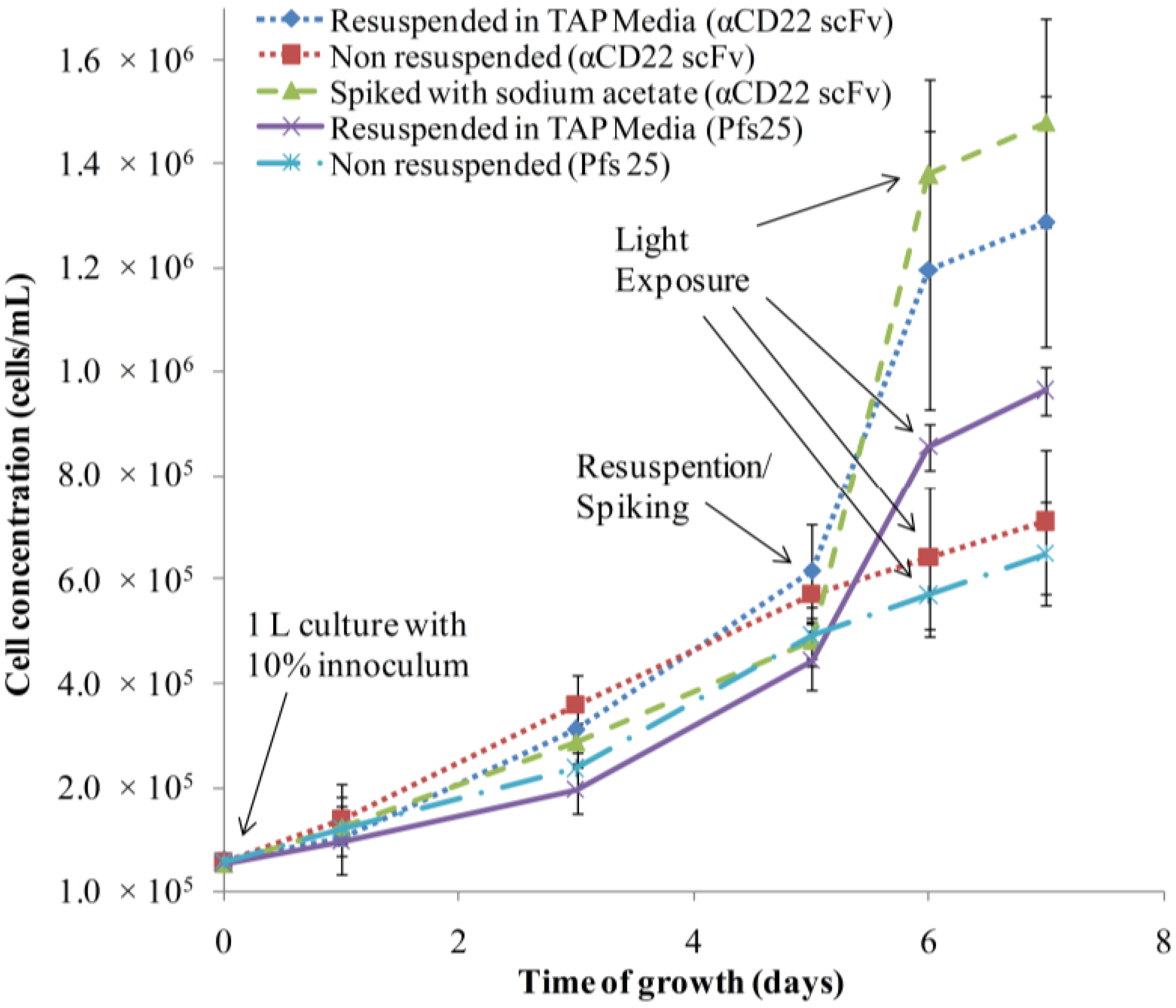
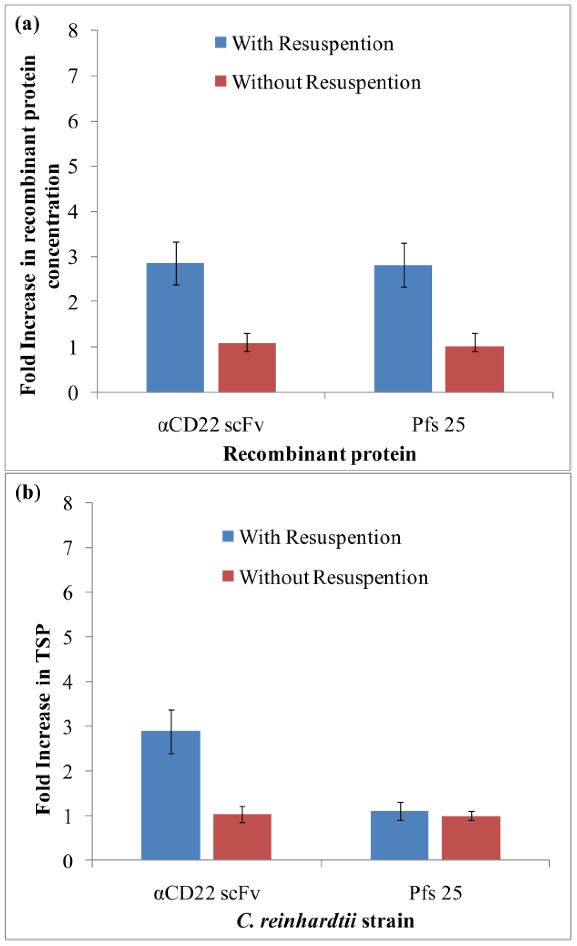
3.1. Algae Cultivation and Accumulation of αCD22scFv and Pfs25

The optimization of algal growth and induced expression conditions could significantly enhance recombinant protein accumulation. Maximal biomass accumulation was achieved after four to five days of continuous growth under heterotrophic conditions (no light exposure) with the cell concentration reaching 5 to 6 × 10⁵ cells/mL (Figure 1). In order to further increase the cell concentration and to provide sufficient nutrients for the subsequent induction phase, cells were resuspended in fresh media at the end of the fifth day and allowed to grow for 24 h before the induction (light exposure) period (Figure 1).
Figure 1. Heterotrophic growth curves of Chlamydomonas reinhardtii expressing αCD22 scFv and Pfs25. The effect of resuspension, spiking and light exposure (101 μmol m−2 s−1) on cell concentration, average of three replicates. TAP: Tris-Acetate-Phosphate.
Processes 02 00625 g001
Twenty-four hours after resuspension (Day 6), the cell number of resuspended cultures increased almost two-fold (1.8-fold for αCD22 scFv and 1.5-fold for Pfs25) compared to non-resuspended ones (Figure 1). The exposure of the cultures to light for 24 h (Day 7) increased cell concentrations of resuspended and non-resuspended cultures proportionally, maintaining the two-fold difference. The variability in cell concentrations for resuspended cultures could be due to different cell adaptation periods between (lag phase) the three batches after resuspension.
We tested both 24 h and 48 h incubation after resuspension and found that 24 h was optimal, as no additional increase in cell number was observed. The increase in culture pH to 8.0 near the end of the exponential phase (Day 5) reflects the depletion of acetate (carbon source); therefore, the increase in cell number during resuspension could be attributed to the replenishment of the carbon source in the media. We confirmed the above hypothesis by spiking the culture with sodium acetate (1.68 g/L), which also resulted in about a two-fold increase in the cell concentration. The growth curve of C. reinhardtii after spiking with sodium acetate followed a similar trend as resuspension with fresh TAP media (Figure 1). Thus, cell resuspension is not necessary, and fed-batch cultivation with only acetate addition would be a feasible method to increase algal biomass. At the end of Day 6, wet biomass of resuspended cultures increased 2.65-fold for αCD22 scFv and three-fold for Pfs25 compared to the non-resuspended cultures. The reason for the higher fold increase in wet biomass compared to cell concentration reflects the increase in the cell mass (i.e., cell size) with resuspension.
Total biomass, as measured by optical density at 750 nm, as well as extracted soluble protein (TSP) in cultures increased with increased light exposure, which agrees with the fact that RuBisCO and other light-activated enzymes and pigments are being synthesized under light conditions [19]. In C. reinhardtii, the genes encoding the light harvesting chlorophyll a/b-binding proteins (LHCPs) and the oxygen evolving enhancer (OEE) complex proteins are expressed when cells are shifted from the dark into white light [20]. During light exposure, there was no significant increase in cell concentration, probably due to a higher energy demand for carbon fixation, since the carbon uptake flux is directed through the Calvin cycle [19,21].
We investigated the effect of resuspension and light induction on αCD22 scFv and Pfs25 synthesis and accumulation, because the aim of increasing the cell density was to increase the recombinant protein accumulation per liter of culture. The effect of resuspension on the amount of purified αCD22 scFv and Pfs25 is shown in Figure 2a. The increase of cell concentration resulted in a 2.8-fold increase of αCD22 scFv and a 2.8-fold increase of Pfs25 protein eluted from the affinity resin.
Figure 2. The effect of resuspension on (a) purified recombinant protein (αCD22 scFv and Pfs25) and (b) total soluble protein concentrations. Data in Figure 2 are average of three replicates.
Processes 02 00625 g002
These results also suggest a correlation between total soluble protein and recombinant protein accumulation in crude extracts (Figure 2b). Because resuspension has a positive effect on the overall growth of the algal cells, replenishment of the media also results in an increase in total soluble protein. In the case of αCD22scFv, the increase in purified recombinant protein amount was synchronous with the increase in total soluble protein (TSP). As expected, resuspension resulted in a similar fold increase in recombinant protein accumulation (2.8 ± 0.9) and total soluble protein accumulation (2.9 ± 0.4). However, Pfs25 showed a greater fold increase in purified recombinant protein concentration (2.8 ± 0.5) compared to the (1.1 ± 0.2) fold increase in total soluble protein accumulation. The higher fold increase in the FLAG-purified Pfs25 protein concentration compared to αCD22 scFv might have been the result of Pfs25 aggregation (Figure 3a,b), which often reduces recombinant protein susceptibility to proteases [22].
Figure 3. (a) Coomassie-stained SDS-PAGE of reduced Chlamydomonas reinhardtii Pfs25 purified with FLAG affinity chromatography. Lane 1: molecular weight marker (kDa); Lane 2: 10× diluted clarified algae extract; Lane 3: 10× diluted supernatant after 2 h of binding with FLAG resin; Lane 4: wash 1 before elution 10×; Lane 5: wash 2 before elution 10×; Lanes 6 to 10: pH 3.5 eluted fractions; (b) Western blot analysis of C. reinhardtii Pfs25 using anti-FLAG-AP conjugated antibody. Lane 1: molecular weight marker (kDa); Lane 2: 10× diluted clarified initial extract; Lane 3: 10× diluted supernatant after 2 h of binding with FLAG resin; Lane 4: wash 1 before elution 10×; Lane 5: wash 2 before elution 10×; Lanes 6 to 10: pH 3.5 eluted fractions.
Processes 02 00625 g003

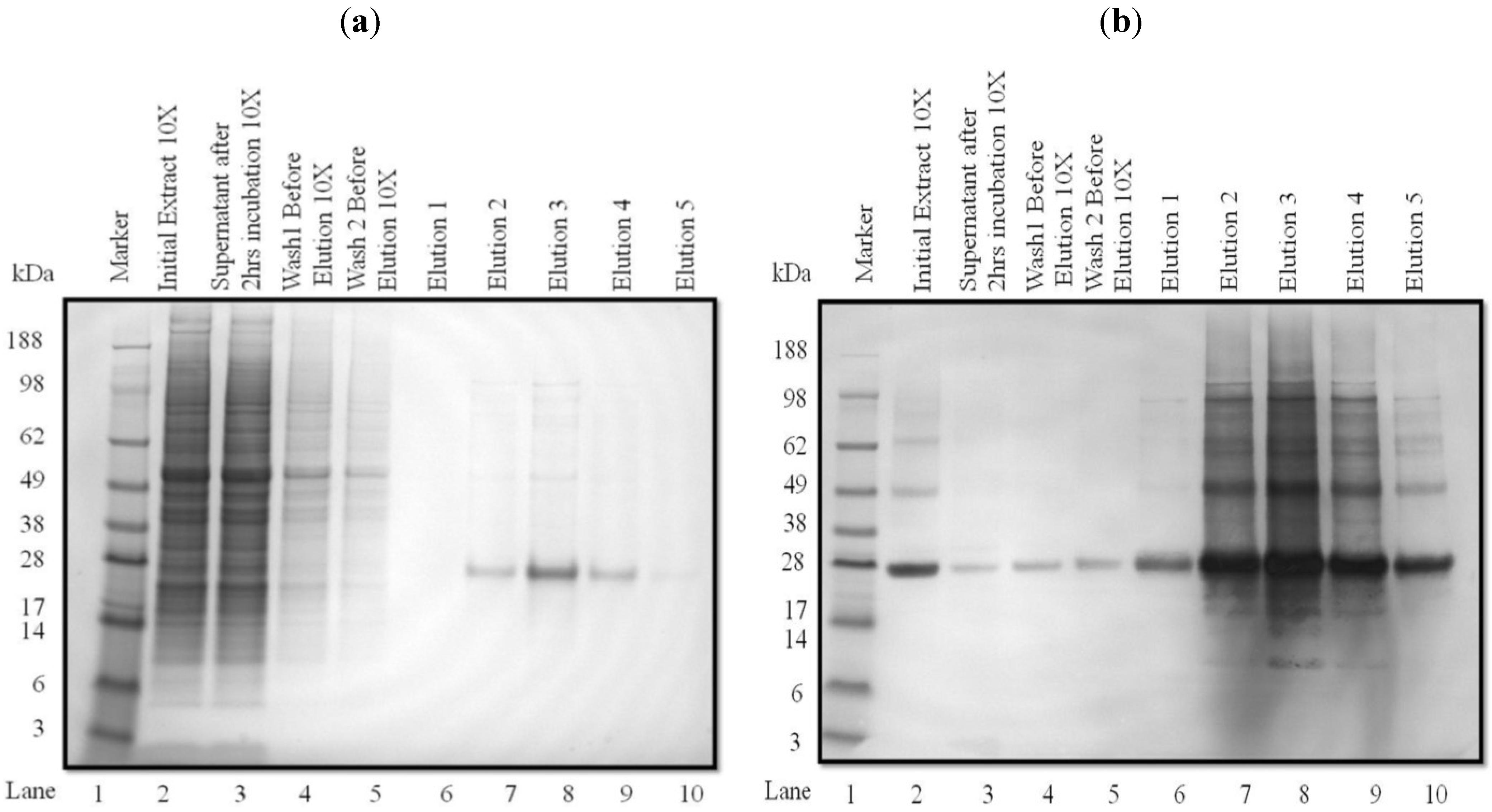
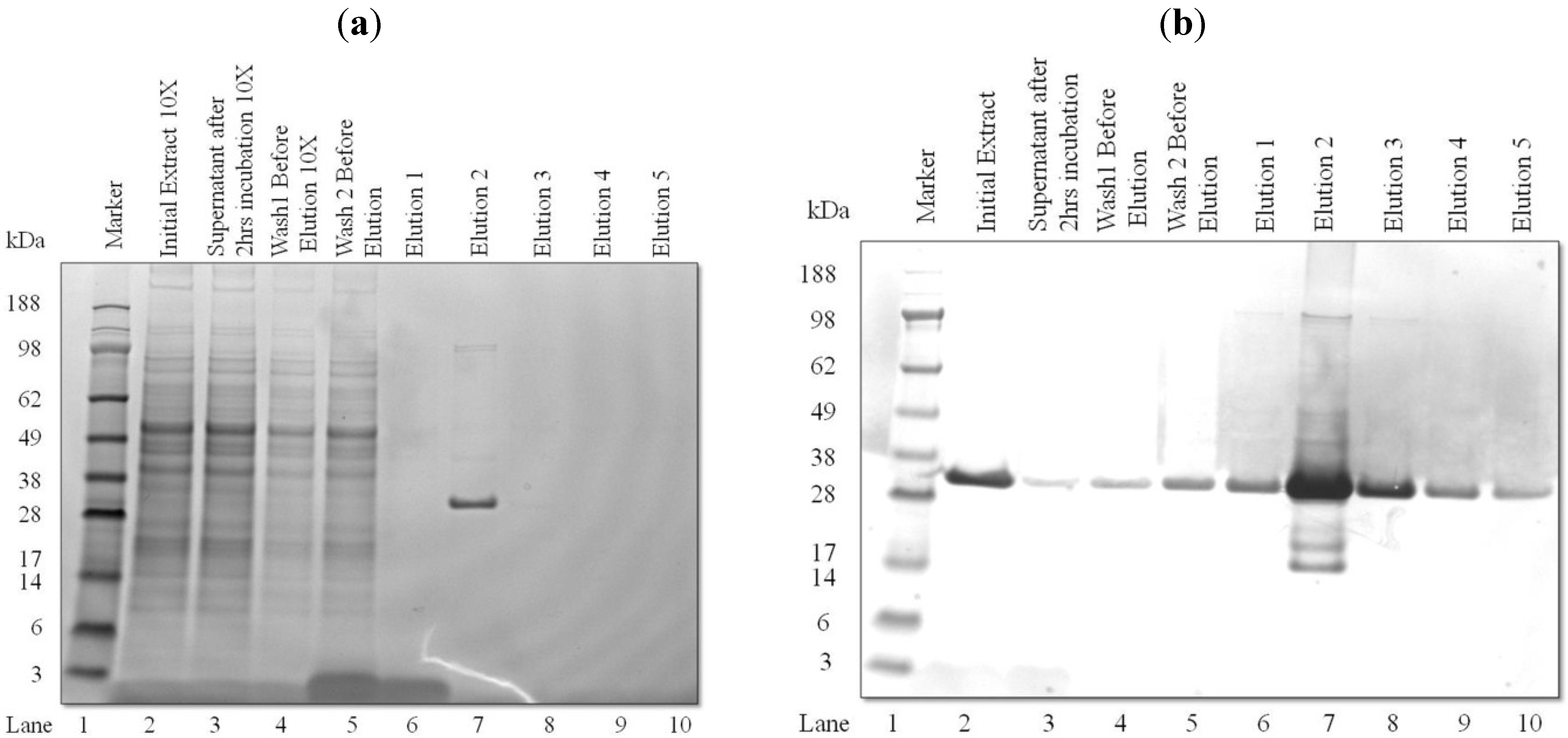
3.2. Purification and Analysis of Recombinant Proteins

Recombinant proteins were purified from clarified crude lysates by FLAG affinity adsorption and analyzed by SDS-PAGE and western blotting (Figure 3a,b). The presence bands of negligible intensity in the supernatant after 2 h of incubation (Lane 3) and washes before elution (Lanes 4–5) at the same size as Pfs25 confirm the efficient binding of FLAG-tagged Pfs25 to the anti-FLAG affinity resin. SDS-PAGE gels and total eluted protein analysis by the Bradford assay indicated that the majority of purified Pfs25 (25 kDa band) eluted in Fractions 2, 3 and 4 (Lanes 7–9). These two fractions typically contained approximately 80% of the total eluted recombinant protein from the resin. The eluted fractions, in both the SDS-PAGE and western blot, show the presence of aggregates, even under reducing conditions (~50 kDa to 100 kDa bands in Figure 3a,b).
The detection of aggregates was not surprising, because Pfs25 is a membrane protein, and similar aggregation has also been observed in yeast-produced Pfs25 [23,24]. In spite of Pfs25 aggregation, FLAG-affinity purified Pfs25 multimers generated an immune response and elicited antibodies with significant levels of transmission blocking activity [4].
The analyses of purified αCD22 scFv revealed no significant aggregation in the eluted fractions by SDS-PAGE (Figure 4a, Lanes 6 to 10) and western blot (Figure 4b). Similar to Pfs25, the insignificant losses in the supernatant (Lane 2) and washes (Lanes 3–4) confirms the efficient binding and elution of FLAG-tagged αCD22 scFv from the anti-FLAG affinity resin. A major 30-kDa band was detected by the SDS-PAGE gel (Figure 4a) and western blot (Figure 4b). Western blot (Figure 4b, Elution 2) revealed the presence of minor degradation products of 14 kDa and 17 kDa in size.
Figure 4. (a) Coomassie-stained SDS-PAGE gels of reduced Chlamydomonas reinhardtii CD22 scFv purified with FLAG affinity chromatography. Lane 1: molecular weight marker (kDa); Lane 2: 10× diluted clarified initial extract; Lane 3: 10× diluted supernatant after 2 h of binding with FLAG resin; Lane 4: wash 1 before elution 10×; Lane 5: wash 2 before elution; Lanes 6 to 10: pH 3.5 eluted fractions; (b) Western blot analysis of C. reinhardtii CD22 scFv using anti-FLAG-AP conjugated antibody. Lane 1: molecular weight marker (kDa); Lane 2: clarified initial extract; Lane 3: supernatant after 2 h of binding with FLAG resin; Lane 4: wash 1 before elution; Lane 5: wash 2 before elution; Lanes 6 to 10: pH 3.5 eluted fractions.
Processes 02 00625 g004

3.3. Light-Induced Accumulation of αCD22scFv

We observed that the recombinant protein accumulated did not increase significantly when the wet biomass concentration increased beyond 4 g/L (>106 cells/mL), and some batches even reported somewhat lower recombinant protein accumulation. The significant (p < 0.05) increase in the cell number that occurred after resuspension raised the question of the cell shading effect on protein accumulation. Because light induction is a unique feature of psbA-driven gene constructs, we hypothesized that recombinant production would be affected by the amount of light (total energy) reaching each cell in the culture. To determine the optimal light conditions required to overcome shading and maximize recombinant protein accumulation, we used only αCD22scFv strain, because extensive aggregation of Pfs25 could obscure the effect of delivered light flux on the recombinant protein yield.
Table 1. The effect of light intensity and duration on biomass harvested, total soluble protein (TSP) and αCD22 scFv protein production. The values given are averages from three replicates ± standard deviations.
Photosynthetic Photon Flux (PPF)Duration101 μmol m−2 s−1300 μmol m−2 s−1
Biomass (g)12 hx 4.0 a ± 0.01x 4.9 a ± 1.5
24 hx 4.0 a ± 0.01x 5.3 a ± 1.0
TSP in algae extract (μg/mL)12 hx 5939 a ± 257x 5485 a ± 802
24 hx 6840 a ± 1167x 5248 a ± 415
αCD22 scFv in wet biomass (μg/g)12 hx 21.2 a ± 1.8x 26.6 a ± 4.3
24 hx 35.1 b ± 5.8y 61.5 b ± 14
αCD22 scFv volumetric conc. (μg/L)12 hx 84.7 a ± 7.1x 135 a ± 66
24 hx 140 a ± 20y 314 b ± 20
αCD22 scFv (%TSP)12 hx 0.07 a ± 0.01x 0.1 a ± 0.03
24 hx 0.12 a ± 0.03y 0.23 b ± 0.05
a,b For each observation, means within a column that are not followed by a common superscript letter are significantly different (p < 0.05); x,y means within a row that are not followed by a common subscript letter are significantly different (p < 0.05).
A 23 full factorial design was performed to determine the effect of the light intensity, light duration and light placement on the volumetric concentration of recombinant protein αCD22scFv. The optimal cell concentration used for light induction was 1 × 10⁵ cells/mL. The placement of the light source (one vs. two sides) did not have a significant effect on the recombinant protein accumulation; however, there was a significant effect (p < 0.05) of light duration and light intensity on recombinant protein accumulation. Because preliminary data with both Pfs25 and αCD22scFv showed no substantial protein accumulation beyond 24 h of light exposure at 101 μmol m−2 s−1, we restricted the further investigation to 12 h and 24 h of light exposure and two levels of light intensity: 101 μmol m−2 s−1 and 300 μmol m−2 s−1. The effect of these two variables is summarized in Table 1. It is evident from the data that a 24-h light duration at both light intensities was optimal for maximum recombinant protein accumulation per gram of wet biomass and unit culture volume. At a light intensity of 101 μmol m−2 s−1, 21.2 ± 1.8 μg/g of αCD22 scFv was recovered after 12 h of light exposure compared to 35.1 ± 5.8 μg/g after 24 h of light exposure. A similar time effect was observed at 300 μmol m−2 s−1; 26.6 ± 4.3 g/g of αCD22 scFv were recovered after 12 h and 61.5 ± 14 μg/g after 24 h. Interestingly, the recombinant protein recovered after 12 h of exposure at a higher light intensity of 300 μmol m2 s−1 (26.6 ± 4.3 μg/g) was not significantly different from the 12 h of light exposure at 101 μmol m−2 s−1 (21. 2 ± 1.8 μg/g). However, after 24 h, the difference in αCD22 scFv accumulation at 300 μmol m−2 s−1 became twice as much as that at 101 μmol m−2 s−1, indicating that both the light intensity and duration of exposure were important factors for the synthesis of recombinant protein. The same conclusion could be reached by comparing αCD22 scFv volumetric concentrations after 12 and 24 h at 300 μmol m−2 s−1 and 101 μmol m−2 s−1, respectively. Interestingly, the effect of the same two factors was different for total soluble protein. Host proteins, such as RuBisCO and other light-activated enzymes listed under TSP in Table 1, were synthesized mainly during the first 12 h of light exposure, and the TSP concentration did not significantly change during the next 12 h. These data suggest that native host proteins were synthesized faster than the recombinant protein and that the 12-h period of light exposure was not sufficient for maximal accumulation of αCD22 scFv. These results corroborate previous data indicating that the translation of the psbA promoter is a rate-limiting step for recombinant protein expression in transgenic C. reinhardtii [25]. The increase of αCD22 scFv accumulation (% TSP) after 24 h at both light intensities (Table 1) reflects the effect of light duration and intensity on continued αCD22 scFv accumulation at constant total soluble protein. The two-fold increase of the recombinant protein fraction in the extracted soluble protein is a desirable product recovery outcome, which is equivalent to achieving a two-fold purification [9].

4. Conclusions

This study provides an account of factors that affect the accumulation of two recombinant proteins in C. reinhardtii chloroplast. Cultivation variables of C. reinhardtii, such as light-induction time and intensity, replenishment with fresh media and culture age, had a significant effect on biomass and recombinant protein accumulation.
Maximum biomass accumulation of transgenic C. reinhardtii was achieved after resuspending the cells in fresh TAP media after five days of heterotrophic growth, which resulted in a 2.8-fold increase in the culture concentration of recombinant therapeutic proteins αCD22 scFv and Pfs25, but this could also be achieved by adding acetate. There was a correlation between total soluble protein and recombinant protein accumulation in crude extracts for αCD22 scFv, but not Pfs25. Resuspension resulted in a similar fold increase in αCD22 scFv accumulation (2.8 ± 0.9) and total soluble protein accumulation (2.9 ± 0.4).
There was also a significant effect of light duration and intensity on the psbA regulated expression of αCD22 scFv protein in the chloroplast. The exposure of light of 300 μmol m−2 s−1 for 24 h resulted in a maximum culture concentration of αCD22 scFv (314.0 ± 20 μg/mL), which is a two-fold increase compared to 101 μmol m−2 s−1. Decoupling of the cell growth and light-induced production of Pfs25 and αCD22 scFv allowed us to demonstrate that native C. reinhardtii proteins accumulate faster (12 h) during the light phase than recombinant proteins, which needed 24 h of light exposure. Thus, an increase in light intensity to 300 μmol m−2 s−1 not only increased the recombinant protein production, but also led to an efficient product recovery with a two-fold increase of the recombinant protein fraction in the extracted soluble protein.
The two proteins were efficiently purified by FLAG affinity adsorption with minimal losses (less than 20%) as judged by western blots. Overall, resuspension and light optimization resulted in a six-fold increase in the recovered recombinant protein in the chloroplasts of C. reinhardtii.

Acknowledgments

The authors acknowledge the funding of this work by the National Science Foundation (Chemical, Bioengineering, Environmental, and Transport Systems Grant #1160117). The authors thank Stephen Mayfield and his research group at the University of California, San Diego, for providing transgenic strains of C. reinhardtii and for their valuable discussions and suggestions. Thanks are also extended to Rebekah Garcia, who helped us with media preparation and cultivation.

Author Contributions

Zivko Nikolov, conceptualized, developed and supervised the overall research plan. He also contributed to the preparation, review and editing of the manuscript. Neera Munjal designed and conducted experiments on the project involving αCD22 scFv. She contributed to the preparation and editing of the manuscript. Katelyn Wilson and Andrea Garzon conducted research on C. reinhardtii expressing Pfs25 strain. James Gregory performed a thorough review of the manuscript and provided advice on the cultivation of Pfs25 strains. All of the authors contributed significantly to the discussions involving the research study.

Conflicts of Interest

The authors declare no conflict of interest.

References

  1. Mayfield, S.P.; Manuell, A.L.; Chen, S.; Wu, J.; Tran, M.; Siefker, D.; Muto, M.; Marin-Navarro, J. Chlamydomonas reinhardtii chloroplasts as protein factories. Curr Opin Biotechnol. 2007, 18, 126–133. [Google Scholar] [CrossRef] [PubMed]
  2. Rasala, B.A.; Muto, M.; Lee, P.A.; Jager, M.; Cardoso, R.M.F.; Behnke, C.A.; Kirk, P.; Hokanson, C.A.; Crea, R.; Mendez, M.; et al. Production of therapeutic proteins in algae, analysis of expression of seven human proteins in the chloroplast of Chlamydomonas reinhardtii. Plant Biotechnol. J. 2010, 8, 719–733. [Google Scholar] [CrossRef] [PubMed]
  3. Tran, M.; Van, C.; Barrera, D.J.; Pettersson, P.L.; Peinado, C.D.; Bui, J.; Mayfield, S.P. Production of unique immunotoxin cancer therapeutics in algal chloroplasts. Proc. Natl. Acad. Sci. USA 2012, 110, 15–22. [Google Scholar] [CrossRef]
  4. Gregory, J.A.; Li, F.; Tomosada, L.M.; Cox, C.J.; Topol, A.B.; Vinetz, J.M.; Mayfield, S.P. Algae-produced Pfs25 elicits antibodies that inhibit malaria transmission. PLoS One 2012, 7. [Google Scholar] [CrossRef]
  5. Lauersen, K.J.; Berger, H.; Mussgnug, J.H.; Kruse, O. Efficient recombinant protein production and secretion from nuclear transgenes in Chlamydomonas reinhardtii. J. Biotechnol. 2013, 167, 101–110. [Google Scholar] [CrossRef] [PubMed]
  6. Mayfield, S.P.; Franklin, S.E. Expression of human antibodies in eukaryotic micro-algae. Vaccine 2005, 23, 1828–1832. [Google Scholar] [CrossRef] [PubMed]
  7. Tran, M.; Zhou, B.; Pettersson, P.L.; Gonzalez, M.J.; Mayfield, S.P. Synthesis and assembly of a full-length human monoclonal antibody in algal chloroplasts. Biotechnol. Bioeng. 2009, 104, 663–673. [Google Scholar] [PubMed]
  8. Kaslow, D.C.; Shiloach, J. Production, purification and immunogenicity of a malaria transmission-blocking vaccine candidate: TBV25H expressed in yeast and purified using nickel-NTA agarose. Nat. Biotechnol. 1994, 12, 494–499. [Google Scholar] [CrossRef]
  9. Wilken, L.R.; Nikolov, Z.N. Recovery and purification of plant-made recombinant proteins. Biotechnol. Adv. 2012, 30, 419–433. [Google Scholar] [CrossRef] [PubMed]
  10. Tran, M.; Henry, R.E.; Siefker, D.; Van, C.; Newkirk, G.; Kim, J.; Bui, J.; Mayfield, S.P. Production of anti-cancer immunotoxins in algae: Ribosome inactivating proteins as fusion partners. Biotechnol. Bioeng. 2013, 110, 2826–2835. [Google Scholar] [CrossRef] [PubMed]
  11. Manuell, A.L.; Beligni, M.V.; Elder, J.H.; Siefker, D.T.; Tran, M.; Weber, A.; McDonald, T.L.; Mayfield, S.P. Robust expression of a bioactive mammalian protein in Chlamydomonas chloroplast. Plant Biotechnol. J. 2007, 5, 402–412. [Google Scholar] [CrossRef] [PubMed]
  12. Surzycki, R.; Greenham, K.; Kitayama, K.; Dibal, F.; Wagner, R.; Rochaix, J.D.; Ajam, T.; Surzycki, S. Factors effecting expression of vaccines in microalgae. Biologicals 2009, 37, 133–138. [Google Scholar] [CrossRef] [PubMed]
  13. Herman, C.A.; Im, C.S.; Beale, S.I. Light-regulated expression of the gsa gene encoding the chlorophyll biosynthetic enzyme glutamate 1-semialdehyde aminotransferase in carotenoid-deficient Chlamydomonas reinhardtii cells. Plant Mol. Biol. 1999, 39, 289–297. [Google Scholar] [CrossRef] [PubMed]
  14. Bohne, F.; Linden, H. Regulation of carotenoid biosynthesis genes in response to light in Chlamydomonas reinhardtii. Biochim. Biophys. Acta 2002, 1579, 26–34. [Google Scholar]
  15. Barneche, F.; Winter, V.; Crevecoeur, M.; Rochaix, J.D. ATAB2 is a novel factor in the signalling pathway of light-controlled synthesis of photosystem proteins. EMBO J. 2006, 25, 5907–5918. [Google Scholar] [CrossRef] [PubMed]
  16. Franklin, S.; Ngo, B.; Efuet, E.; Mayfield, S.P. Development of a GFP reporter gene for Chlamydomonas reinhardtii chloroplast. Plant J. 2002, 30, 733–744. [Google Scholar] [CrossRef] [PubMed]
  17. Gregory, J.A.; Topol, A.B.; Doerner, D.Z.; Mayfield, S.P. Alga-Produced Cholera Toxin-Pfs25 Fusion Proteins as Oral Vaccines. Appl. Environ. Microbiol. 2013, 79, 3917–3925. [Google Scholar] [CrossRef] [PubMed]
  18. Bradford, M.M. A rapid and sensitive method for the quantitation of microgram quantities of protein utilizing the principle of protein-dye binding. Anal. Biochem. 1976, 72, 248–254. [Google Scholar] [CrossRef] [PubMed]
  19. Boyle, N.; Morgan, J. Flux balance analysis of primary metabolism in Chlamydomonas reinhardtii. BMC Syst. Biol. 2009, 3. [Google Scholar] [CrossRef]
  20. Malnoë, P.; Mayfield, S.P.; Rochaix, J.D. Comparative analysis of the biogenesis of photosystem II in the wild-type and Y-1 mutant of Chlamydomonas reinhardtii. J. Cell Biol. 1988, 106, 609–616. [Google Scholar] [CrossRef] [PubMed]
  21. Griffiths, M.J.; Garcin, C.; van Hille, R.P.; Harrison, S.T.L. Interference by pigment in the estimation of microalgal biomass concentration by optical density. J. Microbiol. Methods 2011, 85, 119–123. [Google Scholar] [CrossRef] [PubMed]
  22. Villaverde, A.; Carrió, M.M. Protein aggregation in recombinant bacteria: Biological role of inclusion bodies. Biotechnol. Lett. 2003, 25, 1385–1395. [Google Scholar] [CrossRef] [PubMed]
  23. Miles, A.P.; Zhang, Y.; Saul, A.; Stowers, A.W. Large-scale purification and characterization of malariavaccine candidate antigen Pvs25H for use in clinical trials. Protein Expr. Purif. 2002, 25, 87–96. [Google Scholar] [CrossRef] [PubMed]
  24. Miyata, T.; Harakuni, T.; Tsuboi, T.; Sattabongkot, J.; Kohama, H.; Tachibana, M.; Matsuzaki, G.; Torii, M.; Arakawa, A. Plasmodium vivax ookinete surface protein Pvs25 linked to cholera toxin B subunit induces potent transmission-blocking immunity by intranasal as well as subcutaneous immunization. Infect. Immun. 2010, 78, 3773–3782. [Google Scholar] [CrossRef] [PubMed]
  25. Coragliotti, A.; Beligni, M.; Franklin, S.; Mayfield, S.P. Molecular factors affecting the accumulation of recombinant proteins in the Chlamydomonas reinhardtii chloroplast. Mol. Biotechnol. 2011, 48, 60–75. [Google Scholar] [CrossRef] [PubMed]

