Article

(18-Crown-6)potassium(I) Trichlorido[28-acetyl-3-(tris-(hydroxylmethyl)amino-ethane)betulinic ester-κN]platinum(II): Synthesis and In Vitro Antitumor Activity

by
Goran N. Kaluđerović
1,*,
Mirna Bulatović
2,
Tamara Krajnović
2,
Reinhard Paschke
3,
Bojana B. Zmejkovski
4,
Danijela Maksimović-Ivanić
2 and
Sanja Mijatović
2,*
1
Department of Bioorganic Chemistry, Leibniz Institute of Plant Biochemistry, Weinberg 3, D-06120 Halle (Saale), Germany
2
Institute for Biological Research “Siniša Stanković” University of Belgrade, Bulevar despota Stefana 142, 11060 Belgrade, Serbia
3
Biozentrum, Martin-Luther-Universität Halle-Wittenberg, Weinbergweg 22, D-06120 Halle, Germany
4
Department of Chemistry, Institute of Chemistry, Technology and Metallurgy, University of Belgrade, Studentski trg 14, 11000 Belgrade, Serbia
*
Authors to whom correspondence should be addressed.
Inorganics 2017, 5(3), 56; https://doi.org/10.3390/inorganics5030056
Submission received: 28 June 2017 / Revised: 16 August 2017 / Accepted: 16 August 2017 / Published: 19 August 2017
(This article belongs to the Special Issue Tumor Inhibiting Metal Complexes)

Abstract

:
Synthesis of platinum(II) conjugate with acetylated betulinic acid tris(hydroxymethyl)aminomethane ester (BATRIS) is presented (BATRISPt). HR-ESI-MS and multinuclear NMR spectroscopy, as well as elemental analysis were used for characterization of BATRISPt. Cytotoxicity (3-(4,5-dimethythiazol-2-yl)-2,5-diphenyltetrazolium bromide (MTT), crystal violet (CV), and sulforhodamine B (SRB) assays) of BA, BATRIS, BATRISPt, and cisplatin were assessed on seven different tumor cell lines: melanoma B16, colon HCT116 and DLD-1, adenocarcinoma HeLa, breast MCF-7, and anaplastic thyroid tumor 8505C and SW1736; as well as normal MRC-5 fibroblasts. Furthermore, the effect of the mentioned compounds on the apoptosis (Annexin V/PI assay) and autophagy induction (acridine orange (AO) assay) as well as caspase 3, 8, and 9 activation were investigated on the selected B16 melanoma cell line. BATRISPt showed lower activity than BA, BATRIS, or cisplatin. All tested compounds triggered apoptosis in B16 cells. Induction of autophagy was observed in B16 cells exposed only to BATRIS. On the other hand, new conjugate activates caspases 8 and 9 in B16 cells with higher impact than BATRIS or cisplatin alone.

Graphical Abstract

1. Introduction

Metal-based drugs (e.g., cisplatin, oxaliplatin), natural products and their derivatives (e.g., paclitaxel, doxorubicin), or even their combination (e.g., 5-fluorouracil, oxaliplatin, irinotecan) are frequently used as chemotherapeutic drugs for cancer treatment [1,2,3,4]. However, along with killing the cancer cells, such drugs cause severe side effects [5]. While the combined application of few drugs is a common approach in the treatment of cancer, and numerous experimental data both in vitro and in vivo have confirmed the efficacy of such treatment [6,7,8], the conjugation of platinum(II) complexes to biologically (antitumor) active agents is not as profoundly investigated [9,10,11,12]. The first approach, combinational chemotherapy, depends on the biological properties of the single components. For instance, how such compounds can be delivered, metabolized, and how and to which magnitude they may enter the tumor cells. On the other side, two components covalently linked to each other—double drug [13], presumably—may possess different behavior than single components alone.
Even lower in activity than paclitaxel or doxorubicin [14,15], a cell-type-specific antitumor active compound betulinic acid (BA) has drawn more attention in the last few years and is still explored by many groups [16,17,18,19,20,21,22,23,24]. BA is a naturally occurring pentacyclic triterpenoid which is isolated from the bark of several species of plants (e.g., white birch). Betulinic acid and its derivatives present compounds with numerous biological activities—for instance, antioxidant, antiviral, antimicrobial, anti-inflammatory, antidiabetic, immunomodulatory, etc. [25,26]. However, investigations on betulinic acid in the last few years have been pointed toward the field of antitumor properties [27,28,29,30,31].
We recently reported the synthesis and characterization of the new series of betulinic acid–cisplatin conjugates, which are both apoptosis inducing agents [32]. However, their cytotoxicity was found to be lower than parental drugs cisplatin and betulinic acid. Herein we report on the synthesis, characterization, and in vitro activity of acetylated betulinic acid derivative tris(hydroxymethyl)aminomethane (TRIS) ester conjugated with PtCl3-fragment.

2. Results and Discussion

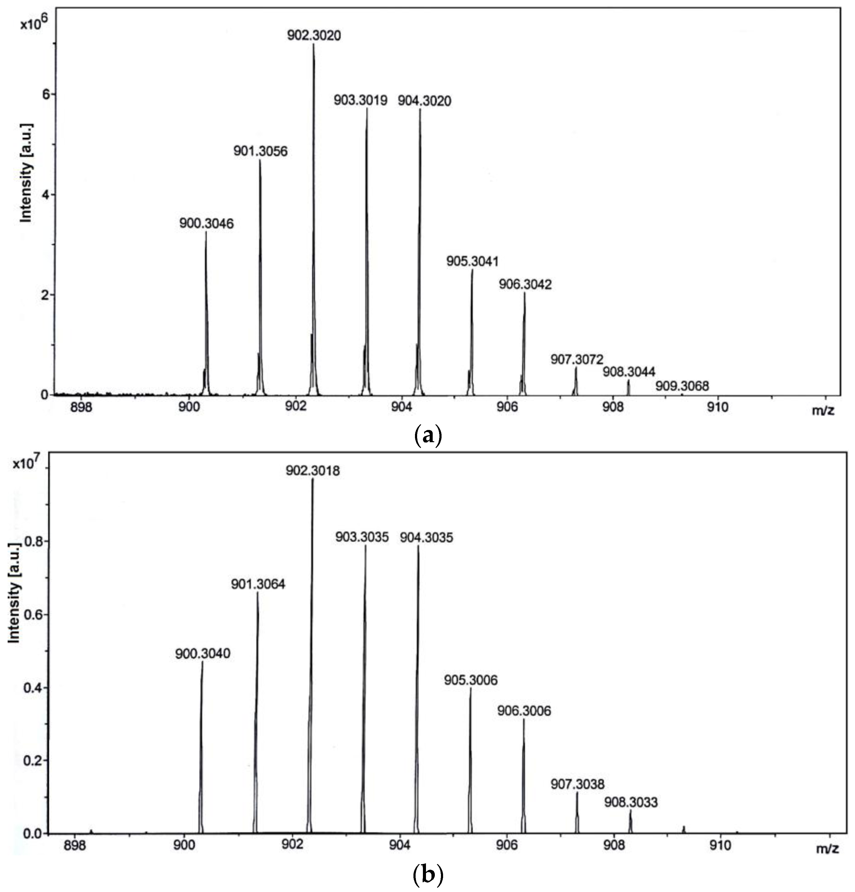
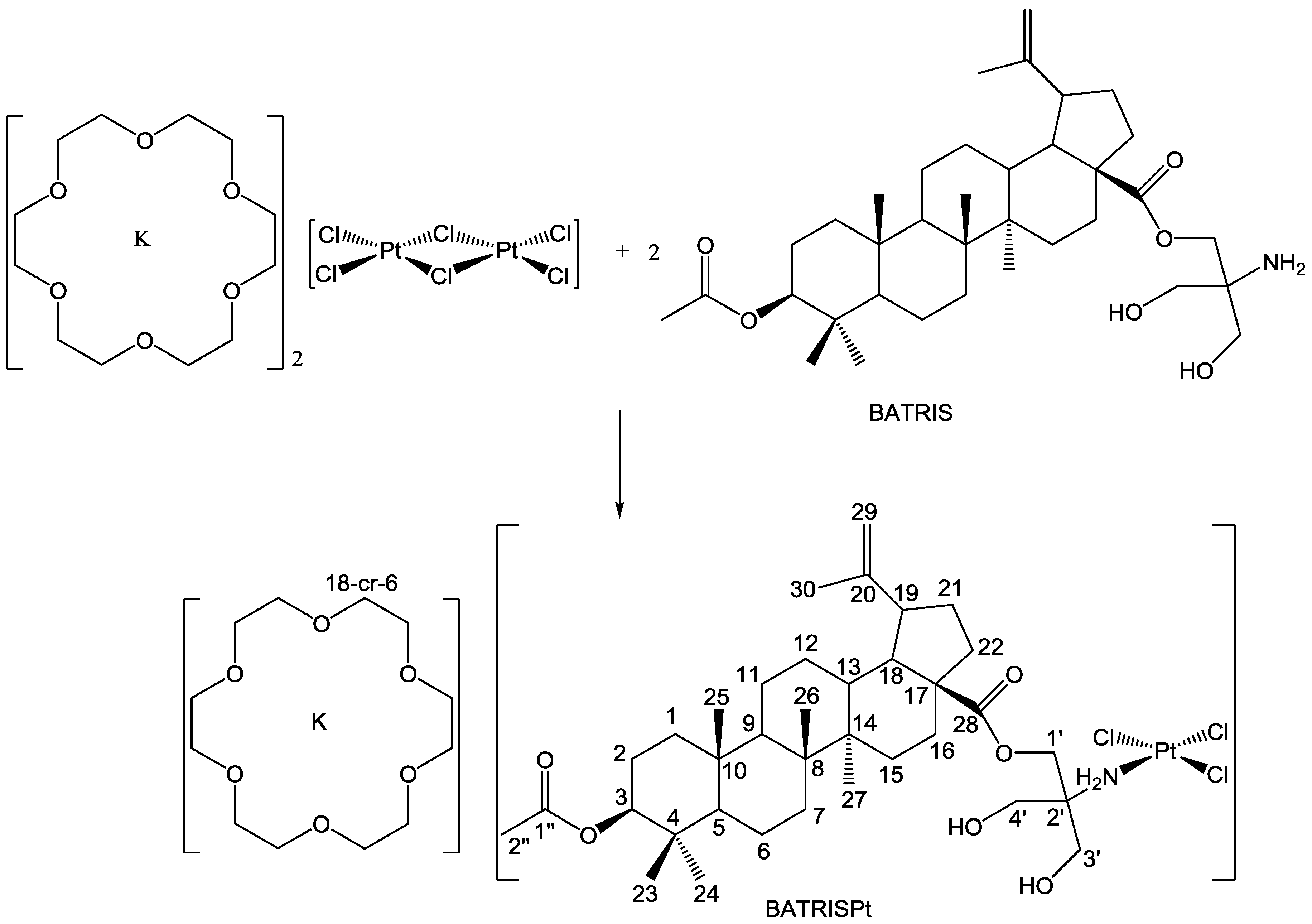
2.1. Synthesis and Characterization

The reaction of dichloromethane solution of acetylated betulinic acid tris(hydroxymethyl)aminomethane ester (BATRIS) and [K(18-cr-6)]2[Pt2Cl6] afforded BATRISPt complex (Scheme 1) in almost quantitative yield. This complex was isolated as a yellow solid.
The isolated compound was characterized with elemental analysis, HR-ESI-MS (High-resolution electrospray ionisation mass spectrometry), as well as multinuclear NMR spectroscopy. The stoichiometric formula of BATRISPt is in agreement with elemental analysis. HR-ESI-MS recorded in negative ion mode gave evidence for the molecular composition of the platinum(II) anionic complex. Namely, the {BATRISPt–[K(18-cr-6)]} ion (m/z = 902.30213) with the characteristic expected isotope pattern was detected (Figure A1 in Appendix A). In positive ion mode, [K(18-cr-6)]+ ion was detected at m/z = 303.12055.
In the 1H and 13C NMR spectra of BASTRISPt, chemical shifts arising from lupane moiety were found at expected values [24]. Hydrogen atoms from the TRIS moiety in the 1H NMR spectrum showed coordination-induced shifts up to 0.5 ppm (C3’H and C4’H). Similar behavior upon coordination was observed for TRIS moiety in 13C NMR spectrum; for example, C2’ atom is deshielded for ca. 14 ppm in comparison to the same carbon atom in BATRIS [30]. Chemical shift at 70.7 ppm corresponds to the carbon atoms from 18-cr-6, while appropriate hydrogen atoms at 3.61 ppm in the 1H-NMR spectrum were observed [33]. The 195Pt NMR spectrum showed exclusively the presence of one complex with the resonance at −1921 ppm. The chemical shift is in good agreement with those observed for [Cl3,N] donor set; e.g., K[PtCl3(NH3)] complex δ is −1884 ppm [34].
The stability of the BASTRISPt complex was evaluated using 1H-NMR spectroscopy. The complex was dissolved in DMSO-d6 and recorded over time. Additionally, the same experimental settings were used for BATRISPt dissolved in CDCl3. As expected, it could be proven that BATRISPt did not decompose in CDCl3 over the investigated period of time (0, 4, and 24 h). The 1H NMR spectrum of BATRISPt in DMSO-d6—recorded immediately after dissolution—gave a comparable chemical shifts pattern to one obtained in CDCl3. Direct comparison of the resonances of the hydrogen atoms belonging to TRIS moiety from BATRIS (in DMSO-d6) or BATRISPt (0, 4 and 24 h) clearly pointed out that proton resonances from the free ligand in BATRISPt were not observed. Thus, it could be clearly demonstrated that ligand remained coordinated to platinum center. Moreover, there was no precipitate during the investigated reaction time. 1H NMR spectrum in DMSO-d6 after 4 h showed decrease (ca. 25%) in intensities of the proton resonances near platinum(II) coordination sphere (C3’H2 and C4’H2), indicating possible coordination of solvent molecules. After 24 h, the substitution of Cl ligands with DMSO-d6 in BATRISPt took place to a greater extent (ca. 45%). Furthermore, this confirms that in the coordination sphere of platinum(II), only substitution of Cl ligand(s) occurs. Nevertheless, for in vitro studies, stock solutions of DMSO were prepared freshly and diluted immediately with nutrition medium to various working concentrations in order to hinder solvolysis.

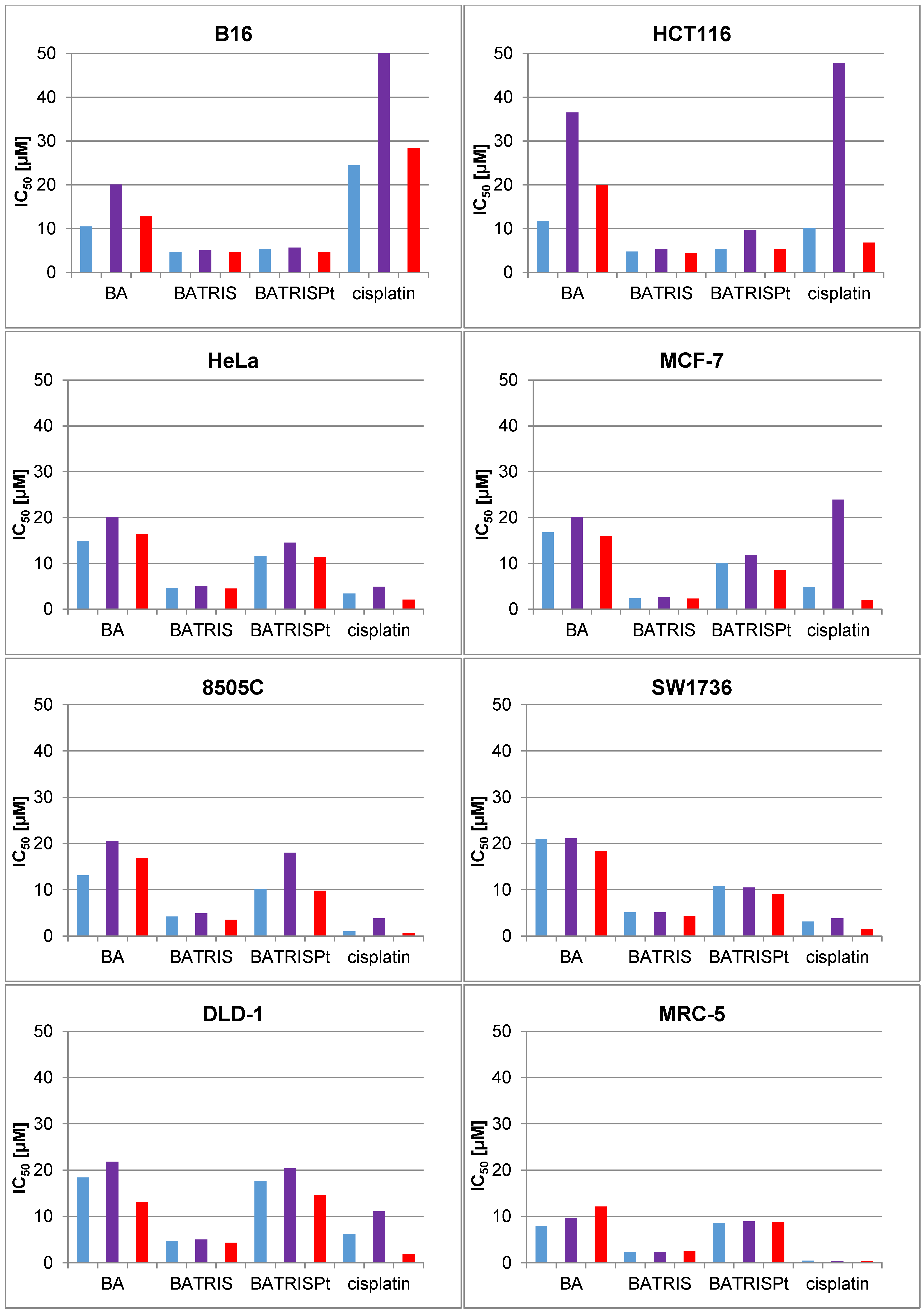
2.2. Cytotoxicity of BATRISPt

Initially it was reported that betulinic acid is selectively cytotoxic for the melanoma cell lines, with IC50 values ranging from 2.4 to 10.3 µM (1.1–4.8 µg/mL) [35]. Since then, a growing number of tumor cells derived from colon, prostate, breast, lung, squamous, and glioma were tested, while the corresponding IC50 values for different cancer cells were around 43 µM (20 µg/mL), whereas normal cells were unaffected by BA treatment [36]. However, limited solubility and sometimes modest activity represent a challenge for different modification of this compound. The idea reported herein concerns a hybrid molecule comprised of two well-known apoptosis-inducing entities.
Seven different tumor cell lines (melanoma B16, colon HCT116 and DLD-1, adenocarcinoma HeLa, breast MCF-7, and anaplastic thyroid tumor 8505C and SW1736) were used for the assessment of viability upon exposure to BA, BATRIS, BATRISPt, and cisplatin. For determination of the viability, three different tests were applied. The MTT (3-(4,5-dimethythiazol-2-yl)-2,5-diphenyltetrazolium bromide) test is based on the measurement of mitochondrial respiration, crystal violet (CV) on the quantity of nucleic acids while sulforhodamine B (SRB) on the protein content. Therefore, by comparing the data obtained by those assays, initial information about the mechanism of drug action can be obtained. Cells were exposed to drugs for 96 h, and subsequently IC50 values were assessed (Figure 1).
As presented in Figure 1, it is obvious that all tested cell lines were sensitive to the treatment with BA. IC50 values obtained by MTT and SRB are in concordance and mostly significantly lower than those obtained by CV assay. The discrepancy in IC50 values obtained by these tests indicated that these agents probably blocked cell cycle division in G2M or S phase of the cell cycle. Therefore, the quantity of stained DNA in cell cultures is not a reflection of the number of viable cells. Detected IC50 values are in the range of that previously reported in the literature [27,28,29,30,31]. As already shown in the literature [30], modification of BA with TRIS (→ BATRIS) strongly amplified the efficacy of BA in all tested cells, and IC50 values obtained by applied methods were well synchronized, suggesting that this chemical intervention changed the mode of drug action.
In order to intensify the cytotoxicity of BATRIS, PtCl3-moiety was conjugated with the BATRIS (→ BATRISPt). However, unexpectedly, BATRISPt was less potent than BATRIS and BA, as well as cisplatin. The obtained data showed that decreased efficacy of conjugate compound BATRISPt might be caused by the activation of some cytoprotective mechanism inside of the cell, which in final point neutralizes the toxicity of the treatment. Data presented herein are in concordance with recently reported results, where different betulinic acid derivatives conjugated with PtCl2-fragment were tested [32]. However, strong selectivity of some of these synthetic compounds apart from lower cytotoxicity indicated that they might have potential as anticancer agents.
On the other hand, treatment of human immortalized fibroblasts MRC-5 with cisplatin alone resulted in completely abolished viability showing even worse effect in comparison to tumor cells. Similarly, viability of normal fibroblast was even more affected by BA and BATRIS in comparison to cancer cells. This data opposed to previously observed high selectivity of BA for cancer cells [32] and definitely require further confirmation in diverse types of normal cells. Conjugate BATRISPt was found to be less toxic to primary cells in comparison to maternal drugs. Even though the IC50 values are in line or even higher with one observed for few cell lines, selectivity was proven for B16 and HCT116 cell lines, indicating that in spite of lower cytotoxicity, conjugated drug deserves further evaluation.

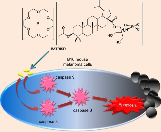
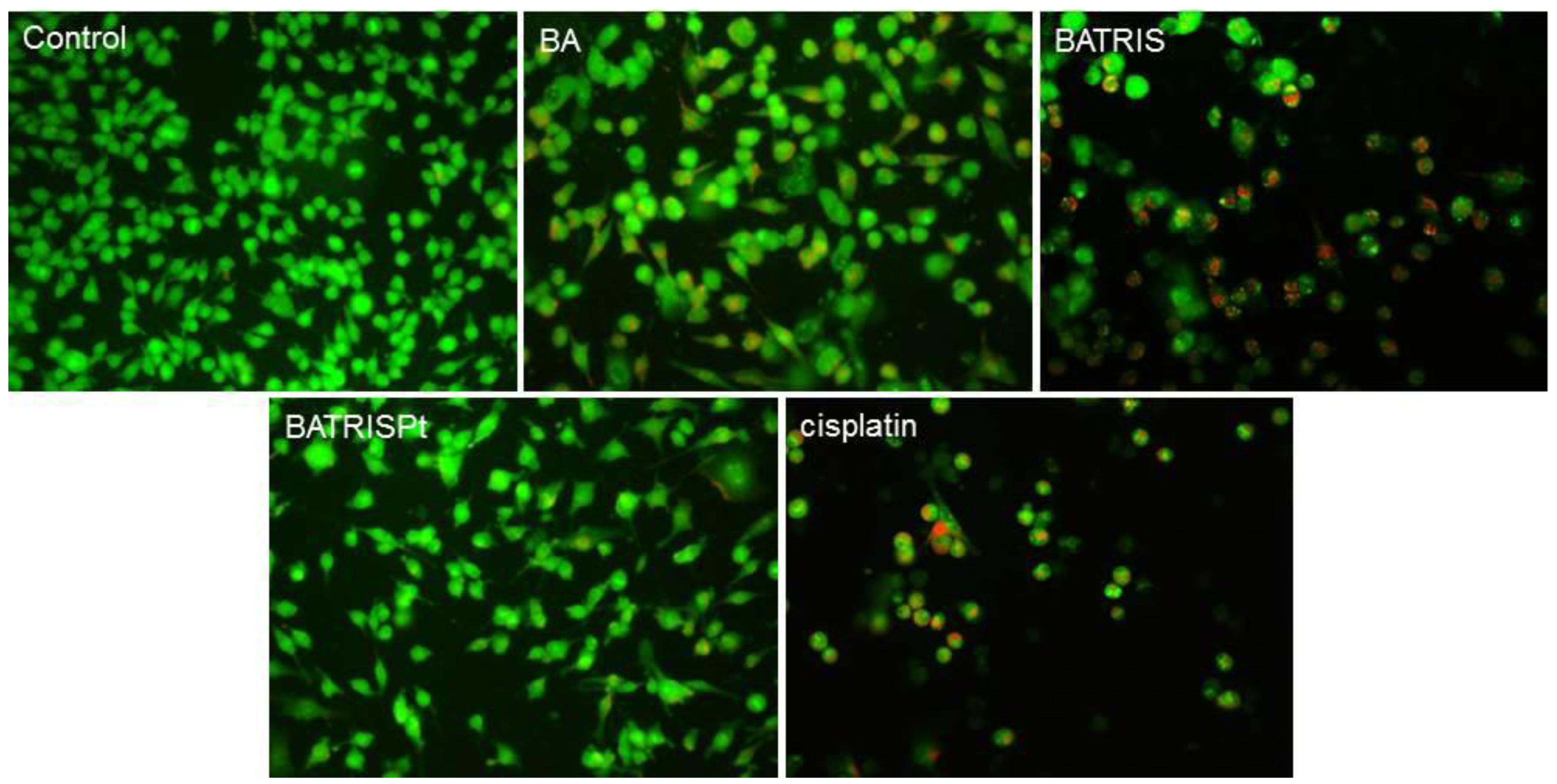
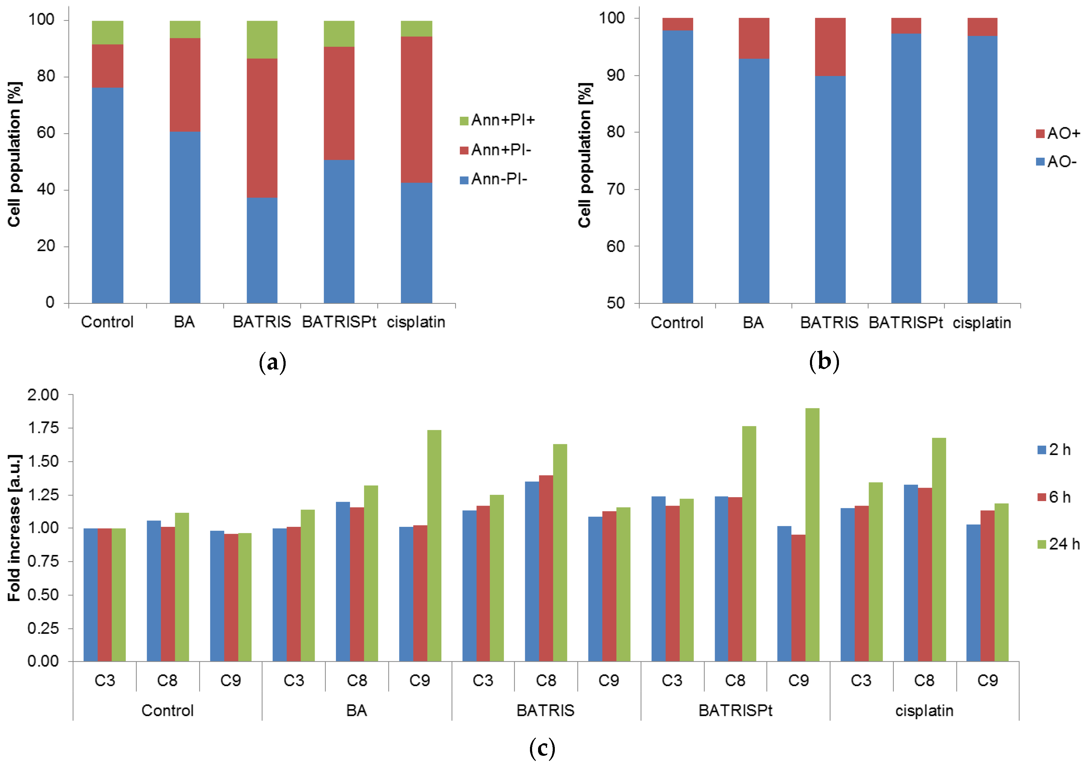
2.3. Mechanism of Action

To evaluate the reason for neutralization of BATRIS toxicity in BATRISPt, B16 cells were treated with compounds for 48 h and further stained by acridine orange/ethidium bromide (AO/EtBr) (Figure 2). Analysis of morphological changes induced by the treatments showed that BATRIS as well as cisplatin dramatically decreased the number of viable cells. Numerous rounded cells and cells with picknotic nuclei and condensed chromatin were visible, indicating that both compounds induced apoptotic cell death [37].
The fact that BA induced apoptosis in several different cancer cell lines through multiple pathways is well described. BA treatment caused distinct morphological changes like cell shrinkage, DNA fragmentation, nuclear condensation, and membrane blebbing in sensitive cells [38]. Several studies revealed that BA directly targeted mitochondria, resulting in decreased mitochondrial membrane potential, up-regulation of death receptors, and interactions with other agents. Accumulated experimental evidence showed that the activation of caspases, MAP kinase cascade, the modulation of NF-κB signaling, intensified production of reactive oxygen species, and the inhibition of topoisomerase I are behind the tumoricidal action of BA. In concordance, by data of viability tests, conjugate BATRISPt was remarkably less potent since a significantly higher number of cells was present in culture exposed to this compound. Volume and size of the cells were enhanced in comparison to control. Their morphology, described as flat with dendritic-like processes, could be the sign of more differentiated phenotype or senescence state [39].
Furthermore, annexin V (Ann)/propidium iodide (PI) staining was performed. As seen in Figure 3a, B16 cells exposed to all treatments were conducted to apoptosis. In concordance with cell viability decrease, the percentage of early apoptotic cells was the highest in the presence of BATRIS and cisplatin in cell culture. However, treatment with BATRISPt resulted in a lower amount of early apoptotic cells (Ann+PI), confirming once more that the combination of two cytotoxic molecules, instead of amplification, minor inhibition of the activity occurs. Knowing that autophagy can confront the apoptosis and is often responsible for decreased efficacy of the apoptosis-inducing agents, B16 treated cells were stained by AO. Crosstalk between apoptosis and autophagy may be crucial for the balance between cell survival and death. Triggering of autophagy by tumor cells as a cytoprotective mechanism able to suppress apoptosis induced by chemotherapy seems to be a general phenomenon and one of the major problems in cancer therapy efficacy. Cytoprotective effects due to intensified autophagy were reported in different tumor types exposed to gefitinib, erlotinib, and celecoxib, suggesting that the concomitant use of autophagy inhibitors with chemotherapy might be beneficial [40]. On the other hand, while increasing evidence showed that autophagy may be responsible for cell death in certain settings, it is clear that this process and its interplay with apoptosis determines the fate of cells upon therapy [41].
The intensity of the red fluorescence correlates with the presence of acidic vesicles recognized as autophagosomes [42]. Surprisingly, autophagy was the most intensive in cultures exposed to BATRIS, since the addition of the PtCl3-moiety abrogated this process, bringing it to the level of cisplatin alone (Figure 3b). It is indicative that in this experimental setting the process of autophagy serves as a dying process rather than a cytoprotective one in BATRIS-exposed cells. In addition, the decreased amount of autophagy resulted in diminished toxicity in BATRISPt-treated cultures. To explore a possible connection between caspase activation and observed effect, the activity of caspase 3, 8, 9 were analyzed (Figure 3c).
Paradoxically, BATRISPt, even with the worst anticancer potential in this study, activated caspases 8 and 9 more powerfully than other compounds. Recently, it has been described that caspases are important mediators in crosstalk between autophagy and apoptosis [43]. Activated caspases are able to cleave some autophagic proteins like Beclin-1, p62, Atg3, Atg4D, Atg5, Atg7, and AMBRA1, leading to the inactivation of autophagy. Caspase 8 cleaves Atg3, targeting the conserved LETD sequence (Atg3 amino acids 166–169), shutting down autophagic activity. On the other hand, caspases are able to cleave some pro-autophagic proteins, transforming them into pro-apoptotic proteins. In addition, autophagy can modulate the activity of caspases. Caspase 8 is known as an essential trigger involved in death receptor-induced apoptosis, but nowadays the increasing evidence indicates that caspase 8 also participates in regulating autophagy [43]. Since it has been described that caspases are also involved in autophagy inhibition and that we hypothesized that autophagy contributes to the dying process in BATRIS-treated cells, it could be possible that the activated caspases by the BATRISPt down-regulated autophagy and subsequently protected cells against autophagy-mediated destruction [43,44,45]. According to this, diminished toxicity is probably the result of confrontation between programmed cell death apoptosis and autophagic cell death.

3. Materials and Methods

3.1. General Remarks

Commercially-pure chemicals were used as received from distributor. [K(18-cr-6)]2[Pt2Cl6] was synthesized according to the reported procedure [33]. The elemental analysis (C, H, N) was carried out on a CHNS-93 (LECO) elemental analyzer. High-resolution ESI mass spectra were obtained from a Bruker Apex III Fourier transform ion cyclotron resonance (FT-ICR) mass spectrometer (Bruker Daltonics, Bremen, Germany) equipped with an Infinity cell, a 7.0 T superconducting magnet (Bruker), an rf-only hexapole ion guide, and an external APOLLO electrospray ion source (Agilent, off-axis spray). The sample solutions were introduced continuously via a syringe pump with a flow rate of 120 µL·h−1. The data were evaluated using Bruker Compass DataAnalysis 4.0 (Bremen, Germany). The 1H, 13C, and 195Pt NMR spectra were recorded on a VXR 400 NMR spectrometer in CDCl3 at 27 °C. Chemical shifts in the 1H and 13C NMR spectra are relative to CHCl3 (δ = 7.24 ppm) and CDCl3 (δ = 77.0 ppm) as internal references. The 195Pt NMR chemical shift was determined using H2[PtCl6] as external standard (δ = 0 ppm). For stability studies, BATRISPt was dissolved in CDCl3 or DMSO-d6, and 1H NMR spectra were recorded over 0 h, 4 h, and 24 h on a Bruker Avance III 500 (Bremen, Germany); chemical shifts were referenced to internal standard TMS (tetramethylsilane).

3.2. Synthesis of the Platinum(II) Complex, BATRISPt

[K(18-cr-6)]2[Pt2Cl6] (50 mg, 0.04 mmol) and BATRIS (50 mg, 0.08 mmol) were stirred in CH2Cl2 (10 mL) for 3 h. The color of the reaction mixture changed from pink to yellow. Afterwards, solvent was evaporated and product was dried in vacuum. Yield: 49 mg (98%). Anal. Calc. for C48H83Cl3KNO12Pt (1206.72): C, 47.78; H, 6.93; N, 1.16. Found: C, 47.14; H, 6.63; N, 1.11. HR-ESI-MS m/z: 902.30213 {BATRISPt–[K(18-cr-6)]}, calcd. for [C36H5935Cl3NO6195Pt] 900.30611; 303.12055 [K(18-cr-6)]+, calcd. for [C12H24O6K]+ 303.12045. Numeration of H and C atoms is shown in Scheme 1. 1H NMR (400 MHz, CDCl3): δ 0.76 (m, 1H, C5H), 0.77 (s,3H, C24H3), 0.84 (s, 3H, C23H3), 0.88 (s, 3H, C25H3), 0.93 and 1.81 (m, 2H, C1H2), 0.96 (s, 3H, C26H3), 1.03 and 1.84 (m, 2H, C15H2), 1.19 and 1.87 (m, 2H, C12H2), 1.30 and 1.61 (m, 2H, C11H2), 1.35 and 1.52 (m, 2H, C7H2), 1.38 and 1.56 (m, 2H, C6H2), 1.41 (m, 1H, C9H), 1.42 and 2.17 (m, 2H, C16H2), 1.54 and 2.22 (m, 2H, C21H2), 1.62 and 2.24 (m, 2H, C22H2), 1.76 (s, 3H, C30H3), 1.78 (m, 2H, C2H2), 1.81 (m, 1H, C18H), 2.08 (s, 3H, C2′′H3), 2.09 (m, 1H, C13H), 2.89 (m, 1H, C19H), 3.61 (s, 24H, C18-cr-6H), 4.08 (m, 2H, C3′H2 and m, 2H, C4′H2), 4.24 (dd, 2H, C1′H2), 4.37 (m, 1H, C3H), 4.53 (s, H, C29H), 4.65 (s, H, C29H). 13C NMR (100 MHz, CDCl3): δ 15.0 (C27), 15.2 (C24), 15.5 (C25), 17.2 (C26), 18.3 (C6), 19.0 (C29), 20.3 (C11), 22.7 (C2′′), 24.5 (C2), 26.9 (C12), 28.7 (C23), 29.5 (C21), 31.1 (C15), 33.2 (C16), 35.9 (C7), 36.0 (C22), 36.1 (C4), 36.8 (C10), 37.2 (C13), 37.4 (C1), 39.7 (C8), 41.4 (C14), 45.9 (C18), 48.4 (C19), 49.5 (C9), 55.7 (C5), 62.0 (C17), 54.4 (C1′), 61.5 and 61.6 (C3′ and C4′), 70.7 (C18-cr-6), 79.9 (C2′), 108.8 (C30), 149.3 (C20), 170.0 (C1′′), 174.5 (C28). 195Pt NMR (86 MHz, CDCl3): δ −1921 ppm.

3.3. Preparation of Drug Solution

Compound BA, BATRIS, and BATRISPt (all at concentration 20 mM) were dissolved in DMSO. Before the treatment, working solutions were made in culture medium. Cisplatin stock solution was prepared immediately before treatment in DMF (20 mM). The working solutions were obtained from diluting appropriate stock solutions in nutrition medium (HEPES-buffered (4-(2-hydroxyethyl)-1-piperazineethanesulfonic acid) RPMI-1640 medium supplemented with 10% heat-inactivated fetal calf serum (FCS), 2 mM l-glutamine, 0.01% sodium pyruvate, penicillin (100 U/mL) and streptomycin (100 μg/mL)). The final concentrations of DMSO and DMF did not exceed 0.5% (the concentration non-toxic to the cells).

3.4. Cell Culture and Conditions

Culture medium RPMI-1640 was obtained from Biowest (Superlab, Belgrade, Serbia). Fetal calf serum (FCS), phosphate-buffered saline (PBS), dimethyl sulfoxide (DMSO), crystal violet (CV) and propidium iodide (PI) were obtained from Sigma (St. Louis, MO, USA). 3-(4,5-dimethythiazol-2-yl)-2,5-diphenyltetrazolium bromide (MTT) was obtained from AppliChem (Promedia doo, Kikinda, Serbia). Annexin V-FITC (AnnV) was from BD (Pharmingen, San Diego, CA, USA) and specific substrate of caspases 3 (Ac-DEVD-pNA), 8 (Ac-IETD-pNA), and 9 (Ac-LEHD-pNA) were from Axxora (Loerrach, Germany). Pen-Strep solution was from Biological Industries (Cromwell, CT, USA). The cell lines 8505C, A253, A549, A2780, and DLD-1 were included in this study. Cell lines HCT116, DLD-1, HeLa, MCF-7, 8505C, and SW1736 were kindly provided by Dr. Thomas Müller (Department of Hematology/Oncology, Martin Luther University of Halle-Wittenberg, Germany), while B16 and MRC-5 as kind gift from Prof. Siniša Radulović (Institute for Oncology and Radiology of Serbia). Cells were grown in nutrition medium at 37 °C in a humidified atmosphere with 5% CO2. Density of cells at seeding time in 6- and 96-well plates was as previously described [46].

3.5. MTT and CV Assays for Cellular Viability

Cells were incubated in the presence of experimental compounds during 96 h with subsequent estimation of cellular viability. For MTT test, cells were incubated with MTT solution (0.5 mg/mL) at 37 °C for around 1 h till formazan crystals were made. Dye was discarded, and produced formazan was dissolved in DMSO. For CV test, first fixation of cells with 4% paraformaldehyde for 10 min at room temperature was done, and then cells were stained with 2% CV solution for 15 min. Finally, cells were washed with tap water, air-dried, and the dye was dissolved in 33% acetic acid. The absorbance was measured with an automated microplate reader at 540 nm with a reference wavelength of 670 nm. The IC50 values were calculated using four-parameter logistic function and presented as mean from three independent experiments.

3.6. SRB Assay

SRB assay was performed as reported previously [47]. For SRB assay cells were fixed with 10% of TCA (trichloroacetic acid) for 2 h at 4 °C, washed in distilled water, and stained with 0.4% SRB solution for 30 min at r.t. Thereafter, cells were washed and dried overnight. The dye was dissolved in 10 mM TRIS Buffer. The absorbance was measured with an automated microplate reader (Tecan Spectra, Crailsheim, Germany) at 540 nm with a reference wavelength of 670 nm. The results were expressed as the percentage of control, nontreated cells. The IC50 values were calculated using four-parameter logistic function and presented as mean from three independent experiments.

3.7. Annexin V-FITC/PI and Acridin Orange Assays

Cells were cultivated with IC50 doses of experimental drugs for 48 h. Cells were stained with AnnV-FITC/PI following manufacturer’s instructions. For AO staining, cells were incubated for 15 min at 37 °C in 1 µg/mL of dye solution, washed, and resuspended in PBS. Cells were analyzed with Attune flow cytometer (Applied Biosystems, Foster City, CA, USA).

3.8. Caspase 3, 8, and 9 Activation Assay

Activity of caspases 3, 8, and 9 was measured using the caspase substrate cleavage assay. After exposure to IC50 concentrations of investigated compounds, cells were sampled 2 h, 6 h, and 24 h for cleavage of caspases following manufacturer’s instructions. A specific substrate of caspases 3, 8, and 9 were used. Extinction of released p-nitroaniline was measured at 405 nm (Tecan Spectra, Crailsheim, Germany) and activity of caspases 3, 8, and 9 was evaluated by optical density (OD) ratio of treated/untreated samples [48].

4. Conclusions

In this paper, synthesis and characterization of platinum(II) conjugate with BATRIS is reported. Prepared complex was obtained in almost quantitative yield. Its structure was confirmed with HR-ESI-MS and multinuclear NMR spectroscopy. In vitro investigations on seven different tumor cell lines showed that BATRISPt exhibited lower activity than parental drugs BA, BATRIS, or cisplatin alone. MRC-5 normal fibroblasts were strongly sensitive to cisplatin, BA, and BATRIS, and displayed limited selectivity toward distinct malignant phenotype such as B16 and HCT116. However, BATRISPt is triggering apoptosis in B16 cells with lower impact than BA, BATRIS, or cisplatin. Autophagy was detected in the B16 cell treated with BATRIS, however with its platinum(II) conjugate this process was not confirmed. Activation of caspases 8 and 9 was more profound in B16 cells upon treatment with BATRISPt than BATRIS or cisplatin alone. In summary, obtained data indicated that the idea of hybrid molecule creation through binding of two strong cytotoxic agents with the aim of amplifying their efficacy will not obviously result in a desirable outcome. On the contrary, overlapping of the two cytotoxic mechanisms might lead to their neutralization and further point out caution in therapeutic protocols based on combined treatments.

Acknowledgments

We would like to thank J. Schmidt and A. Laub (Leibniz Institute of Plant Biochemistry, Halle) for HR-ESI-MS measurement and analysis of the sample. This research was partly supported by the Ministry of Education, Science and Technological Development of the Republic of Serbia, grant numbers 173013 and 172035, and of the German Academic Exchange Service (DAAD).

Author Contributions

Goran N. Kaluđerović, Danijela Maksimović-Ivanić, Reinhard Paschke and Sanja Mijatović conceived and designed the experiments; Goran N. Kaluđerović, Mirna Bulatović, Tamara Krajnović and Bojana B. Zmejkovski performed the experiments; Goran N. Kaluđerović, Danijela Maksimović-Ivanić and Sanja Mijatović analyzed the data; Goran N. Kaluđerović, Danijela Maksimović-Ivanić and Sanja Mijatović wrote the paper.

Conflicts of Interest

The authors declare no conflict of interest.

Appendix A

Figure A1. HR-ESI-MS of BATRISPt negative mode: (a) measured; (b) calculated spectra for {BATRISPt–[K(18-cr-6)]} ion.
Inorganics 05 00056 g004

References

  1. Stage, T.B.; Bergmann, T.K.; Kroetz, D.L. Clinical Pharmacokinetics of Paclitaxel Monotherapy: An Updated Literature Review. Clin. Pharmacokinet. 2017. [Google Scholar] [CrossRef] [PubMed]
  2. Kaluđerović, G.N.; Paschke, R. Anticancer metallotherapeutics in preclinical development. Curr. Med. Chem. 2011, 18, 4738–4752. [Google Scholar] [CrossRef] [PubMed]
  3. Cohen, R.; Cervera, P.; Svrcek, M.; Pellat, A.; Dreyer, C.; de Gramont, A.; André, T. BRAF-Mutated Colorectal Cancer: What Is the Optimal Strategy for Treatment? Curr. Treat. Options Oncol. 2017, 18, 9. [Google Scholar] [CrossRef] [PubMed]
  4. Waqar, S.N.; Morgensztern, D. Treatment advances in small cell lung cancer (SCLC). Pharmacol. Ther. 2017. [Google Scholar] [CrossRef] [PubMed]
  5. Kung, M.-L.; Hsieh, C.-W.; Tai, M.-H.; Weng, C.-H.; Wu, D.-C.; Wu, W.-J.; Yeh, B.-W.; Hsieh, S.-L.; Kuo, C.-H.; Hung, H.-S.; et al. Nanoscale characterization illustrates the cisplatin-mediated biomechanical changes of B16-F10 melanoma cells. Phys. Chem. Chem. Phys. PCCP 2016, 18, 7124–7131. [Google Scholar] [CrossRef] [PubMed]
  6. Krajnović, T.; Kaluđerović, G.N.; Wessjohann, L.A.; Mijatović, S.; Maksimović-Ivanić, D. Versatile antitumor potential of isoxanthohumol: Enhancement of paclitaxel activity in vivo. Pharmacol. Res. 2016, 105, 62–73. [Google Scholar] [CrossRef] [PubMed]
  7. Zervoudakis, A.; Boucher, T.; Kemeny, N.E. Treatment Options in Colorectal Liver Metastases: Hepatic Arterial Infusion. Visc. Med. 2017, 33, 47–53. [Google Scholar] [CrossRef] [PubMed]
  8. Bukowska, B.; Gajek, A.; Marczak, A. Two drugs are better than one. A short history of combined therapy of ovarian cancer. Contemp. Oncol. Pozn. Pol. 2015, 19, 350–353. [Google Scholar] [CrossRef] [PubMed]
  9. Intini, F.P.; Zajac, J.; Novohradsky, V.; Saltarella, T.; Pacifico, C.; Brabec, V.; Natile, G.; Kasparkova, J. Novel Antitumor Platinum(II) Conjugates Containing the Nonsteroidal Anti-inflammatory Agent Diclofenac: Synthesis and Dual Mechanisms of Antiproliferative Effects. Inorg. Chem. 2017, 56, 1483–1497. [Google Scholar] [CrossRef] [PubMed]
  10. Ma, J.; Yang, X.; Hao, W.; Huang, Z.; Wang, X.; Wang, P.G. Mono-functionalized glycosylated platinum(IV) complexes possessed both pH and redox dual-responsive properties: Exhibited enhanced safety and preferentially accumulated in cancer cells in vitro and in vivo. Eur. J. Med. Chem. 2017, 128, 45–55. [Google Scholar] [CrossRef] [PubMed]
  11. Liu, R.; Fu, Z.; Zhao, M.; Gao, X.; Li, H.; Mi, Q.; Liu, P.; Yang, J.; Yao, Z.; Gao, Q. GLUT1-mediated selective tumor targeting with fluorine containing platinum(II) glycoconjugates. Oncotarget 2017, 8, 39476–39496. [Google Scholar] [CrossRef] [PubMed]
  12. Qin, X.; Fang, L.; Chen, F.; Gou, S. Conjugation of platinum(IV) complexes with chlorambucil to overcome cisplatin resistance via a “joint action” mode toward DNA. Eur. J. Med. Chem. 2017, 137, 167–175. [Google Scholar] [CrossRef] [PubMed]
  13. Smith, R.A.; Yuan, H.; Weissleder, R.; Cantley, L.C.; Josephson, L. A Wortmannin−Cetuximab as a Double Drug. Bioconjug. Chem. 2009, 20, 2185–2189. [Google Scholar] [CrossRef] [PubMed]
  14. Denel-Bobrowska, M.; Marczak, A. Structural modifications in the sugar moiety as a key to improving the anticancer effectiveness of doxorubicin. Life Sci. 2017, 178, 1–8. [Google Scholar] [CrossRef] [PubMed]
  15. Onrubia, M.; Cusidó, R.M.; Ramirez, K.; Hernández-Vázquez, L.; Moyano, E.; Bonfill, M.; Palazon, J. Bioprocessing of plant in vitro systems for the mass production of pharmaceutically important metabolites: Paclitaxel and its derivatives. Curr. Med. Chem. 2013, 20, 880–891. [Google Scholar] [CrossRef] [PubMed]
  16. Ackermann, A.; Karagöz, A.Ç.; Ghoochani, A.; Buchfelder, M.; Eyüpoglu, I.; Tsogoeva, S.B.; Savaskan, N. Cytotoxic profiling of artesunic and betulinic acids and their synthetic hybrid compound on neurons and gliomas. Oncotarget 2017. [Google Scholar] [CrossRef]
  17. Gupta, N.; Rath, S.K.; Singh, J.; Qayum, A.; Singh, S.; Sangwan, P.L. Synthesis of novel benzylidene analogues of betulinic acid as potent cytotoxic agents. Eur. J. Med. Chem. 2017, 135, 517–530. [Google Scholar] [CrossRef] [PubMed]
  18. Biswas, R.; Chanda, J.; Kar, A.; Mukherjee, P.K. Tyrosinase inhibitory mechanism of betulinic acid from Dillenia indica. Food Chem. 2017, 232, 689–696. [Google Scholar] [CrossRef] [PubMed]
  19. Navanesan, S.; Abdul Wahab, N.; Manickam, S.; Cheow, Y.L.; Sim, K.S. Intrinsic capabilities of Leptospermum javanicum in inducing apoptosis and suppressing the metastatic potential of human lung carcinoma cells. Chem. Biol. Interact. 2017, 273, 37–47. [Google Scholar] [CrossRef] [PubMed]
  20. Luo, R.; Fang, D.; Chu, P.; Wu, H.; Zhang, Z.; Tang, Z. Multiple molecular targets in breast cancer therapy by betulinic acid. Biomed. Pharmacother. 2016, 84, 1321–1330. [Google Scholar] [CrossRef] [PubMed]
  21. Ali-Seyed, M.; Jantan, I.; Vijayaraghavan, K.; Bukhari, S.N.A. Betulinic Acid: Recent Advances in Chemical Modifications, Effective Delivery, and Molecular Mechanisms of a Promising Anticancer Therapy. Chem. Biol. Drug Des. 2016, 87, 517–536. [Google Scholar] [CrossRef] [PubMed]
  22. Bache, M.; Zschornak, M.P.; Passin, S.; Kessler, J.; Wichmann, H.; Kappler, M.; Paschke, R.; Kaluđerović, G.N.; Kommera, H.; Taubert, H.; et al. Increased betulinic acid induced cytotoxicity and radiosensitivity in glioma cells under hypoxic conditions. Radiat. Oncol. 2011, 6, 111. [Google Scholar] [CrossRef] [PubMed]
  23. Blazevski, J.; Petkovic, F.; Momcilovic, M.; Paschke, R.; Kaluđerović, G.N.; Mostarica Stojkovic, M.; Miljkovic, D. Betulinic acid regulates generation of neuroinflammatory mediators responsible for tissue destruction in multiple sclerosis in vitro. Acta Pharmacol. Sin. 2013, 34, 424–431. [Google Scholar] [CrossRef] [PubMed]
  24. Kommera, H.; Kaluđerović, G.N.; Kalbitz, J.; Paschke, R. Lupane Triterpenoids-Betulin and Betulinic acid derivatives induce apoptosis in tumor cells. Investig. New Drugs 2011, 29, 266–272. [Google Scholar] [CrossRef] [PubMed]
  25. Zhang, X.; Hu, J.; Chen, Y. Betulinic acid and the pharmacological effects of tumor suppression (Review). Mol. Med. Rep. 2016, 14, 4489–4495. [Google Scholar] [CrossRef] [PubMed]
  26. Rastogi, S.; Pandey, M.M.; Kumar Singh Rawat, A. Medicinal plants of the genus Betula—Traditional uses and a phytochemical–pharmacological review. J. Ethnopharmacol. 2015, 159, 62–83. [Google Scholar] [CrossRef] [PubMed]
  27. Willmann, M.; Wacheck, V.; Buckley, J.; Nagy, K.; Thalhammer, J.; Paschke, R.; Triche, T.; Jansen, B.; Selzer, E. Characterization of NVX-207, a novel betulinic acid-derived anti-cancer compound. Eur. J. Clin. Investig. 2009, 39, 384–394. [Google Scholar] [CrossRef] [PubMed]
  28. Kallifatidis, G.; Hoy, J.J.; Lokeshwar, B.L. Bioactive natural products for chemoprevention and treatment of castration-resistant prostate cancer. Semin. Cancer Biol. 2016, 40, 160–169. [Google Scholar] [CrossRef] [PubMed]
  29. Kommera, H.; Kaluđerović, G.N.; Kalbitz, J.; Paschke, R. Synthesis and Anticancer Activity of Novel Betulinic acid and Betulin Derivatives. Arch. Pharm. (Weinh.) 2010, 343, 449–457. [Google Scholar] [CrossRef] [PubMed]
  30. Kommera, H.; Kaluđerović, G.N.; Kalbitz, J.; Draeger, B.; Paschke, R. Small structural changes of pentacyclic lupane type triterpenoid derivatives lead to significant differences in their anticancer properties. Eur. J. Med. Chem. 2010, 45, 3346–3353. [Google Scholar] [CrossRef] [PubMed]
  31. Kommera, H.; Kaluđerović, G.N.; Dittrich, S.; Kalbitz, J.; Draeger, B.; Mueller, T.; Paschke, R. Carbamate derivatives of betulinic acid and betulin with selective cytotoxic activity. Bioorg. Med. Chem. Lett. 2010, 20, 3409–3412. [Google Scholar] [CrossRef] [PubMed]
  32. Emmerich, D.; Vanchanagiri, K.; Baratto, L.C.; Schmidt, H.; Paschke, R. Synthesis and studies of anticancer properties of lupane-type triterpenoid derivatives containing a cisplatin fragment. Eur. J. Med. Chem. 2014, 75, 460–466. [Google Scholar] [CrossRef] [PubMed]
  33. Gerisch, M.; Heinemann, F.W.; Markgraf, U.; Steinborn, D. Synthese und Struktur der Kronenetherkomplexe von Kaliumhexachlorodipalladat(II) und-diplatinat(II). Z. Anorg. Allg. Chem. 1997, 623, 1651–1656. [Google Scholar] [CrossRef]
  34. Qu, Y.; Farrell, N.; Valsecchi, M.; de Greco, L.; Spinelli, S. Multinuclear 195Pt and 15N magnetic resonance spectroscopic studies of the reaction of K[PtCl3(NH3)] with KI and amines. Magn. Reson. Chem. 1993, 31, 920–924. [Google Scholar] [CrossRef]
  35. Pisha, E.; Chai, H.; Lee, I.S.; Chagwedera, T.E.; Farnsworth, N.R.; Cordell, G.A.; Beecher, C.W.; Fong, H.H.; Kinghorn, A.D.; Brown, D.M. Discovery of betulinic acid as a selective inhibitor of human melanoma that functions by induction of apoptosis. Nat. Med. 1995, 1, 1046–1051. [Google Scholar] [CrossRef] [PubMed]
  36. Zuco, V.; Supino, R.; Righetti, S.C.; Cleris, L.; Marchesi, E.; Gambacorti-Passerini, C.; Formelli, F. Selective cytotoxicity of betulinic acid on tumor cell lines, but not on normal cells. Cancer Lett. 2002, 175, 17–25. [Google Scholar] [CrossRef]
  37. Kroemer, G.; Galluzzi, L.; Vandenabeele, P.; Abrams, J.; Alnemri, E.S.; Baehrecke, E.H.; Blagosklonny, M.V.; El-Deiry, W.S.; Golstein, P.; Green, D.R.; et al. Nomenclature Committee on Cell Death 2009 Classification of cell death: Recommendations of the Nomenclature Committee on Cell Death 2009. Cell Death Differ. 2009, 16, 3–11. [Google Scholar] [CrossRef] [PubMed]
  38. Alakurtti, S.; Mäkelä, T.; Koskimies, S.; Yli-Kauhaluoma, J. Pharmacological properties of the ubiquitous natural product betulin. Eur. J. Pharm. Sci. 2006, 29, 1–13. [Google Scholar] [CrossRef] [PubMed]
  39. Bulatović, M.Z.; Maksimović-Ivanić, D.; Bensing, C.; Gómez-Ruiz, S.; Steinborn, D.; Schmidt, H.; Mojić, M.; Korać, A.; Golić, I.; Pérez-Quintanilla, D.; et al. Organotin(IV)-loaded mesoporous silica as a biocompatible strategy in cancer treatment. Angew. Chem. Int. Ed. Engl. 2014, 53, 5982–5987. [Google Scholar] [CrossRef] [PubMed]
  40. El-Khattouti, A.; Selimovic, D.; Haikel, Y.; Hassan, M. Crosstalk between Apoptosis and Autophagy: Molecular Mechanisms and Therapeutic Strategies in Cancer. J. Cell Death 2013, 2013, 37–55. [Google Scholar] [CrossRef]
  41. Bonora, M.; Wieckowsk, M.R.; Chinopoulos, C.; Kepp, O.; Kroemer, G.; Galluzzi, L.; Pinton, P. Molecular mechanisms of cell death: Central implication of ATP synthase in mitochondrial permeability transition. Oncogene 2015, 34, 1608. [Google Scholar] [CrossRef] [PubMed]
  42. Mizushima, N. Autophagy: Process and function. Genes Dev. 2007, 21, 2861–2873. [Google Scholar] [CrossRef] [PubMed]
  43. Wu, H.; Che, X.; Zheng, Q.; Wu, A.; Pan, K.; Shao, A.; Wu, Q.; Zhang, J.; Hong, Y. Caspases: A molecular switch node in the crosstalk between autophagy and apoptosis. Int. J. Biol. Sci. 2014, 10, 1072–1083. [Google Scholar] [CrossRef] [PubMed]
  44. Li, X.; Su, J.; Xia, M.; Li, H.; Xu, Y.; Ma, C.; Ma, L.; Kang, J.; Yu, H.; Zhang, Z.; et al. Caspase-mediated cleavage of Beclin1 inhibits autophagy and promotes apoptosis induced by S1 in human ovarian cancer SKOV3 cells. Apoptosis Int. J. Program. Cell Death 2016, 21, 225–238. [Google Scholar] [CrossRef] [PubMed]
  45. Nikoletopoulou, V.; Markaki, M.; Palikaras, K.; Tavernarakis, N. Crosstalk between apoptosis, necrosis and autophagy. Biochim. Biophys. Acta Mol. Cell Res. 2013, 1833, 3448–3459. [Google Scholar] [CrossRef] [PubMed]
  46. Kommera, H.; Kaluđerović, G.N.; Bette, M.; Kalbitz, J.; Fuchs, P.; Fulda, S.; Mier, W.; Paschke, R. In vitro anticancer studies of alpha- and beta-d-glucopyranose betulin anomers. Chem. Biol. Interact. 2010, 185, 128–136. [Google Scholar] [CrossRef] [PubMed]
  47. Skehan, P.; Storeng, R.; Scudiero, D.; Monks, A.; McMahon, J.; Vistica, D.; Warren, J.T.; Bokesch, H.; Kenney, S.; Boyd, M.R. New colorimetric cytotoxicity assay for anticancer-drug screening. J. Natl. Cancer Inst. 1990, 82, 1107–1112. [Google Scholar] [CrossRef] [PubMed]
  48. Dietrich, A.; Mueller, T.; Paschke, R.; Kalinowski, B.; Behlendorf, T.; Reipsch, F.; Fruehauf, A.; Schmoll, H.-J.; Kloft, C.; Voigt, W. 2-(4-(Tetrahydro-2H-pyran-2-yloxy)-undecyl)-propane-1,3-diamminedichloroplatinum(II): A novel platinum compound that overcomes cisplatin resistance and induces apoptosis by mechanisms different from that of cisplatin. J. Med. Chem. 2008, 51, 5413–5422. [Google Scholar] [CrossRef] [PubMed]
Scheme 1. Synthesis of BATRISPt complex and numeration of H and C atoms.
Inorganics 05 00056 sch001
Figure 1. IC50 values (µM) (SD < 10%) of the various tumor cells and normal fibroblasts MRC-5 treated with investigated compounds determined with MTT (), CV () and SRB () assays (96 h). IC50 > 50 µM. BA: betulinic acid; BATRIS: acetylated betulinic acid tris(hydroxymethyl)aminomethane ester; BATRISPt: platinum(II) conjugate with BATRIS; CV: crystal violet; MTT: 3-(4,5-dimethythiazol-2-yl)-2,5-diphenyltetrazolium bromide.
Inorganics 05 00056 g001
Figure 2. Morphological and apoptosis evaluation of B16 cells (untreated and treated).
Inorganics 05 00056 g002
Figure 3. B16 cells treated with investigated compounds: (a) apoptosis; (b) autophagy; and (c) caspase activation.
Inorganics 05 00056 g003

