Case Report

Magnet-Retained Two-Mini-Implant Overdenture: Clinical and Mechanical Consideration

1
Department of Oral & Maxillofacial Prosthodontics, Tokushima University, Graduate School of Biomedical Sciences, 3-18-15, Kuramoto, Tokushima 770-8504, Japan
2
Department of Prosthodontics, Sri Hasanamba Dental College and Hospital, Vidyanagar, Hassan, Karnataka 537201, India
3
Private clinic, Jl, Hayam Wuruk #96, Denpasar, Bali 80235, Indonesia
4
Visiting lecturer at Faculty of Dentistry, Hang Tuah University, Surabaya 60111, Indonesia
*
Author to whom correspondence should be addressed.
These authors contributed equally to this work.
Dent. J. 2016, 4(4), 35; https://doi.org/10.3390/dj4040035
Submission received: 4 August 2016 / Revised: 20 September 2016 / Accepted: 29 September 2016 / Published: 10 October 2016
(This article belongs to the Special Issue Advances in Implant Dentistry)

Abstract

:
Two-implant overdentures have become the accepted treatment for restoring mandibular edentulism. The dimensions of regular implants sometimes limit their use, such as in the case of narrow ridges. Mini-implants with reduced diameters (less than 3.0 mm) enable insertion into narrow ridges. A magnet-retained two-mini-implant overdenture system was developed and is described in this paper. Additionally, we describe a clinical mandibular procedure using the system and evaluate its biomechanical performance.

1. Introduction

After the Mcgill and York consensus, two-implant overdentures (2-IOD) have become the accepted treatment of choice for restoring mandibular edentulism due to the favorable results, including improvement in oral function and patient satisfaction [1,2,3]. However, selection of the appropriate type of implant and connectors to assure retention and stability are still controversial due to the resorption pattern of the mandibular arch.
Despite the advantages of two-implant overdentures, they may show limitations in some cases. Patients who could benefit from implant therapy may reject them due to fear of oral surgery or other psychological issues [4]. Cost plays an important role for some patients, systemic diseases can restrict operative procedures, and a long duration of treatment is undesirable, especially for the elderly [5]. Furthermore, the dimensions of standard implants limit their use, particularly in cases involving narrow ridges [6].
Mini-implants are an alternative to standard fixtures; their reduced diameter (less than 3.0 mm) enables insertion into narrow ridges [7,8,9]. The main advantages of mini-implants are that they are minimally invasive, cost-effective, and require a short treatment duration; this motivates patients to choose implants for the stabilization of their dentures [10]. The use of proper attachments can enhance retention and stability of the overdenture.
Several types of attachments are employed for implant overdenture, such as splinting (bar-clip constructions with various bar-shape designs, compared with various ball-type attachments, magnet attachments, and attachments with telescopic copings) [11,12]. Splinting by bar-clip is most common, but ball and magnetic attachments have more advantages compared with bar-clip attachments, such as decreased procedure time, easier cleaning, and lower component costs [13,14,15].
A magnet-retained mini-implant overdenture system was developed; here, we describe a mandibular clinical procedure and evaluate the biomechanical performance.

2. Case Presentation

A magnet-retained two-mini-implant overdenture was installed in a 67-year-old female patient with natural dentition in the maxilla and total edentulism in the mandible (Figure 1). The patient wore a bilateral fixed partial denture between the canines and a bilateral removable partial denture in the mandible. The lower anterior teeth were extracted due to periodontal disease, and the removable partial denture was affixed to the complete denture. The patient had complete denture instability and needed higher mastication performance and denture stability.
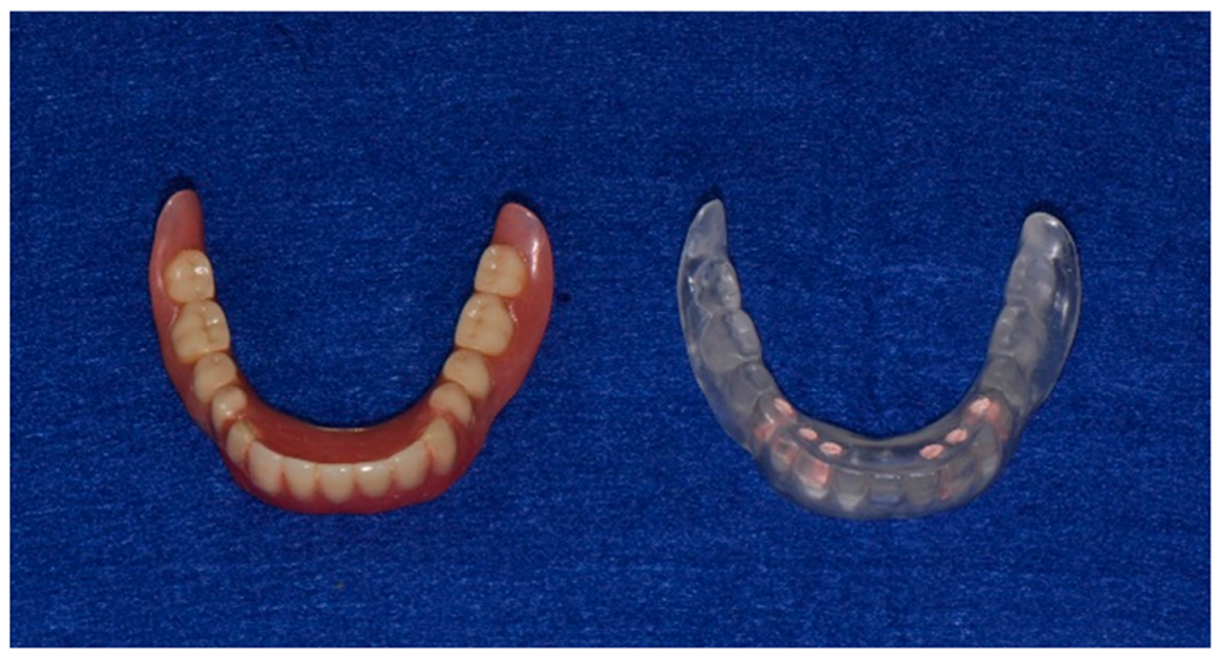
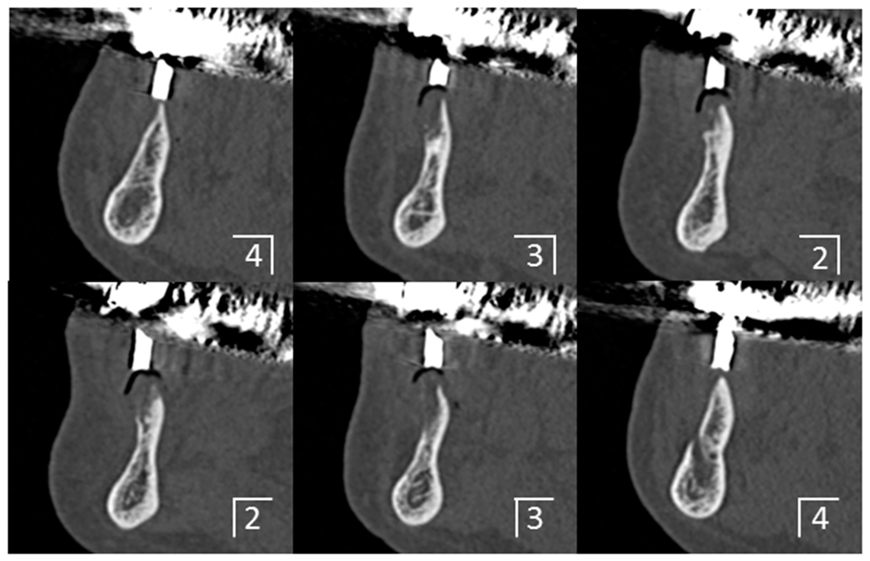
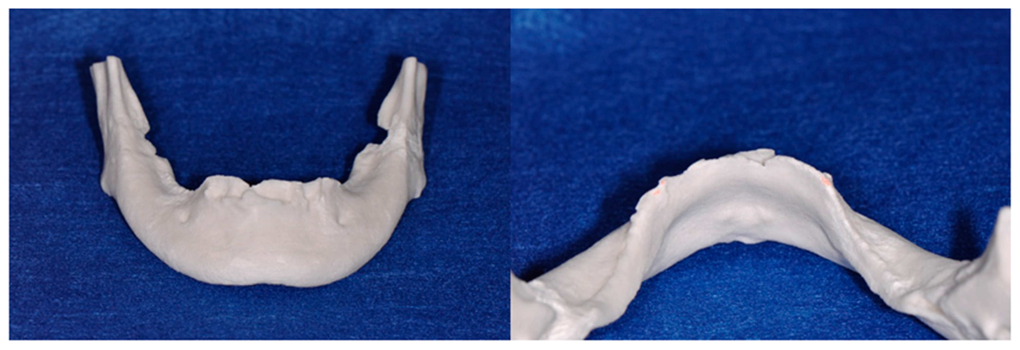
A medical computer tomography (CT) examination was performed for the placement of a diagnostic denture (Figure 2 and Figure 3), and a plaster model of the mandible was then fabricated from CT data using a three-dimensional printer (Figure 4). The bone shape was examined using the CT images and plaster model, and a thin residual ridge was found around the anterior region.
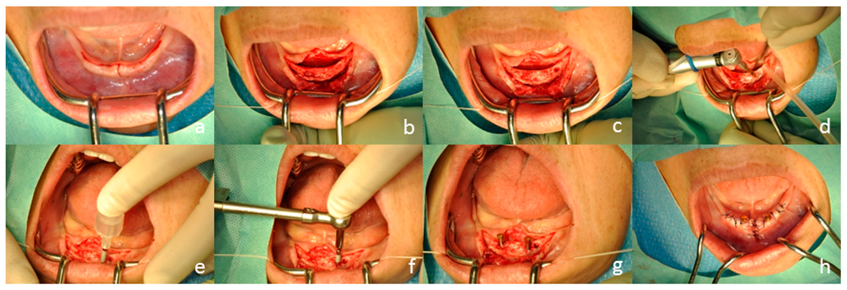
Two mini-implants (Magfit® MIP, Aichi Steel, Aichi, Japan & Platon Japan, Tokyo, Japan) were placed in the anterior region of the mandible. After local anesthesia, the periosteal flap was opened and minimal osetoplasty was done prior to placement of the implant. The mini-implants were placed at the planned positions according to the manufacturer’s directions using the preoperative examinations, and a dome-type keeper was connected to the mini-implant (Figure 5 and Figure 6).
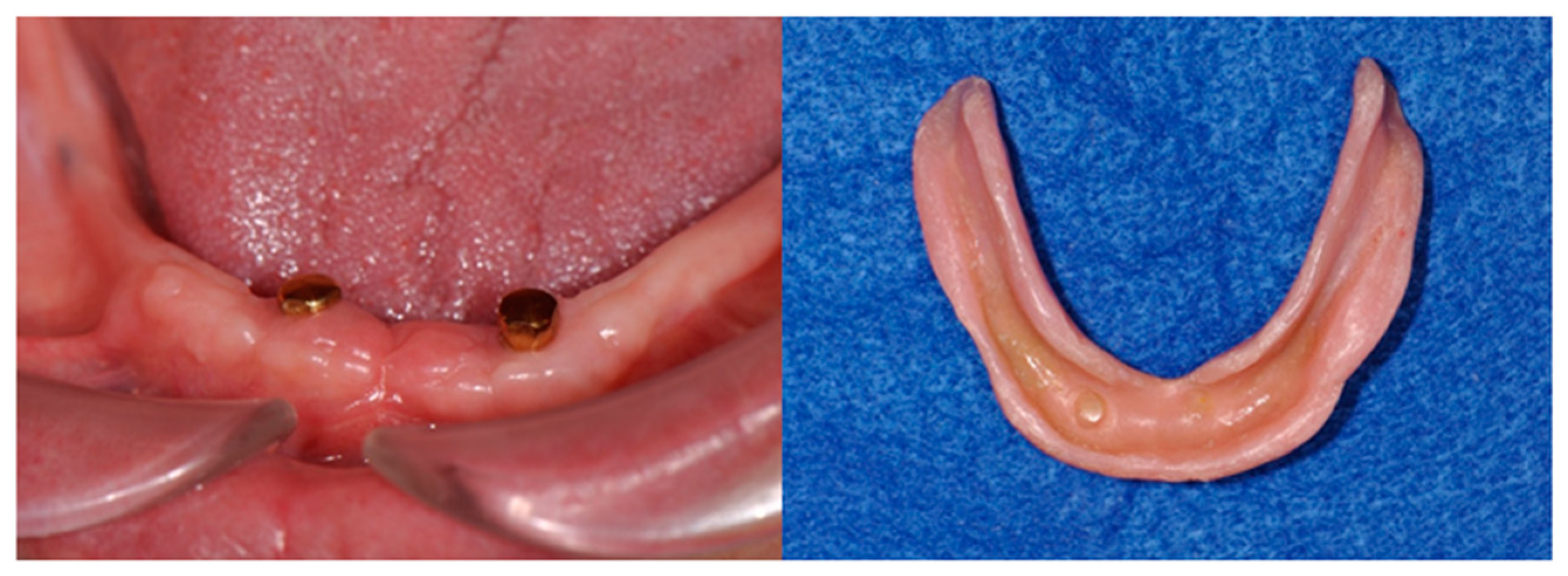
After surgical installation, the denture was fixed so that the implant placement region was relieved by the application of a tissue conditioning material (Figure 7).
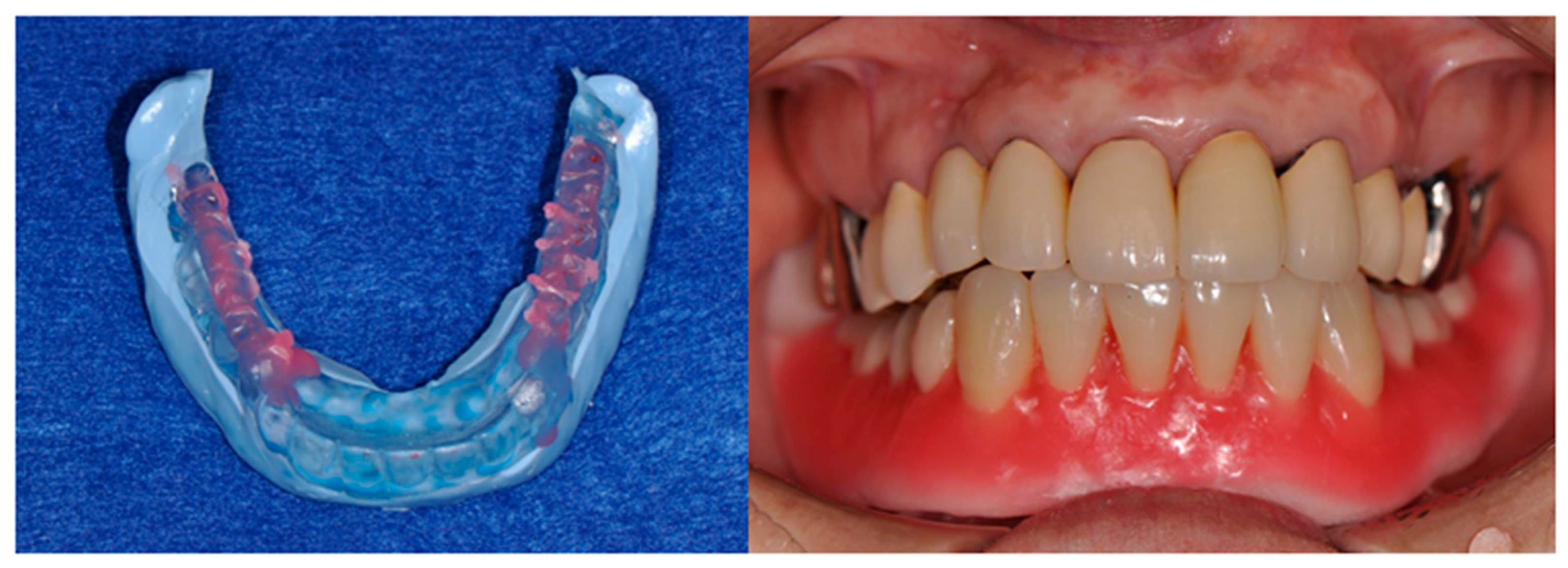
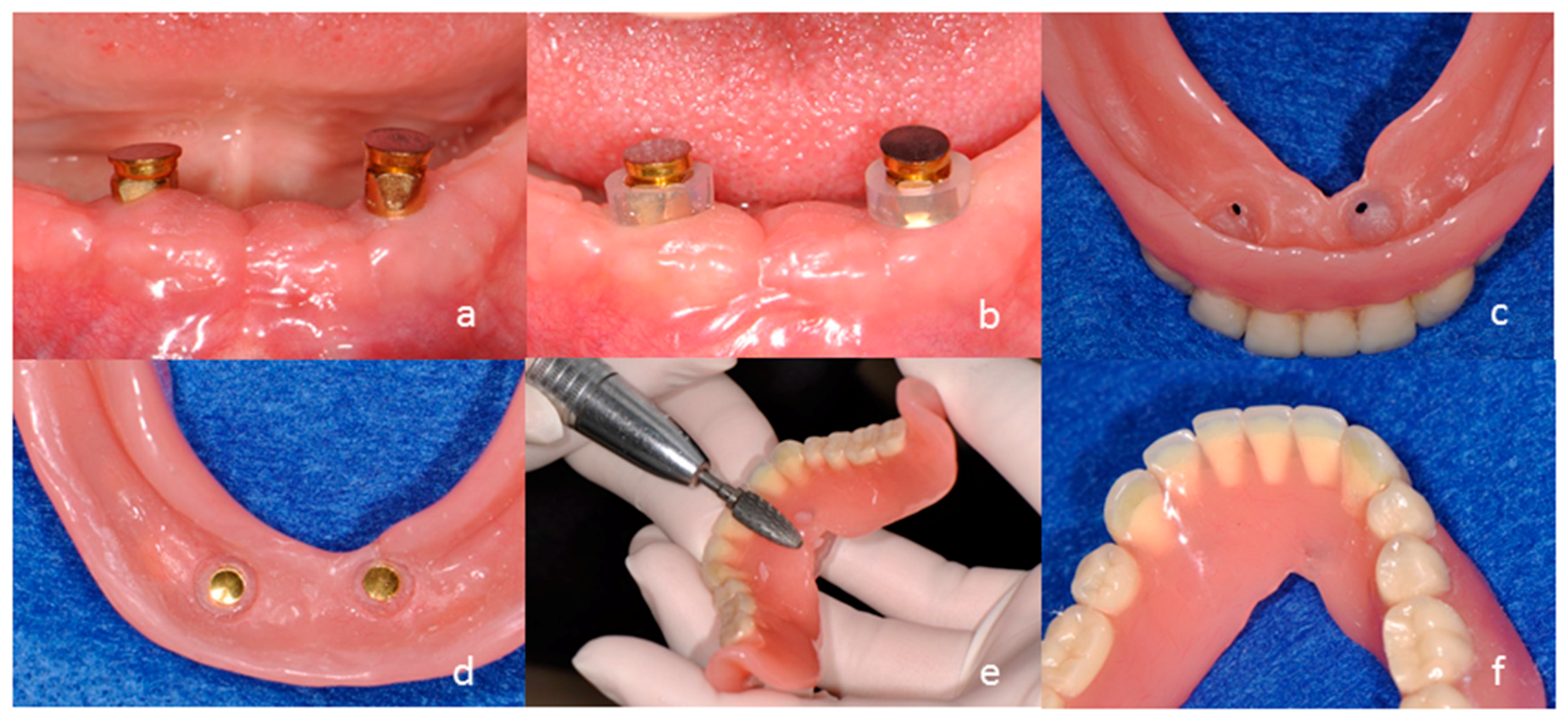
Three months after the procedure, the final impression and interocclusal record of the duplicated denture were obtained (Figure 8). A new magnet-retained two-mini-implant overdenture with hard artificial teeth (ENDURA, Shofu, Kyoto, Japan) was manufactured by conventional denture fabrication. Two magnet assemblies with concave surfaces having a complementary relationship with the dome-type keepers were placed on the mini-implant. The two magnet assemblies were connected to the denture base to hold the complete overdenture in position on the mucosa. Both the mucosa and implants provided support, retention, and stability (Figure 9).
The patient was satisfied with masticatory performance and appearance with the magnet-retained two-mini-implant overdenture.

3. Clinical and Mechanical Considerations

A 2-IOD was built as the primary choice for mandibular edentulism [16,17]. In this case, the patient rejected either treatment option of a fixed four-implant bridge due to cost concerns or an additional surgery such as bore grafting before the placement of two regular implants. Therefore, we decided to use only two mini-implants for this case. Although there are a few reports on the clinical outcome of the 2-IOD using mini-implants [18,19], the outcome is still unclear. Mini-implants may cause mechanical overstressing of the occlusion due to the narrow implant body form [20].
The success of implant overdenture depends on three conditions: implant positioning, selection of attachment, and the splinting between the attachment and denture base [21,22]. Mini-implants can be inserted in narrow ridges via minimally invasive surgery, and are appropriately positioned when parallel to each other and perpendicular to the occlusal plane.
We recommend splinting of the implants in the overdenture because of decreasing the stress around the implants [23]. However, even if the implants are splinted, occlusal stress is not evenly distributed between the implants [24]. The center implant is most likely to be affected by stress, and that implant may have a worse prognosis than the adjacent implants [25]. The load control to each implant should be addressed with considerations for occlusal scheme and contact.
The stress of the implant is the inverse square of the implant diameter [26]. Assuming that a regular implant has a diameter of 3.8 mm and a mini-implant has a diameter of 2.7 mm, the stress of a mini-implant will be about twice as large as that of a regular implant. If assumed that the threshold of bone resorption based on Frost’s theory is 1000 με (20 MPa) [27], the occlusal force is supported around the neck of a 3.75-mm-diameter implant with a 2-mm width and bone contact ratio is 50%; thus, the acceptable amount of occlusal force is ~200 N and 100 N for regular- and mini-implants, respectively. In the previous study, maximum bite force in mandibular two-implant overdenture with a bar attachment was reported around 100 N [28]. Considering this fact, we predict that the load against a mini-implant will not exceed 100 N during conventional mastication, but may occur due to denture movement; special care should be paid to this case.
Magnetic attachment is effective for avoiding excessive horizontal force against the implant, but provides no buffering for axial movements and may require gaps between the keeper and the magnet assembly [29]. The magnet assembly should be installed on a denture base using any load greater than 0 N; this is called “after settling down the denture delivery”. Clinically, the role of the mini-implant is to provide slight support and retention, and these functions were maintained during this study.
Implant overdentures require a level of maintenance that far exceeds that of fixed implant denture. We should give constant attention to the occlusal changes although the hard resin artificial teeth with high strength and wear resistance were used for the implant overdenture. In addition, we have to pay attention to the denture movements by making the implants as a rotation fulcrum, as the alveolar ridge under the denture base area is absorbed for a long term. When the implant overdenture loses soft tissue support and begins to move more freely, relining is applied as maintenance, and the magnetic attachment functions successfully for a long time.

Author Contributions

Yuichi Ishida: case preparation; Yuichi Ishida and H. S. Kiran Kumar: manuscript preparation; Takaharu Goto, Megumi Watanabe, Rudi Wigianto, and Tetsuo Ichikawa: critical discussion and correction of the manuscript; Tetsuo Ichikawa: correspondence.

Conflicts of Interest

Rudi Wigianto and Tetsuo Ichikawa had received travel fees for the regular meetings of International Symposium and Workshop on Magnetic Dentistry (IRPMD) from Aichi steel. The other authors declare that there is no conflict of interests regarding the publication of this paper.

References

  1. Vercruyssen, M.; Marcelis, K.; Coucke, W.; Naert, I.; Quirynen, M. Long-term, retrospective evaluation (implant and patient-centred outcome) of the two-implants-supported overdenture in the mandible. Part 1: Survival rate. Clin. Oral Implant. Res. 2010, 21, 357–365. [Google Scholar] [CrossRef] [PubMed]
  2. Thomason, J.M.; Kelly, S.A.; Bendkowski, A.; Ellis, J.S. Two implant retained overdentures—A review of the literature supporting the McGill and York consensus statements. J. Dent. 2012, 40, 22–34. [Google Scholar] [CrossRef] [PubMed]
  3. Geckili, O.; Cilingir, A.; Erdogan, O.; Kesoglu, A.C.; Bilmenoglu, C.; Ozdiler, A.; Bilhan, H. The influence of momentary retention forces on patient satisfaction and quality of life of two-implant-retained mandibular overdenture wearers. Int. J. Oral Maxillofac. Implant. 2015, 30, 397–402. [Google Scholar] [CrossRef] [PubMed]
  4. Assunção, W.G.; Zardo, G.G.; Delben, J.A.; Barão, V.A. Comparing the efficacy of mandibular implant-retained overdentures and conventional dentures among elderly edentulous patients: Satisfaction and quality of life. Gerodontology 2007, 24, 235–238. [Google Scholar] [CrossRef] [PubMed]
  5. Mundt, T.; Polzer, I.; Samietz, S.; Grabe, H.J.; Messerschmidt, H.; Dören, M.; Schwarz, S.; Kocher, T.; Biffar, R.; Schwahn, C. Socioeconomic indicators and prosthetic replacement of missing teeth in a working-age population: Results of the Study of Health in Pomerania (SHIP). Community Dent. Oral Epidemiol. 2009, 37, 104–115. [Google Scholar] [CrossRef] [PubMed]
  6. Clavero, J.; Lundgren, S. Ramus or chin grafts for maxillary sinus inlay and local onlay augmentation: Comparison of donor site morbidity and complications. Clin. Implant Dent. Relat. Res. 2003, 5, 154–160. [Google Scholar] [CrossRef] [PubMed]
  7. Sebastian, B. Mini-implants: New possibilities in interdisciplinary treatment approaches. Case Rep. Dent. 2014, 2014. [Google Scholar] [CrossRef] [PubMed]
  8. Preoteasa, E.; Imre, M.; Preoteasa, C.T. A 3-year follow-up study of overdentures retained by mini-dental implants. Int. J. Oral Maxillofac. Implant. 2014, 29, 1170–1176. [Google Scholar] [CrossRef] [PubMed]
  9. De Souza, R.F.; Ribeiro, A.B.; Della Vecchia, M.P.; Costa, L.; Cunha, T.R.; Reis, A.C.; Albuquerque, R.F., Jr. Mini vs. Standard Implants for Mandibular Overdentures: A Randomized Trial. J. Dent. Res. 2015, 94, 1376–1384. [Google Scholar] [CrossRef] [PubMed]
  10. Griffitts, T.M.; Collins, C.P.; Collins, P.C. Mini dental implants: An adjunct for retention, stability, and comfort for the edentulous patient. Oral Surg. Oral Med. Oral Pathol. Oral Radiol. Endodontol. 2005, 100, 81–84. [Google Scholar] [CrossRef] [PubMed]
  11. Ichikawa, T.; Horiuchi, M.; Wigianto, R.; Matsumoto, N. In vitro study of mandibular implant-retained overdentures: The influence of stud attachments on load transfer to the implant and soft tissue. Int. J. Prosthodont. 1996, 9, 394–399. [Google Scholar] [PubMed]
  12. Andreiotelli, M.; Att, W.; Strub, J.R. Prosthodontic complications with implant overdentures: A systematic literature review. Int. J. Prosthodont. 2010, 23, 195–203. [Google Scholar] [PubMed]
  13. Naert, I.; Gizani, S.; Vuylsteke, M.; Van Steenberghe, D. A 5-year prospective randomized clinical trial on the influence of splinted and unsplinted oral implants retaining a mandibular overdenture: Prosthetic aspects and patient satisfaction. J. Oral Rehabil. 1999, 26, 195–202. [Google Scholar] [CrossRef] [PubMed]
  14. Heckmann, S.M.; Winter, W.; Meyer, M.; Weber, H.P.; Wichmann, M.G. Overdenture attachment selection and the loading of implant and denture-bearing area. Part 2: A methodical study using five types of attachment. Clin. Oral Implant. Res. 2001, 12, 640–647. [Google Scholar] [CrossRef]
  15. Goodacre, C.J.; Bernal, G.; Rungcharassaeng, K.; Kan, J.Y. Clinical complications with implants and implant prostheses. J. Prosthet. Dent. 2003, 90, 121–132. [Google Scholar] [CrossRef]
  16. Feine, J.S.; Carlsson, G.E.; Awad, M.A.; Chehade, A.; Duncan, W.J.; Gizani, S.; Head, T.; Lund, J.P.; MacEntee, M.; Mericske-Stern, R.; et al. The McGill consensus statement on overdentures. Int. J. Prosthodont. 2002, 15, 413–414. [Google Scholar] [PubMed]
  17. Thomason, J.M.; Feine, J.; Exley, C.; Moynihan, P.; Müller, F.; Naert, I.; Ellis, J.S.; Barclay, C.; Butterworth, C.; Scott, B.; et al. Mandibular two implant-supported overdentures as the first choice standard of care for edentulous patients—The York Consensus Statement. Br. Dent. J. 2009, 207, 185–186. [Google Scholar] [CrossRef] [PubMed]
  18. Tomasi, C.; Idmyr, B.O.; Wennström, J.L. Patient satisfaction with mini-implant stabilized full dentures. A 1-year prospective study. J. Oral Rehabil. 2013, 40, 526–534. [Google Scholar] [CrossRef] [PubMed]
  19. Sfeir, E.; Nassif, N.; Moukarzel, C. Use of mini dental implants in ectodermal dysplasia children: Follow-up of three cases. Eur. J. Paediatr. Dent. 2014, 15, 207–212. [Google Scholar] [PubMed]
  20. Himmlová, L.; Dostálová, T.; Kácovský, A.; Konvicková, S. Influence of implant length and diameter on stress distribution: A finite element analysis. J. Prosthet. Dent. 2004, 91, 20–25. [Google Scholar] [CrossRef] [PubMed]
  21. MacEntee, M.I.; Walton, J.N.; Glick, N. A clinical trial of patient satisfaction and prosthodontic needs with ball and bar attachments for implant-retained complete overdentures: Three-year results. J. Prosthet. Dent. 2005, 93, 28–37. [Google Scholar] [CrossRef] [PubMed]
  22. Duran, I.; Yilmaz, B.; Ural, C. A technique for removing implant-retained denture: Direct relining complication. Case Rep. Dent. 2015, 2015, 1–3. [Google Scholar] [CrossRef] [PubMed]
  23. Jofre, J.; Cendoya, P.; Munoz, P. Effect of splinting mini-implants on marginal bone loss: A biomechanical model and clinical randomized study with mandibular overdentures. Int. J. Oral Maxillofac. Implant. 2010, 25, 1137–1144. [Google Scholar]
  24. Naert, I.; Koutsikakis, G.; Quirynen, M.; Duyck, J.; van Steenberghe, D.; Jacobs, R. Biologic outcome of implant-supported restorations in the treatment of partial edentulism. Part 2: A longitudinal radiographic study. Clin. Oral Implant. Res. 2002, 13, 390–395. [Google Scholar] [CrossRef]
  25. Nissan, J.; Ghelfan, O.; Gross, M.; Chaushu, G. Analysis of load transfer and stress distribution by splinted and unsplinted implant-supported fixed cemented restorations. J. Oral Rehabil. 2010, 37, 658–662. [Google Scholar] [CrossRef] [PubMed]
  26. Baggi, L.; Cappelloni, I.; Di Girolamo, M.; Maceri, F.; Vairo, G. The influence of implant diameter and length on stress distribution of osseointegrated implants related to crestal bone geometry: A three-dimensional finite element analysis. J. Prosthet. Dent. 2008, 100, 422–431. [Google Scholar] [CrossRef] [Green Version]
  27. Frost, H.M. Bone’s mechanostat: A 2003 update. Anat. Rec. A Discov. Mol. Cell Evol. Biol. 2003, 275, 1081–1101. [Google Scholar] [CrossRef] [PubMed]
  28. Elsyad, M.A.; Khairallah, A.S. Chewing efficiency and maximum bite force with different attachment systems of implant overdentures: A crossover study. Clin Oral Implant. Res. 2016. [Google Scholar] [CrossRef] [PubMed]
  29. Goto, T.; Nagao, K.; Ishida, Y.; Tomotake, Y.; Ichikawa, T. Influence of matrix attachment installation load on movement and resultant forces in implant overdentures. J. Prosthodont. 2015, 24, 156–163. [Google Scholar] [CrossRef] [PubMed]
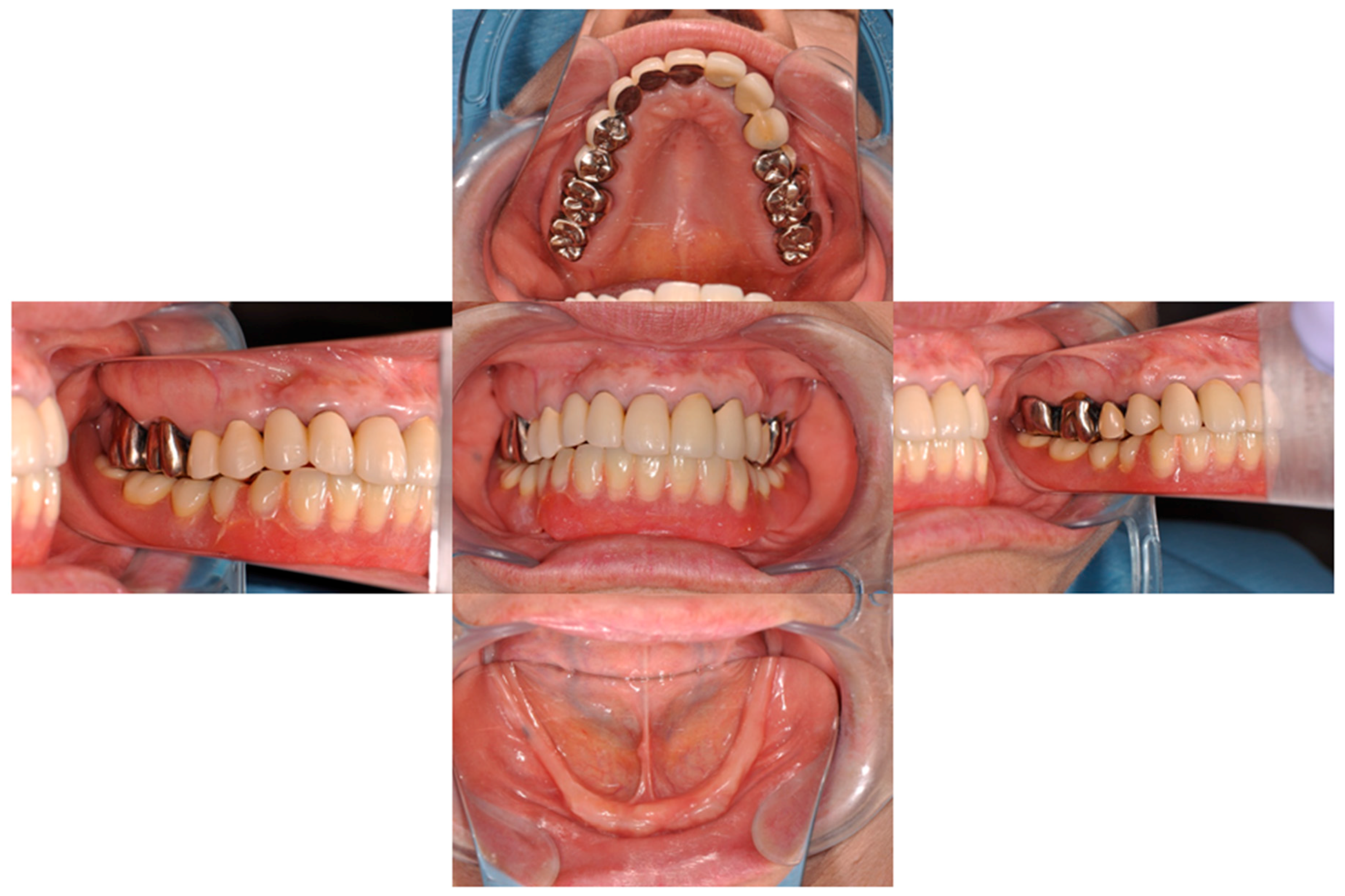
Figure 1. Preoperative view of oral cavity.
Dentistry 04 00035 g001
Figure 2. Wearing denture (Left) and diagnostic denture embedded guttapercha points in the 432┬234 (34,33,32,42,43,44) regions (Right).
Dentistry 04 00035 g002
Figure 3. Preoperative computer tomograpy images taken in placing the diagnostic denture. Cross-sectional images at each position.
Dentistry 04 00035 g003
Figure 4. The plaster model of mandible fabricating by a three-dimensional printer on the CT data. Front (Left) and rear (Right) views.
Dentistry 04 00035 g004
Figure 5. Surgical installation. Incision and periosteal flap was opened. As alveolar plasty is required in the preoperative examination, wider and longitudinal incisions were also added (a,b); alveolar plasty (c) and drilling (d) were performed. Two implant cavities were drilled parallel to each other. Importantly, the perforation through lingual cortical bone was avoided. Implant placement proceeded with hand motion (e) and torque wrench (f); Final torque at the initial fixation was 25 N. Dome-type keepers were placed (g); and the flap was closed with suturing (h). The implant was positioned so that the top of dome-type keeper was the same height as the mocosa.
Dentistry 04 00035 g005
Figure 6. Drilling steps for preparing the implant cavity: Perform cortical bone using a guide drill with less than 700–800 rpm under saline irrigation (a); Confirm the direction and position of implant cavity using guide pin (b); Prepare the implant cavity using starting drill (φ1.8 mm, c) and implant drill (φ2.1 mm, d); Use implant drill (φ2.4 mm, e); if the bone is dense, and implant placement (f). Reproduced with permission from (Aichi Steel, Aichi, Japan) & (Platon Japan, Tokyo, Japan).
Dentistry 04 00035 g006
Figure 7. Three-month postoperative view of mandible (Left), and the tissue conditioning material was relined under denture base of the wearing denture (Right).
Dentistry 04 00035 g007
Figure 8. The completion of final impression and interocclusal records using the duplicated denture (Left). Wax lower denture was fabricated according to the morphology of duplicated denture (Right).
Dentistry 04 00035 g008
Figure 9. Installation of magnet assembly to the new overdenture using an auto-curing acrylic resin. Place the metal-adhesion-treated magnet assembly (a); Set the silicone tube on the keeper (b); Make a hole though denture base (c); Apply the resin material and fix the magnet assembly to the denture base (d); Remove the extra resin (e); Polish (f). The magnet assemble should be fixed to the denture base with the load, or after the overdenture is settled down to the oral mucosa.
Dentistry 04 00035 g009