
The Role of the Component Metals in the Toxicity of Military-Grade Tungsten Alloy

Abstract
:1. Introduction
2. Experimental Section
2.1. Animals
2.2. Metal Pellets
2.3. Pellet-Implantation Procedures
2.4. Experimental Groups
2.5. Hematology
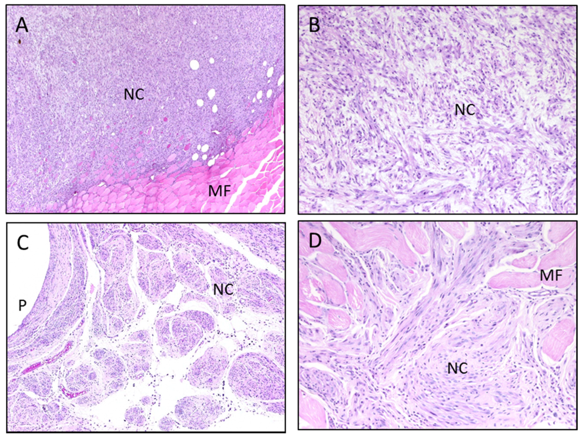
2.6. Pathology
2.7. Sample Preparation for Metal Analysis
2.8. Metal Analysis
2.9. Statistical Analysis
3. Results and Discussion
3.1. Pellet Formulations

| Group | Metal (%) | |||
|---|---|---|---|---|
| Pellet | W | Ni | Co | Ta |
| Ta | - | - | - | 100 |
| WTa | 91.1 | - | - | 8.9 |
| NiTa | - | 6.0 | - | 94.0 |
| CoTa | - | - | 2.9 | 97.1 |
| WNiTa | 91.1 | 6.0 | - | 2.9 |
| WCoTa | 91.1 | - | 2.9 | 6.0 |
| NiCoTa | - | 6.0 | 2.9 | 91.1 |
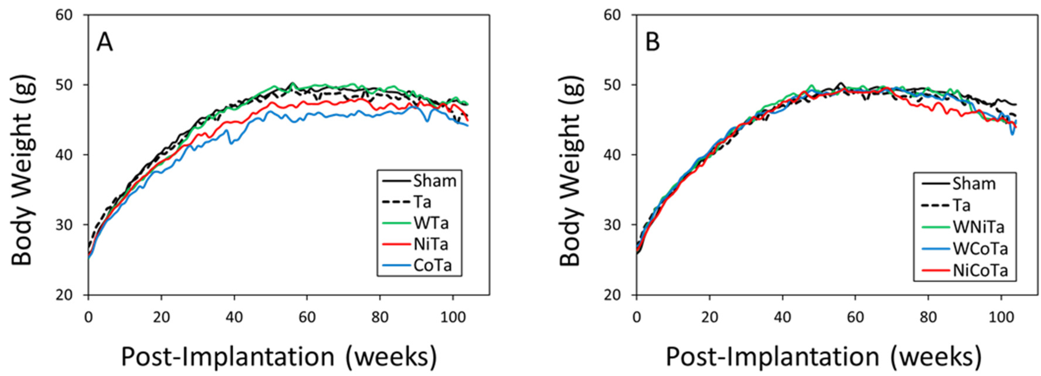
3.2. Pellet Implantation

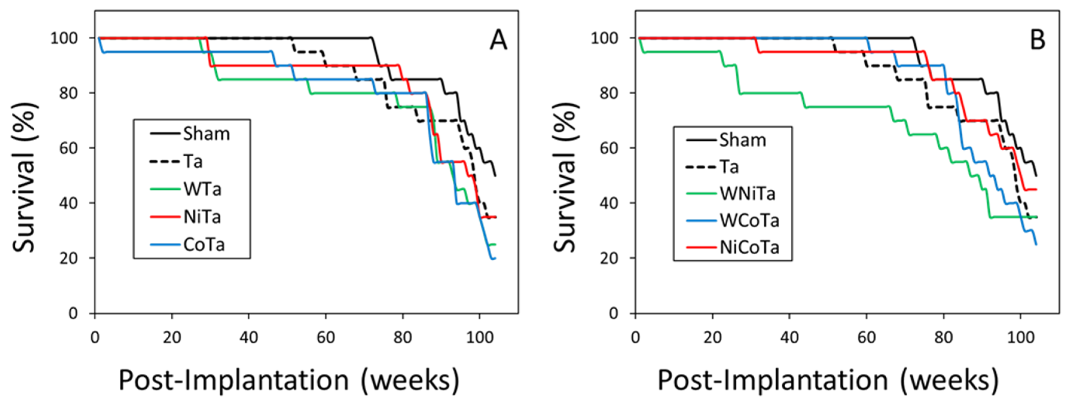
3.3. Survival Rates and Necropsy Findings

| Group | Median Survival (Weeks) |
|---|---|
| Sham | 104 |
| Ta | 99 |
| WTa | 93 * |
| NiTa | 97 |
| CoTa | 93 * |
| WNiTa | 87 * |
| WCoTa | 92 |
| NiCoTa | 100 |
| Tissue | ||||||
|---|---|---|---|---|---|---|
| Group | Liver | Lung | Spleen | Kidney | Testes | Skeletal Muscle |
| Sham | 8 | 4 | 1 | - | - | - |
| Ta | 8 | 2 | 1 | 2 | - | - |
| WTa | 8 | 6 | - | - | - | 1 |
| NiTa | 8 | 5 | 1 | - | - | - |
| CoTa | 7 | 2 | 2 | - | - | 4 |
| WNiTa | 5 | 1 | 1 | 1 | 1 | - |
| WCoTa | 7 | - | 2 | - | - | 1 |
| NiCoTa | 6 | 1 | 3 | 1 | - | - |

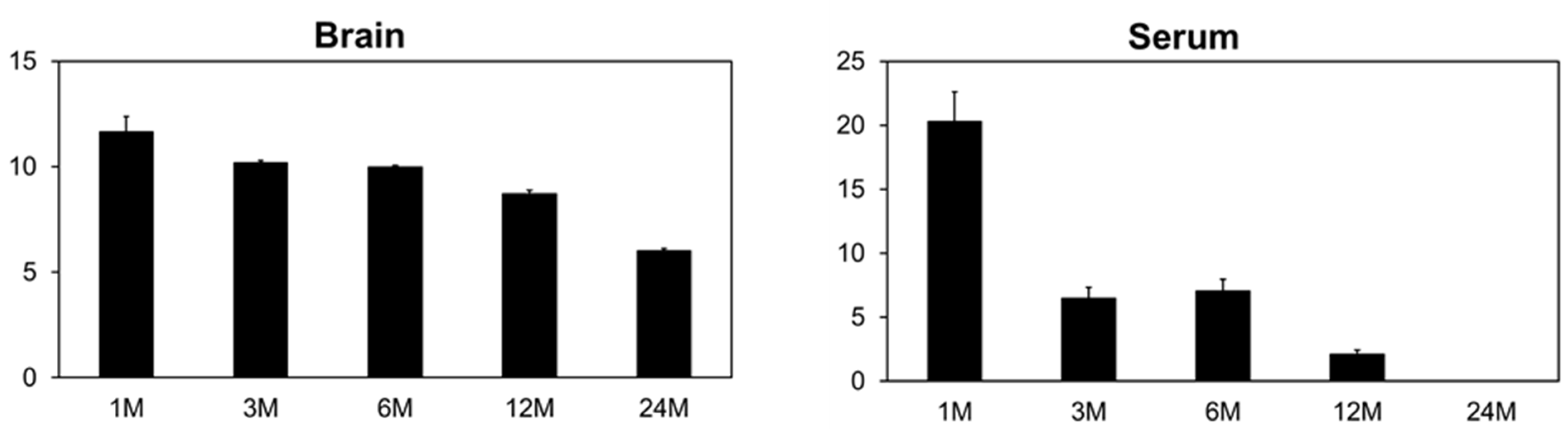
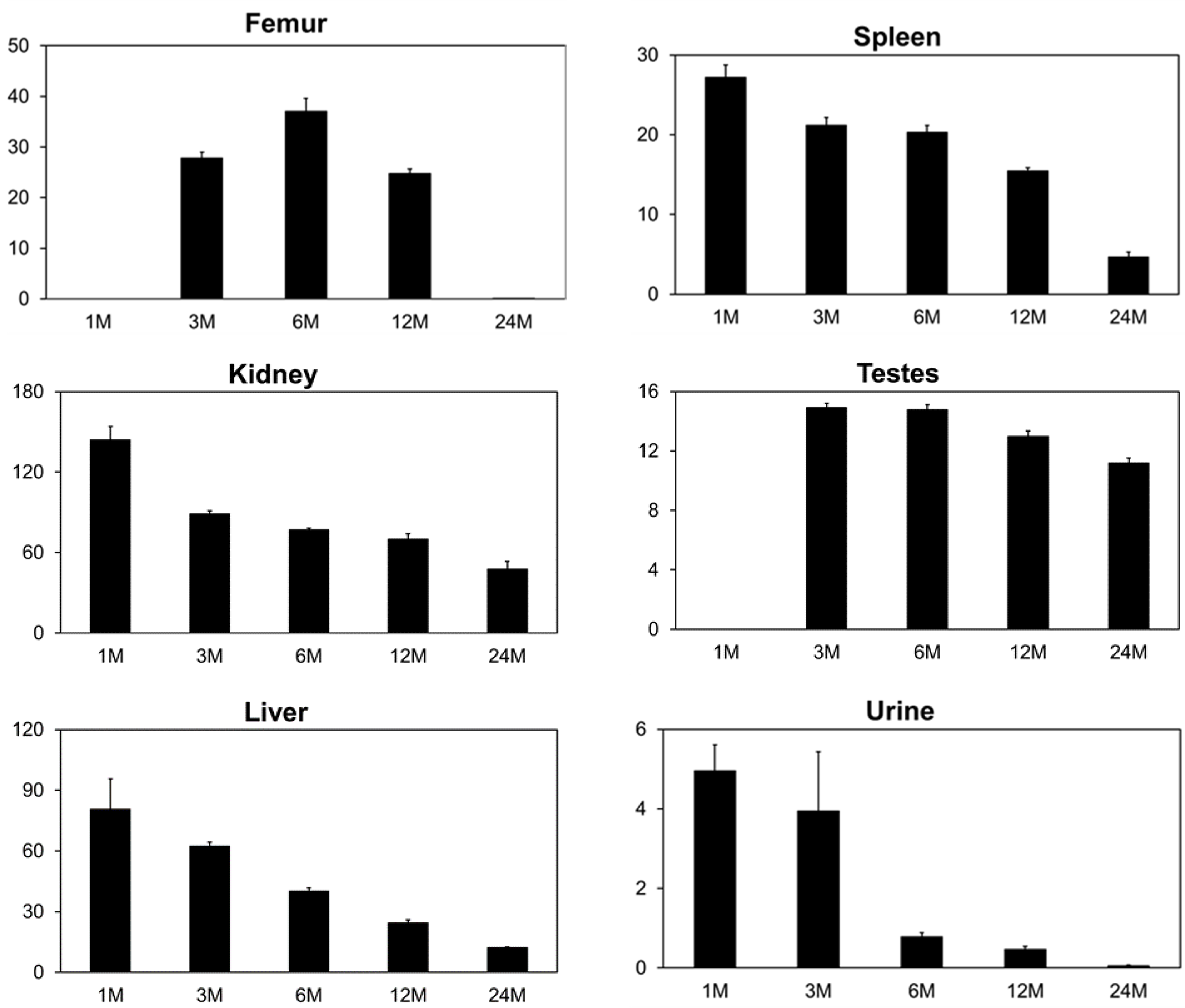
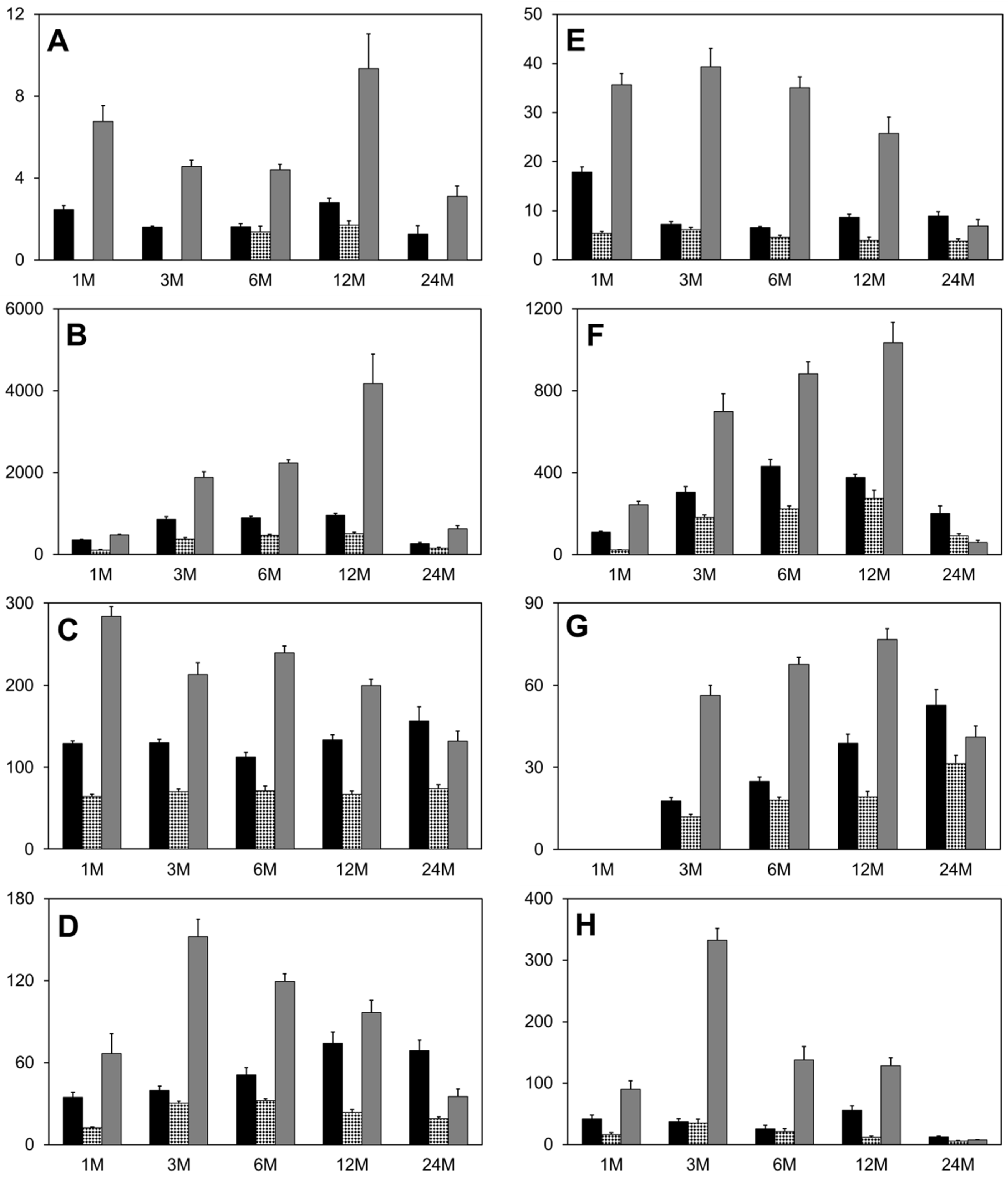
3.4. Tissue Metal Analysis
| Instrument Parameters | |
| Nebulizer type | Concentric |
| Spray chamber | Conical, with impact bead |
| Sampler cone | Platinum, 1-mm orifice diameter |
| Skimmer cone | Platinum, 0.7-mm orifice diameter |
| Sample uptake rate | 1.0 mL/min |
| Sample read delay | 60 s |
| Plasma Conditions | |
| RF power | 1400 W |
| Plasma argon gas flow | 13.0 L/min |
| Auxiliary argon gas flow | 0.80 L/min |
| Nebulizer gas flow | 0.94 L/min |
| Mass Spectrometer Settings | |
| Scanning mode | Peak jump |
| Sweeps | 100 |
| Dwell time | 600 µs |
| Channels/mass | 1 |
| Acquisition time | 18 s |
| Number of readings/replicate | 5 |
| Number of replicates | 2 |


| Group | Time Post-Implantation | ||||
|---|---|---|---|---|---|
| 1 M | 3 M | 6 M | 12 M | 24 M | |
| Ta | 98.7 ± 0.2 | 102.0 ± 1.1 | 97.9 ± 0.7 | 100.1 ± 0.2 | 101.9 ± 1.3 |
| WTa | 94.3 ± 3.0 | 92.6 ± 1.5 | 88.9 ± 1.2 | 79.8 ± 2.4 | 74.8 ± 4.8 |
| NiTa | 94.6 ± 1.2 | 94.0 ± 1.2 | 98.8 ± 1.0 | 96.6 ± 0.7 | 98.1 ± 1.0 |
| CoTa | 95.2 ± 1.0 | 94.0 ± 0.5 | 98.5 ± 0.4 | 97.2 ± 1.0 | 101.0 ± 0.6 |
| WNiTa | 97.2 ± 0.9 | 98.8 ± 1.4 | 92.8 ± 1.9 | 84.7 ± 1.6 | 77.2 ± 1.8 |
| WCoTa | 91.9 ± 1.0 | 86.2 ± 0.6 | 73.9 ± 1.2 | 48.4 ± 3.1 | 34.8 ± 2.1 |
| NiCoTa | 95.0 ± 0.5 | 97.0 ± 0.9 | 93.8 ± 0.7 | 94.7 ± 0.6 | 95.4 ± 0.7 |
4. Conclusions
Acknowledgments
Author Contributions
Conflicts of Interest
Backmatter
References
- McClain, D.E.; Benson, K.A.; Dalton, T.K.; Ejnik, J.; Emond, C.A.; Hodge, S.J.; Kalinich, J.F.; Landauer, M.R.; Livengood, D.R.; Miller, A.C.; et al. Health effects of embedded depleted uranium. Mil. Med. 2002, 167, 117–119. [Google Scholar] [PubMed]
- Van der Voet, G.B.; Todorov, T.I.; Centeno, J.A.; Jonas, W.; Ives, J.; Mullick, F.G. Metals and health: A clinical toxicological perspective on tungsten and review of the literature. Mil. Med. 2007, 172, 1002–1005. [Google Scholar] [CrossRef] [PubMed]
- United States Fish and Wildlife Service. Nontoxic Shot Regulations for Hunting Waterfowl and Coots in the U.S. Available online: www.fws.gov/migratorybirds/CurrentBirdIssues/nontoxic.htm (accessed on 3 November 2015).
- Kraabel, B.J.; Miller, M.W.; Getzy, D.M.; Ringelman, J.K. Effects of embedded tungsten-bismuth-tin shot and steel shot on mallards (Anas platyrhynchos). J. Wildl. Dis. 1996, 32, 1–8. [Google Scholar] [CrossRef] [PubMed]
- Kelly, M.E.; Fitzgerald, S.D.; Aulerich, R.J.; Balander, R.J.; Powell, D.C.; Stickle, R.L.; Stevens, W.; Cray, C.; Tempelman, R.J.; Bursian, S.J. Acute effects of lead, steel, tungsten-iron, and tungsten-polymer shot administered to game-farm mallards. J. Wildl. Dis. 1998, 34, 673–687. [Google Scholar] [CrossRef] [PubMed]
- Mitchell, R.R.; Fitzgerald, S.D.; Aulerich, R.J.; Balander, R.J.; Powell, D.C.; Tempelman, R.J.; Cray, C.; Stevens, W.; Bursian, S.J. Hematological effects and metal residue concentrations following chronic dosing with tungsten-iron and tungsten-polymer shot in adult game-farm mallards. J. Wildl. Dis. 2001, 37, 459–467. [Google Scholar] [CrossRef] [PubMed]
- Mitchell, R.R.; Fitzgerald, S.D.; Aulerich, R.J.; Balander, R.J.; Powell, D.C.; Tempelman, R.J.; Stevens, W.; Bursian, S.J. Reproductive effects and duckling survivability following chronic dosing with tungsten-iron and tungsten-polymer shot in adult game-farm mallards. J. Wildl. Dis. 2001, 37, 468–474. [Google Scholar] [CrossRef] [PubMed]
- Mitchell, R.R.; Fitzgerald, S.D.; Aulerich, R.J.; Balander, R.J.; Powell, D.C.; Tempelman, R.J.; Stickle, R.L.; Stevens, W.; Bursian, S.J. Health effects following chronic dosing with tungsten-iron and tungsten-polymer shot in adult game-farm mallards. J. Wildl. Dis. 2001, 37, 451–458. [Google Scholar] [CrossRef] [PubMed]
- Brewer, L.; Fairbrother, A.; Clark, J.; Amick, D. Acute toxicity of lead, steel, and an iron-tungsten-nickel shot to mallard ducks (Anas platyrhynchos). J. Wildl. Dis. 2003, 39, 638–648. [Google Scholar] [CrossRef] [PubMed]
- Oak Ridge National Laboratories. Application of Life Cycle Analysis: The Case of Green Bullets; ORNL/CP-98264; Oak Ridge National Laboratories: Oak Ridge, TN, USA, 1998.
- Kalinich, J.F.; Emond, C.A.; Dalton, T.K.; Mog, S.R.; Coleman, G.D.; Kordell, J.E.; Miller, A.C.; McClain, D.E. Embedded weapons-grade tungsten alloy shrapnel rapidly induces metastatic high-grade rhabdomyosarcomas in F344 rats. Environ. Health Perspect. 2005, 113, 729–734. [Google Scholar] [CrossRef] [PubMed]
- Schuster, B.E.; Roszell, L.E.; Murr, L.E.; Ramirez, D.A.; Demaree, J.D.; Klotz, B.R.; Rosencrance, A.B.; Dennis, W.E.; Bao, W.; Perkins, E.J.; et al. In vivo corrosion, tumor outcome, and microarray gene expression for two types of muscle-implanted tungsten alloys. Toxicol. Appl. Pharmacol. 2012, 265, 128–138. [Google Scholar] [CrossRef] [PubMed]
- Emond, C.A.; Vergara, V.B.; Lombardini, E.D.; Mog, S.R.; Kalinich, J.F. Induction of rhabdomyosarcoma by embedded military-grade tungsten/nickel/cobalt not by tungsten/nickel/iron in the B6C3F1 mouse. Int. J. Toxicol. 2015, 34, 44–54. [Google Scholar] [CrossRef] [PubMed]
- Hockley, A.D.; Goldin, J.H.; Wake, M.J.C.; Iqbal, J. Skull repair in children. Pediatr. Neurosurg. 1990, 16, 271–275. [Google Scholar] [CrossRef] [PubMed]
- Johansson, C.B.; Hansson, H.A.; Albrektsson, T. Qualitative interfacial study between bone and tantalum, niobium or commercially pure titanium. Biomaterials 1990, 11, 277–280. [Google Scholar] [CrossRef]
- Strecker, E.P.; Hagan, B.; Liermann, D.; Schneider, B.; Wolf, H.R.; Wambsganss, J. Iliac and femoropopliteal vascular occlusive disease treated with flexible tantalum stents. Cardiovasc. Interv. Radiol. 1993, 16, 158–164. [Google Scholar] [CrossRef]
- Institute for Laboratory Animal Research; National Research Council. Guide for the Care and Use of Laboratory Animals, 8th ed.; National Academies Press: Washington, DC, USA, 2010. [Google Scholar]
- Slot, C. Plasma creatinine determination. A new and specific Jaffe reaction method. Scand. J. Clin. Lab. Investig. 1965, 17, 381–387. [Google Scholar] [CrossRef] [PubMed]
- Heinegard, D.; Tiderstrom, G. Determination of serum creatinine by a direct colorimetric method. Clin. Chim. Acta 1973, 43, 305–310. [Google Scholar]
- Emond, C.A.; Kalinich, J.F. Biokinetics of embedded surrogate radiological dispersal device material. Health Phys. 2012, 102, 124–136. [Google Scholar] [CrossRef] [PubMed]
- Pellmar, T.C.; Fuciarelli, A.F.; Ejnik, J.W.; Hamilton, M.; Hogan, J.; Strocko, S.; Emond, C.; Mottaz, H.M.; Landauer, M.R. Distribution of uranium in rats implanted with depleted uranium pellets. Toxicol. Sci. 1999, 49, 29–39. [Google Scholar] [CrossRef] [PubMed]
- Chandra, M.; Frith, C.H. Spontaneous neoplasms in B6C3F1 mice. Toxicol. Lett. 1992, 60, 91–98. [Google Scholar] [CrossRef]
- Haseman, J.K.; Hailey, J.R.; Morris, R.W. Spontaneous neoplasm incidences in Fischer 344 rats and B6C3F1 mice in two-year carcinogenicity studies: A National Toxicology Program update. Toxicol. Pathol. 1998, 26, 428–441. [Google Scholar] [CrossRef] [PubMed]
- Harris, R.M.; Williams, T.D.; Hodges, N.J.; Waring, R.H. Reactive oxygen species and oxidative DNA damage mediate the cytotoxicity of tungsten-nickel-cobalt alloys in vitro. Toxicol. Appl. Pharmacol. 2011, 250, 19–28. [Google Scholar] [CrossRef] [PubMed]
- Miller, A.C.; Mog, S.; McKinney, L.-A.; Luo, L.; Allen, J.; Xu, J.; Page, N. Neoplastic transformation of human osteoblast cells to the tumorigenic phenotype by heavy metal-tungsten alloy particles: Induction of genotoxic effects. Carcinogenesis 2001, 22, 115–125. [Google Scholar] [CrossRef] [PubMed]
- Harris, R.M.; Williams, T.D.; Waring, R.H.; Hodges, N.J. Molecular basis of carcinogenicity of tungsten alloy particles. Toxicol. Appl. Pharmacol. 2015, 283, 223–233. [Google Scholar] [CrossRef] [PubMed]
- Guilbert, C.; Kelly, A.D.R.; Petruccelli, L.A.; Lemaire, M.; Mann, K.K. Exposure to tungsten induces DNA damage and apoptosis in developing B lymphocytes. Leukemia 2011, 25, 1900–1904. [Google Scholar] [CrossRef] [PubMed]
- Kelly, A.D.R.; Lemaire, M.; Young, Y.K.; Eustache, J.H.; Guilbert, C.; Molina, M.F.; Mann, K.K. In vivo tungsten exposure alters B-cell development and increases DNA damage in murine bone marrow. Toxicol. Sci. 2013, 131, 434–446. [Google Scholar] [CrossRef] [PubMed]
- Laulicht, F.; Brocato, J.; Cartularo, L.; Vaughan, J.; Wu, F.; Kluz, T.; Sun, H.; Oksuz, B.A.; Shen, S.; Peana, M.; et al. Tungsten-induced carcinogenesis in human bronchial epithelial cells. Toxicol. Appl. Pharmacol. 2015, 288, 33–39. [Google Scholar] [CrossRef] [PubMed]
- Osawa, R.; Abe, R.; Inokuma, D.; Yokota, K.; Ito, H.; Nabeshima, M.; Shimizu, H. Chain saw blade granuloma: Reaction to a deeply embedded metal fragment. Arch. Dermatol. 2006, 142, 1079–1080. [Google Scholar] [CrossRef] [PubMed]
- Saruwatari, H.; Kamiwada, R.; Matsushita, S.; Hashiguchi, T.; Kawai, K.; Kanekura, T. Tungsten granuloma attributable to a piece of lawn-mower blade. Clin. Exp. Dermatol. 2009, 34, e268–e269. [Google Scholar] [CrossRef] [PubMed]
- Bolt, A.M.; Sabourin, V.; Molina, M.F.; Police, A.M.; Silva, L.F.N.; Plourde, D.; Lemaire, M.; Ursini-Siegel, J.; Mann, K.K. Tungsten targets the tumor microenvironment to enhance breast cancer metastasis. Toxicol. Sci. 2015, 143, 165–177. [Google Scholar] [CrossRef] [PubMed]
© 2015 by the authors; licensee MDPI, Basel, Switzerland. This article is an open access article distributed under the terms and conditions of the Creative Commons Attribution license (http://creativecommons.org/licenses/by/4.0/).
Share and Cite
Emond, C.A.; Vergara, V.B.; Lombardini, E.D.; Mog, S.R.; Kalinich, J.F. The Role of the Component Metals in the Toxicity of Military-Grade Tungsten Alloy. Toxics 2015, 3, 499-514. https://doi.org/10.3390/toxics3040499
Emond CA, Vergara VB, Lombardini ED, Mog SR, Kalinich JF. The Role of the Component Metals in the Toxicity of Military-Grade Tungsten Alloy. Toxics. 2015; 3(4):499-514. https://doi.org/10.3390/toxics3040499
Chicago/Turabian StyleEmond, Christy A., Vernieda B. Vergara, Eric D. Lombardini, Steven R. Mog, and John F. Kalinich. 2015. "The Role of the Component Metals in the Toxicity of Military-Grade Tungsten Alloy" Toxics 3, no. 4: 499-514. https://doi.org/10.3390/toxics3040499