Article

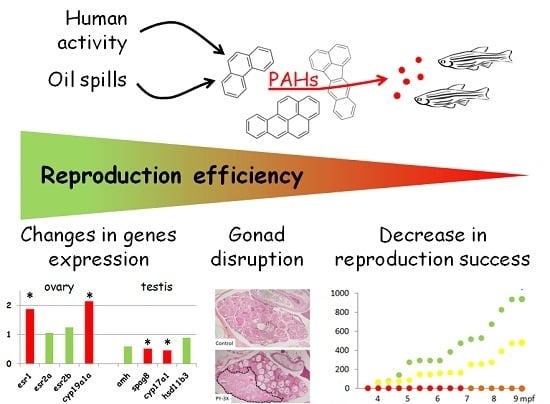
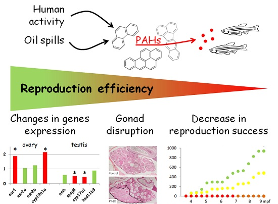
Fish Reproduction Is Disrupted upon Lifelong Exposure to Environmental PAHs Fractions Revealing Different Modes of Action

1
Ifremer, Ecotoxicology Laboratory, Place Gaby Coll, F-17137 L’Houmeau, France
2
INRA UMR703, APEX, Oniris, F-44307 Nantes, France
3
Oniris, École Nationale vétérinaire, Agro-Alimentaire et de L’alimentation Nantes-Atlantique, LUNAM Université, F-44307 Nantes, France
4
Department of Science and Technology, University of Bordeaux 1, EPOC, UMR CNRS 5805, F-33405 Talence, France
5
Ifremer, Fisheries Laboratory, Place Gaby Coll, F-17137 L’Houmeau, France
6
INRA LPGP, Campus de Beaulieu, F-35042 Rennes, France
7
Ifremer, Laboratoire Adaptation et Adaptabilité des Animaux et des Systèmes, UMR MARBEC, Route de Maguelone, F-34250 Palavas les Flots, France
*
Author to whom correspondence should be addressed.
Toxics 2016, 4(4), 26; https://doi.org/10.3390/toxics4040026
Submission received: 18 July 2016 / Revised: 17 October 2016 / Accepted: 19 October 2016 / Published: 28 October 2016
(This article belongs to the Special Issue Zebrafish as a Model for Assessing Chemical Toxicity)

Abstract

:
Polycyclic aromatic hydrocarbons (PAHs) constitute a large family of organic pollutants emitted in the environment as complex mixtures, the compositions of which depend on origin. Among a wide range of physiological defects, PAHs are suspected to be involved in disruption of reproduction. In an aquatic environment, the trophic route is an important source of chronic exposure to PAHs. Here, we performed trophic exposure of zebrafish to three fractions of different origin, one pyrolytic and two petrogenic. Produced diets contained PAHs at environmental concentrations. Reproductive traits were analyzed at individual, tissue and molecular levels. Reproductive success and cumulative eggs number were disrupted after exposure to all three fractions, albeit to various extents depending on the fraction and concentrations. Histological analyses revealed ovary maturation defects after exposure to all three fractions as well as degeneration after exposure to a pyrolytic fraction. In testis, hypoplasia was observed after exposure to petrogenic fractions. Genes expression analysis in gonads has allowed us to establish common pathways such as endocrine disruption or differentiation/maturation defects. Taken altogether, these results indicate that PAHs can indeed disrupt fish reproduction and that different fractions trigger different pathways resulting in different effects.

Graphical Abstract

1. Introduction

In ecology, recruitment is defined as the ability of one individual to contribute to the next generation. From birth, this includes a large number of abilities such as to develop, to grow, to meet congeners and to reproduce. Beyond intrinsic characteristics, genetically defined, many external factors can impede recruitment. Among them, xenobiotics could act on virtually all functions or important steps [1].
Among xenobiotics, polycyclic aromatic hydrocarbons (PAHs) form a diverse family of compounds containing at least two aromatic rings [2]. PAHs are increasingly emitted in the environment as a consequence of human activities [3,4]. There are two major types of environmental PAHs, petrogenic and pyrolytic, which enter the environment through different routes. Petrogenic PAHs are abundant in oils and enter the aquatic environment due to harbor activity or as a consequence of oil spills. Pyrolytic PAHs result from the incomplete combustion of organic matter, including fossil fuel; they enter aquatic environments through the deposition of atmospheric emissions directly on water or on the ground followed by soil runoff. Because PAHs are hydrophobic molecules, they are found to be associated with suspended particulate matter in water, such that they tend to accumulate in sediments; consequently, sediments constitute major sinks and can also act as secondary sources for aquatic systems contamination [5]. Monitoring networks have documented PAH concentrations of up to 50 μg·g−1 dry weight (dw) in sediment from various affected aquatic ecosystems although the concentrations in highly contaminated areas are more commonly in the 10 μg·g−1 range [6,7,8,9,10,11].
Thanks to its biological characteristics, including a short life cycle, the zebrafish makes a particularly good case study for performing longitudinal studies along the life cycle. This applies in the case of reproduction for which it is possible in some months to monitor the gonads’ development and maturation, endocrine regulation and reproductive effort. In addition, the large numbers of tools developed to study zebrafish physiology allows for a refined analysis [12,13].
Reproduction is a key function to any species survival, but, at the same time, it is the target of some xenobiotics. Reproduction disruption may be the results of direct insults such as reproduction hormones modification in the case of endocrine disruptor compounds or may be the indirect consequence of general physiological degradation including growth or behavioral defects [14]. Additional complexity is due to the fact that reproduction is the ultimate step in a long-term process, from germ-cells’ migration in embryos to gametes’ maturation in reproducing adults, and that all these steps can be disrupted throughout life and eventually result in reproduction failure. Complexity is also due to the fact that, in the environment, xenobiotics are present as complex mixtures of compounds which, taken separately, can have pro- or anti-estrogenic properties [15]. PAHs have been shown to interfere with reproduction in fish and other animals, including invertebrates [16,17,18,19,20,21,22,23,24,25]. From a mechanistic point of view, many elements remain unclear; however, PAHs have been shown to produce endocrine disruption in fish, which has been revealed by disruption of circulating steroid hormones level in males and females [16,26,27,28,29,30].
In previous articles we have reported physiological disruptions observed in zebrafish after long-term diet exposure to environmentally relevant mixtures of polycyclic aromatic hydrocarbons (PAHs) and demonstrated disruption of growth, behavior and survival along with an increase in tumorigenesis [31,32,33]. In order to evaluate the reprotoxicity of environmental PAHs mixtures, zebrafish were exposed through diet from the first meal (5-days post fertilization (dpf)) and for the 9 following months, i.e., from larvae to reproducing adults. Three complex fractions of PAHs, representative of environmental situations, were used to spike the food: a pyrolitic (PY) fraction from sediment sampled in the Seine Estuary; and two petrogenic fractions, one from a heavy oil “Erika” (HO) and one from a light crude oil “Arabian light” (LO). We analyzed the consequences of this exposure on reproduction evaluated at functional, organ and molecular levels.

2. Material and Methods

2.1. PAH-Contaminated Diet Preparation

Three aromatic fractions were used for exposures: (i) A pyrolytic fraction (PY) extracted from sediments collected in a polluted site of the Seine Estuary (Oissel, France) and (ii) two petrogenic fractions obtained from Erika fuel (heavy oil; HO) and Arabian Light crude oil (LO). PAHs extractions were performed as previously described [7]. Zebrafish TU strain (ZFIN ID: 76 ZDB-GENO-990623-3) was used and larvae were exposed from their first meal (at 5 dpf) onward using spiked food pellets [33]. Three concentrations, 0.3X, 1X and 3X were used with the 1X concentration corresponding to the 16 PAHs used as indicators by the US Environmental Protection Agency (US-EPA) at 5 μg·g−1 dw food, representative of the concentrations found in mollusks in the Seine Estuary. Although it does not give an accurate representation of the fractions’ compositions, this subset of PAHs has been used for comparison with concentrations found in the environment (see in Table S1 a detailed description of actual fractions compositions). Diets are named after the origin of the fraction and its concentration: the 1X pyrolytic fraction diet will be named PY-1X. For each exposure, a fourth control treatment was included corresponding to the plain food treated as spiked-food with dichloromethane which was used as carrier solvent for PAHs spiking. Because the fraction extracted from the original sample is a mixture of PAHs, the term mixture will also be used more or less synonymously with fraction.

2.2. Reproduction Monitoring

Fish were reared in triplicates as batches of 25–30 individuals in 10 L tanks (see [33] for further details). Onset and spawning effort were determined by placing two spawning boxes (AquaSchwartz, Göttingen, Germany) at 17:00 on the bottom of each 10 L tank from 4 to 6 months post fertilization (mpf) in PY fish, from 4 to 7 mpf in HO and from 5 to 9 in LO fish. In total, 19 to 87 spawning assays (solicitation) were performed depending on fraction and concentration (Table 1). Further monitoring of reproduction effort was performed with cross-pairing for LO and HO (without HO-3X) using one exposed fish and one control fish pairwise mated overnight in a spawning box (AquaSchwartz, Germany) outside of the tank. In both cases (group and pairwise mating), spawning occurs the following morning progressively after a few minutes of light illumination. One hour later, eggs were collected, cleaned, sorted (into fertilized or none fertilized), and counted. The monitoring allowed us to assess spawning success (ratio of number of spawns obtained/number of assays), egg production (cumulative number of fertilized egg over observation time further normalized by the number of assays and the number of females in the tank) and spawn fertilization rate.

2.3. Histological Analysis

Fish were sampled at 9 mpf for all diets except for HO which were sampled at 7 mpf because of high mortality in HO-3X condition. Control fish were systematically sampled in parallel. Fish were euthanized with a lethal dose of benzocaine (250 μg·L−1 of a 10% solution in 100% ethanol; Sigma-Aldrich, Lyon, France) and fixed in 4% buffered formalin after ventral incision of the abdomen for larger individuals [34]. After 24 h of fixation, fins, most scales and the caudal peduncle were carefully removed and samples were dehydrated in graded ethanol solutions and embedded in paraffin. Step sectioning was performed as already published [35]. Serial sagittal step sections were cut from the left side of the fish. Four step sections from each adult fish were mounted on glass slides, one from the eye anterior chamber level, one from the eye posterior chamber level, one just medial to the eye, and one at the midline. Sections were routinely stained with haematoxylin-eosin-saffron (HES). A first histological analysis was performed to qualify structure and morphology on the gonads and also to identify lesions. The second step consisted of follicle staging (functional characterization). All oocytes present on mid-level section and on sections medial to the eye were counted along with identification of their morphological stage (F1 to F4) using middle magnification [36]. Individual frequencies of each type of follicles were calculated using ratio to the total ovarian area.

2.4. qPCR Analysis

Ovaries and testis were sampled on 9mpf animals from each fraction and concentration except HO-3X (N = 5 per case) euthanized as described above and immediately frozen in liquid nitrogen and stored at −80 °C until used. Total RNAs were extracted using Trizol Reagent (Invitrogen, Carlsbad, CA, USA) following the manufacturer’s instructions. Total RNA concentration was quantified by spectrophotometry at 260 nm. Furthermore, purity of RNAs was verified by measuring the A260/A230 nm and A260/A280 nm ratios. To remove the possibility of genomic DNA contamination, RNA sample was digested by RNase-free DNase I (Promega, Madison, WI, USA) and then purified. First-strand cDNA was synthetized from a total of 1 µg RNA using reaction mix including 500 ng of oligo(dT)15, 250 ng of random hexamerprimers (Promega, Madison, WI, USA) using M-MLV Reverse Transcriptase (Promega, Madison, WI, USA) following manufacturer’s instructions. Reaction was incubated for 1 h at 42 °C and inactivated by heating for 15 min at 70 °C and cDNA stored at −20 °C until a real-time PCR analysis. PCR primers were designed with Primer 3 software. Real-time PCR reactions were performed using Fast SYBR Green Master Mix 5X (ThermoFisher, Illkirch, France) and in a StepOnePlus instrument (ThermoFisher, Illkirch, France) following the manufacturer’s instructions. Gene’s expressionwere quantified from the threshold cycle (CT) number and normalized to four housekeeping genes (eef1, βactin, g6pd and gapdh). Gene expressions were analyzed in females (esr1, esr2a, esr2b, cyp19a1a) and males (amh, spag8, cyp17a1, hsd11b3) using REST-2009 software (Qiagen, Dusseldorf ,Germany).

2.5. Statistical Analysis

Within each fraction (PY, LO, HO), fertilization rate frequencies were calculated per classes (20% wide) and Chi2 analysis performed to compare eggs fertilization rate as well as spawning success between concentrations using Prism 6.0 (Graphpad, La Jolla, CA, USA ).
Comparing fertilization rates in cross-pairings was performed using non-parametric test, Kruskal-Wallis, whereas GLMs were calculated to compare egg production within each fraction (PY, HO or LO) with concentration as fixed factors (Control, 0.3X, 1X and 3X). For PY ovary morphology, surface of degenerating areas expressed as percentage of total ovarian surface were compared between concentrations (Control, 0.3X, 1X and 3X) using ANOVA. For ovary quality evaluation within each fraction, concentration and follicles stages (F1 to F4) were used as a fixed factor and fish as random factor. Interaction between concentrations and stages was also analyzed. Post hoc tests were performed with Newman-Keuls in all cases. These statistical analyses were performed with Statistica 9.0 (Statsoft, Tulsa, OK, USA) software.
All statistical analyses were carried out at a 95% level of significance and only the fixed factor and the interaction between them are presented in the text. The results reported in text and all figures are means ± SEM.

3. Results

This work is part of a larger program that included analysis of modification of several physiological variables following exposure to the same diets. Chemical analysis of fractions, diets and metabolites have been presented previously [33]. Briefly, PY fraction was characterized by a high proportion of heavy PAHs (82%) and almost no methylated derivatives (<5%). HO fraction contained moderate levels of heavy PAHs (31%) and methylated derivatives (45%). LO fraction contained a very low level of heavy PAHs (6%) and a high level of methylated ones (59%). As mentioned above, proportions of light, heavy and methylated PAHs differ notably between fractions and spiked diets. Total PAHs concentrations based on 22 parents HAPs (including the 16 EPA PAHs) and 8 methylated PAHs are presented in detail in Table S1. Briefly, for PY, HO and LO respectively PAHs parents’ concentrations in 1X diets were 5.5 ± 1.4, 2.6 ± 0.2 and 2.7 ± 0.2 µg·g−1 dw and 0.3 ± 0.1, 2.1 ± 0.2 and 4.0 ± 0.3 µg·g−1 dw for methylnaphthalenes and methylphenanthrenes. This resulted in a similar total concentration of quantified PAHs (included methylated) in the three 1X diets: 5.8 ± 1.4, 4.7 ± 0.4 and 6.7 ± 0.3 µg·g−1 dw for PY, HO and LO respectively. Since PAHs is a family of hundreds of compounds, it is not possible to quantify all individual PAHs and therefore we acknowledge that the above concentrations only partially describe the fractions used. Quantification of hydroxylated metabolites in dietary exposed 15 dpf-larvae confirmed an effective exposure of fish, and results were consistent with diet composition [33].

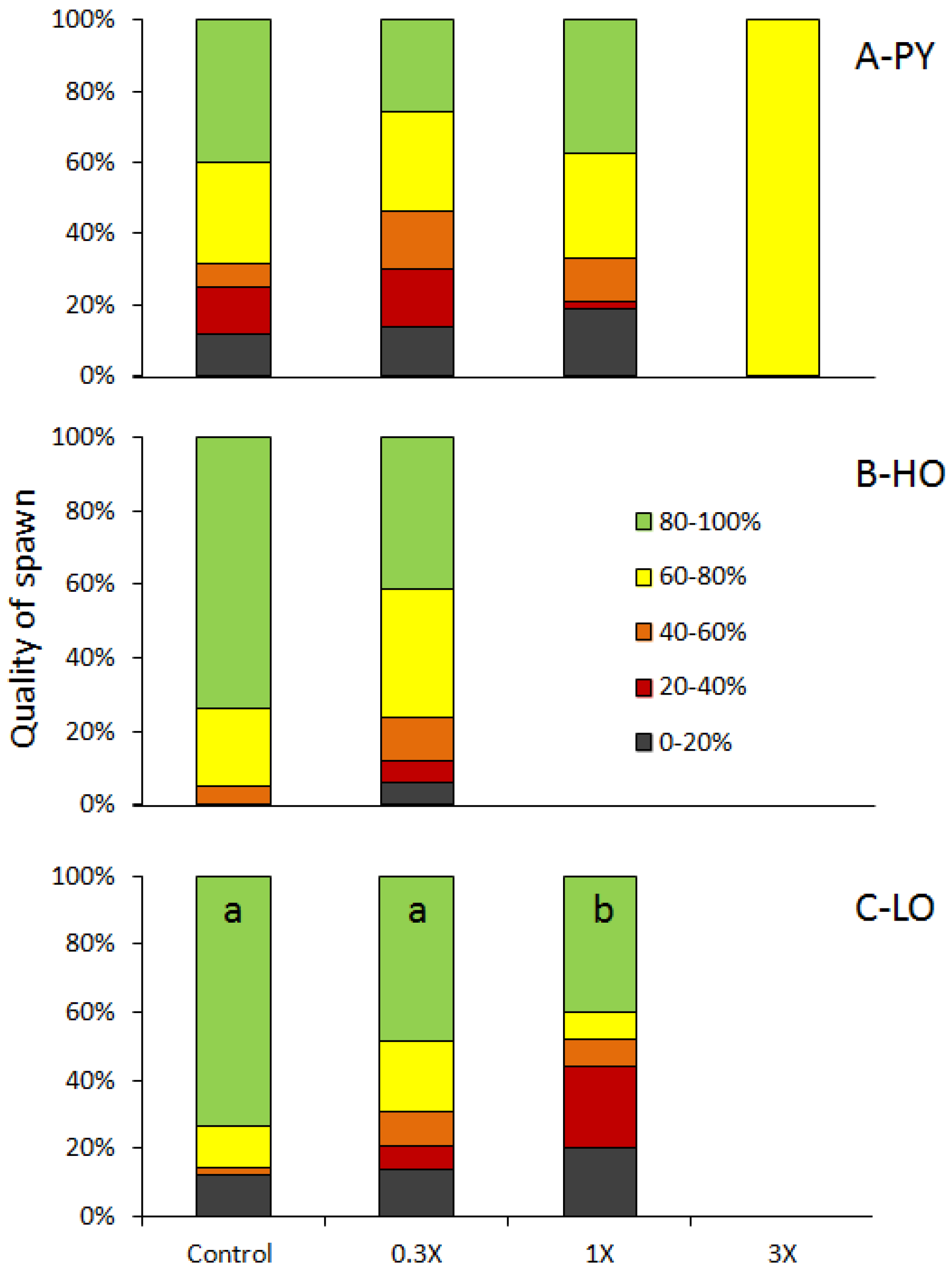
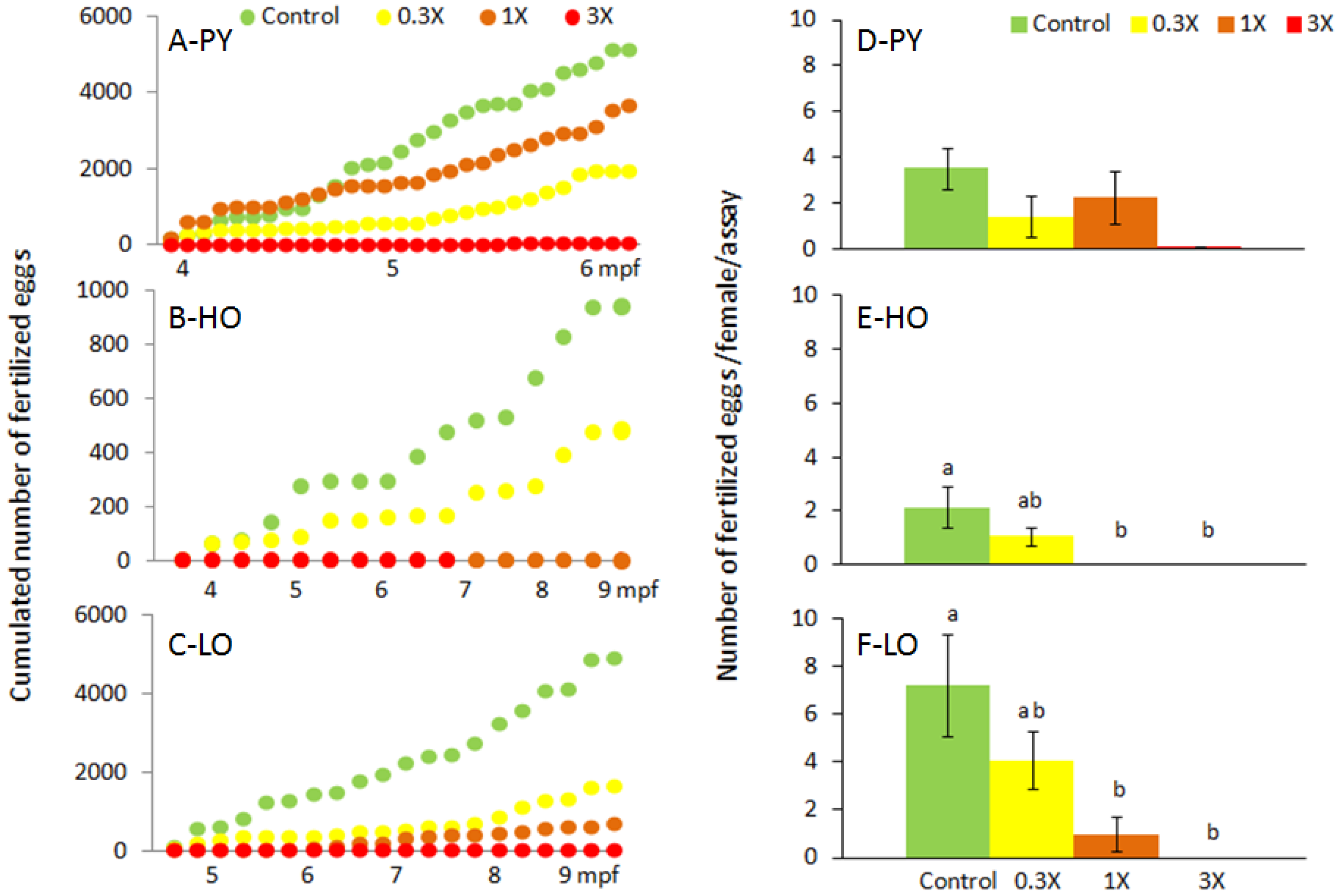
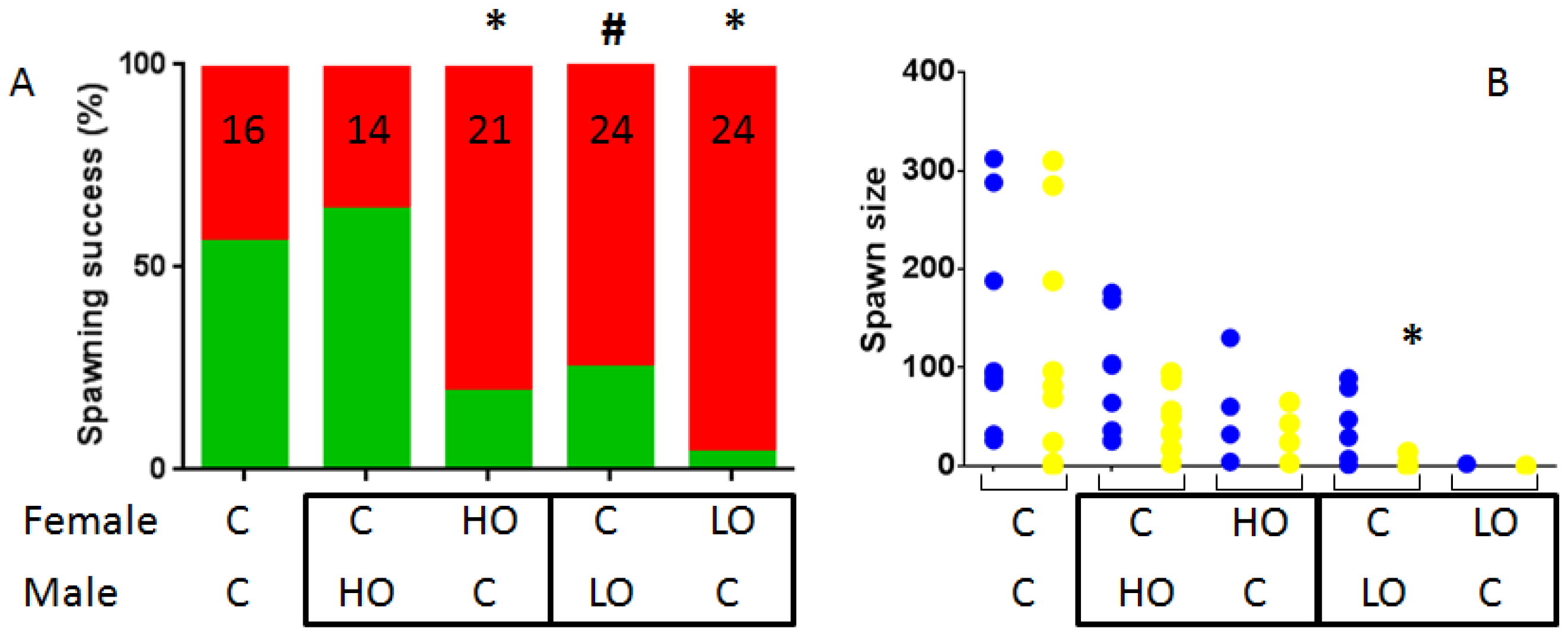
3.1. Reproduction Monitoring

Experimental groups showed varied sex-ratio with females to males (F/M) ratio ranging from 3.32 (PY Control) to 0.51 (HO-1X) while the overall mean ratio was 1.15. When considering only Control conditions, HO-Control had a F/M ratio of 0.54 to PY-Control (F/M = 3.32) or LO-Control (F/M = 1.19). The varying sex ratio arose from the initial creation of the replicate at embryonic stage and the fact that reared replicate composition was not changed over time.
In a first step, group spawns were monitored directly in the rearing tanks starting at 4 mpf for PY and HO fish and at 5 mpf for LO fish. Results are summarized in Table 1. A significant number of spawns were obtained for all diets but 3X concentration for all three fractions with only one spawn was obtained for PY-3X over 87 assays and none for HO-3X and LO-3X. The spawning success rate was significantly reduced after exposure to all three PAHs fractions (Chi2; p < 0.001). In more detail, this rate was reduced for all fractions and all concentrations compared to their respective control (p < 0.05) except for HO-0.3X (p = 0.81).
The cumulative number of fertilized eggs was monitored over time (Figure 1A–C).
The number of fertilized eggs spawned per female and assay was significantly reduced for fish exposed to petrogenic diets for 1X and 3X concentrations (Figure 1D–F). The 0.3X was intermediate. In the case of PY-3X, a trend was only obtained (p = 0.078) using the GLM analysis despite the fact that only one spawn was obtained with a hundred-fold reduction of the number of fertilized eggs per female per spawn.
Spawns mean fertilization rates ranged from 66.0% to 77.1% for PY fraction, from 81.8 to 88.1 for HO fraction and from 51.3 to 78.3 for LO fraction with a trend to follow a dose-effect decrease for this latter fraction. When classified according to their fertilization rates in 20% wide classes (Figure 2), this trend was confirmed (overall Chi2 = 18.48; p = 0.02). LO-0.3X spawns tended to have a lower quality compared to Control (Chi2 = 7.17; p = 0.13) while LO-1X spawn quality was significantly reduced compared to Control (Chi2 = 14.60; p = 0.006). GLM analysis indicated that these trends and differences were due to the reduction of the spawn number in class 80%–100% for LO-0.3X and LO-1X (p = 0.09 and p = 0.05 respectively).
In order to evaluate if reproduction disruption could be ascribed to a particular sex and so gain insight into mechanisms, we performed crosses between exposed and non-exposed fish. This was performed using HO-1X and LO-3X fish. Contrary to what was observed with homogenous fish, we obtained spawns for all crosses involving contaminated fish albeit with various success (Figure 3). Spawning success for female Control/male Control crosses was obtained in 9 assays out of 16 (56.3%). Spawning success for female Control/male HO-1X crosses was not different (64.3%; p = 0.72). In contrast, in female HO-1X/male Control crosses, the success was significantly reduced compared to Control (19.0%; p < 0.05). In the case of LO-3X crosses, the situation was different with a trend to decrease for female Control/male LO-3X (25.0%; p = 0.09) and a very significant decrease for the opposite cross female LO-3X/male Control with only one spawn obtained out of 24 assays (4.2%; p < 0.001). When further qualifying the obtained spawns, only one spawn of the female LO-3X/male Control cross was excluded from the analysis; the only significant difference obtained was the number of fertilized eggs obtained in the female Control/male LO-3X crosses which was significantly reduced (KW = 14.95; p = 0.02 and post-hoc test p = 0.04), resulting in a fertilization rate of 7% which is approximately tenfold lower compared to other conditions (60%–84%).

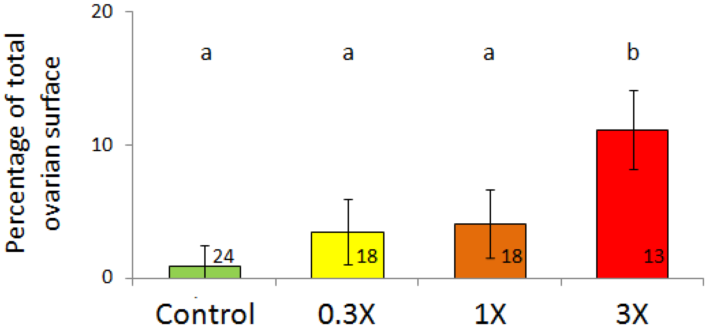
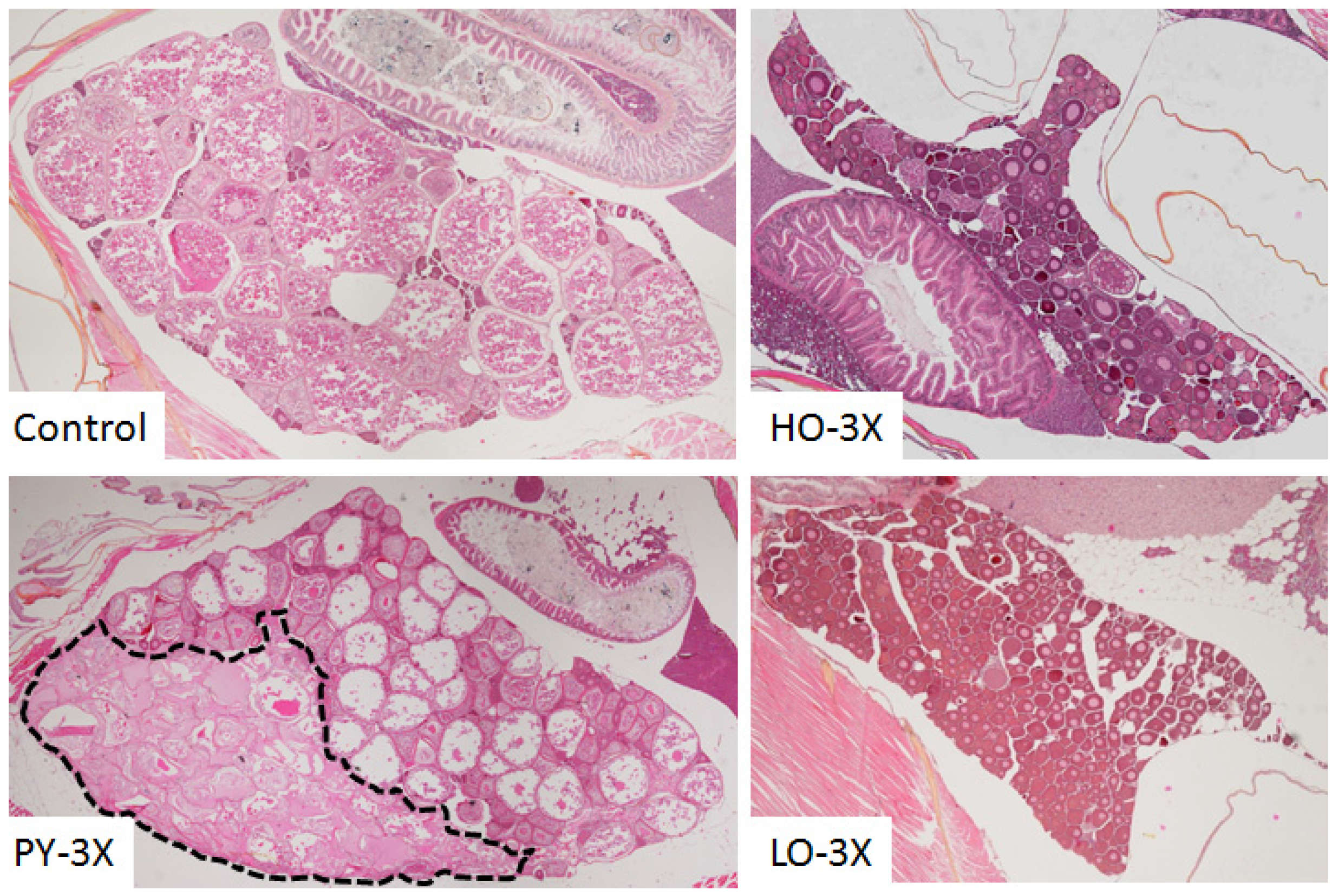
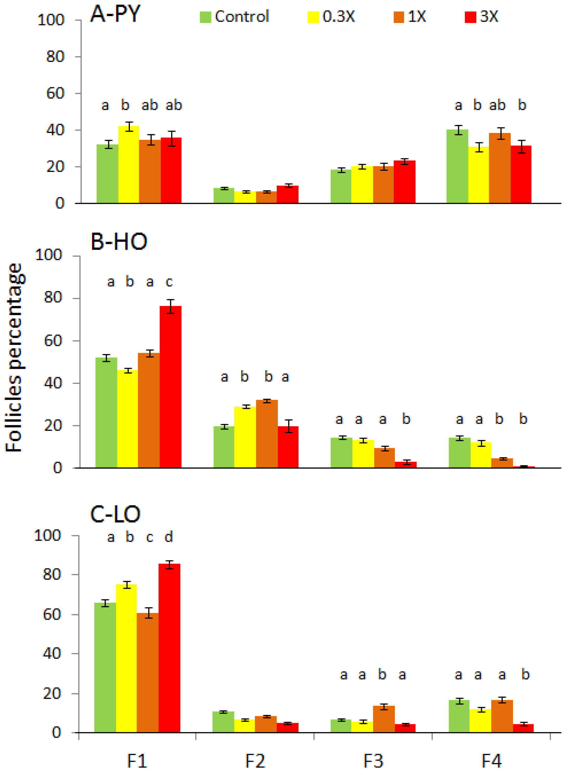
3.2. Histological Analysis

Histological analyses were performed to assess the ovaries’ structure, morphology and functionality. The ovaries’ overall structure was affected in female exposed but these disruptions differed depending on the fraction (Figure 4).
Ovaries from control females presented an even distribution of follicles at all stages of maturation. All stages of follicles were also found in the ovary from females exposed to PY-3X diet which presented large areas of disrupted morphology which did not correspond to atretic follicles but rather degenerating areas. The surface percentage of these degenerating areas increased with PY concentration (Figure 5; F = 4.81; p = 0.007). This kind of degenerating area was not found in ovaries from HO-3X and LO-3X fish.
Ovaries from females exposed to petrogenic fractions presented a significantly reduced number of maturing follicles (Figure 4). To quantify this disruption, follicles were counted according to their stage (Figure 6). In PY ovaries at 9 mpf, a slight increase in the proportion of F1 was observed in PY-0.3X ovaries. In contrast, a slight reduction of F4 was observed for PY-0.3X and PY-3X fish. More important disruptions were observed in HO and LO females. A significant increase of F1 was observed in HO-3X and LO-3X and to a lesser extent in LO-0.3X. In contrast, a slight decrease of F1 was observed in HO-0.3X and LO-1X. The percentage of some intermediate follicles was increased, F2 for HO-0.3X and HO-1X as well as F3 for LO-1X or decreased F3 for HO-1X. The case of F4 is clearer with a significant decrease of mature follicles in ovaries from HO-1X, HO-3X and LO-3X.
Testis structure and morphology were also analyzed at the same age. Testicular hypoplasia (Figure 7A,B) has been observed in males exposed to HO-1X (n = 3; 6.4% of analysed males), in HO-3X (n = 9; 42.9%) as well as in LO-3X (n = 2; 7.4%). In addition to these functional disruptions, some seminoma have been identified in 1X or 3X concentrations in all fractions while none has been found in control fish. These very invasive tumors were described in more detail in Larcher et al. [31]. Finally, one ovotestis has been identified in HO-3X treatment (Figure 7C).

3.3. Genes Expression Analysis

Expression of genes encoding estrogens receptors (esr1, esr2a and esr2b) and ovarian aromatase (cyp19a1a) was assessed in ovaries sampled at 9 mpf (Table 2). Regarding estrogens receptors, the esr1 gene is up-regulated in ovaries of PY-1X (p < 0.05) and HO-0.3X (p < 0.05) while esr2a is up-regulated in HO-0.3X (p < 0.01) and HO-1X (p < 0.05). In contrast, esr2b is down-regulated in LO-1X (p < 0.05) and just above significance threshold for LO-3X (p = 0.053). Ovarian aromatase is also up-regulated in ovaries of PY-1X (p < 0.05) and HO-0.3X (p < 0.05) but down-regulated in ovaries of LO-3X.
In testis, genes expression has been monitored for two genes involved in the steroidogenesis pathway (cyp17a1 and hsd11b3) and two genes are an indicator of spermatogenesis (amh and spag8; Table 3). In testis of exposed fish, spag8 is down-regulated in PY-0.3X (p < 0.01) and PY-1X (p < 0.01) while cyp17a1 is down-regulated in PY-0.3X (p < 0.01), HO-0.3X (p < 0.01) and H0-1X (p < 0.01). In contrast, cyp17a1 is up-regulated in L0-1X (p < 0.01) as well as amh in L0-0.3X (p < 0.05) and L0-1X (p < 0.01) and just above the significance threshold for LO-3X (p = 0.065).
Fold change is indicated relative to ovaries sampled in Control females. The 95% confidence interval provided by REST software is provided (95% CI). Values in bold are significantly different compared to expression in Control fish.
Fold change is indicated relative to testis sampled in Control males. The 95% confidence interval provided by REST software is provided (95% CI). Values in bold are significantly different compared to expression in Control fish.

4. Discussion

4.1. Reproductive Disruptions

Several reproductive traits monitored in this study were disrupted upon lifelong exposure to PAHs fractions all contributing to a reduction in reproduction success. Some differences were observed between fractions and will be specifically discussed later on. The most affected trait is the spawning success evaluated as the number of spawns collected over a period of time. For all fractions, a complete failure of spawning was observed for the 3X concentration (except PY-3X; 1 spawn out of 87 attempts). In addition, zero spawn was obtained for HO-1X. Otherwise, a general trend towards a dose-effect reduction in spawning success could be observed.
Tank replicates were established before hatching and remained unchanged during the whole experiment in order not to perturb group composition in each rearing tank. This was a methodological choice based on the fact that visual and olfactive cues have been shown to be important for reproduction [37]. Because sex is plastic in zebrafish [38], this could result in groups with biased sex-ratio which was indeed observed in our experimental groups with a females to males (F/M) ratio ranging from 3.32 (PY Control) to 0.51 (HO-1X) while the overall mean ratio was 1.15. These differences in sex-ratio may explain some differences in terms of spawning success between diets. When considering only Control conditions, HO-Control with a F/M ratio of 0.54 had a 51% spawning success compared to PY-Control (F/M = 3.32; Success = 69%) or LO-Control (F/M = 1.19; Success = 77%) which suggests that if a low ratio could result in a lower spawning success there is no obvious correlation. Considering relative sex-ratio and spawning success, this may have only lead to an underestimation of spawning success for L0-0.3X condition. To further characterize reproduction traits, spawn’s size normalized per number of females and successful assays in each tank revealed an additional disruption for LO-1X, while spawn’s size from HO- and LO-0.3X were intermediate between Control and 1X conditions. Finally, fertilization rates decreased significantly for LO-1X spawns. Altogether this resulted in a dramatic decrease in reproductive output for exposed fish, as evaluated by the cumulative number of produced fertilized eggs. The analysis of all those traits are rarely considered altogether, especially in such kinds of ecotoxicological studies. Therefore, results will be globally discussed in the light of relevant studies which have, however, mostly been performed using individual PAHs. Indeed, exposures to individual PAHs have been shown to reduce the number of eggs in several fish species [19,39,40,41,42] while an exposure to benzo[a]pyrene produced a decreased fertilization rate in mummichog [43]. In contrast, an exposure of Japanese medaka to phenanthrene produced no modification of the daily number of eggs spawned and fertilization rate did not change, with the exception of one intermediate concentration [17].
The paucity of information on reproduction functionality is puzzling owing the number of studies reporting the disruption of biomarkers related to reproduction after exposure to PAHs. If some studies have reported no gonadal disruptions [17,43], some experimental exposure to PAHs as well as analysis of fish collected in the field in PAH-contaminated areas revealed disruption of gonadal structure or reproduction-related biomarkers, which could help to understand the presently observed reproductive defects. For example, some studies showed that fish exposure to PAHs results in gonadal structure disruption indicative of maturation defects in females [39,40,44] and in males [44,45]. Our results are in agreement with these articles, revealing a decrease in ovary maturation as shown by a decrease in mature follicles (F4) proportion in female exposed to 3X concentration for all fractions and post-vitellogenic follicles (F3) for HO-3X females. In the case of PY females, we have also identified degeneration areas in ovaries, the relative surface of which increased with PY concentration. In the case of males, the observed defects (hypoplasia in a small proportion of LO-3X and HO-1X and in a high proportion (42%) of HO-3X males) have already been mentioned in cod after exposure to produced water [44]. Further, one ovo-testis was also found in one individual morphologically identified as a male. With regard to this latter observation, the occurrence of such histological disruption may have been underestimated because during sampling we targeted females and males on the basis of clear morphological differences; therefore, individuals which could not be ascribed to a sex were not sampled. Finally, in addition to these maturation defects, seminoma have been observed in PY-3X, HO-1X and LO-3X as previously reported [31].
Crosses performed between Control and exposed fish (HO-1X and LO-3X) suggest that spawning success is mainly driven by females, at least for the petrogenic fractions investigated. Beyond strictly reproductive defects, this could be due to behavioral disruptions which we have previously reported [32].
Overall, the present results showed that lifelong exposures to environmentally relevant PAHs fractions lead to reproductive dysfunctions and that these defects depended on the fraction used. Differences could be seen both on defects’ severity with petrogenic fractions producing more dramatic effects than PY fraction, and on defects nature with some specific insults such as ovaries degeneration after PY exposure or fertilization rate decrease after LO exposure. Such differences suggest the triggering of different molecular mechanisms.

4.2. Underlying Mechanisms

Reproduction defects can be the result of a general physiological dysfunction. In regard to this, it should be said that several physiological traits have been measured on the same fish and that, beyond the behavioral disruptions already mentioned, growth has been shown to be reduced after exposure, as well as the expression of some digestive enzyme and the ability to cope with food deprivation [33]. This reduced growth (along with disrupted condition indices) has already been observed upon exposure to PAHs [24,46,47,48] and could also have contributed to reduced reproduction output.
Exposure to PAHs usually results in a decrease in estradiol (E2) and 11-ketotestosterone (T), this has been observed after exposure to individual compounds [26,43,49] and more complex mixtures such as Produced water [44]. However, in other cases, an increase is observed [50] or E2 is reduced while T is increased [41]. In some cases, the situation is more complex with non-monotonic responses [27,45]. Finally, in some cases no change is observed after exposure to individual compounds or complex mixtures [16,51]. Altogether, these results suggest that PAHs can have endocrine disrupter activity while the estrogenic and/or anti-estrogenic property is not clear. In our cases, E2, T and vitellogenin circulating concentrations have been measured but no clear conclusion could be drawn (data not shown). In order to gain insight into molecular mechanisms underlying the observed reproduction defects, we have analyzed the expression of a set of genes involved in different functions (steroid pathways, gonad differentiation and maintenance) in gonads sampled at 9 mpf. Gene’s expression changes not follow a dose-response scheme, in particular very few changes were observed with the 3X concentration (not performed for HO since no more fish were available at this age likely resulting from an excessive toxicity). According to esr regulation by E2 [52], the activation in ovaries of esr1 and esr2a expression in PY-1X and HO-0.3X and 1X and of cyp19a1a in PY-1X as well as the decrease in esr2b in LO-1X are indicative of an estrogenic activity of these fractions in females. Another explanation could be that AhR directly activates the expression of estrogen receptors [53,54]. The reduction of cyp17a1 expression in testis of PY-0.3X and HO males is also indicative of estrogenic activity in males according to the known regulation of Cyp17a1 protein by E2 in zebrafish [55]. In testis, spag8 is expressed in the acrosomal region of spermatozoa and in germ cells starting in spermatocytes [56] and its expression is positively regulated by androgen receptor agonists and negatively by antagonists [57]. The observed decrease in spag8 expression may be indicative of anti-androgen activity in PY exposed fish. This combination of estrogenic and anti-androgenic activity may explain the gonadal degeneration in PY females’ ovaries; indeed both an excess of estrogen and a decrease in testosterone have been shown to promote ovarian structural disruption [58,59,60]. Besides, it has been shown in mouse that PAHs exposure promoted ovarian failure through AhR-mediated bax activation [61]. This pathway could underpin the specific disruption observed in PY ovaries since PY fraction contains high proportion of high molecular weight PAHs which are supposed to be AhR ligands. This mechanism could also explain why, while in HO and LO the decrease in the number of mature follicle is compensated by a higher proportion of pre-vitellogenic follicles, this is not the case in PY-3X ovaries. Indeed, it has also been shown that PAHs triggering AhR such as PY fraction (and HO to a lower extent) also specifically destroys primordial oocytes [62]. On the male side, the regulation of amh expression in testis is of particular interest. Indeed, in mammals, this gene is expressed during gonadal development and later almost exclusively in Sertoli cells during fetal and prepubertal stages. Later on, amh remains expressed but at a lower level in relation with spermatogenesis. The up-regulation of amh in testis of LO males could indicate a persistent defect in testis differentiation and/or spermatogenesis and may explain the specific and dramatic lower fertilization rates observed in pair-wise crosses between females Control and males LO-3X. Altogether, these results are evocative of endocrine disruption but also suggest that in some cases, reproductive dysfunction may be the consequence of gonad differentiation and/or maturation defects leading to a high proportion of immature individuals. In this context, the single ovo-testis observed in HO-3X may be seen as a result of an incomplete switch from ovary-like pre-differentiation gonad to testis rather than a sex-reversal. As in the case of reproductive traits, molecular analyses point out differences between fraction’s effects, and PY and HO appear to share common pathways likely triggered by AhR while LO fraction would be acting through a yet-unidentified pathway.

4.3. Environmental Relevance

Field monitoring as well as environmental assessment after an oil spill have demonstrated the important consequences of PAHs exposure on ecosystems in general and fish in particular [63,64,65]. Identified effects cover a wide range of physiological functions or processes and include reproduction or reproductive traits defects. In the case of an oil spill, the situation is simpler since PAHs source is clearly identified. However, oil denominations cover a large range of mixtures, the compositions of which depend on extraction localization as well as refining level. This is exemplified by the two petrogenic fractions used in this study. HO fraction contained 31% of high molecular weight PAHs while they represent only 6% of LO fraction but both contained high proportion of methylated PAHs (45%–59%) [33]. The situation is less simple in areas where PAHs sources are diverse, often resulting from human activity. In this case, PAHs can be of both pyrolytic and petrogenic origin and enter aquatic environments through the deposition of atmospheric emissions or from soil runoff. PY fraction extracted from sediment collected in the Seine River is representative of such PAHs mixture encountered in urbanized areas. PY fraction contained a very high proportion of high molecular weight PAHs (82%) and very few methylated derivatives (5%) [33]. Another major difference with oil spill is that, in the latter case, PAHs emission is constant, leading to chronic exposure. Nonetheless, after acute exposure during an oil spill, and despite cleaning operations, a secondary chronic exposure to lower level of PAHs persist over a long time period as this has been extensively studied after the Exxon Valdez spill [64] converging toward similar issues as in urbanized areas.
The concentration of PAHs in the biota therefore varies depending on the surrounding concentrations, in particular in sediment, and on a trophic level. For example, the total concentration of US-EPA 16 PAHs in the copepod Eurytemora affinis in the Seine Estuary can reach 3.9 µg·g−1 [66] and in mussels can be as high as 1.6 µg·g−1 [67]. In the case of acute accidental exposure, the concentration in mussels can be even higher, for example 3.0 µg·g−1 after the Erika oil spill [68] and above 14 µg·g−1 after Exxon Valdez oil spill [69]. PAHs concentrations in 1X diets used within the frame of this study are in the (4.7–6.7 µg.g−1 dw) range [33] and therefore fall into concentration encountered in contaminated areas. Fish can be exposed to PAHs through several routes including the trophic route, which has been shown to be an important route [9,10,11].

5. Conclusions

Field monitoring has revealed correlation between PAHs concentrations in the environment (or metabolites in individuals) whatever PAHs origin, and reproductive defects in fish [11,20,21,22,23,24,25,29,63,70,71]. This kind of monitoring analysis is essential for the environment but biomarkers often only allow raw estimates of disruptions and whatever actual reproduction output cannot be measured. Effects on population are therefore often only reached through modeling approaches [1,72]. Moreover, we believe that complementary approaches such as the one presented here, are suitable to investigate environmental exposure to complex mixtures and that, even if we conducted controlled experimental exposures, our results are pinpointing serious alteration of the recruitment success in the wild fish population. Further characterizations of the pathways are needed to refine and develop smarter biomarkers and we believe that zebrafish could provide a useful toolbox for this purpose as well as for transgenerational effects assessment since offspring insult has been shown upon exposure to PAHs [41,46,73].

Supplementary Materials

The following are available online at www.mdpi.com/2305-6304/4/4/26/s1. Table S1: Detailed concentration of individual PAHs in produced diets.

Acknowledgments

We thank Didier Leguay for his help and Patrick Babin for fruitful discussions. This study was financially supported by the ANR project ConPhyPoP (CES 09_002) and CPER A2E. This project is co-financed by the European Union with the European Fund of Regional Development. Doctoral grant was received from the Région Poitou-Charentes and Institut Français de Recherche pour l’Exploitation de la Mer by C.V. This work was part of the LABEX COTE cluster of excellence continental to coastal ecosystems.

Author Contributions

T.L., H.B., M.-L.B. and X.C. conceived and designed the experiments; All Authors contributed to the experiments; C.V., T.L., B.D., H.B., M.-L.B. and X.C. analyzed the data; C.V., M.-L.B. and X.C. wrote the paper.

Conflicts of Interest

The authors declare no conflict of interest.

References

  1. Rochette, S.; Rivot, E.; Morin, J.; Mackinson, S.; Riou, P.; Le Pape, O. Effect of nursery habitat degradation on flatfish population: Application to Solea solea in the eastern channel (western europe). J. Sea Res. 2010, 64, 34–44. [Google Scholar] [CrossRef]
  2. Latimer, J.S.; Zheng, J. The sources, transport, and fate of PAHs in the marine environment. In PAHs: An Ecotoxicological Perspective; John Wiley & Sons, Ltd.: Chichester, UK, 2003; pp. 7–33. [Google Scholar]
  3. Shen, H.; Huang, Y.; Wang, R.; Zhu, D.; Li, W.; Shen, G.; Wang, B.; Zhang, Y.; Chen, Y.; Lu, Y.; et al. Global atmospheric emissions of polycyclic aromatic hydrocarbons from 1960 to 2008 and future predictions. Environ. Sci. Technol. 2013, 47, 6415–6424. [Google Scholar] [CrossRef] [PubMed]
  4. Zhang, Y.; Tao, S. Global atmospheric emission inventory of polycyclic aromatic hydrocarbons (PAHs) for 2004. Atmos. Environ. 2009, 43, 812–819. [Google Scholar] [CrossRef]
  5. Hylland, K. Polycyclic aromatic hydrocarbon (PAH) ecotoxicology in marine ecosystems. J. Toxicol. Environ. Health Part A 2006, 69, 109–123. [Google Scholar] [CrossRef] [PubMed]
  6. Baumard, P.; Budzinski, H.; Garrigues, P.; Sorbe, J.C.; Burgeot, T.; Bellocq, J. Concentrations of PAHs (polycyclic aromatic hydrocarbons) in various marine organisms in relation to those in sediments and to trophic level. Mar. Pollut. Bull. 1998, 36, 951–960. [Google Scholar] [CrossRef]
  7. Cachot, J.; Geffard, O.; Augagneur, S.; Lacroix, S.; Le Menach, K.; Peluhet, L.; Couteau, J.; Denier, X.; Devier, M.H.; Pottier, D.; et al. Evidence of genotoxicity related to high PAH content of sediments in the upper part of the seine estuary (Normandy, France). Aquat. Toxicol. 2006, 79, 257–267. [Google Scholar] [CrossRef] [PubMed]
  8. Benlahcen, K.T.; Chaoui, A.; Budzinski, H.; Bellocq, J.; Garrigues, P. Distribution and sources of polycyclic aromatic hydrocarbons in some mediterranean coastal sediments. Mar. Pollut. Bull. 1997, 34, 298–305. [Google Scholar] [CrossRef]
  9. Johnson, L.L.; Ylitalo, G.M.; Arkoosh, M.R.; Kagley, A.N.; Stafford, C.; Bolton, J.L.; Buzitis, J.; Anulacion, B.F.; Collier, T.K. Contaminant exposure in outmigrant juvenile salmon from pacific northwest estuaries of the united states. Environ. Monit. Assess. 2007, 124, 167–194. [Google Scholar] [CrossRef] [PubMed]
  10. Varanasi, U.; Casillas, E.; Arkoosh, M.R.; Hom, T.; Misitano, D.; Brown, D.W.; Chan, S.-L.; Collier, T.K.; McCain, B.B.; Stein, J.E. Contaminant Exposure and Associated Biological Effects in Juvenile Chinook Salmon (Oncorhynchus tshawytscha) from Urban and Nonurban Estuaries of Puget Sound; NOAA Fisheries: Seattle, WA, USA, 1993; p. 112. [Google Scholar]
  11. Yanagida, G.K.; Anulacion, B.F.; Bolton, J.L.; Boyd, D.; Lomax, D.P.; Paul Olson, O.; Sol, S.Y.; Willis, M.; Ylitalo, G.M.; Johnson, L.L. Polycyclic aromatic hydrocarbons and risk to threatened and endangered chinook salmon in the lower columbia river estuary. Arch. Environ. Contam. Toxicol. 2012, 62, 282–295. [Google Scholar] [CrossRef] [PubMed]
  12. He, J.H.; Gao, J.M.; Huang, C.J.; Li, C.Q. Zebrafish models for assessing developmental and reproductive toxicity. Neurotoxicol. Teratol. 2014, 42, 35–42. [Google Scholar] [CrossRef] [PubMed]
  13. Hinton, D.E.; Kullman, S.W.; Hardman, R.C.; Volz, D.C.; Chen, P.-J.; Carney, M.; Bencic, D.C. Resolving mechanisms of toxicity while pursuing ecotoxicological relevance? Mar. Pollut. Bull. 2005, 51, 635–648. [Google Scholar] [CrossRef] [PubMed]
  14. Leon-Olea, M.; Martyniuk, C.J.; Orlando, E.F.; Ottinger, M.A.; Rosenfeld, C.S.; Wolstenholme, J.T.; Trudeau, V.L. Current concepts in neuroendocrine disruption. Gen. Comp. Endocrinol. 2014, 203, 158–173. [Google Scholar] [CrossRef] [PubMed]
  15. Kloas, W.; Urbatzka, R.; Opitz, R.; Wurtz, S.; Behrends, T.; Hermelink, B.; Hofmann, F.; Jagnytsch, O.; Kroupova, H.; Lorenz, C.; et al. Endocrine disruption in aquatic vertebrates. Ann. N. Y. Acad. Sci. 2009, 1163, 187–200. [Google Scholar] [CrossRef] [PubMed]
  16. Seruto, C.; Sapozhnikova, Y.; Schlenk, D. Evaluation of the relationships between biochemical endpoints of PAH exposure and physiological endpoints of reproduction in male california halibut (Paralichthys californicus) exposed to sediments from a natural oil seep. Mar. Environ. Res. 2005, 60, 454–465. [Google Scholar] [CrossRef] [PubMed]
  17. Horng, C.Y.; Lin, H.C.; Lee, W. A reproductive toxicology study of phenanthrene in medaka (Oryzias latipes). Arch. Environ. Contam. Toxicol. 2010, 58, 131–139. [Google Scholar] [CrossRef] [PubMed]
  18. Tollefsen, K.E.; Sundt, R.C.; Beyer, J.; Meier, S.; Hylland, K. Endocrine modulation in atlantic cod (Gadus morhua L.) exposed to alkylphenols, polyaromatic hydrocarbons, produced water, and dispersed oil. J. Toxicol. Environ. Health A 2011, 74, 529–542. [Google Scholar] [CrossRef] [PubMed]
  19. Tilghman Hall, A.; Oris, J.T. Anthracene reduces reproductive potential is maternally transferred during long-term exposure in fathead minnows. Aquat. Toxicol. 1991, 19, 249–264. [Google Scholar] [CrossRef]
  20. Bugel, S.M.; White, L.A.; Cooper, K.R. Impaired reproductive health of killifish (Fundulus heteroclitus) inhabiting Newwark bay, NJ, a chronically contaminated estuary. Aquat. Toxicol. 2010, 96, 182–193. [Google Scholar] [CrossRef] [PubMed]
  21. Casillas, E.; Misitano, D.; Johnson, L.L.; Rhodes, L.D.; Collier, T.K.; Stein, J.E.; McCain, B.B.; Varanasi, U. Inducibility of spawning and reproductive success of female english sole (Praphrys vetulus) from urban and nonurban areas of Puget Sound, Washington. Mar. Environ. Res. 1991, 31, 99–122. [Google Scholar] [CrossRef]
  22. Johnson, L.L.; Stein, J.E.; Collier, T.K.; Casillas, E.; Varanasi, U. Indicators of reproductive development in prespawning female winter flounder (Pleuronectes americanus) from urban and non-urban estuaries in the northeast United States. Sci. Total Environ. 1994, 141, 241–260. [Google Scholar] [CrossRef]
  23. Kiceniuk, J.W.; Khan, R.A. Effect of petroleum hydrocarbons on atlantic cod, Gadus morhua, following chronic exposure. Can. J. Zool. 1987, 65, 490–494. [Google Scholar] [CrossRef]
  24. Ridgway, L.L.; Chapleau, F.; Comba, M.E.; Backus, S.M. Population characteristics and contaminant burdens of the white sucker (Catostomus commersoni) from the St. Lawrence river near Cornwall, Ontario and Massena, New York. J. Great Lakes Res. 1999, 25, 567–582. [Google Scholar] [CrossRef]
  25. Tetreault, G.R.; McMaster, M.E.; Dixon, D.G.; Parrott, J.L. Using reproductive endpoints in small forage fish species to evaluate the effects of Athabasca oil sands activities. Environ. Toxicol. Chem. 2003, 22, 2775–2782. [Google Scholar] [CrossRef] [PubMed]
  26. Monteiro, P.R.; Reis-Henriques, M.A.; Coimbra, J. Plasma steroid levels in female flounder (Platichthys flesus) after chronic dietary exposure to single polycyclic aromatic hydrocarbons. Mar. Environ. Res. 2000, 49, 453–467. [Google Scholar] [CrossRef]
  27. Arukwe, A.; Nordtug, T.; Kortner, T.M.; Mortensen, A.S.; Brakstad, O.G. Modulation of steroidogenesis and xenobiotic biotransformation responses in zebrafish (Danio rerio) exposed to water-soluble fraction of crude oil. Environ. Res. 2008, 107, 362–370. [Google Scholar] [CrossRef] [PubMed]
  28. Evanson, M.; Van Der Kraak, G.J. Stimulatory effects of selected PAHs on testosterone production in goldfish and rainbow trout and possible mechanisms of action. Comp. Biochem. Physiol. C Toxicol. Pharmacol. 2001, 130, 249–258. [Google Scholar] [CrossRef]
  29. Sol, S.Y.; Johnson, L.L.; Horness, B.H.; Collier, T.K. Relationship between oil exposure and reproductive parameters in fish collected following the Exxon Valdez oil spill. Mar. Pollut. Bull. 2000, 40, 1139–1147. [Google Scholar] [CrossRef]
  30. Truscott, B.; Walsh, J.M.; Burton, M.P.; Payne, J.F.; Idler, D.R. Effect of acute exposure to crude petroleum on some reproductive hormones in salmon and flounder. Comp. Biochem. Physiol. C Comp. Pharmacol. Toxicol. 1983, 75, 121–130. [Google Scholar] [CrossRef]
  31. Larcher, T.; Perrichon, P.; Vignet, C.; Ledevin, M.; Le Menach, K.; Lyphout, L.; Landi, L.; Clerandeau, C.; Lebihanic, F.; Menard, D.; et al. Chronic dietary exposure of zebrafish to PAH mixtures results in carcinogenic but not genotoxic effects. Environ. Sci. Pollut. Res. Int. 2014, 21, 13833–13849. [Google Scholar] [CrossRef] [PubMed]
  32. Vignet, C.; Le Menach, K.; Lyphout, L.; Guionnet, T.; Frere, L.; Leguay, D.; Budzinski, H.; Cousin, X.; Begout, M.L. Chronic dietary exposure to pyrolytic and petrogenic mixtures of PAHs causes physiological disruption in zebrafish—Part II: Behavior. Environ. Sci. Pollut. Res. Int. 2014, 21, 13818–13832. [Google Scholar] [CrossRef] [PubMed]
  33. Vignet, C.; Le Menach, K.; Mazurais, D.; Lucas, J.; Perrichon, P.; Le Bihanic, F.; Devier, M.H.; Lyphout, L.; Frere, L.; Begout, M.L.; et al. Chronic dietary exposure to pyrolytic and petrogenic mixtures of PAHs causes physiological disruption in zebrafish—Part I: Survival and growth. Environ. Sci. Pollut. Res. Int. 2014, 21, 13804–13817. [Google Scholar] [CrossRef] [PubMed]
  34. Daouk, T.; Larcher, T.; Roupsard, F.; Lyphout, L.; Rigaud, C.; Ledevin, M.; Loizeau, V.; Cousin, X. Long-term food-exposure of zebrafish to PCB mixtures mimicking some environmental situations induces ovary pathology and impairs reproduction ability. Aquat. Toxicol. 2011, 105, 270–278. [Google Scholar] [CrossRef] [PubMed]
  35. Spitsbergen, J.M.; Tsai, H.W.; Reddy, A.; Miller, T.; Arbogast, D.; Hendricks, J.D.; Bailey, G.S. Neoplasia in zebrafish (Danio rerio) treated with 7,12-dimethylbenz[a]anthracene by two exposure routes at different developmental stages. Toxicol. Pathol. 2000, 28, 705–715. [Google Scholar] [CrossRef] [PubMed]
  36. Koç, N.D.; Aytekin, Y.; Yüce, R. Ovary maturatıon stages and histological investigation of ovary of the zebrafish (Danio rerio). Braz. Arch. Biol. Technol. 2008, 51, 513–522. [Google Scholar]
  37. Gerlach, G.; Lysiak, N. Kin recognition and inbreeding avoidance in zebrafish, Danio rerio, is based on phenotype matching. Anim. Behav. 2006, 71, 1371–1377. [Google Scholar] [CrossRef]
  38. Siegfried, K.R.; Nusslein-Volhard, C. Germ line control of female sex determination in zebrafish. Dev. Biol. 2008, 324, 277–287. [Google Scholar] [CrossRef] [PubMed]
  39. Brown-Peterson, N.J.; Manning, C.S.; Brouwer, M.; Griffitt, R.J. Effects of pyrene exposure on sheepshead minnow (Cyprinodon variegatus) reproduction. J. Toxicol. Environ. Health Part A 2013, 76, 842–852. [Google Scholar] [CrossRef] [PubMed]
  40. Sun, L.; Zuo, Z.; Chen, M.; Chen, Y.; Wang, C. Reproductive and transgenerational toxicities of phenanthrene on female marine medaka (Oryzias melastigma). Aquat. Toxicol. 2015, 162, 109–116. [Google Scholar] [CrossRef] [PubMed]
  41. Pollino, C.A.; Georgiades, E.; Holdway, D.A. Physiological changes in reproductively active rainbowfish (Melanotaenia fluviatilis) following exposure to naphthalene. Ecotoxicol. Environ. Saf. 2009, 72, 1265–1270. [Google Scholar] [CrossRef] [PubMed]
  42. Hoffmann, J.L.; Oris, J.T. Altered gene expression: A mechanism for reproductive toxicity in zebrafish exposed to benzo[a]pyrene. Aquat. Toxicol. 2006, 78, 332–340. [Google Scholar] [CrossRef] [PubMed]
  43. Booc, F.; Thornton, C.; Lister, A.; Maclatchy, D.; Willett, K.L. Benzo[a]pyrene effects on reproductive endpoints in Fundulus heteroclitus. Toxicol. Sci. 2014, 140, 73–82. [Google Scholar] [CrossRef] [PubMed]
  44. Sundt, R.C.; Bjorkblom, C. Effects of produced water on reproductive parameters in prespawning atlantic cod (Gadus morhua). J. Toxicol. Environ. Health A 2011, 74, 543–554. [Google Scholar] [CrossRef] [PubMed]
  45. Sun, L.; Zuo, Z.; Luo, H.; Chen, M.; Zhong, Y.; Chen, Y.; Wang, C. Chronic exposure to phenanthrene influences the spermatogenesis of male Sebastiscus marmoratus: U-shaped effects and the reason for them. Environ. Sci. Technol. 2011, 45, 10212–10218. [Google Scholar] [CrossRef] [PubMed]
  46. Holth, T.F.; Nourizadeh-Lillabadi, R.; Blaesbjerg, M.; Grung, M.; Holbech, H.; Petersen, G.I.; Alestrom, P.; Hylland, K. Differential gene expression and biomarkers in zebrafish (Danio rerio) following exposure to produced water components. Aquat. Toxicol. 2008, 90, 277–291. [Google Scholar] [CrossRef] [PubMed]
  47. Meador, J.P.; Sommers, F.C.; Ylitalo, G.M.; Sloan, C.A. Altered growth and related physiological responses in juvenile chinook salmon (Oncorhynchus tshawytscha) from dietary exposure to polycyclic aromatic hydrocarbons (PAHs). Can. J. Fish. Aquat. Sci. 2006, 63, 2364–2376. [Google Scholar] [CrossRef]
  48. Rice, C.A.; Myers, M.S.; Willis, M.L.; French, B.L.; Casillas, E. From sediment bioassay to fish biomarker—Connecting the dots using simple trophic relationships. Mar. Environ. Res. 2000, 50, 527–533. [Google Scholar] [CrossRef]
  49. Kennedy, C.J.; Smyth, K.R. Disruption of the rainbow trout reproductive endocrine axis by the polycyclic aromatic hydrocarbon benzo[a]pyrene. Gen. Comp. Endocrinol. 2015, 219, 102–111. [Google Scholar] [CrossRef] [PubMed]
  50. Rodas-Ortiz, J.P.; Ceja-Moreno, V.; Chan-Cocom, M.E.; Gold-Bouchot, G. Vitellogenin induction and increased plasma 17beta-estradiol concentrations in male nile tilapia, Oreochromis niloticus, exposed to organochlorine pollutants and polycyclic aromatics hydrocarbons. Bull. Environ. Contam. Toxicol. 2008, 81, 543–547. [Google Scholar] [CrossRef] [PubMed]
  51. Tintos, A.; Gesto, M.; Miguez, J.M.; Soengas, J.L. Beta-naphthoflavone and benzo(a)pyrene treatment affect liver intermediary metabolism and plasma cortisol levels in rainbow trout oncorhynchus mykiss. Ecotoxicol. Environ. Saf. 2008, 69, 180–186. [Google Scholar] [CrossRef] [PubMed]
  52. Menuet, A.; Le Page, Y.; Torres, O.; Kern, L.; Kah, O.; Pakdel, F. Analysis of the estrogen regulation of the zebrafish estrogen receptor (ER) reveals distinct effects of eralpha, erbeta1 and erbeta2. J. Mol. Endocrinol. 2004, 32, 975–986. [Google Scholar] [CrossRef] [PubMed]
  53. Abdelrahim, M.; Ariazi, E.; Kim, K.; Khan, S.; Barhoumi, R.; Burghardt, R.; Liu, S.; Hill, D.; Finnell, R.; Wlodarczyk, B.; et al. 3-methylcholanthrene and other aryl hydrocarbon receptor agonists directly activate estrogen receptor alpha. Cancer Res. 2006, 66, 2459–2467. [Google Scholar] [CrossRef] [PubMed]
  54. Liu, S.; Abdelrahim, M.; Khan, S.; Ariazi, E.; Jordan, V.C.; Safe, S. Aryl hydrocarbon receptor agonists directly activate estrogen receptor alpha in mcf-7 breast cancer cells. Biol. Chem. 2006, 387, 1209–1213. [Google Scholar] [CrossRef] [PubMed]
  55. Hinfray, N.; Nobrega, R.H.; Caulier, M.; Baudiffier, D.; Maillot-Marechal, E.; Chadili, E.; Palluel, O.; Porcher, J.M.; Schulz, R.; Brion, F. Cyp17a1 and cyp19a1 in the zebrafish testis are differentially affected by oestradiol. J. Endocrinol. 2013, 216, 375–388. [Google Scholar] [CrossRef] [PubMed]
  56. Wu, H.; Chen, Y.; Miao, S.; Zhang, C.; Zong, S.; Koide, S.S.; Wang, L. Sperm associated antigen 8 (SPAG8), a novel regulator of activator of crem in testis during spermatogenesis. FEBS Lett. 2010, 584, 2807–2815. [Google Scholar] [CrossRef] [PubMed]
  57. Garcia-Reyero, N.; Villeneuve, D.L.; Kroll, K.J.; Liu, L.; Orlando, E.F.; Watanabe, K.H.; Sepulveda, M.S.; Ankley, G.T.; Denslow, N.D. Expression signatures for a model androgen and antiandrogen in the fathead minnow (Pimephales promelas) ovary. Environ. Sci. Technol. 2009, 43, 2614–2619. [Google Scholar] [CrossRef] [PubMed]
  58. Van den Belt, K.; Wester, P.W.; van der Ven, L.T.; Verheyen, R.; Witters, H. Effects of ethynylestradiol on the reproductive physiology in zebrafish (Danio rerio): Time dependency and reversibility. Environ. Toxicol. Chem. 2002, 21, 767–775. [Google Scholar] [CrossRef] [PubMed]
  59. Weber, L.P.; Hill, R.L., Jr.; Janz, D.M. Developmental estrogenic exposure in zebrafish (Danio rerio): II. Histological evaluation of gametogenesis and organ toxicity. Aquat. Toxicol. 2003, 63, 431–446. [Google Scholar] [CrossRef]
  60. Forsgren, K.L.; Young, G. Stage-specific effects of androgens and estradiol-17beta on the development of late primary and early secondary ovarian follicles of coho salmon (Oncorhynchus kisutch) in vitro. Biol. Reprod. 2012, 87, 64. [Google Scholar] [CrossRef] [PubMed]
  61. Matikainen, T.; Perez, G.I.; Jurisicova, A.; Pru, J.K.; Schlezinger, J.J.; Ryu, H.Y.; Laine, J.; Sakai, T.; Korsmeyer, S.J.; Casper, R.F.; et al. Aromatic hydrocarbon receptor-driven bax gene expression is required for premature ovarian failure caused by biohazardous environmental chemicals. Nat. Genet. 2001, 28, 355–360. [Google Scholar] [CrossRef] [PubMed]
  62. Mattison, D.R. Morphology of oocyte and follicle destruction by polycyclic aromatic hydrocarbons in mice. Toxicol. Appl. Pharmacol. 1980, 53, 249–259. [Google Scholar] [CrossRef]
  63. Johnson, L.L.; Arkoosh, M.R.; Bravo, C.F.; Collier, T.K.; Krahn, M.M.; Meador, J.P.; Myers, M.S.; Reichert, W.L.; Stein, J.E. The effects of polycyclic aromatic hydrocarbons in fish from Puget Sound, Washington. In The Toxicology of Fishes; CRC Press: Boca Raton, FL, USA, 2008; pp. 877–923. [Google Scholar]
  64. Peterson, C.H.; Rice, S.D.; Short, J.W.; Esler, D.; Bodkin, J.L.; Ballachey, B.E.; Irons, D.B. Long-term ecosystem response to the Exxon Valdez oil spill. Science 2003, 302, 2082–2086. [Google Scholar] [CrossRef] [PubMed]
  65. Collier, T.K.; Anulacion, B.F.; Arkoosh, M.R.; Dietrich, J.P.; Incardona, J.P.; Johnson, L.L.; Ylitalo, G.M.; Myers, M.S. Effects on fish of polycyclic aromatic hydrocarbons (PAHs) and naphthenic acid exposures. In Organic Chemical Toxicology of Fishes; Tierney, K.B., Ed.; Elsevier: London, UK, 2014; Volume 33, pp. 195–255. [Google Scholar]
  66. Cailleaud, K.; Forget-Leray, J.; Souissi, S.; Hilde, D.; LeMenach, K.; Budzinski, H. Seasonal variations of hydrophobic organic contaminant concentrations in the water-column of the seine estuary and their transfer to a planktonic species Eurytemora affinis (Calanoida, Copepoda). Part 1: PCBs and PAHs. Chemosphere 2007, 70, 270–280. [Google Scholar] [CrossRef] [PubMed]
  67. Le Goff, J.; Gallois, J.; Pelhuet, L.; Devier, M.H.; Budzinski, H.; Pottier, D.; André, V.; Cachot, J. DNA adduct measurements in zebra mussels, Dreissena polymorpha, Pallas: Potential use for genotoxicant biomonitoring of fresh water ecosystems. Aquat. Toxicol. 2006, 79, 55–64. [Google Scholar] [CrossRef] [PubMed]
  68. Jeanneret, H.; Chantereau, S.; Belliaeff, B.; Ratiskol, G.; Allenou, J.-P.; Piclet, G. Suivi sanitaire et environnemental des conséquences de la marée noire de l’Erika. In Proceedigns of the Colloque SAFERSEAS: Les Leçons Techniques de l’Erika et des Autres Accidents, Brest, France, 11–16 March 2002.
  69. Payne, J.R.; Driskell, W.B.; Short, J.W.; Larsen, M.L. Long term monitoring for oil in the Exxon Valdez spill region. Mar. Pollut. Bull. 2008, 56, 2067–2081. [Google Scholar] [CrossRef] [PubMed]
  70. Sol, S.; Johnson, L.; Boyd, D.; Olson, O.; Lomax, D.; Collier, T. Relationships between anthropogenic chemical contaminant exposure and associated changes in reproductive parameters in male english sole (Parophrys vetulus) collected from Hylebos waterway, Puget Sound, Washington. Arch. Environ. Contam. Toxicol. 2008, 55, 627–638. [Google Scholar] [CrossRef] [PubMed]
  71. Johnson, L.L.; Landahl, J.T.; Kubin, L.A.; Horness, B.H.; Myers, M.S.; Collier, T.K.; Stein, J.E. Proceedings of the third international symposium on flatfish ecology, part II: Assessing the effects of anthropogenic stressors on puget sound flatfish populations. J. Sea Res. 1998, 39, 125–137. [Google Scholar] [CrossRef]
  72. Heintz, R.A. Chronic exposure to polynuclear aromatic hydrocarbons in natal habitats leads to decreased equilibrium size, growth, and stability of pink salmon populations. Integr. Environ. Assess. Manag. 2007, 3, 351–363. [Google Scholar] [CrossRef] [PubMed]
  73. Vignet, C.; Joassard, L.; Lyphout, L.; Guionnet, T.; Goubeau, M.; Le Menach, K.; Brion, F.; Kah, O.; Chung, B.C.; Budzinski, H.; et al. Exposures of zebrafish through diet to three environmentally relevant mixtures of PAHs produce behavioral disruptions in unexposed F1 and F2 descendant. Environ. Sci. Pollut. Res. Int. 2015, 22, 16371–16383. [Google Scholar] [CrossRef] [PubMed] [Green Version]
Figure 1. Monitoring of group-spawning. Cumulated number of fertilized eggs over-time (AC) and number of fertilized eggs normalized to the number of spawning assays and the number of females in each tank (DF) are presented for fish exposed through diet to PY (A,D), HO (B,E) and LO (C,F). In (DF) mean ± SEM. Different letters indicate significant difference (p < 0.05). Note that in (AC), x- and y-axes scales are different between conditions.
Toxics 04 00026 g001
Figure 2. Spawns quality. Group-spawns obtained from adults exposed to pyrolitic (PY) (A), heavy oil (HO) (B) and light oil (LO) (C) diets were classified according to their fertilization rates (see insert in (B)). In (C), different letters indicate significant difference (p < 0.05).
Toxics 04 00026 g002
Figure 3. Qualification of Control/contaminated spawns. Spawning success of pairwise mating using fish from different conditions. Successful spawns are indicated in green while unsuccessful ones are in red. The number of assays is indicated in each bar (A); Number of eggs (blue) and fertilized eggs (yellow) of each successful spawn (B). * indicates success differing from Control homogenous pairs (p < 0.05) and # trend to a difference (p < 0.1).
Toxics 04 00026 g003
Figure 4. Ovary morphology. Representative pictures of ovaries sampled from Control, PY-3X, HO-3X and LO-3X females. The dotted line delimits disrupted area in PY-3X ovary.
Toxics 04 00026 g004
Figure 5. Surface of degenerating areas in PY fish ovaries (% of total ovarian surface). Mean ± SEM, different letters indicate significant difference (p < 0.05). The number of individuals analyzed is indicated in each bar.
Toxics 04 00026 g005
Figure 6. Follicles distribution according to their stage. Follicles were classified according to their maturation: pre-vitellogenic stages (F1 and F2); post-vitellogenic stage (F3) and mature follicles (F4). Percentage of follicles in each class, mean ± SEM. Different letters indicate significant difference compared to the relative control (p < 0.05).
Toxics 04 00026 g006
Figure 7. Testis functional disruptions. Testicular hypoplasia revealed by the low number of spermatozoa (A) or their absence (B). Ovotestis observed in HO-3X with coexistence of immature follicles (arrowhead) and spermatogonia (arrow) (C).
Toxics 04 00026 g007
Table 1. Summary of group spawning monitoring.
FiguresPY 1HO 2LO 3
Control0.3X1X3XControl0.3X1X3XControl0.3X1X3X
Males19313643463947ND31552527
Females63394438253024ND37293725
Assays878787873737371953535353
Spawns60433511917004129250
Eggs (total)688029214836481065582006254249813070
Eggs (fert.)5144192736733747658200489416566700
Fert. rate (%)74.866.076.077.188.181.8--78.366.351.3-
ND, not determined because of the small size of those fish. Spawns row indicates the number of successful spawns. Numbers in bold indicate success rates significantly different from Control group (p < 0.05); 1 PY, pyrolytic; 2 HO, heavy oil; 3 LO, Light crude oil.
Table 2. qPCR genes expression analysis in ovary of exposed fish.
PY0.3X1X3X
Fold Change95% CIFold Change95% CIFold Change95% CI
esr10.8660.467–1.5231.8710.738–5.3301.520.471–6.460
esr2a1.2250.329–3.3301.0470.254–3.2570.7980.214–2.638
esr2b0.9060.329–2.0091.240.531–2.9901.0280.269–4.046
cyp19a1a1.2650.373–5.1672.1420.632–7.3521.0940.287–4.970
HO0.3X1X3X
Fold Change95% CIFold Change95% CIFold Change95% CI
esr13.1331.517–9.6971.2570.488–4.096Not done-
esr2a5.0293.323–9.4523.0431.182–5.624Not done
esr2bNot done Not done Not done
cyp19a1a1.6660.573–3.2211.2650.686–3.414Not done
LO0.3X1X3X
Fold Change95% CIFold Change95% CIFold Change95% CI
esr10.890.339–2.8690.5990.182–2.5010.6420.220–2.272
esr2a0.8470.120–8.1240.9510.354–4.4181.260.378–8.276
esr2b0.7470.315–2.5300.4550.125–1.8700.4860.185–1.661
cyp19a1a1.0130.041–5.0640.4830.054–3.5900.3360.061–1.206
Table 3. qPCR genes expression analysis in testis of exposed fish.
PY0.3X1X3X
Fold Change95% CIFold Change95% CIFold Change95% CI
amh0.7770.484–1.1890.5940.439–0.8031.4220.729–3.459
spag80.4490.280–0.7760.5090.382–0.6770.7510.483–1.109
cyp17a10.5280.207–1.6230.4470.329–0.6090.9480.385–1.948
hsd11b30.6320.337–1.2410.8850.552–1.4370.7630.472–1.334
HO0.3X1X3X
Fold Change95% CIFold Change95% CIFold Change-
amh0.8850.614–1.5340.7390.191–1.600Not done-
spag81.4960.764–2.8571.3810.600–3.074Not done
cyp17a10.230.091–0.7080.3540.208–0.750Not done
hsd11b30.9170.578–1.6440.930.603–1.611Not done
LO0.3X1X3X
Fold Change95% CIFold Change95% CIFold Change95% CI
amh2.1091.031–3.9781.7231.369–2.0611.8641.173–3.549
spag81.1270.800–1.6531.0780.995–1.1780.6670.311–1.074
cyp17a10.9770.470–2.1231.391.008–1.9711.1040.574–2.174
hsd11b31.3620.700–2.2301.3310.943–1.9950.9180.541–1.530

