Evaluation of the Pharmaceutical Properties and Value of Astragali Radix
by

Amy G. W. Gong
1,2,3,*, 
Ran Duan
1,2,
Huai Y. Wang
1,2,
Xiang P. Kong
1,2, 
Tina T. X. Dong
1,2,
Karl W. K. Tsim
1,2 and
Kelvin Chan
1,4,5,6,* 1
Shenzhen Key Laboratory of Edible and Medicinal Bioresources, SRI, The Hong Kong University of Science and Technology, Shenzhen 518057, China
2
Division of Life Science, Center for Chinese Medicine, The Hong Kong University of Science and Technology, Clear Water Bay, Hong Kong 100044, China
3
Department of Pharmaceutical Sciences, Zunyi Medical University, Zhuhai Campus, Zhuhai 519041, China
4
School of Pharmacy & Biomolecular Sciences, Liverpool John Moores University, Liverpool L3 3 AF, UK
5
National Institute of Complementary Medicine, Western Sydney University, Sydney, NSW 2560, Australia
6
Faculty of Science, University of Technology Sydney, Ultimo, NSW 2007, Australia
*
Authors to whom correspondence should be addressed.
Medicines 2018, 5(2), 46; https://doi.org/10.3390/medicines5020046
Submission received: 9 April 2018
/
Revised: 3 May 2018
/
Accepted: 16 May 2018
/
Published: 21 May 2018
(This article belongs to the Special Issue Bioactivities and Medical Use of Herbs and Plants)
Abstract
:Astragali Radix (AR), a Chinese materia medica (CMM) known as Huangqi, is an important medicine prescribed in herbal composite formulae (Fufang) by Traditional Chinese medicine (TCM) practitioners for thousands of years. According to the literature, AR is suggested for patients suffering from “Qi”- and “Blood”-deficiencies, and its clinical effects are reported to be related to anti-cancer cell proliferation, anti-oxidation, relief of complications in cardiovascular diseases, etc. The underlying cell signaling pathways involved in the regulation of these various diseases are presented here to support the mechanisms of action of AR. There are two botanical sources recorded in China Pharmacopoeia (CP, 2015): Astragalus membranaceus (Fisch.) Bge. Var. mongohlicus, (Bge.) Hsiao, and Astragalus membranaceus (Fisch.) Bge. (Fam. Leguminosae), whose extracts of dried roots are processed via homogenization-assisted negative pressure cavitation extraction. Geographic factors and extraction methods have impacts on the pharmaceutical and chemical profiles of AR. Therefore, the levels of the major bioactive constituents of AR, including polysaccharides, saponins, and flavonoids, may not be consistent in different batches of extract, and the pharmaceutical efficacy of these bioactive ingredients may vary depending on the source. Therefore, the present review mainly focuses on the consistency of the available sources of AR and extracts and on the investigation of the biological functions and mechanisms of action of AR and of its major bioactive constituents. Furthermore, it will also include a discussion of the most popular AR composite formulae to further elucidate their chemical and biological profiles and understand the pharmaceutical value of AR.
1. Introduction
Huangqi (Astragali Radix, AR) is one of the most popular herbal medicines in traditional Chinese medicine (TCM), firstly recorded in “Shennong Bencao Jing” (translated as “The Devine Farmer’s Materia Medica”, which is the Classic of Herbal Medicine in ancient TCM practice). AR tastes sweet and tepid and is extensively used to tonify spleen and lung functions according to the TCM treatment principles. TCM considers that AR can enrich “Qi” (vital energy), treat stagnant blood flow due to qi deficiency, and improve “Yin” deficiency by promoting diuresis to remove oedema due to inadequate transformation of dampness and “Qi”. In the modern pharmaceutical principle, AR enhances the immune system and the blood circulation. Details of these properties and their mechanisms of action will be reviewed and summarized in the following paragraphs.
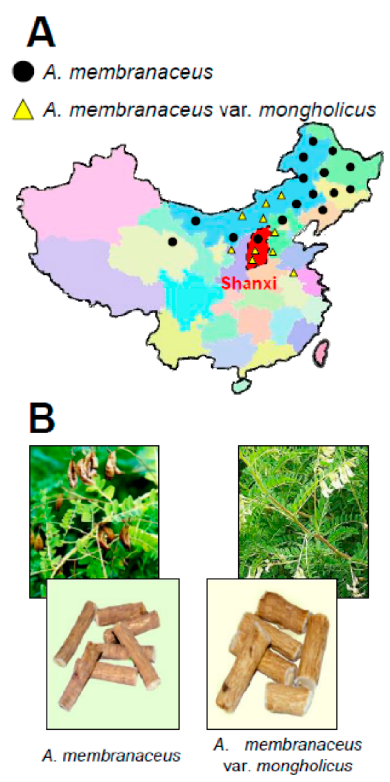
The genus Astragalus L. consists of 278 species, 2 subspecies, 35 varieties, and 2 forma found predominantly within China [1,2]. Two authentic botanical sources of AR recorded in Chinese Pharmacopoeia are Astragalus membranaceus (Fischer) Bunge and A. membranaceus (Fisch.) Bunge var. mongholicus (Bunge) P. K. Hsiao (CP, 2015). A. membranaceus and A. membranaceus var. mongholicus are mainly cultured in Inner Mongolia, Shanxi, Gansu, and Heilongjiang provinces in the north and the northeast parts of mainland China (Figure 1) [2,3]. In fact, A. membranaceus and A. membranaceus var. mongholicus share a basic morphology (Figure 1). Furthermore, A. membranaceus and A. membranaceus var. mongholicus share close similarities of chemical components, i.e., isoflavonoids, saponins, polysaccharides, γ-aminobutyric acid (GABA), and various trace elements, and of genetic components, represented by the internal transcribed spacer sequences of nuclear ribosomal DNA [2,4,5]. Indeed, random amplified polymorphic DNA primers were employed to reveal DNA fingerprinting differences between A. membranaceus and A. membranaceus var. mongholicus. The primer pairs HG3 and HG4 are specific for A. membranaceus, whereas HG7 and HG8 can identify A. membranaceus var. mongholicus [6]. Furthermore, microscopic characteristics, such as layers of phellem, continuing lignified xylem bundles within spring wood, and lignified parenchyma cells in the central part of the xylem, are other major tools to differentiate these two species from others [7]. Besides, annual rings can be identified in the roots of both species and provide critical information for determining the age of a sample [8]. Therefore, microscope analysis could be a key technique to examine the differences between A. membranaceus and A. membranaceus var. mongholicus.
2. Chemical Determination of Different Plant Parts of Astragali Radix
Historically, the root, but not other plant parts, of A. membranaceus has pharmaceutical properties. Kim and co-workers compared the chemical and genetic composition of aerial parts, including flower and stems, and root parts of A. membranaceus. A total of 10 mevalonate pathways involved in astragaloside biosynthesis were found and identified by Illumina/Solexa HiSeq2000 [9]. The accumulation rates of astragalosides were different in various plant organs. Most genes were highly expressed in the root rather than in the stem and leaf [9]. In particular, the concentration of astragaloside IV was distributed in the following order: root (0.58 mg/g) > flower (0.27 mg/g) > stem (0.23 mg/g) > leaf (0.04 mg/g) [10]. In line with this report, the content of calycosin-7-O-β-d-glucoside content was enriched as follows: root (4.88 μg/g) > stem (3.86 μg/g) > leaf (2.0 μg/g) > flower (not detected) [9]. These chemical results support fully the pharmaceutical values of the root of A. membranaceus.
In fact, the roots of A. membranaceus are divided into three major classes: seedling roots, adventitious roots, and hairy roots. Their study revealed that the total content of astragalosides found in the adventitious root was the lowest, whereas the seedling root had the highest content [11]. However, calycosin, one major bioactive isoflavonoid, and calycosin-7-O-β-d-glucoside accumulated the most in the seedling root [11]. Interestingly, the total concentration of astragaloside in the periderm was about eight-fold higher in the cortex and about 30-fold higher than in the xylem [12]. The dry weight percentages of total saponins in primary roots were ~40% in the periderm, ~50% in the cortex, and 9.30% in the xylem, respectively [12].
3. The Optimization of Extraction of Astragali Radix
In order to search the best extraction method for AR, the roles of background electrolyte concentrations, organic solvents, pH, surfactant concentrations, temperature, and voltage on the separation procedure were systematically identified and compared [8]. The optimized extract condition was found to be the micellar phase containing 100 mM sodium cholate, 25% (v/v) acetonitrile, and 20 mM H3BO3 buffer at pH 9.2. Furthermore, repeatability parameters, i.e., intra-day and inter-day precisions, were determined and resulted to be below 4.42% [8]. In 2014, Kim et al. reported that the eight constituents isolated from an 80% methanol AR extract showed a better inhibition of matrix metalloproteinases (MMP) production in IL-1-induced osteoarthritis models, than a water extract of AR [13].
The novel strategy involving pressurized liquid extraction (PLE), microwave-assisted acidic hydrolysis (MAAH), and comprehensive chromatography served as an effective method to increase the polysaccharide extraction yield. The quantification of twelve saccharides, i.e., glucose, free sucrose, fructose, etc. was performed by gas chromatography–mass spectrometry (GC–MS) and HPLC. The results showed that A. membranaceus dried powder contained about 108.5 mg/g free sucrose and lesser amounts of glucose (9.6–26.0 mg/g) and fructose (8.7–22.9 mg/g). Hence, this extraction method was much more efficient than the traditional extraction method [14]. Homogenization-assisted negative pressure cavitation extraction (HNPCE) is another effective and innovative method to extract polysaccharides from AR. The optimal extraction parameters were determined as: homogenization time 70 s, negative pressure −0.068 MPa, extraction time 53 min at 64.8 °C, which increased the polysaccharide yield around 15% [15]. Yin et al. (2012a) have established a green and interesting method based on an aqueous diphase solvent system, consisting of PEG1000-MgSO4-H2O, which purified polysaccharides by using 76.5% of galacturonic acid, 7.7% of galactose, 4.2% of arabinose, etc. [16]. Therefore, we suggest to employ HNPCE to generate the polysaccharide-enriched AR fractions.
4. The Pharmaceutical Value of AR Extract and AR Major Ingredients
AR is one of the most popular herbal medicines used worldwide, possessing tonic, hepatoprotective, diuretic, and expectorant properties, according to the Chinese Pharmacopoeia (CP Volume 1 of 2015 Edition) [2]. It has been shown to have anti-oxidation properties, regulate the immune function, mitigate cardiovascular diseases, inhibit liver fibrosis, and other pharmaceutical functions [17].
4.1. The Anti-Oxidative Actions of Astragali Radix and Its Major Constituents
Oxidative stress plays a key role in the pathogenesis of various diseases, and anti-oxidants compounds could protect cells and tissues from oxidative stress by removing reactive oxygen species [18]. The anti-oxidative functions of AR and of its pharmaceutical constituents are summarized in Table 1. In 1999, Toda & Shirataki indicated that the anti-oxidative functions of AR were superior or similar to those of the positive controls butyl hydroxytoluene and α-tocopherol [19]. Furthermore, AR was reported to adapt to water stress during the growth season by enhancing the activity of anti-oxidant enzymes and accumulating osmotic agents [20]. The AR extract could mitigate the telomere shortening rate of lung diploid fibroblasts 2BS, which is hypothesized to be related to a reduction of DNA damage and the improvement of DNA repair ability [21]. Moreover, the declined telomere shortening rate, the diminished DNA damage, as well as the upregulated DNA repair ability triggered by AR extract are believed to be responsible for the delay of replicative senescence [21]. Animal studies have shown, by histopathology and immunohistochemistry, upregulated lipid peroxidation levels in the cerebral cortex, hippocampus regions, and injured brain tissue in a whisker rat model of removal-induced psycho-emotional stress [22]. However, the oral administration of AR extract could significantly decrease the content of these biomarkers [22]. The activation of NF-κB was predominant in the untreated group, while it was significantly suppressed by the application of an AR water extract in cerebral cortex and hippocampus regions [22].
The anti-oxidant activities of three new flavonoids, including one aurone and two chalcones, were investigated in 2,2-diphenyl-1-(2,4,6-trinitrophenyl) hydrazyl free radical scavenging assays [23]. The IC50 of two chalcones were 35.9 ± 1.1 and 12.2 ± 1.1 µM, respectively [23]. The anti-oxidative functions of formononetin and ononin were much stronger than those of tert-Butylhydroquinon (TBHQ) or butylated hydroxyanisole (BHA) when determined by 2,2-Diphenyl-1-Picrylhydrazyl (DPPH), 2,2′-azino-bis(3-ethylbenzothiazoline-6-sulphonic acid (ABTS), ferric reducing ability of plasma (FRAP), and lipid peroxidation inhibition assays [24,25,26]. Indeed, the combination of formononetin, ononin, calycosin, and calycosin-7-O-β-d-glucoside also exhibited anti-oxidative therapeutic effects in anemic rats, as indicated by the serum levels of SOD and GSH-Px [27].
Astragalosides also showed pharmaceutical value as anti-oxidants (Table 1) and could decrease the level of high-mobility group box 1 protein, a highly expressed protein that regulates acute inflammation in mouse models [28]. On the other hand, astragalosides prevented renal and mitochondrial dysfunctions through their anti-oxidative effects in crush syndrome rat models [29]. This modulatory function was hypothesized to be related to the TLR4/NF-κB pathway [30]. In addition, astragalosides were shown to improve pulmonary inflammatory diseases by interacting with autophagy and PERK-eIF2α pathways [31].
The polysaccharide-enriched AR fractions showed promising therapeutic effects as anti-oxidants both in vivo and in vitro [32,33,34]. In hyperlipidemia and oxidative stress-induced rat models, the serum levels of SOD and GSH-Px activity declined after oral administration of a polysaccharide-enriched AR fraction for four weeks [33]. In vitro studies have shown that AR polysaccharides decreased SOD, GSH-Px, and catalase activities in cultured H2O2-induced MRC-5 cells mimicking oxidative damage models [34]. Another research group reported that a polysaccharide-enriched constituent also played a part in the therapeutic effects on oxidative damage in the skeletal muscles of rats after exhaustive exercise [35]. The rats were randomly divided into four groups. Group 1 was the control group, and groups 2 to 4 received different doses of AR polysaccharides for 30 successive days. Skeletal muscle samples were collected to analyze enzymatic activities. The results indicated that the polysaccharide-treated groups had significantly decreased MDA, SOD, GSH-Px, and CAT contents compared to the negative control [35]. The in vivo anti-aging functions of the polysaccharides were determined by Li et al. [36]. The back of the neck of mice was injected with D-Gal for seven weeks to mimic aging. After polysaccharide treatment at the dose of 200 and 300 mg/kg/day, the serum levels of SOD, CAT, GSH-Px, and anti-hydroxyl radical activity were dramatically upregulated in a dose-dependent manner [32].
4.2. The Immune Functions of Astragali Radix and Its Biological Ingredients
AR extracts have shown considerable immunomodulatory properties both in vitro and in vivo [37] (Table 2). After oral and intracolonic treatments with AR extracts, a decrease in colonic lesion areas and histological damage score and an amelioration of the colonic myeloperoxidase activity were detected in Sprague–Dawley rats [38,39]. Western blot data demonstrated that AR was capable of diminishing the levels of some cytokines, i.e., TNF-α and IL-1β, and of other immune-specific regulators, such as. P-selectin and ICAM-1 [38,39]. Furthermore, AR was also reported to have non-specific immune effects on tilapia [40]. The plasma levels of extracellular superoxide anion production and phagocytosis were examined after feeding three-month-old tilapia with an AR extract. The results showed that the AR extract was capable of modulating the innate immune system by upregulating lysozyme activity instead of altering the respiratory burst activity [40]. Clinically, leukocyte, platelet, and cytokine responses as well as body temperature and blood pressure were examined in healthy individuals after consuming an extract of A. membranaceus [41]: monocytes, neutrophils, and lymphocytes counts were increased in a dose-dependent manner after 8–12 h from the extract administration. Dynamic changes in the concentration of circulating cytokines, i.e., interferon-γ, TNF-α, IL-13, IL-6, and soluble IL-2R, were observed [41].
An aqueous extract of AR could trigger apoptosis of H22 tumor cells by enhancing Bax/Bcl-2 ratios, which was confirmed both by immunoblotting and flow cytometry [42]. Inhibition of tumor cell proliferation was detected in various cell lines. The inhibition rates of AR extracts in AGS, KATOIII, HT29, MDA231, MEL7, and MEL14 cells were 68.25%, 62.36%, 22.8%, 27.69%, 2.85%, and 5.14%, respectively [43]. AR suppressed DNA synthesis by 87.33% at the concentration of 50 μg/mL in AGS cell line. The effects of a co-treatment of AR with conventional cancer therapy were determined on the 798 breast cancer patients. AR combined with conventional therapy is believed to be efficacious in improving the quality of life in late-stage breast cancer patients and in decreasing the number of hot flashes [44].
After oral administration of AR polysaccharides in cyclophosphamide-induced rat, spleen weight, peripheral white blood cell counts, and T cell and B cell proliferation responses were significantly upregulated [45]. Furthermore, splenic nature killer cell activity and peritoneal macrophage phagocytosis were also dramatically increased [45]. In humans, the intake of AR extract promoted immunomodulating effects by upregulating the proliferation of peripheral blood mononuclear cells, as well as inducing interleukin production [46]. Dose-dependent stimulations of IL-10, IL-12, and IL-2 were markedly observed after application of AR polysaccharides, compared to a negative control [46]. The bioactive constituents of AR also showed the ability to induce TNF, GM-CSF, and NO productions in cultured macrophages via the activation of the NF-κB pathway [47]. Moreover, AR-enriched polysaccharides could increase the sensitivity to cisplatin (DDP) and vinorelbine in lung cancer patient, prolong their life span, and increase their life quality by mitigating the toxicity of the chemotherapeutic drugs [48]. In a clinical trial, 136 patients suffering from non-small-cell lung cancer (NSCLC) were enrolled for the duration of two years [48]. Patients were randomized into two groups: an anti-cancer drug group (DDP and vinorelbine) and an anti-cancer drug–AR polysaccharide co-treatment group. After two years of treatment, several indexes showed significant differences, i.e., physical function (p = 0.01), nausea and vomiting (p < 0.001), fatigue (p < 0.001), pain (p = 0.007), and loss of appetite (p = 0.023) [48]. In nude mice, the AR-generated polysaccharides were capable of decreasing tumor sizes and increase the expressions of the apoptosis markers cleaved-caspase 3/9 [49]. Increased IgM antibody production in aged mice receiving AR polysaccharides was also confirmed [50]. In aged mice, i.e., 36- and 60-week-old mice, the antibody levels were significantly decreased to about 70% and 60%, respectively, compared to those of 10-week-old mice. IgM production was significantly upregulated in 36- and 60-week-old mice after treated with AR [50]. The immune function enhancing properties of AR polysaccharides could be interpreted. One the other hand, the role of AR-derived polysaccharides in auto-immune diseases has been explored. Chronic inflammation is believed to be the major component of autoimmune diseases development [51,52]. In vitro data revealed that the minimum anti-inflammatory concentration of polysaccharides was 100 μg/mL in LPS-induced Caco-2 cells [53]. The anti-inflammatory effect was revealed in cultured LPS-inducted macrophages by detecting NO and the protein expression levels of IL-1β, IL-6, and TNF-α [54,55]. A polysaccharide-enriched AR fraction suppressed IL-1β level by ∼20%, IL-6 expression by ∼15%, and TNF-α level by ∼25% in LPS-induced macrophages, respectively [55]. Reduced cell accumulation, swelling, joints arthritic index, and serum concentrations of TNF-α and IL1-β were observed in arthritis rat models [56].
The effects of flavonoids derived from AR on the immune functions were determined in Raw 264.7 cells [57]. Four newly isolated compounds, i.e., (−)-methylinissolin 3-O-β-d-(6′-acetyl)-glucoside, (−)-methylinissolin 3-O-β-d-{6′-[(E)-but-2-enoyl]}-glucoside, and calycosin 7-O-β-d-(6″-acetyl)-glucoside and (−)-methylinissolin 3-O-β-d-glucoside, showed inhibitory effects on NO production in Raw 264.7 cells after LPS-induced chronic inflammation [57]. The inhibitory effects of the newly identified compounds isoliquiritigenin and liquiritigenin on LPS-stimulated bone marrow-derived dendritic cells were investigated [58]. These two compounds exhibited inhibitory effects on LPS-induced IL-6 and IL-12 productions, with IC50 values ranging from 2.7 μM to 6.1 μM [58]. Isoliquiritigenin also showed a moderate suppression function on LPS-stimulated TNF-α production, with an IC50 value of 20.1 μM. Furthermore, the AR flavonoids calycosin and calycosin-7-O-β-d-glucoside accelerated glomerular endothelial cell apoptosis rate [59]. Immunomodulatory functions of AR flavonoids were also well reported in vivo [60]. AR flavonoids could promote lymphocyte proliferation, increase T cell number, modulate T cell subsets disorders, and elevate LAK activity induced by IL-2 in immunosuppressed mice [60,61,62,63].
4.3. Protective Effects on Cardiovascular Diseases
The AR herbal extract dramatically decreased total cholesterol and LDL cholesterol and aortic fatty streak area levels; on the other hand, it exhibited the potential ability of increasing HDL cholesterol levels in atherosclerotic rabbits [64]. Besides, the therapeutic functions of AR on atherosclerosis in the aortic endothelium were determined on apolipoprotein E-deficient (apoE−/−) mice [65]. Upregulated expression of VCAM-1 and phosphorylation of NF-κB were the hallmarks of AR, as indicated in the model group mice: Immunofluorescence analysis confirmed the reduced expression of the adhesion molecules and the expression of macrophages in the aortic endothelium in AR-treated apoE−/− mice [65]. In vitro data showed that this ancient herbal medicine could scavenge superoxide and hydroxyl radicals in a concentration- and time-dependent manner [66]. Furthermore, it could effectively suppress free radical formation in ischemia-reperfusion models [64].
AR showed promising effects in improving biochemical and histological changes of heart failure [66]. The therapeutic activities were categorized as: suppressing lipid accumulation via adenosine monophosphate-activated protein kinase activation, increasing LDL receptor expression, alleviating lipid peroxidation, and decreasing inflammatory cytokines production levels [66]. Clinical data indicated that circulating endothelial cells (CEC) and production of endothelin-1 (ET-1) and malondialdehyde (MDA) in the internal jugular vein of Binswanger’s disease patients were dramatically increased after AR treatment, compared to the negative control [67]. Nevertheless, serum NO concentration significantly declined in the AR group [67]. AR is believed to be effective in protecting vascular endothelial cells in Binswanger’s disease patients.
4.4. Therapeutic Effects of Astragali Radix on Liver Fibrosis
Intraperitoneal injection of 50% CCl4 twice a week for two months and intraperitoneal injection of dimethylnitrosamine (DMN) are the typical methods for inducing liver fibrosis in Sprague–Dawley rats [68,69]. The hallmarks of liver cirrhosis include high concentrations of aspartate aminotransferase (AST), alanine aminotransferase (ALT), hexadecenoic acid (HA), laminin (LN), procollagen type III (PCIII), hydroxyproline (Hyp), GSH-Px, MDA, SOD, and transforming growth factor β1 (TGF-β1) in the serum. AR could significantly reduece the high levels of these biomarkers in the serum [69]. Hematoxylin–eosin and Masson’s trichrome staining, the classical staining methods for monitoring histopathological changes, confirmed that after administration of AR, the damage to the liver function was decreased [69]. Studies have shown that AR was able to suppress TGF-β1, α-SMA, collagen I, and collagen III expression, block the phosphorylation of Smad2/3, and enhance the expression Smad7, the specific inhibitor of TGF-β1 [69].
4.5. The Erythropoietic Functions of Astragali Radix and Its Major Constituents
The angiogenic function of the polysaccharide-enriched AR fraction was revealed in zebrafish. A cocktail containing 300 nM VEGFR tyrosine kinase inhibitor II was applied to Tg(fli-1a: EGFP)y1 and Tg(fli-1a:nEGFP)y7 embryos for 3 h, to induce the loss of blood vessels. After challenging with a polysaccharide-enriched AR fraction, a rescue effect was shown by a statistically significant increase of blood vessels in a dose-dependent manner [70].
Furthermore, our group has investigated the erythropoietic functions of the four major flavonoid constituents of AR (formononetin, ononin, calycosin, and calycosin-7-O-β-d-glucoside) by monitoring the expression of erythropoietin (EPO) and its upstream regulator, hypoxia-induced factor (HIF-1α) in cultured HEK293T cells. These four flavonoids could upregulate EPO and HIF-1α at both the transcriptional and the translational levels [71], as shown in the cyclophosphamide-induced anemic rats after treatment with the combined flavonoids; enhanced content of red blood cells, white blood cells, and hemoglobin, and increased hematocrit were significantly observed [27].
4.6. Other Pharmaceutical Properties of AR and Its Ingredients
The anti-obesity function of a polysaccharide-enriched AR decoction has been also revealed both in vivo and in vitro [72,73]. Decrease in body weight, improvement in insulin sensitivity, and a mitigation of fatty liver were recorded after polysaccharide administration to type 2 diabetes rats [72]. Agyemang and co-workers observed that AR polysaccharides suppressed the crucial regulators of endoplasmic reticulum stress, such as PERK, ATF-6, and XBP1 in type 2 diabetes rat [73]. Other groups revealed that the AR polysaccharides had a negative role in modulating the GLUT4/PKB glucose transportation pathway in insulin-resistant KKAy mice [74].
The anti-cancer function of AR saponins has been demonstrated, as shown in Table 3. AR saponins inhibited cancer cell proliferation both in cell and in animal models. In HT29 colon adenocarcinoma cells, treatment with AR saponins upregulated cleaved PARP, caspase 8, and NAG-1 levels [75,76]. A total AR saponin fraction triggered PTEN expression and downregulated mTOR expression via blocking NFκB and DNA binding activity [76]; it decreased the levels of VEGF and bFGF in a time- and dose-dependent manner [77]. In vivo studies were performed in colon cancer cell HT29-xenografted nude mice [75,77]. The tumor regression rate was about 35% after treatment with AR saponins, without alterations of mice body weight [75]. The translational levels of p-Akt, p-mTOR, VEGF, VEGFR1, and VEGFR2 were decreased in experimental rat tumor tissues [77]. The co-treatment of astragaloside IV with indoleamine 2,3-dioxygenase (IDO), a tryptophan-catabolizing enzyme triggering immune tolerance, could enhance the immune response by suppressing regulatory T cells and enhancing cytotoxic T lymphocytes; therefore, astragaloside IV might be effective in blocking cancer cell proliferations [78,79].
Astragaloside II was capable of inducing bone matrix formation and remodeling, as shown in Table 3. Astragaloside II exhibited osteogenic functions throughout the whole osteoblast differentiation process [80]. In bone development, astragaloside II induced the expression of master bone matrix regulators, such as BMP-2, Smad1/5/8, ERK1/2 and p38, and Cbfa1/Runx2 [80].
The pain reliving pharmaceutical value of AR was also widely reported. Administration of AR extracts significantly suppressed oxaliplatin-trigged hypersensitivity and promoted rescue mechanisms, preventing damages of the nervous tissue and the triggering of chronic pain [81]. Oxaliplatin enhanced the superoxide anion production both in the stable cell line SH-SY5Y and in primary cortical astrocytes. Administration of AR extracts showed protective effects against oxaliplatin-induced lipid peroxidation, carbonylated proteins, and DNA oxidation [82]. Furthermore, a single administration of a hydroalcoholic AR extract dramatically suppressed both sodium mono-iodoacetate- and complete Freund’s adjuvant-induced pain, with over 70% and 90% of pain relief, in rat models with articular damage resembling osteoarthritis and rheumatoid arthritis, respectively [83].
5. Discussion
This review has pointed out the importance of obtaining reliable starting materials for herbal extracts of AR, which are suitable for screening its major bioactive chemicals. It has also summarized the pharmaceutical properties of the CMM, its concentrated extracts, and their individual chemical constituents and purified polysaccharides. These pharmaceutical activities may be useful for the further development of good quality herbal medicines useful in areas where conventional pharmaceutical medicines experience difficulties in controlling chronic diseases, or as nutraceuticals for good health. However, a single CMM is less efficient than well-used composite formulae consisting of several CMMs in combination, because of the multi-targeting properties and synergic or complementing effects of the combined ingredients.
In the clinical practice of TCM, AR is often combined with other CMMs in composite herbal formulae (Fufang) for the treatment of various diseases, according to patients’ individual constitution and symptoms. Indeed, its roles in the different prescription formula are not the same. The principles for composing a TCM prescription, as first described in the Huang Di Nei Jing (The Inner Canon of the Yellow Emperor), stipulate that the prescription may include four different CMMs. They are the Emperor or Principal (Jun CMM), the Adjuvant (Chen CMM), the Assistant (Zuo CMM), and the Guide (Shi CMM), according to the different roles they play in the prescription. At times, some CMM may have two or more roles depending on their diverging properties. The four CMMs, supplementing one another, exert a curative role together. However, not every prescription is composed of four kinds of CMMs; there may be less or more than four, with the Emperor CMM being the dominant one, depending on the diseases, the characteristics of the CMMs, and the therapeutic needs. AR is often used as the Emperor CMM with little or no toxicity in most TCM formulae. The following three examples illustrate the works our team has researched in detail.
Yu Ping Feng San (YPFS), an ancient TCM prescription, consists of AR, Atractyldis Macrocephalae Rhizoma, and Saposhnikoviae Radix; here, AR plays the major functions in YUPS by enriching “Yang Qi” of spleen and lungs and tonifying “Wei Qi” of the stomach [84,85]. Our research team observed that YPFS was capable of modulating the immune system and it exhibited the possibility of enhancing the innate immune system against bacterial and viral invasions [84]. The co-treatment of YPFS and Cisplatin (DDP) showed potential efficacy in reducing DDP-resistance in NSCLC cells by increasing the intracellular DDP content [85]. In vivo data showed that the co-treatment of DDP and YPFS reduced tumor size by more than 80% in tumor-bearing mice, compared to DDP alone [85].
Buyang Huanwu prescription (BYHWD) is another popular ancient formula consisting of seven CMMs (AR, Angelica Sinensis Radix [ASR], Chuanxiong Radix, Paeoniae Rubra Radix, Persicae Semen, Carthami Flos, and Pheretima) which is designed to treat complications after cerebrovascular accident, paralysis, stroke, and their related complications. In this formula, AR also plays the Emperor function. The administration of BYHWD in chronic denervation models significantly increased the axonal regenerative abilities and neurite outgrowth [86]. This formula could also reduce cerebral ischemia/reperfusion damages in animal experiments [87,88,89].
Danggui Buxue Tang (DBT), consisting of two CMMs (AR and ASR), is a widely prescribed classical and simple formula. The pharmaceutical value of this prescription has been reported widely [90]. AR acts as the Emperor role. Previous studies have confirmed that DBT is able to improve the cardiovascular function by stimulating NO production in endothelia cells [54]. Immune enhancing functions of DBT were detected both in vitro and in vivo, as observed in cultured T lymphocytes; after application of DBT, cell proliferation was markedly induced, with the release of IL-2, -6, and -10, and the phosphorylation of ERK [60]. On the other hand, DBT could not only withstand significantly the reduction of blood cells by immune mediation, but also stimulate the growth of bone marrow cells and increase the weight of haemopoietic progenitors in the bone marrow in rats [91]. Furthermore, DBT treatment showed a greater improvement of clinical symptoms, such as decreased skin thickness and scratching behavior in a rat model and decline of the total serum IgE levels and mast cells counts, when compared to the control group and to the single-AR-extract-treated group. The levels of cytokines and inflammatory mediators were significantly decreased in the DBT groups [92]. In vivo monitoring of the biomarkers together with in vitro investigations of cell expression/signaling responses are required to elucidate the biological complexity of the composite herbal formula.
The research on composite formulae is only possible with the advances in biomedical, chemical, and computational technology, by employing multidisciplinary approaches for investigating evidence-based aspects of TCM practice. Research linking a specified starting CMM, the relatively new systems biology, and experience-based TCM principles is vital to elucidate the complexity of TCM prescriptions [93]. More research is needed to probe the role of each chemical present in AR and its composite prescriptions, using systems approaches. This will be the future direction for the advancement of TCM research.
6. Conclusions
The chemical compositions of various plant parts of A. membranaceus and A. membranaceus var. mongholicus were compared. Homogenization-assisted negative pressure cavitation extraction is considered the optimal extraction method for polysaccharide-enriched AR to obtain consistent chemical ingredients for biological investigations. The pharmaceutical values of AR were summarized. The key values include immunomodulation, anti-oxidation activation, cardiovascular protection, inhibition of liver fibrosis, stimulation of blood regeneration, anti-obesity action, and pain-relieving properties. We believe that AR is one the most important natural medicines of TCM herbal composite formulae, and its promising biological functions should be further explored and tested for clinical application in conditions where conventional medicines may not be efficacious.
Acknowledgments
This work was supported by Hong Kong Research Grants Council Theme-based Research Scheme (T13-607/12R), TUYF15SC01, Shenzhen Science and Technology Committee Research Grant (CKFW2016082916015476, JCYJ20170413173747440, ZDSYS201707281432317, JCYJ20160229205726699, JCYJ20160229205812004, JCYJ20160229210027564 and 20170326).
Conflicts of Interest
The authors declare no conflict of interest.
Abbreviations
| ABTS | 2,2′-azino-bis(3-ethylbenzothiazoline-6-sulphonic acid |
| ALT | Alanine aminotransferase |
| AR | Astragali Radix |
| ASR | Angelica Sinensis Radix |
| AST | Aspartate aminotransferase |
| BYHWD | Buyang Huanwu decoction |
| CEC | Circulating endothelial cells |
| CMM | Chinese Materia Medica |
| DBT | Danggui Buxue Tang |
| DDP | Cisplatin |
| DMN | Dimethylnitrosamine |
| DPPH | 2,2-Diphenyl-1-Picrylhydrazyl |
| EF | Epimedii Folium |
| EPO | Erythropoietin |
| ET-1 | Endothelin-1 |
| FRAP | Ferric reducing ability of plasma |
| GABA | γ-aminobutyric acid |
| HA | Hexadecenoic acid |
| HIF-1α | Hypoxia-induced factor |
| HNPCE | Homogenization-assisted negative pressure cavitation extraction |
| Hyp | Hydroxyproline |
| IDO | Indoleamine 2,3-dioxygenase |
| LN | Laminin |
| MAAH | Microwave-assisted acidic hydrolysis |
| MDA | Malondialdehyde |
| MMP | Matrix metalloproteinases |
| NSCLC | Non-small-cell lung cancer |
| PCIII | Procollagen type III |
| PLE | Pressurized liquid extraction |
| TCM | Traditional Chinese medicine |
| YPFS | Yu Ping Feng San |
References
- Fu, K.T. Flora of Reipublicae Popularis Sinicae; Science Press: Beijing, China, 1998; Volume 42, pp. 78–347. [Google Scholar]
- Ma, X.Q.; Shi, Q.; Duan, J.A.; Dong, T.T.; Tsim, K.W. Chemical analysis of Radix Astragali (Huangqi) in China: A comparison with its adulterants and seasonal variations. J. Agric. Food Chem. 2002, 50, 4861–4866. [Google Scholar] [CrossRef] [PubMed]
- Cui, Y.L. Pharmacopoeia of the People’s Republic of China; Chemical Industry Press: Beijing, China, 1990; pp. 127–274. [Google Scholar]
- Ma, X.Q.; Duan, J.A.; Zhu, D.Y.; Dong, T.T.; Tsim, K.W. Species identification of Radix Astragali (Huangqi) by DNA sequence of its 5S-rRNA spacer domain. Phytochemistry 2000, 54, 363–368. [Google Scholar] [CrossRef]
- Liu, J.; Chen, H.B.; Guo, B.L.; Zhao, Z.Z.; Liang, Z.T.; Yi, T. Study of the relationship between genetics and geography in determining the quality of Astragali Radix. Biol. Pharm. Bull. 2011, 34, 1404–1412. [Google Scholar] [CrossRef] [PubMed]
- Liu, T.H.; Lin, H.M.; Wu, R.Y. Identification of Astragalus medicines using scar markers. J. Food Drug Anal. 2008, 16, 57–62. [Google Scholar]
- Yu, K.Z.; Liu, J.; Guo, P.L.; Zhao, Z.Z.; Hong, H.; Chen, H.B.; Cai, S.Q. Microscopic research on a multi-source traditional Chinese medicine, Astragali Radix. J. Nat. Med. 2014, 68, 340–350. [Google Scholar] [CrossRef] [PubMed]
- Song, Y.; Li, P.; Wang, D.; Cheng, Y.Y. Micellar electrokinetic chromatography for the quantitative analysis of flavonoids in the Radix of Astragalus membranaceus var. mongholicus. Planta Med. 2008, 74, 84–89. [Google Scholar] [CrossRef] [PubMed]
- Kim, Y.; Thwe, A.A.; Li, X.; Tuan, P.A.; Lee, S.; Lee, J.W.; Arasu, M.V.; Al-Dhabi, N.A.; Park, S.U. Accumulation of astragalosides and related gene expression in different organs of Astragalus membranaceus Bge. var mongholicus (Bge.). Molecules 2014, 19, 10922–10935. [Google Scholar] [CrossRef] [PubMed]
- Kim, Y.; Thwe, A.A.; Li, X.; Tuan, P.A.; Zhao, S.; Park, C.G.; Lee, J.W.; Park, S.U. Accumulation of flavonoids and related gene expressions in different organs of Astragalus membranaceus Bge. Appl. Biochem. Biotechnol. 2014, 173, 2076–2085. [Google Scholar] [CrossRef] [PubMed]
- Park, Y.J.; Thwe, A.A.; Li, X.H.; Kim, Y.J.; Kim, J.K.; Arasu, M.V.; AI-Dhabi, N.A.; Park, S.U. Triterpene and flavonoid biosynthesis and metabolic profiling of hairy roots, adventitious roots, and seedling roots of Astragalus membranaceus. J. Agric. Food Chem. 2015, 63, 8862–8869. [Google Scholar] [CrossRef] [PubMed]
- Kwon, H.J.; Hwang, J.; Lee, S.K.; Park, Y.D. Astragaloside content in the periderm, cortex, and xylem of Astragalus membranaceus root. J. Nat. Med. 2013, 67, 850–855. [Google Scholar] [CrossRef] [PubMed]
- Kim, G.; Choi, S.; Lee, D.; Noh, H.; Lee, S.; Choi, J.; Kim, S. Effects of ethanol extracts and compounds from Astragali Radix on chondrocytes and MIA-induced osteoarthritis model in rat. Planta Med. 2014, 80, 1493–1494. [Google Scholar] [CrossRef]
- Lv, G.P.; Hu, D.J.; Cheong, K.I.; Li, Z.Y.; Qing, X.M.; Zhao, J.; Li, S.P. Decoding glycome of Astragalus membranaceus based on pressurized liquid extraction, microwave-assisted hydrolysis and chromatographic analysis. J. Chromatogr. A 2015, 1409, 19–29. [Google Scholar] [CrossRef] [PubMed]
- Jiao, J.; Wei, F.Y.; Gai, Q.Y.; Wang, W.; Luo, M.; Fu, Y.J.; Ma, W. A pilot-scale homogenization-assisted negative pressure cavitation extraction of Astragalus polysaccharides. Int. J. Biol. Macromol. 2014, 67, 189–194. [Google Scholar] [CrossRef] [PubMed]
- Yin, J.Y.; Jiang, Z.H.; Yu, H.; Xie, M.Y.; Hsiao, W.L.; Lu, A.P.; Han, Q.B. A new application of an aqueous diphase solvent system in one-step preparation of polysaccharide from the crude water extract of Radix Astragali by high-speed counter-current chromatography. J. Chromatogr. A 2012, 1262, 92–97. [Google Scholar] [CrossRef] [PubMed]
- Fu, J.; Wang, Z.H.; Huang, L.F.; Zheng, S.H.; Wang, D.M.; Chen, S.L.; Zhang, H.T.; Yang, S.H. Review of the botanical characteristics, phytochemistry, and pharmacology of Astragalus membranaceus (Huangqi). Phytother. Res. 2014, 28, 1275–1283. [Google Scholar] [CrossRef] [PubMed]
- Shahzad, M.; Shabbir, A.; Wojcikowski, K.; Wohlmuth, H.; Gobe, G.C. The antioxidant effects of Radix Astragali (Astragalus membranaceus and related species) in protecting tissues from injury and disease. Curr. Drug Targets 2016, 17, 1331–1340. [Google Scholar] [CrossRef] [PubMed]
- Toda, S.; Shirataki, Y. Inhibitory effects of Astragali Radix, a crude drug in oriental medicines, on lipid peroxidation and protein oxidative modification by copper. J. Ethnopharmacol. 1999, 68, 331–333. [Google Scholar] [CrossRef]
- Jia, X.; Sun, C.S.; Li, G.Y.; Li, G.B.; Chen, G.L. Effects of progressive drought stress on the physiology, antioxidative enzymes and secondary metabolites of Radix Astragali. Acta Physiol. Plant 2015, 37, 26. [Google Scholar] [CrossRef]
- Wang, P.; Zhang, Z.; Sun, Y.; Liu, X.; Tong, T. The two isomers of HDTIC compounds from Astragali Radix slow down telomere shortening rate via attenuating oxidative stress and increasing DNA repair ability in human fetal lung diploid fibroblast cells. DNA Cell Biol. 2010, 29, 33–39. [Google Scholar] [CrossRef] [PubMed]
- Kim, H.G.; Lee, J.S.; Choi, M.K.; Han, J.M.; Son, C.G. Ethanolic extract of Astragali Radix and Salviae Radix prohibits oxidative brain injury by psycho-emotional stress in whisker removal rat model. PLoS ONE 2014, 9, e98329. [Google Scholar] [CrossRef] [PubMed]
- Xiao, C.J.; Zhang, Y.; Qiu, L.; Dong, X.; Jiang, B. Schistosomicidal and antioxidant flavonoids from Astragalus englerianus. Planta Med. 2014, 80, 1727–1731. [Google Scholar] [CrossRef] [PubMed]
- Pu, W.J.; Wang, D.M.; Zhou, D. Structural characterization and evaluation of the antioxidant activity of phenolic compounds from Astragalus taipaishanensis and their structure-activity relationship. Sci. Rep. 2015, 5, 13914. [Google Scholar] [CrossRef] [PubMed]
- Chen, C.Y.; Zu, Y.G.; Fu, Y.J.; Luo, M.; Zhao, C.J.; Wang, W.; Zhao, B.S.; Li, J.; Efferth, T. Preparation and antioxidant activity of Radix Astragali residues extracts rich in calycosin and formononetin. Biochem. Eng. J. 2011, 56, 84–93. [Google Scholar] [CrossRef]
- He, Y.X.; Shi, H.L.; Huang, F.; Liu, H.S.; Wu, H.; Zhang, B.B.; Dou, W.; Wu, X.J.; Wang, Z.T. Astragalosides from Radix Astragali benefits experimental autoimmune encephalomyelitis in C57BL/6 mice at multiple levels. BMC Complement. Altern. Med. 2014, 14, 313. [Google Scholar] [CrossRef] [PubMed]
- Zhang, L.; Gong, A.G.; Riaz, K.; Deng, J.Y.; Ho, C.M.; Lin, H.Q.; Dong, T.T.; Lee, Y.K.; Tsim, K.W. A novel combination of four flavonoids derived from Astragali Radix relieves the symptoms of cyclophosphamide-induced anemic rats. FEBS Open Biol. 2017, 7, 318–323. [Google Scholar] [CrossRef] [PubMed]
- Li, J.; Huang, L.; Wang, S.; Zhang, Z. Astragaloside IV attenuates inflammatory reaction via activating immune function of regulatory T-cells inhibited by HMGB1 in mice. Pharm. Biol. 2016, 54, 3217–3225. [Google Scholar] [CrossRef] [PubMed]
- Murata, I.; Abe, Y.; Yaginuma, Y.; Yodo, K.; Kamakari, Y.; Miyazaki, Y.; Baba, D.; Shinoda, Y.; Iwasaki, T.; Takahashi, K.; et al. Astragaloside-IV prevents acute kidney injury and inflammation by normalizing muscular mitochondrial function associated with a nitric oxide protective mechanism in crush syndrome rats. Ann. Intensive Care 2017, 7, 90. [Google Scholar] [CrossRef] [PubMed]
- Yang, J.; Wang, H.X.; Zhang, Y.J.; Yang, Y.H.; Lu, M.L.; Zhang, J.; Li, S.T.; Zhang, S.P.; Li, G. Astragaloside IV attenuates inflammatory cytokines by inhibiting TLR4/NF-кB signaling pathway in isoproterenol-induced myocardial hypertrophy. J. Ethnopharmacol. 2013, 150, 1062–1070. [Google Scholar] [CrossRef] [PubMed]
- Dong, Z.; Zhou, J.; Zhang, Y.; Chen, Y.; Yang, Z.; Huang, G.; Chen, Y.; Yuan, Z.; Peng, Y.; Cao, T. Astragaloside-IV alleviates heat-induced inflammation by inhibiting endoplasmic reticulum stress and autophagy. Cell. Physiol. Biochem. 2017, 42, 824–837. [Google Scholar] [CrossRef] [PubMed]
- Li, R.; Chen, W.C.; Wang, W.P.; Tian, W.Y.; Zhang, X.G. Antioxidant activity of Astragalus polysaccharides and anti-tumor activity of the polysaccharides and siRNA. Carbohydr. Polym. 2010, 82, 240–244. [Google Scholar] [CrossRef]
- Ma, J.W.; Qiao, Z.Y.; Xiang, X. Aqueous extract of Astragalus mongholicus ameliorates high cholesterol diet induced oxidative injury in experimental rats models. J. Med. Plants Res. 2011, 5, 855–858. [Google Scholar]
- Xu, X.Y.; Li, F.; Zhang, X.; Li, P.C.; Zhang, X.; Wu, Z.X.; Li, D.P. In vitro synergistic antioxidant activity and identification of antioxidant components from Astragalus membranaceus and Paeonia lactiflora. PLoS ONE 2014, 9, e96780. [Google Scholar] [CrossRef] [PubMed]
- Deng, Z.H.; Hu, Q.L. Effect of Astragalus membranaceus polysaccharides on oxidative damage in skeletal muscle of exhaustive exercise rats. Afr. J. Agric. Res. 2011, 6, 4086–4090. [Google Scholar]
- Li, X.T.; Zhang, Y.K.; Kuang, H.X.; Jin, F.X.; Liu, D.W.; Gao, M.B.; Liu, Z.; Xin, X.J. Mitochondrial protection and anti-aging activity of Astragalus polysaccharides and their potential mechanism. Int. J. Mol. Sci. 2012, 13, 1747–1761. [Google Scholar] [CrossRef] [PubMed]
- Block, K.I.; Mead, M.N. Immune system effects of Echinacea, Ginseng, and Astragalus: A review integrative cancer therapies. Integr. Cancer Ther. 2003, 2, 247. [Google Scholar] [CrossRef] [PubMed]
- Ko, J.K.; Lam, F.Y.; Cheung, A.P. Amelioration of experimental colitis by Astragalus membranaceus through anti-oxidation and inhibition of adhesion molecule synthesis. World J. Gastroenterol. 2005, 11, 5787–5794. [Google Scholar] [CrossRef] [PubMed]
- Ko, J.K.; Chik, C.W. The protective action of radix Astragalus membranaceus against hapten-induced colitis through modulation of cytokines. Cytokine 2009, 47, 85–90. [Google Scholar] [CrossRef] [PubMed]
- Yin, G.J.; Jeney, G.; Racz, T.; Xu, P.; Jun, X.; Jeney, Z. Effect of two Chinese herbs (Astragalus Radix and Scutellaria Radix) on non-specific immune response of tilapia, Oreochromis niloticus. Aquaculture 2006, 253, 39–47. [Google Scholar] [CrossRef]
- Denzler, K.; Moore, J.; Harrington, H.; Morrill, K.; Huynh, T.; Jacobs, B.; Waters, R.; Langland, J. Characterization of the physiological response following in vivo administration of Astragalus membranaceus. Evid. Based Complement. Altern. Med. 2016, 2016, 6861078. [Google Scholar] [CrossRef] [PubMed]
- Li, L.K.; Kuang, W.J.; Huang, Y.F.; Xie, H.H.; Chen, G.; Zhou, Q.C.; Wang, B.R.; Wan, L.H. Anti-tumor effects of Astragalus on hepatocellular carcinoma in vivo. Indian J. Pharmacol. 2012, 44, 78–81. [Google Scholar] [PubMed]
- Lin, J.; Dong, H.F.; Oppenheim, J.J.; Howard, O.M. Effects of Astragali Radix on the growth of different cancer cell lines. World J. Gastroenterol. 2003, 9, 670–673. [Google Scholar] [CrossRef] [PubMed]
- Kim, W.; Lee, W.B.; Lee, J.W.; Min, B.I.; Baek, S.K.; Lee, H.S.; Cho, S.H. Traditional herbal medicine as adjunctive therapy for breast cancer: A systematic review. Complement. Ther. Med. 2015, 23, 626–632. [Google Scholar] [CrossRef] [PubMed]
- Liu, Y.P.; Qi, Y.G.; Liu, A.J.; Zhang, X.W.; Teng, A.G.; Tang, J.; Zhang, W.J.; Shi, L.G. Protective effect of herbal compound beverage of Radix Astragali on immunosuppressed mice induced by cyclophosphamide. Adv. Mater. Res. 2013, 721, 603–607. [Google Scholar] [CrossRef]
- Yin, J.Y.; Chan, B.C.; Yu, H.; Lau, I.Y.; Han, X.Q.; Cheng, S.W.; Wong, C.K.; Lau, C.B.; Xie, M.Y.; Fung, K.P.; et al. Separation, structure characterization, conformation and immunomodulating effect of a hyperbranched heteroglycan from Radix Astragali. Carbohydr. Polym. 2012, 87, 667–675. [Google Scholar] [CrossRef]
- Zhao, L.H.; Ma, Z.X.; Zhu, J.; Yu, X.H.; Weng, D.P. Characterization of polysaccharide from Astragalus Radix as the macrophage stimulator. Cell. Immunol. 2011, 271, 329–334. [Google Scholar] [CrossRef] [PubMed]
- Guo, L.; Bai, S.P.; Zhao, L.; Wang, X.H. Astragalus polysaccharide injection integrated with vinorelbine and cisplatin for patients with advanced non-small cell lung cancer: Effects on quality of life and survival. Med. Oncol. 2012, 29, 1656–1662. [Google Scholar] [CrossRef] [PubMed]
- Wang, J.; Ito, H. Enhancing effect of antitumor polysaccharide from Astragalus or Radix Hedysarum on C3 cleavage production of macrophages in mice. Jpn. J. Pharmacol. 1988, 51, 432–434. [Google Scholar] [CrossRef]
- Kajimura, K.; Takagi, Y.; Miyano, K.; Sawabe, Y.; Mimura, M.; Sakagami, Y.; Yokoyama, H.; Yoneda, K. Polysaccharide of Astragali Radix enhances IgM antibody production in aged mice. Biol. Pharm. Bull. 1997, 20, 1178–1182. [Google Scholar] [CrossRef] [PubMed]
- Ercolini, A.M.; Miller, S.D. The role of infections in autoimmune disease. Clin. Exp. Immunol. 2009, 155, 1–15. [Google Scholar] [CrossRef] [PubMed]
- Navegantes, K.; Gomes, R.; Pereira, P.; Czaikoski, P.; Heitmann, C.; Azevedo, M.; Monteiro, M. Immune modulation of some autoimmune diseases: The critical role of macrophages and neutrophils in the innate and adaptive immunity. J. Transl. Med. 2017, 15, 36. [Google Scholar] [CrossRef] [PubMed]
- Wang, X.F.; Li, Y.L.; Yang, X.J.; Yao, J.H. Astragalus polysaccharide reduces inflammatory response by decreasing permeability of LPS-infected CaCO2 cells. Int. J. Biol. Macromol. 2013, 61, 347–352. [Google Scholar] [CrossRef] [PubMed]
- Lai, P.K.; Chan, J.Y.; Cheng, L.; Lau, C.P.; Han, S.Q.; Leung, P.C.; Fung, K.P.; Lau, C.B. Isolation of anti-inflammatory fractions and compounds from the root of Astragalus membranaceus. Phytother. Res. 2013, 27, 581–587. [Google Scholar] [CrossRef] [PubMed]
- Gong, A.G.; Zhang, L.M.; Lam, C.T.; Xu, M.L.; Wang, H.Y.; Lin, H.Q.; Dong, T.T.; Tsim, K.W. Polysaccharide of Danggui Buxue Tang, an ancient Chinese herbal decoction, induces expression of pro-inflammatory cytokines possibly via activation of NFκB signaling in cultured Raw 264.7 cells. Phytother. Res. 2017, 31, 274–283. [Google Scholar] [CrossRef] [PubMed]
- Jiang, J.B.; Qiu, J.D.; Yang, L.H.; He, J.P.; Smith, G.W.; Li, H.Q. Therapeutic effects of Astragalus polysaccharides on inflammation and synovial apoptosis in rats with adjuvant-induced arthritis. Int. J. Rheum. Dis. 2010, 13, 396–405. [Google Scholar] [CrossRef] [PubMed]
- Zhang, L.J.; Liu, H.K.; Hsiao, P.C.; Kuo, L.M.; Lee, I.J.; Wu, T.S.; Chiou, W.F.; Kuo, Y.H. New isoflavonoid glycosides and related constituents from Astragali Radix (Astragalus membranaceus) and their inhibitory activity on nitric oxide production. J. Agric. Food Chem. 2011, 59, 1131–1137. [Google Scholar] [CrossRef] [PubMed]
- Li, W.; Sun, Y.N.; Yan, X.T.; Yang, S.Y.; Kim, S.; Lee, Y.M.; Koh, Y.S.; Kim, Y.H. Flavonoids from Astragalus membranaceus and their inhibitory effects on LPS-stimulated pro-inflammatory cytokine production in bone marrow-derived dendritic cells. Arch. Pharm. Res. 2014, 37, 186–192. [Google Scholar] [CrossRef] [PubMed]
- Tang, D.; He, B.; Zheng, Z.G.; Wang, R.S.; Gu, F.; Duan, T.T.; Cheng, H.Q.; Zhu, Q. Inhibitory effects of two major isoflavonoids in Radix Astragali on high glucose-induced mesangial cells proliferation and AGEs-induced endothelial cells apoptosis. Planta Med. 2011, 77, 729–732. [Google Scholar] [CrossRef] [PubMed]
- Jiao, Y.; Wen, J.; Yu, X. Influence of flavonoid of Astragalus membranaceus’s stem and leaves on the function of cell mediated immunity in mice. Chin. J. Integr. Tradit. West. Med. 1999, 19, 356–358. [Google Scholar]
- Gao, Q.T.; Cheung, J.K.; Li, J.; Jiang, Z.Y.; Chu, G.K.; Duan, R.; Cheung, A.W.; Zhao, K.J.; Choi, R.C.; Dong, T.T.; et al. A Chinese herbal decoction, Danggui Buxue Tang, activates extracellular signal-regulated kinase in cultured T-lymphocytes. FEBS Lett. 2007, 581, 5087–5093. [Google Scholar] [CrossRef] [PubMed]
- Wang, D.Q.; Zhuang, Y.; Tian, Y.P.; Thomas, G.N.; Ying, M.Z.; Tomlinson, B. Study of the effects of total flavonoids of Astragalus on atherosclerosis formation and potential mechanisms. Oxid. Med. Cell. Longev. 2012, 2012, 282383. [Google Scholar] [CrossRef] [PubMed]
- Guo, Z.; Xu, H.Y.; Xu, L.; Wang, S.S.; Zhang, X.M. In vivo and in vitro immunomodulatory and anti-inflammatory effects of total flavonoids of Astragalus. Afr. J. Tradit. Complement. Altern. Med. 2016, 13, 60–73. [Google Scholar] [CrossRef] [PubMed]
- Wang, F.; Yu, G.; Liu, S.Y.; Li, J.B.; Wang, J.F.; Bo, L.L.; Qian, L.R.; Sun, X.J.; Deng, X.M. Hydrogen-rich saline protects against renal ischemia/reperfusion injury in rats. J. Surg. Res. 2011, 167, 339–344. [Google Scholar] [CrossRef] [PubMed]
- Yang, H.B.; Yan, J.C.; Su, H.L.; Yuan, W.; Xu, L.J. The effect of CD137-CD137 ligand interaction on the expression of nuclear factor of activated T cells c1 in apolipoprotein E-deficient mice atherosclerotic plaque model. Zhonghua Xin Xue Guan Bing Za Zhi 2012, 40, 775–779. [Google Scholar] [PubMed]
- Yang, Q.Y.; Chen, K.J.; Lu, S.; Sun, H.R. Research progress on mechanism of action of Radix Astragalus in the treatment of heart failure. Chin. J. Integr. Med. 2012, 18, 235–240. [Google Scholar] [CrossRef] [PubMed]
- Chen, C.F.; Jia, H.Y.; Guo, S.S.; Chen, T.H.; Wang, H.D. Protective effects of Radix Astragalus on vascular endothelial cells of patients with Binswanger’s disease. Chin. J. Integr. Med. 1999, 5, 46–49. [Google Scholar]
- Zhou, Y.; Tong, X.; Ren, S.; Wang, X.; Chen, J.; Mu, Y.; Sun, M.; Chen, G.; Zhang, H.; Liu, P. Synergistic anti-liver fibrosis actions of total Astragalus saponins and glycyrrhizic acid via TGF-β1/Smads signaling pathway modulation. J. Ethnopharmacol. 2016, 190, 83–90. [Google Scholar] [CrossRef] [PubMed]
- Liang, X.L.; Yuan, J.Y. Effect of Chinese herbal compound on liver fibrosis in rabbits with schistosomiasis by B-ultrasound. Asian Pac. J. Trop. Med. 2013, 6, 658–662. [Google Scholar] [CrossRef]
- Hu, G.; Mahady, B.; Hoi, M.P.; Wang, Y.H.; Lee, S.M. Polysaccharides from Astragali Radix restore chemical-induced blood vessel loss in zebrafish. Vasc. Cell 2012, 4, 2. [Google Scholar] [CrossRef] [PubMed]
- Zheng, K.Y.; Choi, R.C.; Cheung, A.W.; Guo, A.J.; Bi, C.W.; Zhu, K.Y.; Fu, Q.; Du, Y.; Zhang, W.L.; Zhan, J.Y.; et al. Flavonoids from Radix Astragali induce the expression of erythropoietin in cultured cells: A signaling mediated via the accumulation of hypoxia-inducible factor-1α. J. Agric. Food Chem. 2011, 59, 1697–1704. [Google Scholar] [CrossRef] [PubMed]
- Juan, Y.C.; Kuo, Y.H.; Chang, C.C.; Zhang, L.J.; Lin, Y.Y.; Hsu, C.Y.; Liu, H.K. Administration of a decoction of sucrose- and polysaccharide-rich Radix Astragali (Huang qi) ameliorated insulin resistance and fatty liver but affected Beta-cell function in type 2 diabetic rats. Evid. Based Complement. Altern. Med. 2011, 2011, 349807. [Google Scholar] [CrossRef] [PubMed]
- Agyemang, K.; Han, L.; Liu, E.; Zhang, Y.; Wang, T.; Gao, X. Recent advances in Astragalus membranaceus anti-diabetic research: Pharmacological effects of its phytochemical constituents. Evid. Based Complement. Altern. Med. 2013, 2013, 654643. [Google Scholar] [CrossRef] [PubMed]
- Liu, M.; Wu, K.; Mao, X.; Wu, Y.; Ouyang, J. Astragalus polysaccharide improves insulin sensitivity in KKAy mice: Regulation of PKB/GLUT4 signaling in skeletal muscle. J. Ethnopharmacol. 2010, 127, 32–37. [Google Scholar] [CrossRef] [PubMed]
- Tin, M.; Cho, C.H.; Ko, H. The antitumorigenic potential of total saponins from Radix Astragalus membranaceus as chemotherapeutic adjuvant in treating colon cancer. Acta Pharmacol. Sin. 2006, 27, 68. [Google Scholar]
- Auyeung, K.K.; Mok, N.I.; Wong, C.M.; Cho, C.H.; Ko, J.K. Astragalus saponins modulate mTOR and ERK signaling to promote apoptosis through the extrinsic pathway in HT-29 colon cancer cells. Int. J. Mol. Med. 2010, 26, 341–349. [Google Scholar] [PubMed]
- Law, P.C.; Auyeung, K.K.; Chan, L.Y.; Ko, J.K. Astragalus saponins downregulate vascular endothelial growth factor under cobalt chloride-stimulated hypoxia in colon cancer cells. BMC Complement. Altern. Med. 2012, 12, 160. [Google Scholar] [CrossRef] [PubMed]
- Zhang, A.; Zheng, Y.; Que, Z.; Zhang, L.; Lin, S.; Le, V.; Liu, J.; Tian, J. Astragaloside IV inhibits progression of lung cancer by mediating immune function of Tregs and CTLs by interfering with IDO. J. Cancer Res. Clin. Oncol. 2014, 140, 1883–1890. [Google Scholar] [CrossRef] [PubMed]
- Huang, L.F.; Yao, Y.M.; Li, J.F.; Zhang, S.W.; Li, W.X.; Dong, N.; Yu, Y.; Sheng, Z.Y. The effect of Astragaloside IV on immune function of regulatory T cell mediated by high mobility group box 1 protein in vitro. Fitoterapia 2012, 83, 1514–1522. [Google Scholar] [CrossRef] [PubMed]
- Kong, X.H.; Niu, Y.B.; Song, X.M.; Zhao, D.D.; Wang, J.; Wu, X.L.; Zhang, R.; Mei, Q.B. Astragaloside II induces osteogenic activities of osteoblasts through the bone morphogenetic protein-2/MAPK and Smad1/5/8 pathways. Int. J. Mol. Med. 2012, 29, 1090–1098. [Google Scholar] [PubMed]
- Di Cesare Mannelli, L.; Pacini, A.; Micheli, L.; Femia, A.P.; Maresca, M.; Zanardelli, M.; Vannacci, A.; Gallo, E.; Bilia, A.R.; Caderni, G.; et al. Astragali radix: Could it be an adjuvant for oxaliplatin-induced neuropathy? Sci. Rep. 2017, 7, 42021. [Google Scholar] [CrossRef] [PubMed]
- Di Cesare Mannelli, L.; Zanardelli, M.; Bartolucci, G.; Karioti, A.; Bilia, A.R.; Vannacci, A.; Mugelli, A.; Ghelardini, C. In vitro evidence for the use of astragali radix extracts as adjuvant against oxaliplatin-induced neurotoxicity. Planta Med. 2015, 81, 1045–1055. [Google Scholar] [CrossRef] [PubMed]
- Maresca, M.; Micheli, L.; Cinci, L.; Bili, A.R.; Ghelardini, C.; Di Cesare Mannelli, L. Pain relieving and protective effects of Astragalus hydroalcoholic extract in rat arthritis models. J. Pharm. Pharmacol. 2017, 69, 1858–1870. [Google Scholar] [CrossRef] [PubMed]
- Du, C.Y.; Choi, R.C.; Zheng, K.Y.; Dong, T.T.; Lau, D.T.; Tsim, K.W. Yu Ping Feng San, an ancient Chinese herbal decoction containing Astragali radix, Atractylodis macrocephalae rhizoma and Saposhnikoviae radix, regulates the release of cytokines in murine macrophages. PLoS ONE 2013, 8, e78622. [Google Scholar] [CrossRef] [PubMed]
- Lou, J.S.; Yan, L.; Bi, C.W.; Chan, G.K.; Wu, Q.Y.; Liu, Y.L.; Huang, Y.; Yao, P.; Du, C.Y.; Dong, T.T.; et al. Yu Ping Feng San reverses cisplatin-induced multi-drug resistance in lung cancer cells via regulating drug transporters and p62/TRAF6 signaling. Sci. Rep. 2016, 6, 31926. [Google Scholar] [CrossRef] [PubMed]
- Kim, K.J.; Namgung, U. Facilitating effects of Buyang Huanwu decoction on axonal regeneration after peripheral nerve transection. J. Ethnopharmacol. 2017, 213, 56–64. [Google Scholar] [CrossRef] [PubMed]
- Zhao, L.D.; Wang, J.H.; Jin, G.R.; Zhao, Y.; Zhang, H.J. Neuroprotective effect of Buyang Huanwu Decoction against focal cerebral ischemia/reperfusion injury in rats-Time window and mechanism. J. Ethnopharmacol. 2012, 140, 339–344. [Google Scholar] [CrossRef] [PubMed]
- Mu, Q.C.; Liu, P.; Hu, X.; Gao, H.; Zheng, X.; Huang, H. Neuroprotective effects of Buyang Huanwu decoction on cerebral ischemia-induced neuronal damage. Neural Regen. Res. 2014, 9, 1621–1627. [Google Scholar] [PubMed]
- Yang, J.H.; Gao, F.; Zhang, Y.K.; Liu, Y.; Zhang, D. Buyang Huanwu Decoction (BYHWD) enhances angiogenic effect of mesenchymal stem cell by upregulating VEGF expression after focal cerebral ischemia. J. Mol. Neurosci. 2015, 56, 898–906. [Google Scholar] [CrossRef] [PubMed]
- Lin, H.Q.; Gong, A.G.; Wang, H.Y.; Duan, R.; Dong, T.T.; Zhao, K.J.; Tsim, K.W. Danggui Buxue Tang (Astragali radix and Angelicae sinensis radix) for menopausal symptoms: A review. J. Ethnopharmacol. 2017, 199, 205–210. [Google Scholar] [CrossRef] [PubMed]
- Yang, X.; Huang, C.G.; Du, S.Y.; Yang, S.P.; Zhang, X.; Liu, J.Y.; Luo, X.Q.; Xu, J.H. Effect of Danggui Buxue Tang on immune-mediated aplastic anemia bone marrow proliferation mice. Phytomedicine 2014, 21, 640–646. [Google Scholar] [CrossRef] [PubMed]
- Choi, Y.Y.; Kim, M.H.; Hong, J.; Kim, K.; Yang, W.M. Effect of Dangguibohyul-Tang, a mixed extract of Astragalus membranaceus and Angelica sinensis, on allergic and inflammatory skin reaction compared with single extracts of Astragalus membranous Angelica Sinensis. Evid. Based Complement. Altern. Med. 2016, 2016, 5936354. [Google Scholar] [CrossRef] [PubMed]
- Chan, K. The evolutional development of traditional Chinese medicine (TCM) outside China: Challenges, training, practice, research, and future development. World J. Tradit. Chin. Med. 2016, 2, 6–28. [Google Scholar] [CrossRef]
Figure 1.
Sources of Astragali Radix (AR) and plant morphology. (A) AR produce source is widely present in the mainland of China. Astragalus membranaceus is mainly produced in the northwest and west of China (black dots). A. membranaceus var. mongholicus is greatly produced in the west of China, (yellow triangles). AR collected from Shanxi, China, highlighted in red, is believed to be “Di Dao” material. (B) Authentic plants of A. membranaceus (left) and A. membranaceus var. mongholicus (right).


Table 1.
The anti-oxidative functions of AR and its major constituents.
| Working Parts | Biological Functions | Model | References |
|---|---|---|---|
| Astragaloside | Against oxidation of linoleic acid | In vitro | [19] |
| Enhancing anti-oxidant enzymes activities and accumulating osmotic agents | In vitro | [20] | |
| Improving DNA repair abilities | In vitro | [21] | |
| Upregulating lipideroxidation levels | In vivo | [22] | |
| Flavonoids | Enhancing free radical scavenging activities | In vitro | [23] |
| Stimulating lipid peroxidation inhibition levels | In vitro | [24,25,26] | |
| Decreasing SOD and GSH-Px contents | In vivo | [27] | |
| Saponins | Declining high-mobility group box 1 protein content | In vivo | [28] |
| Preventing renal and mitochondrial oxidative-induced dysfunctions, possibly through the TLR4/NF-κB pathway | In vivo; In vivo | [29,30] | |
| Polysaccharides | Decline of SOD and GSH-Px levels | In vivo | [33,35,36] |
| Decrease of SOD, GSH-Px, and catalase activities | In vitro | [34] |
SOD: Super oxidase dimutase; GSH-Px: Selenium dependent glutathione-peroxidase.
Table 2.
The immunomodulatory functions of AR and its major constituents.
| Working Parts | Biological Functions | Model | References |
|---|---|---|---|
| Astragalosides | Decrease colonic lesion area and histological damage | In vivo | [38,39] |
| Enhance non-specific immune response | In vivo | [40] | |
| Increase of monocytes, neutrophils, and lymphocytes counts | In vivo | [41] | |
| Increase Bax/Bcl-2 ratio | In vitro | [42] | |
| Suppress proliferation of various cancer cell types | In vitro | [43] | |
| Enhance breast cancer patients’ life span and increase their life quality | In vivo | [44] | |
| Polysaccharides | T cell and B cell proliferation | In vivo | [45] |
| Cytokine upregulation | In vitro | [46] | |
| Regulation of GM-CSF and NO productions and modulation of Akt pathway | In vitro | [47] | |
| Prolong cancer patient’s lifespan | In vivo | [48] | |
| Stimulate tumor cell apoptosis | In vitro | [49] | |
| Enhance IgM antibody production | In vivo | [50] | |
| Suppress chronic inflammation cytokine level | In vitro | [56,57,58,59,60] | |
| Increase synovial cell apoptosis rate | In vivo | [61] | |
| Flavonoids | Suppress NO and chronic inflammatory mediator release | In vitro | [62] |
| Inhibit LPS-stimulated cytokine production in bone marrow-derived dendritic cells | In vitro | [58] | |
| Accelerate cancer apoptosis rate | In vitro | [59] | |
| Prolong cancer patient’s lifespan | In vivo | [48] | |
| Stimulate lymphocyte proliferation | In vitro | [60,62,63] |
GM-CSF: Granulocyte-macrophage colony stimulating factors.
Table 3.
Other functions of AR and its major constituents.
| Parts | Biological Functions | Model | References |
|---|---|---|---|
| Polysaccharides | Anti-obesity | In vitro; In vivo | [72,73,74] |
| Saponins | Reduction of tumor size | In vivo | [75] |
| Downregulation of mTOR expression and interference with DNA binding activity | In vitro | [76] | |
| Suppression of VEGF and bFGF levels and downregulation of p-Akt, p-mTaOR, VEGF, VEGFR1, and VEGFR2 | In vitro; In vivo | [77] | |
| Enhancement of immune response | In vitro; In vivo | [78,79] | |
| Induction of BMP-2 and Smad1/5/8 expressions | In vitro | [80] |
© 2018 by the authors. Licensee MDPI, Basel, Switzerland. This article is an open access article distributed under the terms and conditions of the Creative Commons Attribution (CC BY) license (http://creativecommons.org/licenses/by/4.0/).
Share and Cite
MDPI and ACS Style
Gong, A.G.W.; Duan, R.; Wang, H.Y.; Kong, X.P.; Dong, T.T.X.; Tsim, K.W.K.; Chan, K. Evaluation of the Pharmaceutical Properties and Value of Astragali Radix. Medicines 2018, 5, 46. https://doi.org/10.3390/medicines5020046
AMA Style
Gong AGW, Duan R, Wang HY, Kong XP, Dong TTX, Tsim KWK, Chan K. Evaluation of the Pharmaceutical Properties and Value of Astragali Radix. Medicines. 2018; 5(2):46. https://doi.org/10.3390/medicines5020046
Chicago/Turabian StyleGong, Amy G. W., Ran Duan, Huai Y. Wang, Xiang P. Kong, Tina T. X. Dong, Karl W. K. Tsim, and Kelvin Chan. 2018. "Evaluation of the Pharmaceutical Properties and Value of Astragali Radix" Medicines 5, no. 2: 46. https://doi.org/10.3390/medicines5020046
Note that from the first issue of 2016, this journal uses article numbers instead of page numbers. See further details here.