Electrospun Fibers as a Dressing Material for Drug and Biological Agent Delivery in Wound Healing Applications

Abstract
:1. Introduction
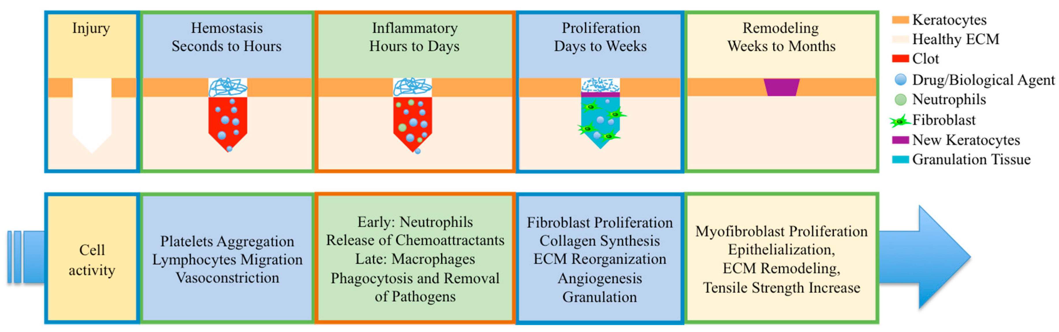
2. Wound Healing
2.1. Classification of Wounds
2.2. Wound Healing Cycles
2.3. Non-Healing Wounds
3. Electrospun Fibers
3.1. Natural Polymers
3.2. Synthetic Polymers
3.3. Electrospinning Parameters
4. Release of Small Molecule Drugs
4.1. Hydrophilic Drugs
4.2. Hydrophobic Drugs
5. Release of Macromolecules
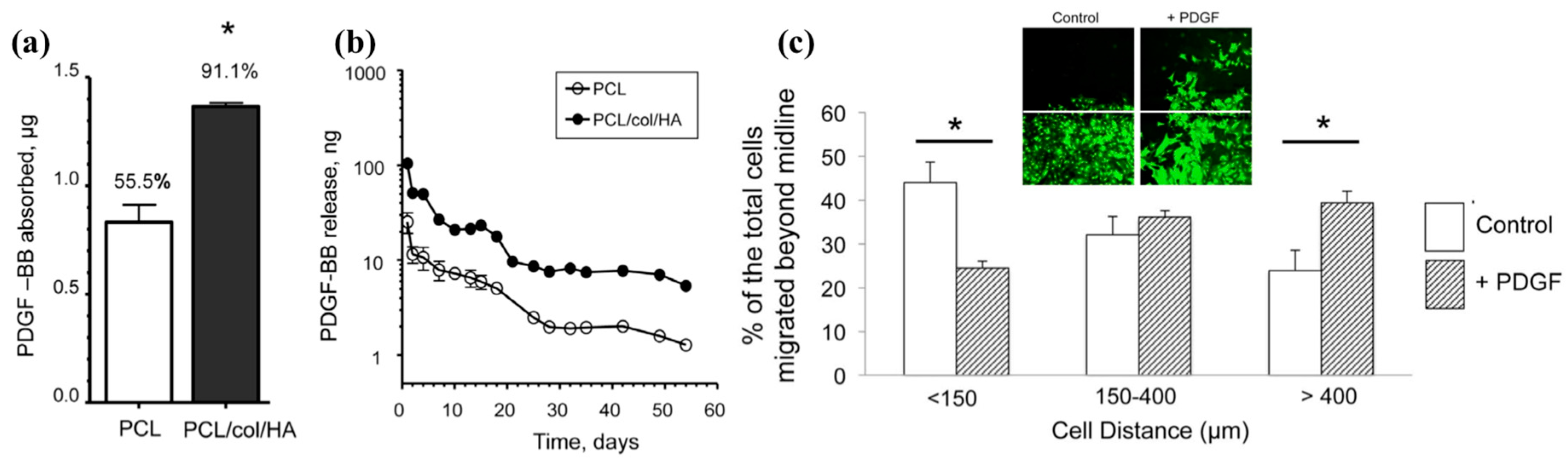
5.1. Growth Factors
5.2. Peptides
6. Release of Gene Vectors
6.1. Non-Viral Genes Vectors
6.2. Non-Viral Genes Vectors Delivered by Fiber Platform
7. Fiber Composites
7.1. Fiber-Micelle Composites
7.2. Fiber-Nanoparticle Composites
8. Conclusions and Future Directions
Conflicts of Interest
References
- Sidgwick, G.P.; McGeorge, D.; Bayat, A. A comprehensive evidence-based review on the role of topicals and dressings in the management of skin scarring. Arch. Dermatol. Res. 2015, 307, 461–477. [Google Scholar] [CrossRef] [PubMed]
- Chou, S.-F.; Carson, D.; Woodrow, K.A. Current strategies for sustaining drug release from electrospun nanofibers. J. Control. Release 2015, 220, 584–591. [Google Scholar] [CrossRef] [PubMed]
- Strodtbeck, F. Physiology of wound healing. Newborn Infant Nurs. Rev. 2001, 1, 43–52. [Google Scholar] [CrossRef]
- Merlin-Manton, E. Wound care: Selecting the right dressings. Pract. Nurse 2017, 47, 28–32. [Google Scholar]
- Tejiram, S.; Kavalukas, S.L.; Shupp, J.W.; Barbul, A. 1-Wound healing. In Wound Healing Biomaterials; Ågren, M.S., Ed.; Woodhead Publishing: Sawston, Cambridge, UK, 2016; pp. 3–39. ISBN 978-1-78242-455-0. [Google Scholar]
- Velnar, T.; Bailey, T.; Smrkolj, V. The wound healing process: An overview of the cellular and molecular mechanisms. J. Int. Med. Res. 2009, 37, 1528–1542. [Google Scholar] [CrossRef] [PubMed]
- Abrigo, M.; McArthur, S.L.; Kingshott, P. Electrospun nanofibers as dressings for chronic wound care: Advances, challenges, and future prospects. Macromol. Biosci. 2014, 14, 772–792. [Google Scholar] [CrossRef] [PubMed]
- Whitney, J.D. Overview: Acute and Chronic Wounds. Nurs. Clin. N. Am. 2005, 40, 191–205. [Google Scholar] [CrossRef] [PubMed]
- Boateng, J.S.; Matthews, K.H.; Stevens, H.N.E.; Eccleston, G.M. Wound healing dressings and drug delivery systems: A review. J. Pharm. Sci. 2008, 97, 2892–2923. [Google Scholar] [CrossRef] [PubMed]
- Arasteh, S.; Kazemnejad, S.; Khanjani, S.; Heidari-Vala, H.; Akhondi, M.M.; Mobini, S. Fabrication and characterization of nano-fibrous bilayer composite for skin regeneration application. Methods 2016, 99, 3–12. [Google Scholar] [CrossRef] [PubMed]
- Lee, Y.-H.; Chang, J.-J.; Yang, M.-C.; Chien, C.-T.; Lai, W.-F. Acceleration of wound healing in diabetic rats by layered hydrogel dressing. Carbohydr. Polym. 2012, 88, 809–819. [Google Scholar] [CrossRef]
- Kasuya, A.; Tokura, Y. Attempts to accelerate wound healing. J. Dermatol. Sci. 2014, 76, 169–172. [Google Scholar] [CrossRef] [PubMed]
- Neuenschwander, P.F.; Jesty, J. Blood coagulation. In Encyclopedia of Life Sciences; John Wiley & Sons, Ltd.: Chichester, UK, 2011; ISBN 978-0-470-01617-6. [Google Scholar]
- Castellanos, G.; Bernabé-García, Á.; Moraleda, J.M.; Nicolás, F.J. Amniotic membrane application for the healing of chronic wounds and ulcers. Placenta 2017, 59, 146–153. [Google Scholar] [CrossRef] [PubMed]
- Hosgood, G. Stages of Wound healing and their clinical relevance. Vet. Clin. N. Am. Small Anim. Pract. 2006, 36, 667–685. [Google Scholar] [CrossRef] [PubMed]
- Reinke, J.M.; Sorg, H. Wound repair and regeneration. Eur. Surg. Res. 2012, 49, 35–43. [Google Scholar] [CrossRef] [PubMed]
- Landén, N.X.; Li, D.; Ståhle, M. Transition from inflammation to proliferation: A critical step during wound healing. Cell. Mol. Life Sci. 2016, 73, 3861–3885. [Google Scholar] [CrossRef] [PubMed]
- Clinical Guidelines (Nursing): Wound Care. Available online: https://www.rch.org.au/rchcpg/hospital_clinical_guideline_index/Wound_care/ (accessed on 19 October 2017).
- Hanna, J.R.; Giacopelli, J.A. A review of wound healing and wound dressing products. J. Foot Ankle Surg. 1997, 36, 2–14. [Google Scholar] [CrossRef]
- Harding, K.G.; Morris, H.L.; Patel, G.K. Healing chronic wounds. BMJ Br. Med. J. Lond. 2002, 324, 160–163. [Google Scholar] [CrossRef]
- Dovi, J.V.; Szpaderska, A.M.; DiPietro, L.A. Neutrophil function in the healing wound: Adding insult to injury? Thromb. Haemost. 2004, 92, 275–280. [Google Scholar] [CrossRef] [PubMed]
- Mathews, V.; Hanson, P.T.; Ford, E.; Fujita, J.; Polonsky, K.S.; Graubert, T.A. Recruitment of bone marrow-derived endothelial cells to sites of pancreatic β-cell injury. Diabetes 2004, 53, 91–98. [Google Scholar] [CrossRef] [PubMed]
- Velazquez, O.C. Angiogenesis and vasculogenesis: Inducing the growth of new blood vessels and wound healing by stimulation of bone marrow–derived progenitor cell mobilization and homing. J. Vasc. Surg. 2007, 45, A39–A47. [Google Scholar] [CrossRef] [PubMed]
- Aramwit, P. 1-Introduction to biomaterials for wound healing. In Wound Healing Biomaterials; Ågren, M.S., Ed.; Woodhead Publishing: Sawston, Cambridge, UK, 2016; pp. 3–38. ISBN 978-1-78242-456-7. [Google Scholar]
- Sood, A.; Granick, M.S.; Tomaselli, N.L. Wound dressings and comparative effectiveness data. Adv. Wound Care 2014, 3, 511–529. [Google Scholar] [CrossRef] [PubMed]
- Liu, M.; Duan, X.-P.; Li, Y.-M.; Yang, D.-P.; Long, Y.-Z. Electrospun nanofibers for wound healing. Mater. Sci. Eng. C 2017, 76, 1413–1423. [Google Scholar] [CrossRef] [PubMed]
- Wang, J.; Windbergs, M. Functional electrospun fibers for the treatment of human skin wounds. Eur. J. Pharm. Biopharm. 2017, 119, 283–299. [Google Scholar] [CrossRef] [PubMed]
- Ahmadi Majd, S.; Rabbani Khorasgani, M.; Moshtaghian, S.J.; Talebi, A.; Khezri, M. Application of Chitosan/PVA Nano fiber as a potential wound dressing for streptozotocin-induced diabetic rats. Int. J. Biol. Macromol. 2016, 92, 1162–1168. [Google Scholar] [CrossRef] [PubMed]
- Chou, S.-F.; Woodrow, K.A. Relationships between mechanical properties and drug release from electrospun fibers of PCL and PLGA blends. J. Mech. Behav. Biomed. Mater. 2017, 65, 724–733. [Google Scholar] [CrossRef] [PubMed]
- Chen, S.; Boda, S.K.; Batra, S.K.; Li, X.; Xie, J. Emerging roles of electrospun nanofibers in cancer research. Adv. Healthc. Mater. 2017. [Google Scholar] [CrossRef] [PubMed]
- Mogoşanu, G.D.; Grumezescu, A.M. Natural and synthetic polymers for wounds and burns dressing. Int. J. Pharm. 2014, 463, 127–136. [Google Scholar] [CrossRef] [PubMed]
- Huang, S.; Fu, X. Naturally derived materials-based cell and drug delivery systems in skin regeneration. J. Control. Release 2010, 142, 149–159. [Google Scholar] [CrossRef] [PubMed]
- Dai, T.; Tanaka, M.; Huang, Y.-Y.; Hamblin, M.R. Chitosan preparations for wounds and burns: Antimicrobial and wound-healing effects. Expert Rev. Anti-Infect. Ther. 2011, 9, 857–879. [Google Scholar] [CrossRef] [PubMed]
- Bano, I.; Arshad, M.; Yasin, T.; Ghauri, M.A.; Younus, M. Chitosan: A potential biopolymer for wound management. Int. J. Biol. Macromol. 2017, 102, 380–383. [Google Scholar] [CrossRef] [PubMed]
- Pakravan, M.; Heuzey, M.-C.; Ajji, A. A fundamental study of chitosan/PEO electrospinning. Polymer 2011, 52, 4813–4824. [Google Scholar] [CrossRef]
- Lu, J.-W.; Zhu, Y.-L.; Guo, Z.-X.; Hu, P.; Yu, J. Electrospinning of sodium alginate with poly(ethylene oxide). Polymer 2006, 47, 8026–8031. [Google Scholar] [CrossRef]
- Topuz, F.; Uyar, T. Electrospinning of gelatin with tunable fiber morphology from round to flat/ribbon. Mater. Sci. Eng. C 2017, 80, 371–378. [Google Scholar] [CrossRef] [PubMed]
- Ostrovidov, S.; Shi, X.; Zhang, L.; Liang, X.; Kim, S.B.; Fujie, T.; Ramalingam, M.; Chen, M.; Nakajima, K.; Al-Hazmi, F.; et al. Myotube formation on gelatin nanofibers—Multi-walled carbon nanotubes hybrid scaffolds. Biomaterials 2014, 35, 6268–6277. [Google Scholar] [CrossRef] [PubMed]
- Zhang, K.; Li, Z.; Kang, W.; Deng, N.; Yan, J.; Ju, J.; Liu, Y.; Cheng, B. Preparation and characterization of tree-like cellulose nanofiber membranes via the electrospinning method. Carbohydr. Polym. 2018, 183, 62–69. [Google Scholar] [CrossRef] [PubMed]
- Bak, S.Y.; Yoon, G.J.; Lee, S.W.; Kim, H.W. Effect of humidity and benign solvent composition on electrospinning of collagen nanofibrous sheets. Mater. Lett. 2016, 181, 136–139. [Google Scholar] [CrossRef]
- Sadeghi-Avalshahr, A.; Nokhasteh, S.; Molavi, A.M.; Khorsand-Ghayeni, M.; Mahdavi-Shahri, M. Synthesis and characterization of collagen/PLGA biodegradable skin scaffold fibers. Regen. Biomater. 2017, 4, 309–314. [Google Scholar] [CrossRef] [PubMed]
- Kutlusoy, T.; Oktay, B.; Apohan, N.K.; Süleymanoğlu, M.; Kuruca, S.E. Chitosan-co-Hyaluronic acid porous cryogels and their application in tissue engineering. Int. J. Biol. Macromol. 2017, 103, 366–378. [Google Scholar] [CrossRef] [PubMed]
- Liu, Y.; Ma, G.; Fang, D.; Xu, J.; Zhang, H.; Nie, J. Effects of solution properties and electric field on the electrospinning of hyaluronic acid. Carbohydr. Polym. 2011, 83, 1011–1015. [Google Scholar] [CrossRef]
- Brenner, E.K.; Schiffman, J.D.; Thompson, E.A.; Toth, L.J.; Schauer, C.L. Electrospinning of hyaluronic acid nanofibers from aqueous ammonium solutions. Carbohydr. Polym. 2012, 87, 926–929. [Google Scholar] [CrossRef]
- Esparza, Y.; Ullah, A.; Boluk, Y.; Wu, J. Preparation and characterization of thermally crosslinked poly(vinyl alcohol)/feather keratin nanofiber scaffolds. Mater. Des. 2017, 133, 1–9. [Google Scholar] [CrossRef]
- Yukseloglu, S.M.; Sokmen, N.; Canoglu, S. Biomaterial applications of silk fibroin electrospun nanofibres. Microelectron. Eng. 2015, 146, 43–47. [Google Scholar] [CrossRef]
- Dias Antunes, M.; da Silva Dannenberg, G.; Fiorentini, Â.M.; Pinto, V.Z.; Lim, L.-T.; da Rosa Zavareze, E.; Dias, A.R.G. Antimicrobial electrospun ultrafine fibers from zein containing eucalyptus essential oil/cyclodextrin inclusion complex. Int. J. Biol. Macromol. 2017, 104, 874–882. [Google Scholar] [CrossRef] [PubMed]
- Deng, L.; Kang, X.; Liu, Y.; Feng, F.; Zhang, H. Characterization of gelatin/zein films fabricated by electrospinning vs solvent casting. Food Hydrocoll. 2018, 74, 324–332. [Google Scholar] [CrossRef]
- Wongkanya, R.; Chuysinuan, P.; Pengsuk, C.; Techasakul, S.; Lirdprapamongkol, K.; Svasti, J.; Nooeaid, P. Electrospinning of alginate/soy protein isolated nanofibers and their release characteristics for biomedical applications. J. Sci. Adv. Mater. Devices 2017, 2, 309–316. [Google Scholar] [CrossRef]
- Okutan, N.; Terzi, P.; Altay, F. Affecting parameters on electrospinning process and characterization of electrospun gelatin nanofibers. Food Hydrocoll. 2014, 39, 19–26. [Google Scholar] [CrossRef]
- Entekhabi, E.; Haghbin Nazarpak, M.; Moztarzadeh, F.; Sadeghi, A. Design and manufacture of neural tissue engineering scaffolds using hyaluronic acid and polycaprolactone nanofibers with controlled porosity. Mater. Sci. Eng. C 2016, 69, 380–387. [Google Scholar] [CrossRef] [PubMed]
- Ma, H.; Shen, J.; Cao, J.; Wang, D.; Yue, B.; Mao, Z.; Wu, W.; Zhang, H. Fabrication of wool keratin/polyethylene oxide nano-membrane from wool fabric waste. J. Clean. Prod. 2017, 161, 357–361. [Google Scholar] [CrossRef]
- Kishimoto, Y.; Morikawa, H.; Yamanaka, S.; Tamada, Y. Electrospinning of silk fibroin from all aqueous solution at low concentration. Mater. Sci. Eng. C 2017, 73, 498–506. [Google Scholar] [CrossRef] [PubMed]
- Tampau, A.; González-Martinez, C.; Chiralt, A. Carvacrol encapsulation in starch or PCL based matrices by electrospinning. J. Food Eng. 2017, 214, 245–256. [Google Scholar] [CrossRef]
- Ranjbar-Mohammadi, M.; Zamani, M.; Prabhakaran, M.P.; Bahrami, S.H.; Ramakrishna, S. Electrospinning of PLGA/gum tragacanth nanofibers containing tetracycline hydrochloride for periodontal regeneration. Mater. Sci. Eng. C 2016, 58, 521–531. [Google Scholar] [CrossRef] [PubMed]
- Ju, J.; Shi, Z.; Fan, L.; Liang, Y.; Kang, W.; Cheng, B. Preparation of elastomeric tree-like nanofiber membranes using thermoplastic polyurethane by one-step electrospinning. Mater. Lett. 2017, 205, 190–193. [Google Scholar] [CrossRef]
- Dorneanu, P.P.; Cojocaru, C.; Olaru, N.; Samoila, P.; Airinei, A.; Sacarescu, L. Electrospun PVDF fibers and a novel PVDF/CoFe2O4 fibrous composite as nanostructured sorbent materials for oil spill cleanup. Appl. Surf. Sci. 2017, 424, 389–396. [Google Scholar] [CrossRef]
- Yan, S.; Li, X.; Dai, J.; Wang, Y.; Wang, B.; Lu, Y.; Shi, J.; Huang, P.; Gong, J.; Yao, Y. Electrospinning of PVA/sericin nanofiber and the effect on epithelial-mesenchymal transition of A549 cells. Mater. Sci. Eng. C 2017, 79, 436–444. [Google Scholar] [CrossRef] [PubMed]
- Son, W.K.; Youk, J.H.; Lee, T.S.; Park, W.H. The effects of solution properties and polyelectrolyte on electrospinning of ultrafine poly(ethylene oxide) fibers. Polymer 2004, 45, 2959–2966. [Google Scholar] [CrossRef]
- Reksamunandar, R.P.; Edikresnha, D.; Munir, M.M.; Damayanti, S. Khairurrijal Encapsulation of β-carotene in poly(vinylpyrrolidone) (PVP) by Electrospinning Technique. Procedia Eng. 2017, 170, 19–23. [Google Scholar] [CrossRef]
- Chou, S.-F.; Gunaseelan, S.; Kiellani, M.H.H.; Thottempudi, V.V.K.; Neuenschwander, P.; Nie, H. A review of injectable and implantable biomaterials for treatment and repair of soft tissues in wound healing. J. Nanotechnol. 2017, 2017, 1–15. [Google Scholar] [CrossRef]
- Ponjavic, M.; Nikolic, M.S.; Nikodinovic-Runic, J.; Jeremic, S.; Stevanovic, S.; Djonlagic, J. Degradation behaviour of PCL/PEO/PCL and PCL/PEO block copolymers under controlled hydrolytic, enzymatic and composting conditions. Polym. Test. 2017, 57, 67–77. [Google Scholar] [CrossRef]
- Luong-Van, E.; Grøndahl, L.; Chua, K.N.; Leong, K.W.; Nurcombe, V.; Cool, S.M. Controlled release of heparin from poly(ε-caprolactone) electrospun fibers. Biomaterials 2006, 27, 2042–2050. [Google Scholar] [CrossRef] [PubMed]
- Lu, L.; Garcia, C.A.; Mikos, A.G. In vitro degradation of thin poly(DL-lactic-co-glycolic acid) films. J. Biomed. Mater. Res. 1999, 46, 236–244. [Google Scholar] [CrossRef]
- Park, T.G. Degradation of poly(lactic-co-glycolic acid) microspheres: Effect of copolymer composition. Biomaterials 1995, 16, 1123–1130. [Google Scholar] [CrossRef]
- Peña, J.; Corrales, T.; Izquierdo-Barba, I.; Doadrio, A.L.; Vallet-Regí, M. Long term degradation of poly(ε-caprolactone) films in biologically related fluids. Polym. Degrad. Stab. 2006, 91, 1424–1432. [Google Scholar] [CrossRef]
- You, Y.; Min, B.-M.; Lee, S.J.; Lee, T.S.; Park, W.H. In vitro degradation behavior of electrospun polyglycolide, polylactide, and poly(lactide-co-glycolide). J. Appl. Polym. Sci. 2005, 95, 193–200. [Google Scholar] [CrossRef]
- Yeganegi, M.; Kandel, R.A.; Santerre, J.P. Characterization of a biodegradable electrospun polyurethane nanofiber scaffold: Mechanical properties and cytotoxicity. Acta Biomater. 2010, 6, 3847–3855. [Google Scholar] [CrossRef] [PubMed]
- Sheikh, F.A.; Zargar, M.A.; Tamboli, A.H.; Kim, H. A super hydrophilic modification of poly(vinylidene fluoride) (PVDF) nanofibers: By in situ hydrothermal approach. Appl. Surf. Sci. 2016, 385, 417–425. [Google Scholar] [CrossRef]
- Li, X.; Kanjwal, M.A.; Lin, L.; Chronakis, I.S. Electrospun polyvinyl-alcohol nanofibers as oral fast-dissolving delivery system of caffeine and riboflavin. Colloids Surf. B Biointerfaces 2013, 103, 182–188. [Google Scholar] [CrossRef] [PubMed]
- Kim, T.G.; Lee, D.S.; Park, T.G. Controlled protein release from electrospun biodegradable fiber mesh composed of poly(ε-caprolactone) and poly(ethylene oxide). Int. J. Pharm. 2007, 338, 276–283. [Google Scholar] [CrossRef] [PubMed]
- Bui, H.T.; Chung, O.H.; Cruz, J.D.; Park, J.S. Fabrication and characterization of electrospun curcumin-loaded polycaprolactone-polyethylene glycol nanofibers for enhanced wound healing. Macromol. Res. 2014, 22, 1288–1296. [Google Scholar] [CrossRef]
- Ahire, J.J.; Robertson, D.D.; van Reenen, A.J.; Dicks, L.M.T. Polyethylene oxide (PEO)-hyaluronic acid (HA) nanofibers with kanamycin inhibits the growth of Listeria monocytogenes. Biomed. Pharmacother. 2017, 86, 143–148. [Google Scholar] [CrossRef] [PubMed]
- Balogh, A.; Farkas, B.; Verreck, G.; Mensch, J.; Borbás, E.; Nagy, B.; Marosi, G.; Nagy, Z.K. AC and DC electrospinning of hydroxypropylmethylcellulose with polyethylene oxides as secondary polymer for improved drug dissolution. Int. J. Pharm. 2016, 505, 159–166. [Google Scholar] [CrossRef] [PubMed]
- Haider, A.; Haider, S.; Kang, I.-K. A comprehensive review summarizing the effect of electrospinning parameters and potential applications of nanofibers in biomedical and biotechnology. Arab. J. Chem. 2015. [Google Scholar] [CrossRef]
- Megelski, S.; Stephens, J.S.; Chase, D.B.; Rabolt, J.F. Micro- and nanostructured surface morphology on electrospun polymer fibers. Macromolecules 2002, 35, 8456–8466. [Google Scholar] [CrossRef]
- Rodoplu, D.; Mutlu, M. Effects of electrospinning setup and process parameters on nanofiber morphology intended for the modification of quartz crystal microbalance surfaces. J. Eng. Fibers Fabr. 2012, 7, 118–123. [Google Scholar]
- Matabola, K.P.; Moutloali, R.M. The influence of electrospinning parameters on the morphology and diameter of poly(vinyledene fluoride) nanofibers- effect of sodium chloride. J. Mater. Sci. 2013, 48, 5475–5482. [Google Scholar] [CrossRef]
- Ball, C.; Chou, S.-F.; Jiang, Y.; Woodrow, K.A. Coaxially electrospun fiber-based microbicides facilitate broadly tunable release of maraviroc. Mater. Sci. Eng. C 2016, 63, 117–124. [Google Scholar] [CrossRef] [PubMed]
- Zamani, M.; Prabhakaran, M.P.; Ramakrishna, S. Advances in drug delivery via electrospun and electrosprayed nanomaterials. Int. J. Nanomed. 2013, 2997–3017. [Google Scholar]
- Contardi, M.; Heredia-Guerrero, J.A.; Perotto, G.; Valentini, P.; Pompa, P.P.; Spanò, R.; Goldoni, L.; Bertorelli, R.; Athanassiou, A.; Bayer, I.S. Transparent ciprofloxacin-povidone antibiotic films and nanofiber mats as potential skin and wound care dressings. Eur. J. Pharm. Sci. 2017, 104, 133–144. [Google Scholar] [CrossRef] [PubMed]
- Li, H.; Williams, G.R.; Wu, J.; Lv, Y.; Sun, X.; Wu, H.; Zhu, L.-M. Thermosensitive nanofibers loaded with ciprofloxacin as antibacterial wound dressing materials. Int. J. Pharm. 2017, 517, 135–147. [Google Scholar] [CrossRef] [PubMed]
- Kabay, G.; Meydan, A.E.; Kaleli Can, G.; Demirci, C.; Mutlu, M. Controlled release of a hydrophilic drug from electrospun amyloid-like protein blend nanofibers. Mater. Sci. Eng. C 2017, 81, 271–279. [Google Scholar] [CrossRef] [PubMed]
- Sohrabi, A.; Shaibani, P.M.; Etayash, H.; Kaur, K.; Thundat, T. Sustained drug release and antibacterial activity of ampicillin incorporated poly(methyl methacrylate)–nylon6 core/shell nanofibers. Polymer 2013, 54, 2699–2705. [Google Scholar] [CrossRef]
- Sultanova, Z.; Kaleli, G.; Kabay, G.; Mutlu, M. Controlled release of a hydrophilic drug from coaxially electrospun polycaprolactone nanofibers. Int. J. Pharm. 2016, 505, 133–138. [Google Scholar] [CrossRef] [PubMed]
- Xue, J.; He, M.; Niu, Y.; Liu, H.; Crawford, A.; Coates, P.; Chen, D.; Shi, R.; Zhang, L. Preparation and in vivo efficient anti-infection property of GTR/GBR implant made by metronidazole loaded electrospun polycaprolactone nanofiber membrane. Int. J. Pharm. 2014, 475, 566–577. [Google Scholar] [CrossRef] [PubMed]
- He, M.; Jiang, H.; Wang, R.; Xie, Y.; Zhao, C. Fabrication of metronidazole loaded poly (ε-caprolactone)/zein core/shell nanofiber membranes via coaxial electrospinning for guided tissue regeneration. J. Colloid Interface Sci. 2017, 490, 270–278. [Google Scholar] [CrossRef] [PubMed]
- Zupančič, Š.; Potrč, T.; Baumgartner, S.; Kocbek, P.; Kristl, J. Formulation and evaluation of chitosan/polyethylene oxide nanofibers loaded with metronidazole for local infections. Eur. J. Pharm. Sci. 2016, 95, 152–160. [Google Scholar] [CrossRef] [PubMed]
- Sadri, M.; Sorkhi, S.A. Preparation and characterization of CS/PEO/cefazolin nanofibers with in vitro and in vivo testing. Nanomedicine Res. J. 2017, 2, 100–110. [Google Scholar]
- Rath, G.; Hussain, T.; Chauhan, G.; Garg, T.; Goyal, A.K. Development and characterization of cefazolin loaded zinc oxide nanoparticles composite gelatin nanofiber mats for postoperative surgical wounds. Mater. Sci. Eng. C 2016, 58, 242–253. [Google Scholar] [CrossRef] [PubMed]
- Kataria, K.; Gupta, A.; Rath, G.; Mathur, R.B.; Dhakate, S.R. In vivo wound healing performance of drug loaded electrospun composite nanofibers transdermal patch. Int. J. Pharm. 2014, 469, 102–110. [Google Scholar] [CrossRef] [PubMed]
- Zhang, H.; Lou, S.; Williams, G.R.; Branford-White, C.; Nie, H.; Quan, J.; Zhu, L.-M. A systematic study of captopril-loaded polyester fiber mats prepared by electrospinning. Int. J. Pharm. 2012, 439, 100–108. [Google Scholar] [CrossRef] [PubMed]
- Zheng, X.-F.; Lu, X.-Y. Measurement and correlation of solubilities of asiaticoside in water, methanol, ethanol, n -propanol, n -butanol, and a methanol + water mixture from (278.15 to 343.15) K. J. Chem. Eng. Data 2011, 56, 674–677. [Google Scholar] [CrossRef]
- Zhu, L.; Liu, X.; Du, L.; Jin, Y. Preparation of asiaticoside-loaded coaxially electrospinning nanofibers and their effect on deep partial-thickness burn injury. Biomed. Pharmacother. 2016, 83, 33–40. [Google Scholar] [CrossRef] [PubMed]
- Mutlu, G.; Calamak, S.; Ulubayram, K.; Guven, E. Curcumin-loaded electrospun PHBV nanofibers as potential wound-dressing material. J. Drug Deliv. Sci. Technol. 2018, 43, 185–193. [Google Scholar] [CrossRef]
- Ranjbar-Mohammadi, M.; Rabbani, S.; Bahrami, S.H.; Joghataei, M.T.; Moayer, F. Antibacterial performance and in vivo diabetic wound healing of curcumin loaded gum tragacanth/poly(ε-caprolactone) electrospun nanofibers. Mater. Sci. Eng. C 2016, 69, 1183–1191. [Google Scholar] [CrossRef] [PubMed]
- Basar, A.O.; Castro, S.; Torres-Giner, S.; Lagaron, J.M.; Turkoglu Sasmazel, H. Novel poly(ε-caprolactone)/gelatin wound dressings prepared by emulsion electrospinning with controlled release capacity of Ketoprofen anti-inflammatory drug. Mater. Sci. Eng. C 2017, 81, 459–468. [Google Scholar] [CrossRef] [PubMed]
- Kenawy, E.-R.; Abdel-Hay, F.I.; El-Newehy, M.H.; Wnek, G.E. Controlled release of ketoprofen from electrospun poly(vinyl alcohol) nanofibers. Mater. Sci. Eng. A 2007, 459, 390–396. [Google Scholar] [CrossRef]
- Liu, L.; Bai, S.; Yang, H.; Li, S.; Quan, J.; Zhu, L.; Nie, H. Controlled release from thermo-sensitive PNVCL-co-MAA electrospun nanofibers: The effects of hydrophilicity/hydrophobicity of a drug. Mater. Sci. Eng. C 2016, 67, 581–589. [Google Scholar] [CrossRef] [PubMed]
- Yu, D.-G.; Yu, J.-H.; Chen, L.; Williams, G.R.; Wang, X. Modified coaxial electrospinning for the preparation of high-quality ketoprofen-loaded cellulose acetate nanofibers. Carbohydr. Polym. 2012, 90, 1016–1023. [Google Scholar] [CrossRef] [PubMed]
- Hamori, M.; Yoshimatsu, S.; Hukuchi, Y.; Shimizu, Y.; Fukushima, K.; Sugioka, N.; Nishimura, A.; Shibata, N. Preparation and pharmaceutical evaluation of nano-fiber matrix supported drug delivery system using the solvent-based electrospinning method. Int. J. Pharm. 2014, 464, 243–251. [Google Scholar] [CrossRef] [PubMed]
- Lin, X.; Tang, D.; Du, H. Self-assembly and controlled release behaviour of the water-insoluble drug nifedipine from electrospun PCL-based polyurethane nanofibres: Self-assembly and release of drug. J. Pharm. Pharmacol. 2013, 65, 673–681. [Google Scholar] [CrossRef] [PubMed]
- Lin, X.; Tang, D.; Gu, S.; Du, H.; Jiang, E. Electrospun poly(N-isopropylacrylamide)/poly(caprolactone)-based polyurethane nanofibers as drug carriers and temperature-controlled release. New J. Chem. 2013, 37, 2433–2439. [Google Scholar] [CrossRef]
- Zahedi, P.; Rezaeian, I.; Jafari, S.H. In vitro and in vivo evaluations of phenytoin sodium-loaded electrospun PVA, PCL, and their hybrid nanofibrous mats for use as active wound dressings. J. Mater. Sci. 2013, 48, 3147–3159. [Google Scholar] [CrossRef]
- Kurczewska, J.; Pecyna, P.; Ratajczak, M.; Gajęcka, M.; Schroeder, G. Halloysite nanotubes as carriers of vancomycin in alginate-based wound dressing. Saudi Pharm. J. 2017, 25, 911–920. [Google Scholar] [CrossRef] [PubMed]
- El-Khordagui, L.; El-Sayed, N.; Galal, S.; El-Gowelli, H.; Omar, H.; Mohamed, M. Photosensitizer-eluting nanofibers for enhanced photodynamic therapy of wounds: A preclinical study in immunocompromized rats. Int. J. Pharm. 2017, 520, 139–148. [Google Scholar] [CrossRef] [PubMed]
- Anstead, G.M.; Hart, L.M.; Sunahara, J.F.; Liter, M.E. Phenytoin in wound healing. Ann. Pharmacother. 1996, 30, 768–775. [Google Scholar] [CrossRef] [PubMed]
- Hokkam, E.; El-Labban, G.; Shams, M.; Rifaat, S.; El-mezaien, M. The use of topical phenytoin for healing of chronic venous ulcerations. Int. J. Surg. 2011, 9, 335–338. [Google Scholar] [CrossRef] [PubMed]
- Lupo, E.; Locher, R.; Weisser, B.; Vetter, W. In vitro antioxidant activity of calcium antagonists against LDL oxidation compared with α-tocopherol. Biochem. Biophys. Res. Commun. 1994, 203, 1803–1808. [Google Scholar] [CrossRef] [PubMed]
- Barrientos, S.; Stojadinovic, O.; Golinko, M.S.; Brem, H.; Tomic-Canic, M. PERSPECTIVE ARTICLE: Growth factors and cytokines in wound healing. Wound Repair Regen. 2008, 16, 585–601. [Google Scholar] [CrossRef] [PubMed]
- Lai, H.-J.; Kuan, C.-H.; Wu, H.-C.; Tsai, J.-C.; Chen, T.-M.; Hsieh, D.-J.; Wang, T.-W. Tailored design of electrospun composite nanofibers with staged release of multiple angiogenic growth factors for chronic wound healing. Acta Biomater. 2014, 10, 4156–4166. [Google Scholar] [CrossRef] [PubMed]
- Choi, J.S.; Choi, S.H.; Yoo, H.S. Coaxial electrospun nanofibers for treatment of diabetic ulcers with binary release of multiple growth factors. J. Mater. Chem. 2011, 21, 5258–5267. [Google Scholar] [CrossRef]
- Phipps, M.; Ma, Y.; Bellis, S. Delivery of platelet-derived growth factor as a chemotactic factor for mesenchymal stem cells by bone-mimetic electrospun scaffolds. PLoS ONE 2012, 7, e40831. [Google Scholar] [CrossRef] [PubMed]
- Kim, I.L.; Pfeifer, C.G.; Fisher, M.B.; Saxena, V.; Meloni, G.R.; Kwon, M.Y.; Kim, M.; Steinberg, D.R.; Mauck, R.L.; Burdick, J.A. Fibrous scaffolds with varied fiber chemistry and growth factor delivery promote repair in a porcine cartilage defect model. Tissue Eng. Part A 2015, 21, 2680–2690. [Google Scholar] [CrossRef] [PubMed]
- Hu, F.; Zhang, X.; Liu, H.; Xu, P.; Doulathunnisa; Teng, G.; Xiao, Z. Neuronally differentiated adipose-derived stem cells and aligned PHBV nanofiber nerve scaffolds promote sciatic nerve regeneration. Biochem. Biophys. Res. Commun. 2017, 489, 171–178. [Google Scholar] [CrossRef] [PubMed]
- Davis, M.E.; Hsieh, P.C.H.; Takahashi, T.; Song, Q.; Zhang, S.; Kamm, R.D.; Grodzinsky, A.J.; Anversa, P.; Lee, R.T. Local myocardial insulin-like growth factor 1 (IGF-1) delivery with biotinylated peptide nanofibers improves cell therapy for myocardial infarction. Proc. Natl. Acad. Sci. USA 2006, 103, 8155–8160. [Google Scholar] [CrossRef] [PubMed]
- Noh, K.H.; Park, Y.M.; Kim, H.S.; Kang, T.H.; Song, K.-H.; Lee, Y.-H.; Byeon, Y.; Jeon, H.N.; Jung, I.D.; Shin, B.C. GM-CSF-loaded chitosan hydrogel as an immunoadjuvant enhances antigen-specific immune responses with reduced toxicity. BMC Immunol. 2014, 15, 1–7. [Google Scholar] [CrossRef] [PubMed]
- Olvera, D.; Sathy, B.N.; Carroll, S.F.; Kelly, D.J. Modulating microfibrillar alignment and growth factor stimulation to regulate mesenchymal stem cell differentiation. Acta Biomater. 2017, 64, 148–160. [Google Scholar] [CrossRef] [PubMed]
- Choi, J.S.; Leong, K.W.; Yoo, H.S. In vivo wound healing of diabetic ulcers using electrospun nanofibers immobilized with human epidermal growth factor (EGF). Biomaterials 2008, 29, 587–596. [Google Scholar] [CrossRef] [PubMed]
- Norouzi, M.; Shabani, I.; Ahvaz, H.H.; Soleimani, M. PLGA/gelatin hybrid nanofibrous scaffolds encapsulating EGF for skin regeneration. J. Biomed. Mater. Res. A 2015, 103, 2225–2235. [Google Scholar] [CrossRef] [PubMed]
- Schneider, A.; Wang, X.Y.; Kaplan, D.L.; Garlick, J.A.; Egles, C. Biofunctionalized electrospun silk mats as a topical bioactive dressing for accelerated wound healing. Acta Biomater. 2009, 5, 2570–2578. [Google Scholar] [CrossRef] [PubMed]
- Gümüşderelioğlu, M.; Dalkıranoğlu, S.; Aydın, R.S.T.; Çakmak, S. A novel dermal substitute based on biofunctionalized electrospun PCL nanofibrous matrix. J. Biomed. Mater. Res. A 2011, 98, 461–472. [Google Scholar] [CrossRef] [PubMed]
- Jin, G.; Prabhakaran, M.P.; Ramakrishna, S. Photosensitive and biomimetic core–shell nanofibrous scaffolds as wound dressing. Photochem. Photobiol. 2014, 90, 673–681. [Google Scholar] [CrossRef] [PubMed]
- Jin, G.; Prabhakaran, M.P.; Kai, D.; Ramakrishna, S. Controlled release of multiple epidermal induction factors through core–shell nanofibers for skin regeneration. Eur. J. Pharm. Biopharm. 2013, 85, 689–698. [Google Scholar] [CrossRef] [PubMed]
- Gil, E.S.; Panilaitis, B.; Bellas, E.; Kaplan, D.L. Functionalized silk biomaterials for wound healing. Adv. Healthc. Mater. 2013, 2, 206–217. [Google Scholar] [CrossRef] [PubMed]
- Mirdailami, O.; Soleimani, M.; Dinarvand, R.; Khoshayand, M.R.; Norouzi, M.; Hajarizadeh, A.; Dodel, M.; Atyabi, F. Controlled release of rhEGF and rhbFGF from electrospun scaffolds for skin regeneration. J. Biomed. Mater. Res. A 2015, 103, 3374–3385. [Google Scholar] [CrossRef] [PubMed]
- Kobsa, S.; Kristofik, N.J.; Sawyer, A.J.; Bothwell, A.L.M.; Kyriakides, T.R.; Saltzman, W.M. An electrospun scaffold integrating nucleic acid delivery for treatment of full thickness wounds. Biomaterials 2013, 34, 3891–3901. [Google Scholar] [CrossRef] [PubMed]
- Zhao, Q.; Lu, W.W.; Wang, M. Modulating the release of vascular endothelial growth factor by negative-voltage emulsion electrospinning for improved vascular regeneration. Mater. Lett. 2017, 193, 1–4. [Google Scholar] [CrossRef]
- Song, D.W.; Kim, S.H.; Kim, H.H.; Lee, K.H.; Ki, C.S.; Park, Y.H. Multi-biofunction of antimicrobial peptide-immobilized silk fibroin nanofiber membrane: Implications for wound healing. Acta Biomater. 2016, 39, 146–155. [Google Scholar] [CrossRef] [PubMed]
- Sebe, I.; Ostorhazi, E.; Fekete, A.; Kovacs, K.N.; Zelko, R.; Kovalszky, I.; Li, W.; Wade, J.D.; Szabo, D.; Otvos, L. Polyvinyl alcohol nanofiber formulation of the designer antimicrobial peptide APO sterilizes Acinetobacter baumannii-infected skin wounds in mice. Amino Acids Vienna 2016, 48, 203–211. [Google Scholar] [CrossRef] [PubMed]
- Lee, Y.J.; Lee, J.-H.; Cho, H.-J.; Kim, H.K.; Yoon, T.R.; Shin, H. Electrospun fibers immobilized with bone forming peptide-1 derived from BMP7 for guided bone regeneration. Biomaterials 2013, 34, 5059–5069. [Google Scholar] [CrossRef] [PubMed]
- Shao, Z.; Zhang, X.; Pi, Y.; Wang, X.; Jia, Z.; Zhu, J.; Dai, L.; Chen, W.; Yin, L.; Chen, H.; et al. Polycaprolactone electrospun mesh conjugated with an MSC affinity peptide for MSC homing in vivo. Biomaterials 2012, 33, 3375–3387. [Google Scholar] [CrossRef] [PubMed]
- Foldvari, M.; Chen, D.W.; Nafissi, N.; Calderon, D.; Narsineni, L.; Rafiee, A. Non-viral gene therapy: Gains and challenges of non-invasive administration methods. J. Control. Release 2016, 240, 165–190. [Google Scholar] [CrossRef] [PubMed]
- Penn, M.; Michler, R.E.; Espinal, E.; McGrath, M.F.; Firstenberg, M.S.; McCarthy, P.M.; Patel, A.N. Stromal cell derived factor-1 over-expression immediately following surgical closure minimizes scar formation. J. Am. Coll. Surg. 2014, 219, e66. [Google Scholar] [CrossRef]
- Ko, J.; Jun, H.; Chung, H.; Yoon, C.; Kim, T.; Kwon, M.; Lee, S.; Jung, S.; Kim, M.; Park, J.H. Comparison of EGF with VEGF non-viral gene therapy for cutaneous wound healing of streptozotocin diabetic mice. Diabetes Metab. J. 2011, 35, 226–235. [Google Scholar] [CrossRef] [PubMed]
- Dou, C.; Lay, F.; Ansari, A.M.; Rees, D.J.; Ahmed, A.K.; Kovbasnjuk, O.; Matsangos, A.E.; Du, J.; Hosseini, S.M.; Steenbergen, C.; et al. Strengthening the skin with topical delivery of keratinocyte growth factor-1 using a novel DNA plasmid. Mol. Ther. 2014, 22, 752–761. [Google Scholar] [CrossRef] [PubMed]
- Lee, S.; Jin, G.; Jang, J.-H. Electrospun nanofibers as versatile interfaces for efficient gene delivery. J. Biol. Eng. 2014, 8, 30. [Google Scholar] [CrossRef] [PubMed]
- Kim, H.S.; Yoo, H.S. MMPs-responsive release of DNA from electrospun nanofibrous matrix for local gene therapy: In vitro and in vivo evaluation. J. Control. Release 2010, 145, 264–271. [Google Scholar] [CrossRef] [PubMed]
- Kim, H.S.; Yoo, H.S. Matrix metalloproteinase-inspired suicidal treatments of diabetic ulcers with siRNA-decorated nanofibrous meshes. Gene Ther. 2013, 20, 378–385. [Google Scholar] [CrossRef] [PubMed]
- Kim, H.S.; Yoo, H.S. In vitro and in vivo epidermal growth factor gene therapy for diabetic ulcers with electrospun fibrous meshes. Acta Biomater. 2013, 9, 7371–7380. [Google Scholar] [CrossRef] [PubMed]
- Luu, Y.K.; Kim, K.; Hsiao, B.S.; Chu, B.; Hadjiargyrou, M. Development of a nanostructured DNA delivery scaffold via electrospinning of PLGA and PLA–PEG block copolymers. J. Control. Release 2003, 89, 341–353. [Google Scholar] [CrossRef]
- Saraf, A.; Baggett, L.S.; Raphael, R.M.; Kasper, F.K.; Mikos, A.G. Regulated non-viral gene delivery from coaxial electrospun fiber mesh scaffolds. J. Control. Release 2010, 143, 95–103. [Google Scholar] [CrossRef] [PubMed]
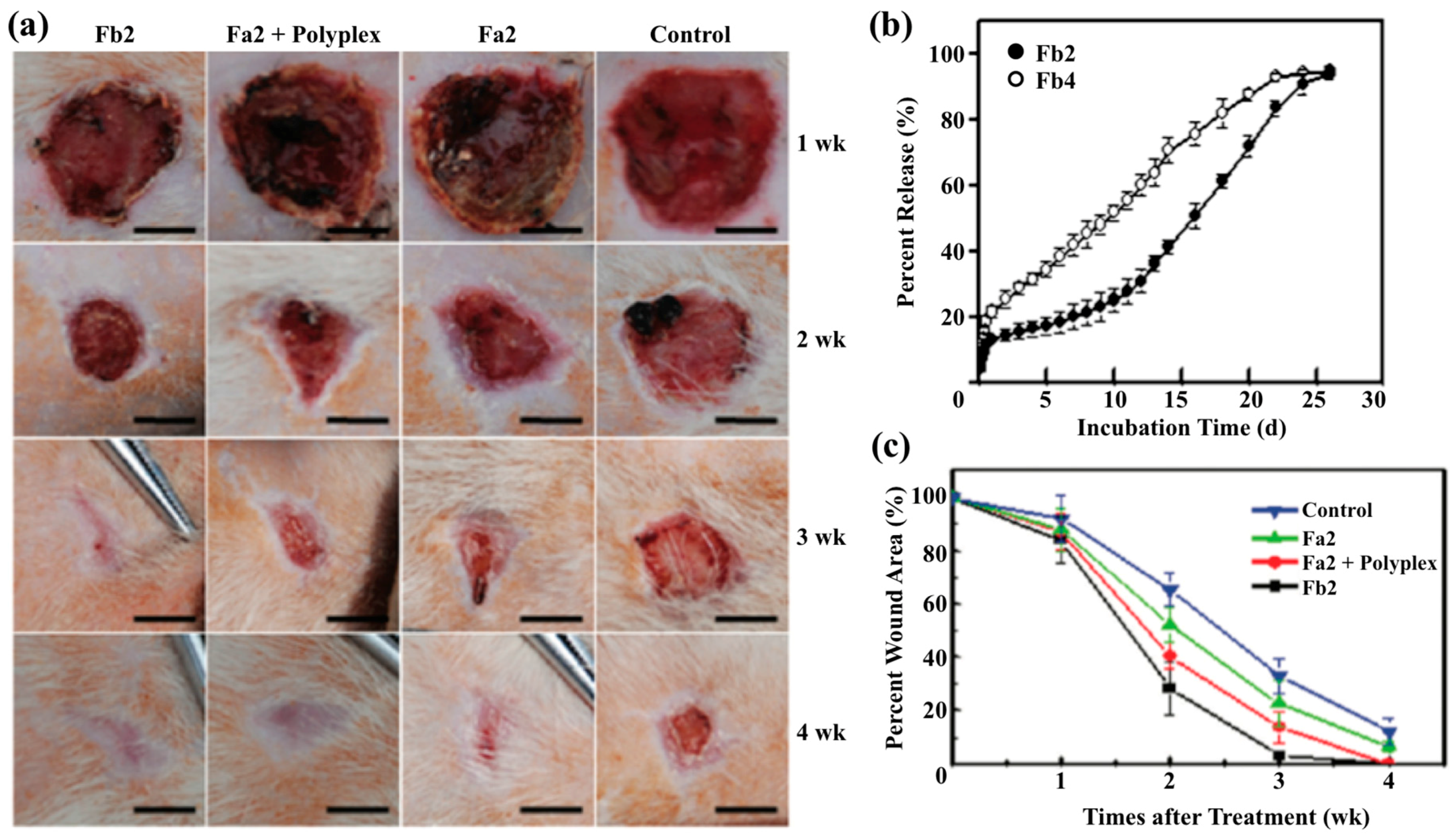
- Yang, Y.; Xia, T.; Chen, F.; Wei, W.; Liu, C.; He, S.; Li, X. Electrospun fibers with plasmid bFGF polyplex loadings promote skin wound healing in diabetic rats. Mol. Pharm. 2012, 9, 48–58. [Google Scholar] [CrossRef] [PubMed]
- He, S.; Xia, T.; Wang, H.; Wei, L.; Luo, X.; Li, X. Multiple release of polyplexes of plasmids VEGF and bFGF from electrospun fibrous scaffolds towards regeneration of mature blood vessels. Acta Biomater. 2012, 8, 2659–2669. [Google Scholar] [CrossRef] [PubMed]
- Kataoka, K.; Harada, A.; Nagasaki, Y. Block copolymer micelles for drug delivery: Design, characterization and biological significance. Adv. Drug Deliv. Rev. 2001, 47, 113–131. [Google Scholar] [CrossRef]
- Kazunori, K.; Glenn S., K.; Masayuki, Y.; Teruo, O.; Yasuhisa, S. Block copolymer micelles as vehicles for drug delivery. J. Control. Release 1993, 24, 119–132. [Google Scholar] [CrossRef]
- Redhead, H.M.; Davis, S.S.; Illum, L. Drug delivery in poly(lactide-co-glycolide) nanoparticles surface modified with poloxamer 407 and poloxamine 908: In vitro characterisation and in vivo evaluation. J. Control. Release 2001, 70, 353–363. [Google Scholar] [CrossRef]
- Thotakura, N.; Dadarwal, M.; Kumar, R.; Singh, B.; Sharma, G.; Kumar, P.; Katare, O.P.; Raza, K. Chitosan-palmitic acid based polymeric micelles as promising carrier for circumventing pharmacokinetic and drug delivery concerns of tamoxifen. Int. J. Biol. Macromol. 2017, 102, 1220–1225. [Google Scholar] [CrossRef] [PubMed]
- Wang, Y.; Ke, X.; Voo, Z.X.; Yap, S.S.L.; Yang, C.; Gao, S.; Liu, S.; Venkataraman, S.; Obuobi, S.A.O.; Khara, J.S.; et al. Biodegradable functional polycarbonate micelles for controlled release of amphotericin B. Acta Biomater. 2016, 46, 211–220. [Google Scholar] [CrossRef] [PubMed]
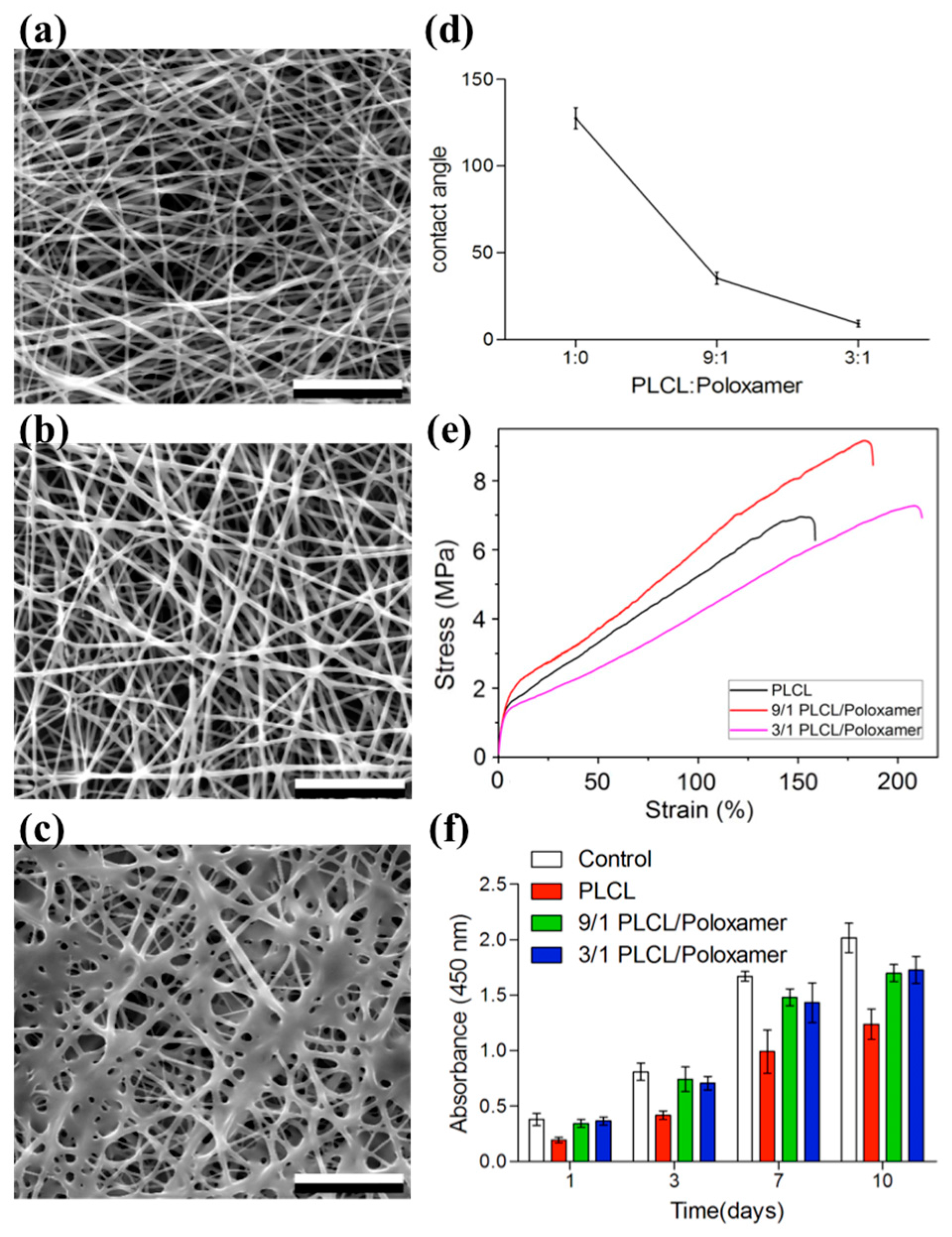
- Pan, J.; Liu, N.; Sun, H.; Xu, F. Preparation and characterization of electrospun PLCL/poloxamer nanofibers and dextran/gelatin hydrogels for skin tissue engineering. PLoS ONE 2014, 9, e112885. [Google Scholar] [CrossRef] [PubMed]
- Gong, C.; Wu, Q.; Wang, Y.; Zhang, D.; Luo, F.; Zhao, X.; Wei, Y.; Qian, Z. A biodegradable hydrogel system containing curcumin encapsulated in micelles for cutaneous wound healing. Biomaterials 2013, 34, 6377–6387. [Google Scholar] [CrossRef] [PubMed]
- Kim, J.H.; Ramasamy, T.; Tran, T.H.; Choi, J.Y.; Cho, H.J.; Yong, C.S.; Kim, J.O. Polyelectrolyte complex micelles by self-assembly of polypeptide-based triblock copolymer for doxorubicin delivery. Asian J. Pharm. Sci. 2014, 9, 191–198. [Google Scholar] [CrossRef]
- Oyarzun-Ampuero, F.; Vidal, A.; Concha, M.; Morales, J.; Orellana, S.; Moreno-Villoslada, I. Nanoparticles for the Treatment of Wounds. Curr. Pharm. Des. 2015, 21, 4329–4341. [Google Scholar] [CrossRef] [PubMed]
- Kalwar, K.; Sun, W.-X.; Li, D.-L.; Zhang, X.-J.; Shan, D. Coaxial electrospinning of polycaprolactone@chitosan: Characterization and silver nanoparticles incorporation for antibacterial activity. React. Funct. Polym. 2016, 107, 87–92. [Google Scholar] [CrossRef]
- Chen, C.-H.; Chen, S.-H.; Shalumon, K.T.; Chen, J.-P. Dual functional core–sheath electrospun hyaluronic acid/polycaprolactone nanofibrous membranes embedded with silver nanoparticles for prevention of peritendinous adhesion. Acta Biomater. 2015, 26, 225–235. [Google Scholar] [CrossRef] [PubMed]
- Ghosh Auddy, R.; Abdullah, M.F.; Das, S.; Roy, P.; Datta, S.; Mukherjee, A. New guar biopolymer silver nanocomposites for wound healing applications. BioMed Res. Int. 2013, 2013, 1–8. [Google Scholar] [CrossRef] [PubMed]
- Hazer, D.B.; Hazer, B.; Dinçer, N. Soft tissue response to the presence of polypropylene-g-poly(ethylene glycol) comb-type graft copolymers containing gold nanoparticles. J. Biomed. Biotechnol. 2011, 2011, 1–7. [Google Scholar] [CrossRef] [PubMed]
- Leu, J.-G.; Chen, S.-A.; Chen, H.-M.; Wu, W.-M.; Hung, C.-F.; Yao, Y.-D.; Tu, C.-S.; Liang, Y.-J. The effects of gold nanoparticles in wound healing with antioxidant epigallocatechin gallate and α-lipoic acid. Nanomed. Nanotechnol. Biol. Med. 2012, 8, 767–775. [Google Scholar] [CrossRef] [PubMed]
- Raguvaran, R.; Manuja, B.K.; Chopra, M.; Thakur, R.; Anand, T.; Kalia, A.; Manuja, A. Sodium alginate and gum acacia hydrogels of ZnO nanoparticles show wound healing effect on fibroblast cells. Int. J. Biol. Macromol. 2017, 96, 185–191. [Google Scholar] [CrossRef] [PubMed]
- Martinez, L.R.; Han, G.; Chacko, M.; Mihu, M.R.; Jacobson, M.; Gialanella, P.; Friedman, A.J.; Nosanchuk, J.D.; Friedman, J.M. Antimicrobial and healing efficacy of sustained release nitric oxide nanoparticles against Staphylococcus aureus skin infection. J. Investig. Dermatol. 2009, 129, 2463–2469. [Google Scholar] [CrossRef] [PubMed]
- Rather, H.A.; Thakore, R.; Singh, R.; Jhala, D.; Singh, S.; Vasita, R. Antioxidative study of cerium oxide nanoparticle functionalised PCL-Gelatin electrospun fibers for wound healing application. Bioact. Mater. 2017. [Google Scholar] [CrossRef]
- Quignard, S.; Coradin, T.; Powell, J.J.; Jugdaohsingh, R. Silica nanoparticles as sources of silicic acid favoring wound healing in vitro. Colloids Surf. B Biointerfaces 2017, 155, 530–537. [Google Scholar] [CrossRef] [PubMed]
- Wu, H.; Li, F.; Wang, S.; Lu, J.; Li, J.; Du, Y.; Sun, X.; Chen, X.; Gao, J.; Ling, D. Ceria nanocrystals decorated mesoporous silica nanoparticle based ROS-Scavenging tissue adhesive for highly efficient regenerative wound healing. Biomaterials 2018, 151, 66–77. [Google Scholar] [CrossRef] [PubMed]
- Wang, C.; Hou, W.; Guo, X.; Li, J.; Hu, T.; Qiu, M.; Liu, S.; Mo, X.; Liu, X. Two-phase electrospinning to incorporate growth factors loaded chitosan nanoparticles into electrospun fibrous scaffolds for bioactivity retention and cartilage regeneration. Mater. Sci. Eng. C 2017, 79, 507–515. [Google Scholar] [CrossRef] [PubMed]
- Gutha, Y.; Pathak, J.L.; Zhang, W.; Zhang, Y.; Jiao, X. Antibacterial and wound healing properties of chitosan/poly(vinyl alcohol)/zinc oxide beads (CS/PVA/ZnO). Int. J. Biol. Macromol. 2017, 103, 234–241. [Google Scholar] [CrossRef] [PubMed]
- Sanad, R.A.-B.; Abdel-Bar, H.M. Chitosan–hyaluronic acid composite sponge scaffold enriched with Andrographolide-loaded lipid nanoparticles for enhanced wound healing. Carbohydr. Polym. 2017, 173, 441–450. [Google Scholar] [CrossRef] [PubMed]
- Blakney, A.; Jiang, Y.; Woodrow, K.; Krogstad, E. Delivery of multipurpose prevention drug combinations from electrospun nanofibers using composite microarchitectures. Int. J. Nanomed. 2014, 2967–2978. [Google Scholar] [CrossRef] [PubMed]
- Krogstad, E.A.; Woodrow, K.A. Manufacturing scale-up of electrospun poly(vinyl alcohol) fibers containing tenofovir for vaginal drug delivery. Int. J. Pharm. 2014, 475, 282–291. [Google Scholar] [CrossRef] [PubMed]


| Classification Method | Subcategory | Characteristics | Examples |
|---|---|---|---|
| Time frame of healing | Acute | Faster healing (5–10 days) | Traumatic wounds, surgical wounds |
| Chronic | Takes long time to heal | Leg ulceration | |
| Wound closing method | Primary intention | Treated by closing the surface around the wound | Traumatic lacerations or surgical |
| Secondary intentions | Treated by filling the gaps with granulating tissue | Leg ulcers, pressure damage, and lacerations | |
| Tertiary intention | Open intentionally to allow for drainage to take place | Abdominal wound | |
| Wound tissue types | Black coloration | Shows black discoloration | Necrotic tissue |
| Green | Shows green discoloration | Infected tissue | |
| Yellow | Shows yellow discoloration | Sloughy tissue | |
| Red | Shows red discoloration | Granulating tissue | |
| Pink | Shows pink discoloration | Epithelial tissue | |
| Depth of wound | Superficial | Affect the epidermis | Abrasions |
| Partial thickness | Affect both the epidermis and the inner dermal layer | Pressure sores and severe scale exits |
| Polymer(s) † | Solvent(s) | Voltage (kV) | Distance (cm) | Flow Rate (mL/h) | Ref. |
|---|---|---|---|---|---|
| Natural | |||||
| Chitosan/PEO | 50% Acetic Acid | 15–35 | 15 | 0.1–2 | [35] |
| Alginate/Soy Protein/PEO | Deionized Water | 15 | 15 | 0.5 | [49] |
| Gelatin | 20% Acetic Acid | 28–35 | 10 | 0.1–1 | [50] |
| Cellulose | Acetic Acid | 30–40 | 15 | 1 | [39] |
| Collagen | PBS/Ethanol | 18 | 15 | 0.3 | [40] |
| Hyaluronic Acid/PCL | Formic Acid/Acetic Acid (75/25) | 13 | 13 | 1 | [51] |
| Keratin/PEO | 88% Formic Acid | 14 | 15 | 0.5 | [52] |
| Silk Fibroin | Lithium Bromide | 15 | 18 | - | [53] |
| Synthetic | |||||
| PCL | Acedic Acid | 9.5–22 | 15 | 0.15–1.2 | [54] |
| PLGA/GT | 1,1,1,3,3,3 hexafluoro-2-propanol | 15 | 15 | 1 | [55] |
| PU | N,N-dimethylformamide | 35–45 | 10–15 | 0.5–1.5 | [56] |
| PVDF | Dimethylformamide and Acetone | 25 | 15 | 0.75 | [57] |
| PVA/Silk Sericin | Deionized Water | 8–12 | 20 | 3 | [58] |
| PEO | Ethanol, Chloroform, and Deionized Water | 13 | 10 | 3 | [59] |
| PVP | Ethanol | 15 | 10 | 1 | [60] |
| Small Molecules Drugs | Agent | Fiber | Release (Units) | Ref. | ||||
|---|---|---|---|---|---|---|---|---|
| Aq. Sol. † (mg/mL) | Log P † | Polymer(s) ‡ | Loading (% w/w) | 1 h | 2 h | Others | ||
| Hydrophilic | ||||||||
| Ciprofloxacin | 1.35 | −0.57 | PVP | 0.4 | - | - | 60% (1 min) | [81] |
| PLCL/PDEGMA | 10 | 12% | 20% | 80% (220 h) | [82] | |||
| PVA/Alginate | - | 30% | 40% | 85% (6 h) | [91] | |||
| Ampicillin | 0.605 | 0.88 | AL-BSA | 5 10 20 | 23% 17% 7% | 37% 25% 10% | 99% (96 h) 81% (96 h) 40% (96 h) | [83] |
| PMMA/Nylon6 | 1–20 | - | - | 30% (6 h) 50% (12 days) | [84] | |||
| PCL | 16.7 | 75% | 80% | 98% (24 h) | [85] | |||
| Captopril | 4.52 | 1.02 | PLLA PLGA PLCL | 10 10 10 | - - - | - - - | 98%(48 h) 100% (48 h) 78% (48 h) | [92] |
| Metronidazole | 5.92 | -0.15 | PCL | 1–40 | - | - | 45% (1 day) 85% (5 days) | [86] |
| PCL | 4.8–14.4 | 20% | 40% | 90% (24 h) | [87] | |||
| Chitosan/PEO | 1 5 15 | 52% 70% 70% | 75% 80% 100% | - - - | [88] | |||
| Cefazolin | 0.487 | −0.4 | Chitosan/PEO | 1 | - | 26% | 65% (24 h) | [89] |
| Gelatin | 10 | 10% | 30 | 95% (17 h) | [90] | |||
| Hydrophobic | ||||||||
| Asiaticoside | [93] | [93] | Alginate/PVA/Chitosan | 2.5 | 20% | 23% | 83%(12 h) | [94] |
| Curcumin | 0.006 | 3.62 | PHBV | 1 3 4.7 | 20% 55% 65% | 40% 65% 67% | 45% (5 h) 70% (5 h) 78% (5 h) | [95] |
| PCL/GT | 3 | - | - | 65% (20 days) | [96] | |||
| Ketoprofen | 0.0213 | 3.29 | PCL/Gelatin | 5 | - | - | 40% (20 h) 80% (45 h) | [97] |
| PVA | 5 | 50% | - | 62% (48 h) | [98] | |||
| PNVCL-co-MAA | 20 | 5% | - | 35% (24 h) | [99] | |||
| Cellulose Acetate | 15 | 10% | - | 60% (48 h) | [100] | |||
| Nifedipine | 0.0177 | 2.49 | Eudragit® | 10 | 40% | 50% | 70% (8 h) | [101] |
| PU | 4.2 | 15% | - | 75% (72 h) | [102] | |||
| PNIPAAm/PU | 12 | 8% | 10% | 23% (30 h) | [103] | |||
| Phenytoin | 0.0711 | 2.26 | PVA PCL PVA/PCL | 2 2 2 | 27% 5% 11% | 29% 8% 15% | 88% (48 h) 16% (48 h) 47% (48 h) | [104] |
| Vancomycin | 0.255 | 1.11 | Alginate | 10 | 10% | - | 60% (48 h) | [105] |
| Methylene Blue | 0.0296 | 3.61 | PHB/PEG | - | 32% | - | 90% (7 days) | [106] |
| Growth Factor | Polymer | Solvent | Cell | Method | Ref. |
|---|---|---|---|---|---|
| EGF | PCL and PCL–PEG/PCL | Methanol/Chloroform | Human Primary Keratinocyte | Immobilization | [119] |
| PLGA and Gelatin | Chloroform/Acetone and Acetic Acid | Human Fibroblasts | Emulsion | [120] | |
| Silk Fibroin | Lithium Bromide | Human Dermal Fibroblasts | Blend | [121] | |
| PCL and PCL/Collagen | DMF/DCM and HFIP | Human Dermal Keratinocyte | Immobilization | [122] | |
| Gelatin/PLA-co-PCL | HFIP | Human Dermal Fibroblasts | Coaxial | [123] [124] | |
| Silk/PEO | Lithium Bromide | - | Blend/Coating | [125] | |
| bFGF/EGF | PCL-PEG | Methanol and Chloroform | Keratinocyte and fibroblast | Coaxial/Immobilization | [112] |
| PLGA/PEO | Chloroform and DMF/Water | Human Skin Fibroblasts | Fiber containing GFs encapsulated microspheres. | [126] | |
| bFGF, EGF, VEGF, PDGF | Collagen-Hyaluronic Acid/Gelatin Nanoparticle | Hyaluronic Acid: NaOH/DMF Collagen: Acetic Acid | HUVEC | Blend: bEGF/EGF In nanoparticle: VEGF/PDGF | [111] |
| PDGF | PCL/Collagen/Hyaluronic Acid | HFP, PBS | MSC | Blend | [113] |
| FGF2 | PHBV, PEO | 2, 2, 2-trifluoroethanol | MSC | FGF2-miR-218 induction on aligned PHBV fibers | [115] |
| KGF | PLA/PCL | Chloroform, Acetone | Fibroblasts | Seeded scaffolds with mouse fibroblast in DMEM with FBS | [127] |
| TGF-β | MeHA, HH, PCL, HA | DI Water | Cartilage | Composite scaffolds of HA and PCL with TGFβ3 | [114] |
| VEGF | PLGA | Water-in-oil emulsions, Dichloromethane, PBS, BSA | HUVEC, Endothelials | PVEES, and NVEES Scaffolds containing VEGF | [128] |
| GM-CSF | Chitosan | HCl | In vivo mouse model | Hydrogels containing ovalbumin and GM-CSF | [117] |
| CTGF | PCL | Chloroform | MSC | Aligned fibers as a guide | [118] |
| Polymeric Micelles | Drug | Functions | Ref. |
|---|---|---|---|
| Poloxamer 407 and 908/PLGA nanoparticles | Rose Bengal Dye | Showed protective effects of Poloxamer 407 and 908 micelles. | [147] |
| Chitosan/Palmitic Acid | Tamoxifen | Release profiles showed much more linear release when encapsulated in micelle structures. | [148] |
| phenylboronic acid-functionalized polycarbonate/PEG (PEG-PBC)/urea-functionalized polycarbonate/PEG (PEG-PUC)/diblock copolymers | Amphotericin B | Used to study delivery of anti-fungal medication. PEG-PBC and diblock copolymers of PEG-PBC and PEG-PUC showed sustained release of drug while PEG-PUC had burst release profile. | [149] |
| Poly(l-aspartic acid)-b-poly(ethylene glycol)-b-poly(l-aspartic acid) (PLD-PEG-PLD) | Doxorubicin | Showed effect pH of release media has on release profiles of doxorubicin loaded PLD-PEG-PLD micelles. Found more acidic environment correlated to higher release rates. | [152] |
| PLCL/poloxamer with dextran/gelatin hydrogel | No Drug | Showed fibers supported cell viability and proliferation when tested with stem cells. Mechanical properties increased with addition of of Poloxamer at 9/1 ratio. | [150] |
| PEG-PCL and PEG-PCL/hydrogel | Curcumin | Micelle structure sustained release 14 days and achieved higher cumulative release rate than micelle/hydrogel. In Vivo model showed micelle. Hydrogel combination produced higher tensile strength and thicker epidermis during wound healing breaking test. Micelle/Hydrogel also showed enhanced wound closure rate. | [151] |
| Nanoparticles | Effects | Functions | Ref. |
|---|---|---|---|
| Silver/guar gum alkylamine | Antibacterial | Exhibited faster would healing rates and improved cosmetic attributes. | [156] |
| Gold | Anti-Inflammatory | Wounds exhibited reduction in inflammatory response. Increase in cell proliferation resulting in reduction of wound healing time in mice. | [157] |
| Zinc Oxide loaded alginate/gun acacia | Antibacterial | Showed that Zinc nanoparticles have antibacterial effects at low levels but can become toxic at high levels. | [159] |
| Nitric Oxide | Antibacterial | Promoted regeneration of dermal architecture through protection of collagen from bacteria. | [160] |
| Cerum Oxide loaded PCL/Gelatin fibers | Reduction of reactive oxygen levels, decreased healing time | Lowered the level of reactive oxygen levels that hinder proper wound healing. | [161] |
| Adhesive nanocomposite made of ultrasmall ceria nanocrystals adhered to the surface of mesoporous silica nanoparticles | Reduction of reactive oxygen levels, decreased healing time | Reduced healing time and scar formation. Stimulated proliferation and cell migration in vivo. | [163] |
| Chitosan nanoparticles with PLLA-CL fibers | Nel-like mlecule-1 growth factor delivery | Dual release system prolonged release of growth factor when compared to plain PLLA-CL fibers. Dual release system Increased cell proliferation in human bone mesenchymal stem cells. | [164] |
| Chitosan/PVA/Zinc Oxide | Decreased wound healing time/Antibacterial | Displayed shorter healing time and almost no bacterial growth in cultured pus from wounds. | [165] |
| Lipid nanocarrier/Hyaluronic Acid/Chitosan | Drug delivery | Prolonged release of Andrographolide combined with depolymerization of chitosan resulted in the reduction of wound healing time. | [166] |
© 2018 by the authors. Licensee MDPI, Basel, Switzerland. This article is an open access article distributed under the terms and conditions of the Creative Commons Attribution (CC BY) license (http://creativecommons.org/licenses/by/4.0/).
Share and Cite
Gizaw, M.; Thompson, J.; Faglie, A.; Lee, S.-Y.; Neuenschwander, P.; Chou, S.-F. Electrospun Fibers as a Dressing Material for Drug and Biological Agent Delivery in Wound Healing Applications. Bioengineering 2018, 5, 9. https://doi.org/10.3390/bioengineering5010009
Gizaw M, Thompson J, Faglie A, Lee S-Y, Neuenschwander P, Chou S-F. Electrospun Fibers as a Dressing Material for Drug and Biological Agent Delivery in Wound Healing Applications. Bioengineering. 2018; 5(1):9. https://doi.org/10.3390/bioengineering5010009
Chicago/Turabian StyleGizaw, Mulugeta, Jeffrey Thompson, Addison Faglie, Shih-Yu Lee, Pierre Neuenschwander, and Shih-Feng Chou. 2018. "Electrospun Fibers as a Dressing Material for Drug and Biological Agent Delivery in Wound Healing Applications" Bioengineering 5, no. 1: 9. https://doi.org/10.3390/bioengineering5010009