Article

Dietary Milk Sphingomyelin Reduces Systemic Inflammation in Diet-Induced Obese Mice and Inhibits LPS Activity in Macrophages

by
Gregory H. Norris
,
Caitlin M. Porter
,
Christina Jiang
and
Christopher N. Blesso
*
Department of Nutritional Sciences, University of Connecticut, Storrs, CT 06269, USA
*
Author to whom correspondence should be addressed.
Both investigators contributed equally to this work.
Beverages 2017, 3(3), 37; https://doi.org/10.3390/beverages3030037
Submission received: 31 May 2017 / Revised: 6 July 2017 / Accepted: 14 July 2017 / Published: 21 July 2017
(This article belongs to the Special Issue Milk: Bioactive Components and Role in Human Nutrition)

Abstract

:
High-fat diets (HFD) increase lipopolysaccharide (LPS) activity in the blood and may contribute to systemic inflammation with obesity. We hypothesized that dietary milk sphingomyelin (SM), which reduces lipid absorption and colitis in mice, would reduce inflammation and be mediated through effects on gut health and LPS activity. C57BL/6J mice were fed high-fat, high-cholesterol diets (HFD, n = 14) or the same diets with milk SM (HFD-MSM, 0.1% by weight, n = 14) for 10 weeks. HFD-MSM significantly reduced serum inflammatory markers and tended to lower serum LPS (p = 0.08) compared to HFD. Gene expression related to gut barrier function and macrophage inflammation were largely unchanged in colon and mesenteric adipose tissues. Cecal gut microbiota composition showed greater abundance of Acetatifactor genus in mice fed milk SM, but minimal changes in other taxa. Milk SM significantly attenuated the effect of LPS on pro-inflammatory gene expression in RAW264.7 macrophages. Milk SM lost its effects when hydrolysis was blocked, while long-chain ceramides and sphingosine, but not dihydroceramides, were anti-inflammatory. Our data suggest that dietary milk SM may be effective in reducing systemic inflammation through inhibition of LPS activity and that hydrolytic products of milk SM are important for these effects.

1. Introduction

Chronic low-grade inflammation is involved in the pathogenesis of cardiovascular disease, type 2 diabetes, and non-alcoholic fatty liver disease [1]. Metabolically related chronic low-grade inflammation may be exacerbated by circulating lipopolysaccharide (LPS), also called “endotoxin”, a pro-inflammatory molecule found in the outer membrane of Gram-negative bacteria [2]. Recognition of bacterial LPS by host cells can trigger a pro-inflammatory signaling cascade mediated by Toll-like receptor-4 (TLR4), a pattern recognition receptor [3]. The human gastrointestinal tract has a “gut barrier” to limit the passage of microbes between host intestinal cells (e.g., tight junctions between enterocytes) and into the bloodstream [4]. These features of the gut barrier reduce permeability to LPS, which may otherwise trigger host inflammation and disease [5]. However, LPS translocation from the gut to circulation is enhanced by the absorption of dietary lipids [6]. High-fat diets (HFD) have been shown to promote inflammation of the distal intestine, causing an impairment of the protective gut barrier, and translocation of LPS into host circulation [7]. Diets high in fat can also alter the gut microbiota [8], resulting in diminished amounts of beneficial bacteria, such as some bifidobacteria, which would promote gut barrier permeability [7]. Additionally, diets rich in triglyceride and cholesterol increase chylomicron production during digestion and absorption. It has been shown that LPS can be incorporated into chylomicrons and transported into the bloodstream of mice [6]. Overall, these effects of HFD result in the paracellular and transcellular translocation of LPS from the gut into the circulation. The presence of LPS in the circulation activates the inflammatory response of the immune system, mainly through the activation of TLR4 [3]. Lipopolysaccharide engages the TLR4/MD-2 receptor complex via LPS-binding protein (LBP) and CD14. Toll-like receptor 4 signaling then activates the nuclear factor-kappa B (NF-κb) transcription factor resulting in the production of pro-inflammatory cytokines, such as tumor necrosis factor alpha (TNF-α) [3]. Macrophages in the colon have been shown to be the first pro-inflammatory immune responders to an HFD [9]; thus, inhibiting LPS translocation and the subsequent inflammatory responses of macrophages shows great potential in reducing the detrimental effects of a diet rich in fat and cholesterol.
Dietary phospholipids, including sphingolipids, show potential in mitigating chronic disease through effects on lipid absorption and inflammation [10,11]. Dietary sphingolipids, which include sphingomyelin (SM), ceramides, and sphingosine, are mainly found in milk, eggs, and soybeans [12]. It is estimated that the average American consumes 0.3 to 0.4 grams of sphingolipids per day [12]. Sphingomyelin is considered a zoochemical, being present in animal cell membranes but absent from plants [13]. Sphingomyelin found in milk is an important component of milk fat globule membranes [14]. Dietary SM and other sphingolipids have been studied for their effects on dyslipidemia because they interfere with the absorption of dietary fat and cholesterol [15,16,17,18,19,20,21]. In addition to effects on lipid absorption, dietary SM may have further bioactive effects by reducing inflammation and LPS activity, potentially influencing chronic disease. Dietary milk SM has been shown to reduce dextran sulfate sodium (DSS)-induced colitis in mice, suggesting anti-inflammatory effects in the gut [22]. However, the effect of dietary SM on colon inflammation is controversial, as exacerbation of colitis in mice has been observed with feeding egg-derived SM [23,24]. Phospholipids and sphingolipids appear to impact LPS activity, as they have been shown to dampen LPS-induced inflammation [25,26]. The presence of LPS in circulation also results in the liver upregulating sphingolipid biosynthesis via the sphingolipid rate-limiting biosynthetic enzyme, serine palmitoyltransferase (SPT), possibly as a compensatory mechanism [27]. We have previously shown that feeding 0.25% (w/w) milk SM reduced serum LPS and altered the gut microbiota in HFD-fed mice after four weeks [28]. We also recently reported that feeding dietary SM (0.1% w/w) attenuated hepatic steatosis and adipose tissue inflammation in diet-induced obese mice [29]. The current study aimed to assess the effects of dietary milk SM on systemic and gut inflammation using a HFD-induced obese mouse model. We also sought to elucidate whether milk SM and its hydrolytic products (ceramides, dihydroceramides, and sphingosine) directly affect the inflammatory response of macrophages stimulated by LPS.

2. Animals and Diets

Male C57BL/6J mice were purchased from the Jackson Laboratory (Bar Harbor, ME, USA) at six weeks of age. Mice were housed in a temperature-controlled room and maintained in a 12 h light/12 h dark cycle within the University of Connecticut-Storrs vivarium. The Animal Care and Use Committee of the University of Connecticut-Storrs approved all procedures used in the current study. Mice were acclimated to the facility for two weeks before being placed on either a lard-based high-fat, high-cholesterol diet (HFD; 60% kcal from fat, 0.15% cholesterol added by weight; n = 14) or HFD supplemented with 0.1% of milk SM added by weight (HFD-MSM; n = 14). Detailed diet compositions are presented in Table 1. Experimental diets were prepared using purified ingredients commercially available from Dyets, Inc. (Bethlehem, PA, USA). Milk SM (bovine; >99% purity) was obtained from Avanti Polar Lipids, Inc. (Alabaster, AL, USA) and then substituted for an equal weight of lard in the treatment group. Accounting for weight gained throughout the study, HFD-MSM provided the equivalent of consuming approximately 405–670 mg milk SM/day in a 70 kg human based on body surface normalization [30].
Mice consumed the diets ad libitum and fresh food was provided twice per week. Body weight was assessed weekly, while food intake was calculated at each feeding. After 10 weeks, mice were fasted for 6–8 h followed by euthanasia and blood collection via cardiac puncture. Blood was allowed to clot at room temperature for 30 min before serum isolation by centrifugation (10,000× g for 10 min at 4 °C) and then stored at −80 °C. Special precautions were taken to minimize endotoxin contamination of the serum samples. Mesenteric adipose, small intestine, and colon tissues were isolated and snap-frozen in liquid nitrogen before storage in −80 °C.

2.1. Serum Biochemical Analysis

Serum IL-6, TNF-α, IFNγ, and MIP-1β were measured by Luminex/xMAP magnetic bead-based multiplexing assay using MAGPIX instrumentation from EMD Millipore (Billerica, MA, USA). Serum LPS was measured using a chromogenic limulus amebocyte lysate (LAL) assay (QCL-1000) obtained from Lonza (Basel, Switzerland).

2.2. Gut Microbiota Analysis

Cecal fecal samples were collected from mice and submitted to the University of Connecticut-Storrs Microbial Analysis, Resources, and Services (MARS) facility for microbiota characterization utilizing 16S V4 analysis. DNA was extracted from 0.25 g of fecal sample using the MoBio PowerMag Soil 96 well kit (MoBio Laboratories, Inc., Carlsbad, CA, USA) according to the manufacturer’s protocol for the Eppendorf epMotion liquid handling robot. DNA extracts were quantified using the Quant-iT PicoGreen kit (ThermoFisher Scientific, Waltham, MA, USA). Partial bacterial 16S rRNA (V4) and fungal ITS2 genes were amplified using 30 ng extracted DNA as template. The V4 region was amplified using 515F and 806R with Illumina adapters and dual indices (8 basepair golay on 3′ [31], and 8 basepair on the 5′ [32]). Samples were amplified in triplicate using Accuprime PFX PCR master mix (ThermoFisher Scientific, Waltham, MA, USA) with the addition of 10 μg BSA (New England BioLabs, Ipswich, MA, USA). The PCR reaction was incubated at 95 °C for 2 min., then 30 cycles of 15 s at 95.0 °C, 1 min. at 55 °C, and 1 min. at 68 °C, followed by final extension at 68 °C for 5 min. PCR products were pooled for quantification and visualization using the QIAxcel DNA Fast Analysis (Qiagen, Hilden, Germany). PCR products were normalized based on the concentration of DNA from 250–400 bp then pooled using the QIAgility liquid handling robot. The pooled PCR products were cleaned using Mag-Bind RXNPure Plus (Omega Bio-Tek, Norcross, GA, USA) according to the manufacturer’s protocol. The cleaned pool was sequenced on the MiSeq using v2 2 x 250 base pair kit (Illumina, Inc, San Diego, CA, USA).
Sequences were demultiplexed using onboard bcl2fastq. Demultiplexed sequences were processed in Mothur v. 1.39.4 following the MiSeq SOP [32]. Exact commands can be found at [33]. Merged sequences that had any ambiguities or did not meet length expectations were removed. Sequences were aligned to the Silva nr_v119 alignment [34]. Identification of operational taxonomic units (OTUs) was done using the RDP Bayesian classifier [35] against the Silva nr_v119 taxonomy database.

2.3. Cell Culture

Murine RAW264.7 macrophages were obtained from ATCC (Manassas, VA, USA) and cultured in a humidified incubator at 37 °C with 5% CO2. Cells were maintained in Dulbecco’s modified Eagle’s medium (4 g/L glucose) containing sodium pyruvate, 10% fetal bovine serum (Hyclone, Logan, UT, USA), 2 mM L-glutamine, 100 U/mL penicillin, 100 μg/mL streptomycin antibiotic (ThermoFisher Scientific, Waltham, MA, USA), and 100 μg/mL Normocin (Invitrogen, Carlsbad, CA, USA). Cell counts and viability were routinely measured using Trypan blue and a TC-20 automated cell counter (Bio-Rad, Hercules, CA, USA).

2.4. Effects of Sphingolipids on LPS Stimulation of RAW264.7 Macrophages

Milk sphingomyelin (bovine), sphingosine (d18:1), C16-ceramide (d18:1/16:0), C24-ceramide (d18:1/24:0), C16-dihydroceramide (d18:0/16:0), and C24-dihydroceramide (d18:0/24:0) were obtained from Avanti Polar Lipids at >99% purity. Milk SM and sphingosine were dissolved in 100% ethanol (EtOH), while ceramides were dissolved in EtOH/dodecane (98.8/0.2, v/v). RAW264.7 cells were seeded in 24-well plates and allowed to adhere overnight prior to experimentation. To examine possible anti-inflammatory effects of milk SM, RAW264.7 macrophages were pre-incubated with milk SM (0.8–8 μg/mL) or EtOH vehicle control for 1 h, followed by a 4 h co-incubation in the presence or absence of 1 ng/mL LPS (E. coli 0111:B4) (Sigma-Aldrich, St. Louis, MO, USA). Since milk SM comprised of a natural mixture of SM species, concentrations are reported in 0.8–8 μg/mL, which is comparable to 1–10 μM of a pure SM species. To determine if effects were dependent on SM hydrolysis, cells were incubated for 2 h with 100 μM imipramine (Sigma-Aldrich, St. Louis, MO, USA) prior to 1 h incubation with milk SM or control ± 4 h co-incubation with LPS. Imipramine has been shown to reduce acid sphingomyelinase (SMase) activity to ~20% of basal levels after 2 h of incubation [36]. To test the effects of various ceramides and sphingosine, RAW264.7 macrophages were incubated for 4 h with ceramides (10 μM), dihydroceramides (10 μM), sphingosine (1–10 µM), or vehicle control ± LPS (1 ng/mL). Ethanol was used as a vehicle control for milk SM and sphingosine treatments, while EtOH/dodecane (98.8/0.2, v/v) was used as a control for ceramide treatments. To test for cytotoxicity, Cell Counting Kit-8 (CCK-8) (Sigma-Aldrich, St. Louis, MO, USA) was used after cells were treated with the different sphingolipids. Sodium dodecyl sulfate (SDS, 50 μM) was used as a cytotoxic positive control. Caspase 3 activity was also determined as an indicator of apoptosis using a colorimetric protease assay kit (ThermoFisher Scientific, Waltham, MA, USA) according to the manufacturer’s instructions. Cisplatin (ThermoFisher Scientific, Waltham, MA, USA) at 50 μM concentration was used on cells as a positive control to induce apoptosis.

2.5. RNA isolation, cDNA Synthesis, and qRT-PCR

Total RNA from small intestine, colon, mesenteric adipose tissue, and RAW264.7 macrophages was isolated using TRIzol reagent (Life Technologies, Carlsbad, CA, USA). RNA was treated with DNase I before reverse transcription using iScript cDNA synthesis kit (Bio-Rad). Real-time qRT-PCR using the iTaq Universal SYBR Green Supermix was performed on a CFX96 real-time-PCR detection system (Bio-Rad). For small intestine, colon, and mesenteric adipose tissues, the geometric mean of the reference genes, glyceraldehyde 3-phosphate dehydrogenase (Gapdh) and beta actin was used to standardize mRNA expression using the 2−ΔΔCt method. The macrophage mRNA was standardized to Gapdh expression. All primer sequences used are listed in Table S1.

2.6. Statistical Analysis

Data were analyzed using GraphPad Prism 6. Means were compared using Student’s t test or one-way analysis of variance with Holm–Sidak post hoc analysis where indicated. For gut microbiota analysis, alpha and beta diversity statistics were calculated by taking the average of 1000 random subsampling to 10,000 reads per sample in Mothur. Non-metric multidimensional scaling (NMS) and Permanova were run in R 3.3.2. A subsampled species matrix was used for indicator species analysis [37]. Figures were drawn in R 3.3.2 using ggplot2 2.2.1 and RColorBrewer 1.1-2. Data are reported as mean ± SEM. Significance is reported as p < 0.05.

3. Results

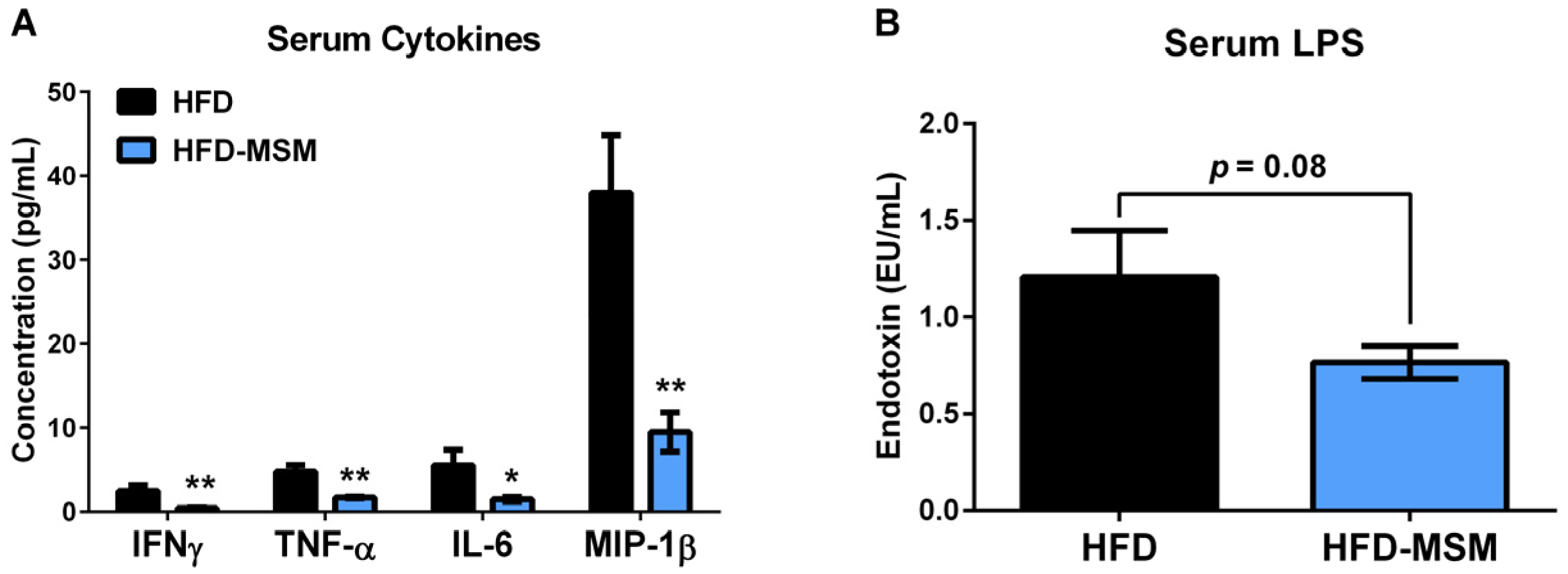
3.1. Dietary Milk SM Reduces Systemic Inflammation and Tends to Lower Circulating LPS

All mice fed the lard-based HFD were obese and insulin-resistant after 10 weeks, while supplementation of 0.1% (w/w) milk SM did not affect food intake, body or tissue weight gain compared to HFD control [29]. However, feeding milk SM strongly decreased serum inflammatory cytokines/chemokines compared to HFD control (Figure 1A). Milk SM tended to reduce LPS compared to HFD control (−36%), but it was not significantly different (p = 0.08) (Figure 1B). Milk SM also tended to increase Niemann-Pick C1-Like 1 (NPC1L1) mRNA expression (p = 0.07) in the small intestine (Figure 2A), which is induced by cellular cholesterol depletion [38]. No other significant changes were observed in the small intestine for gene expression related to lipid absorption. Interestingly, C-C motif chemokine ligand 2 (CCL2) mRNA expression was significantly increased in the colon by milk SM (Figure 2B). However, this did not appear to alter gut health, as mRNA expression of genes related to macrophage infiltration/inflammation (F4/80, Cd68, Cd11c, Tnf) and gut barrier function (Tjp1, Alpi, Ocln) was mostly unaffected by milk SM (Figure 2B). Furthermore, mesenteric adipose tissue mRNA expression related to inflammation was also unchanged, while there was a trend in GLUT4 mRNA expression to be increased by milk SM (p = 0.09) (Figure 2C).

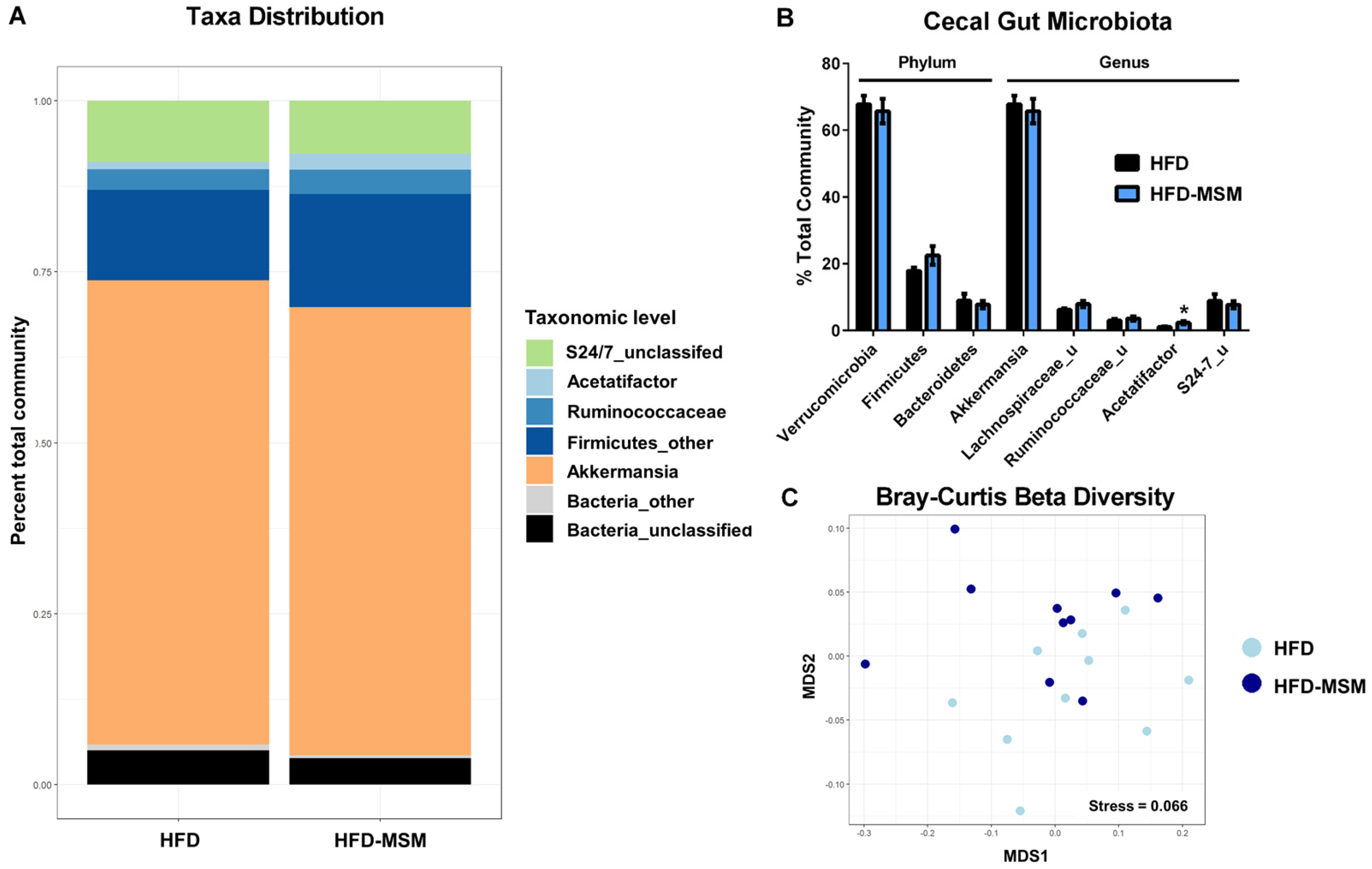
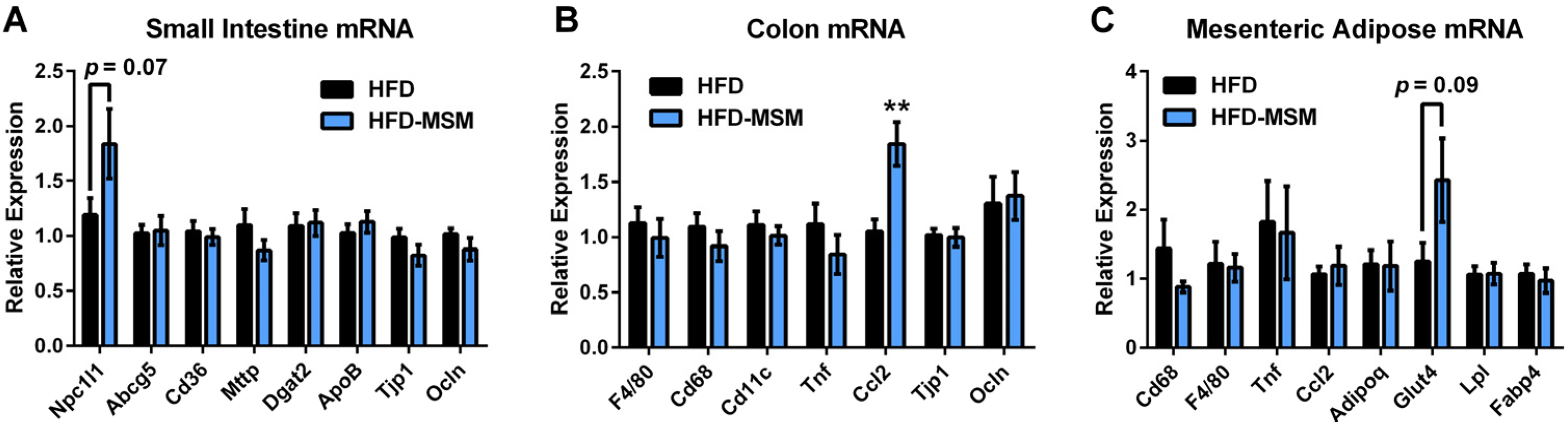
3.2. Gut Microbiota Composition is Mostly Unaffected by 0.1% (w/w) Dietary Milk SM

To determine if alterations in gut microbiota could explain differences in systemic inflammation in the present study, cecal feces microbiota composition was examined by 16S rRNA sequence analysis and results are presented in Figure 3. Verrucomicrobia was the major phylum in cecal feces, but there were no significant differences between groups in its relative abundance or that of its major genus, Akkermansia (Figure 3B). Additionally, no significant differences were observed in the relative abundance of Firmicutes or Bacteroidetes phyla between groups (Figure 3B) or when phyla were grouped as Gram-negative bacteria (HFD: 76.9% ± 1.5% vs. HFD-MSM: 73.5% ± 2.8%, p = 0.31). However, relative abundance of Acetatifactor, a genus of the Firmicutes phylum, was significantly higher in the group fed milk SM compared to HFD control (Figure 3B). Alpha-diversity analysis (diversity within a sample) determined by inverse Simpson index was not significantly different between groups (HFD: 2.19 ± 0.16 vs. HFD-MSM: 2.46 ± 0.32, p = 0.45). Furthermore, beta-diversity analysis (between sample diversity) by Bray–Curtis revealed no significant clustering of samples according to diet (Figure 3C). Therefore, in the context of diet-induced obesity, gut microbiota composition was generally unaffected by supplementation of 0.1% (w/w) milk SM in the diet.

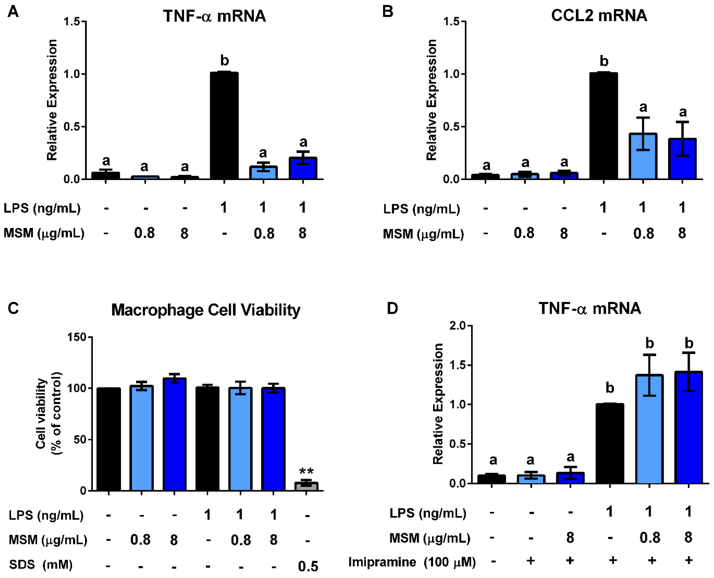
3.3. Milk SM Inhibits LPS Stimulation of Macrophages

Since dietary milk SM may directly influence inflammatory processes beyond inhibiting lipid absorption, we examined its effects on pro-inflammatory gene expression in RAW264.7 macrophages. Increases in TNF-α and CCL2 mRNA with 4 h LPS stimulation were both significantly reduced with milk SM (0.8 and 8 μg/mL) (Figure 4A,B). Milk SM in the absence of LPS did not influence pro-inflammatory gene expression. Furthermore, cell viability was not affected by milk SM at concentrations shown to affect pro-inflammatory gene expression (Figure 4C). With the addition of imipramine, LPS-stimulated inflammation was not reduced in the presence of milk SM (Figure 4D). Imipramine causes proteolytic degradation of acid sphingomyelinase, which hydrolyzes SM to ceramide and phosphorylcholine. This suggests that a hydrolytic product of SM (e.g., ceramide, sphingosine) is important for milk SM’s effect on inflammation.

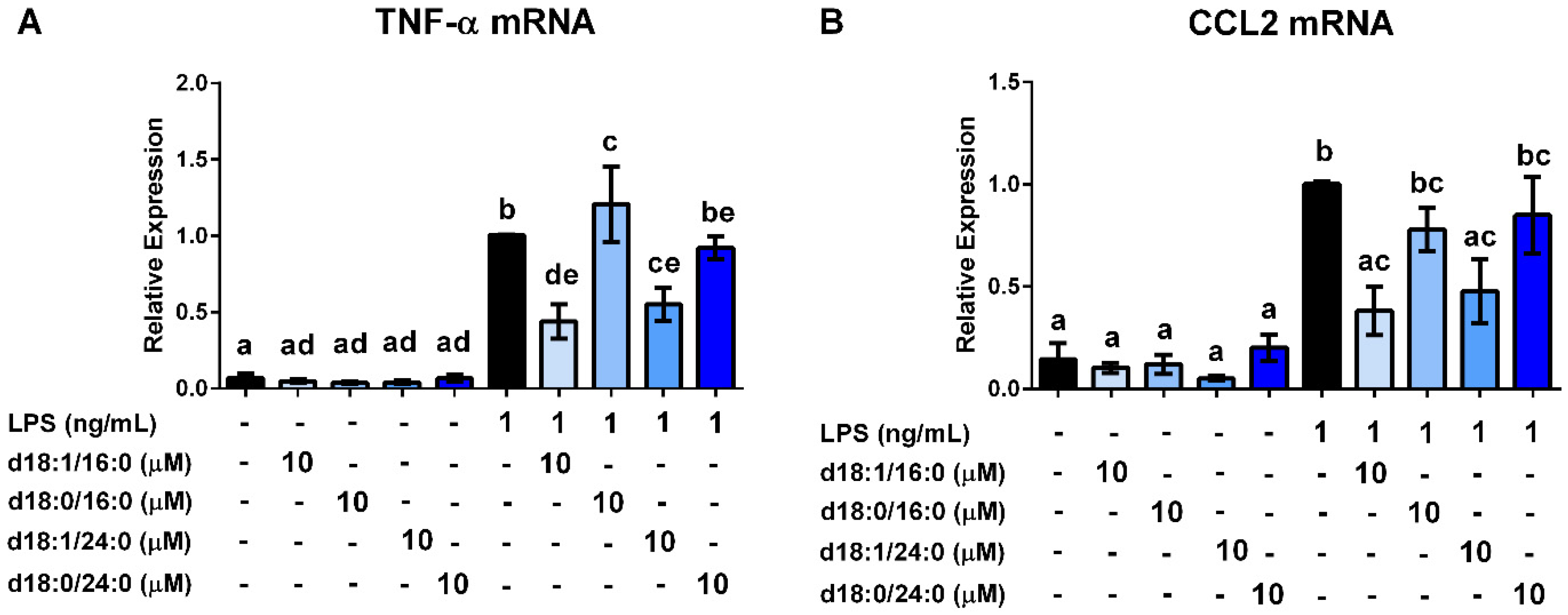
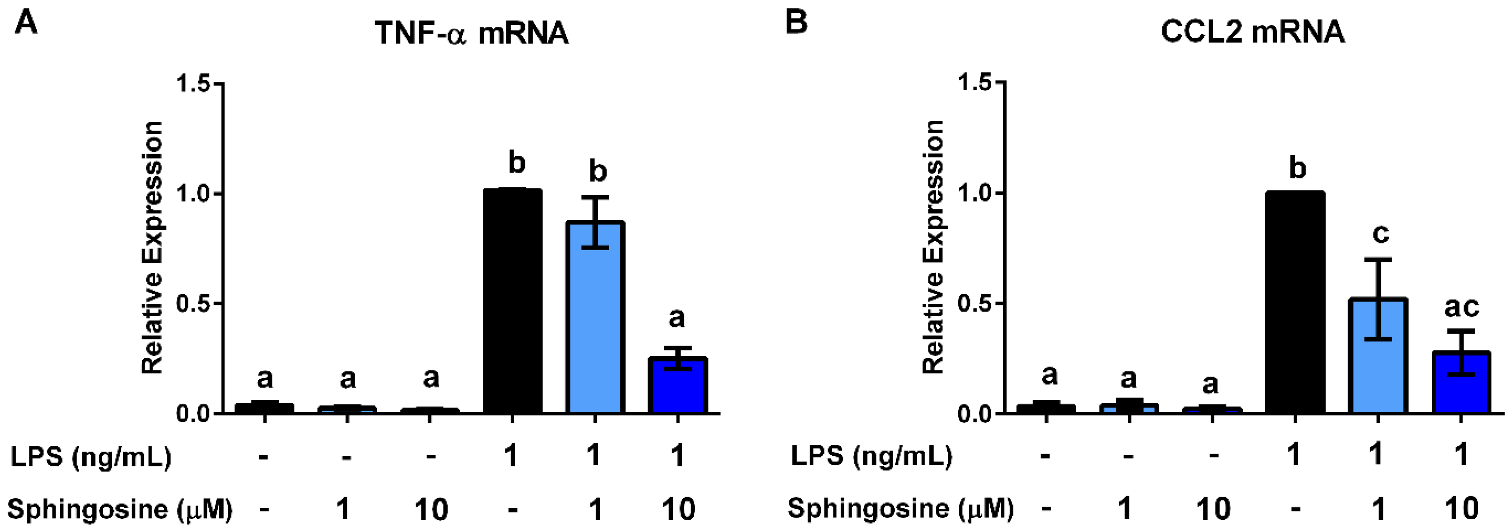
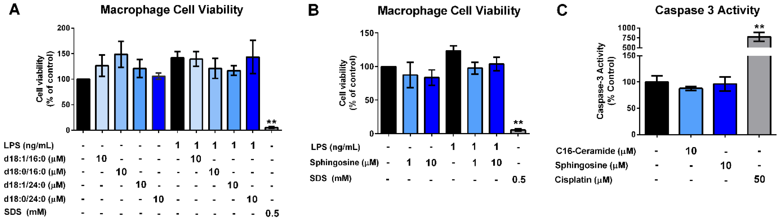
3.4. Ceramides and Sphingosine, but Not Dihydroceramides, Inhibit LPS Stimulation of Macrophages

Since milk SM contains a mixture of SM species with different fatty acid chain lengths and sphingoid backbones, we tested the effects of C16-ceramide, C24-ceramide, C16-dihydroceramide, and C24-dihydroceramide on macrophage pro-inflammatory gene expression (Figure 5A,B). Interestingly, both C16-ceramide and C24-ceramide at 10 μM concentrations significantly reduced TNF-α mRNA (Figure 5A) and CCL2 mRNA (Figure 5B) in LPS-stimulated macrophages, but their corresponding dihydroceramides did not. None of the ceramides tested altered inflammatory gene expression in the absence of LPS. The anti-inflammatory effects of ceramides (sphingosine base), but not dihydroceramides (sphinganine base), suggest that sphingosine is important for bioactivity. Supporting this notion, sphingosine significantly reduced LPS-stimulation of TNF-α mRNA (Figure 6A) and CCL2 mRNA (Figure 6B) in macrophages. Neither ceramides nor sphingosine significantly affected cell viability (Figure 7A,B) or caspase 3 activity (Figure 7C) at the concentrations tested, suggesting that cell death was not the cause of anti-inflammatory effects.

4. Discussion

Chronic low-grade inflammation is a common underlying factor in many diseases afflicting Western societies, including diabetes, non-alcoholic fatty liver disease, and atherosclerosis [39]. Low-grade inflammation in these states may be exacerbated by circulating LPS, also known as endotoxin [2]. Lipopolysaccharide translocation from the gut to circulation is enhanced by lipid absorption [6]. Dietary SM has been studied for its effects on dyslipidemia because it reduces the absorption of other lipids (e.g., fat and cholesterol), and therefore, could potentially influence systemic inflammation. In this study, the addition of 0.1% (w/w) dietary milk SM to a lard-based HFD significantly reduced systemic inflammation markers and tended to lower circulating LPS, but had no effect on gene expression of inflammation or gut barrier function markers in the mesenteric adipose tissue, small intestine, or colon. Furthermore, microbiota composition of cecal feces was mostly unaffected by the addition of milk SM to the HFD, suggesting that gut microbiota compositional differences did not play a major role in altering systemic inflammation markers. Milk SM directly attenuated LPS stimulation of pro-inflammatory gene expression in RAW 264.7 macrophages. Interestingly, hydrolytic products of milk SM (C16-ceramide, C24-ceramide, and sphingosine) showed similar anti-inflammatory effects, whereas the inhibition of SM hydrolysis with imipramine prevented such effects. These results suggest that dietary SM may have additional bioactive effects beyond inhibiting lipid absorption through the lowering of macrophage inflammatory responses, potentially influencing metabolic disease.
In rodent studies, dietary patterns rich in saturated fat and cholesterol, so-called “Western” diets, have been linked to chronic disease through increasing inflammation [40,41]. Detrimental effects of Western diets in mice appear to be influenced by the presence of microbes in the gastrointestinal tract, since depletion of gut microbiota with oral antibiotics reduces the inflammatory response [42]. This is partly related to the effect that Western diets have on increasing LPS in circulation, termed “metabolic endotoxemia”, which has been shown to precede increases in systemic inflammation in animal models [2,42,43]. There is also evidence that diets high in fat and cholesterol increase endotoxin concentration in the bloodstream of humans [44,45]. With the current study, we fed milk SM (0.1%) for 10 weeks to HFD-fed mice (60% kcal as fat; 0.15% cholesterol added) to determine if dietary SM could affect chronic low-grade inflammation. Milk SM strongly reduced serum inflammatory cytokines/chemokines and tended to reduce serum LPS (p = 0.08). Lower serum CCL2 and mRNA expression of inflammation markers in epididymal adipose tissue were also previously shown in mice fed milk SM [29]. We have previously reported that the 4-week dietary supplementation of a higher dose of milk SM (0.25% w/w) reduced circulating LPS by 35% in mice fed a milkfat-based high-fat diet [28]. Although we did not observe any differences in mRNA expression of gut barrier markers in the small intestine, reductions in circulating LPS are expected to be partly due to the inhibitory effect of milk SM on lipid absorption. Supporting this notion, we have previously observed reductions in serum and hepatic lipids with the feeding of milk SM [28,29].
Recently, chronic feeding of HFD to mice was shown to increase colonic pro-inflammatory macrophages and gut inflammation, which promoted LPS translocation, insulin resistance, and adipose tissue inflammation [9]. These effects were mediated by early induction of CCL2 in intestinal epithelial cells, which attracted pro-inflammatory macrophages to colon tissue [9]. In this study, although we observed significantly greater colon CCL2 mRNA expression with milk SM intake, we did not observe any significant changes between groups in gene expression of gut barrier markers or macrophage infiltration/inflammation in colon and mesenteric adipose tissues. This suggests that CCL2 mRNA expression was possibly induced with milk SM because of greater dietary fat/cholesterol reaching the colon, but this did not induce macrophage infiltration and TNF-α expression in the colon and surrounding adipose tissue. Although dietary SM is known to inhibit cholesterol absorption, luminal cholesterol can reciprocally inhibit SM digestion [16]. Feeding mice an HFD has also been shown to reduce the expression of the key enzyme in SM digestion, alkaline sphingomyelinase [46,47]. Therefore, it is possible that the use of a high-fat lard diet with added cholesterol in this study influenced ileal and colonic exposure to SM and bioactive metabolic products (ceramide, sphingosine, and sphingosine-1-phosphate), which in turn could affect colon inflammation [48,49].
The intake of HFD has been shown to negatively alter the gut microbiota [50]. Gut dysbiosis has been linked with endotoxin-mediated chronic disease in mouse models [51,52]. Sphingosine, a hydrolytic product of dietary SM, is known to have bactericidal effects [53]. We have previously reported that supplementation of 0.25% (w/w) milk SM to HFD-fed mice altered the fecal microbiota composition by reducing Gram-negative bacterial phyla and increasing Bifidobacterium [28]. In the current study, however, we did not observe such large changes in gut microbiota composition, possibly related to the lower dose used or the more severe HFD challenge. We observed a significant difference in the relative abundance of Acetatifactor genus, with higher levels in mice fed milk SM. This genus was shown to be isolated from the intestine of obese mice and produces the short-chain fatty acids, acetate and butyrate [54]. The abundance of Acetatifactor in the gut has also been shown to be related to lard intake in mice and correlated with the concentrations of secondary bile acids in mouse cecum [55]. We also have observed increases in Acetatifactor genus with 0.25% (w/w) milk SM feeding on a low-fat diet background (Norris and Blesso, unpublished observations 2016). At this point, it is unclear if these bacteria contribute to the inflammatory changes observed or in response to the effects of milk SM on lipid absorption. Further research is needed to clarify these associations.
Milk SM significantly reduced TNF-α and CCL2 mRNA expression in LPS-stimulated RAW264.7 macrophages. There are several potential mechanisms that could explain this observed effect. First, there could be a direct interaction between milk SM and LPS within the media, where intact SM itself is neutralizing LPS, possibly due to the formation of a complex. In our current study, this would mean that the LPS would be neutralized by SM before it could interact with the cell, as part of a lipid vesicle that masked the reactive lipid-A moiety of LPS. Incubation with 100 μM bovine brain SM was reported to neutralize LPS stimulation of polymorphonuclear leukocytes (PMN) in the presence of soluble CD14 and LBP [25]. Furthermore, SM hydrolysis can yield bioactive products, including ceramide and sphingosine. Imipramine is a tricyclic anti-depressant (TCA) that causes proteolytic degradation of the enzyme acid-SMase [36]. Imipramine may also have other effects besides acid-SMase degradation, as it was shown that acid ceramidase was down-regulated by another TCA, desipramine [56]. Interestingly, the addition of imipramine completely abolished the anti-inflammatory effects of milk SM. This indicates that one of the hydrolytic products of SM (e.g., ceramide, sphingosine, phosphorylcholine) may be responsible for the reduction in inflammation. If milk SM were hydrolyzed by SMase and ceramidase to ceramide and sphingosine, respectively, each of those products could potentially exert anti-inflammatory effects. Longer-chain ceramides may compete with LPS and block TLR4 signaling, as they have been shown to bind to CD14 in monocytes and form a multi-molecular complex that contains CD36, but lacks TLR4 [57]. To confirm this, ceramides containing different amide-linked fatty acid chain lengths (16:0 and 24:0) and sphingoid bases (dihydroceramides) were tested. We used ceramides with 16-carbon and 24-carbon fatty acid chain lengths because these ceramides would be produced from the hydrolysis of the natural mixture of SM species that comprise milk SM. Additionally, these fatty acid chain lengths within the ceramide have the ability to alter its bioactivity [58]. Specifically, it has been shown that intracellular C16-ceramide is particularly pro-apoptotic, whereas C24-ceramide and dihydroceramides antagonize apoptosis induced by shorter ceramides [59,60,61]. Interestingly, both C16-ceramide and C24-ceramide treatment of macrophages resulted in a significant reduction in TNF-α and CCL2 mRNA expression, while dihydroceramides had no effect. None of the ceramides or dihydroceramides affected cell viability or the early apoptosis marker, caspase 3 activity, suggesting that the effects were not due to an induction of cell death. Although intracellular ceramides have been shown to be pro-apoptotic [58], exogenously added long-chain ceramides do not affect viability of macrophages [62]. These data indicate that ceramides contribute to the anti-inflammatory effect of milk SM, and strengthen the findings of the imipramine data. However, since only the sphingosine base containing-ceramides showed anti-inflammatory effects and not sphinganine-containing dihydroceramides, we tested the effects of sphingosine on LPS stimulation of macrophages. We observed similar effects of sphingosine (d18:1) on inhibiting TNF-α and CCL2 mRNA without affecting cell viability or apoptosis, as C16- and C24-ceramides. Sphingosine could display anti-inflammatory effects through the activation of the nuclear receptor, peroxisome proliferator-activate receptor-gamma (PPARγ) [22]. In support of our findings, exogenous C8-ceramide and sphingosine have been previously reported to reduce TNF-α secretion from LPS-stimulated macrophages [63]. Furthermore, both C16-ceramide and membrane-permeable C2-ceramide have been shown to reduce TNF-α secretion from LPS-stimulated macrophages by post-translational mechanisms [64].
In summary, dietary milk SM reduced systemic inflammation in diet-induced obese mice, without altering gene expression of macrophage or barrier function markers in the mesenteric adipose tissue or colon. Milk SM directly attenuated LPS stimulation of pro-inflammatory gene expression in RAW 264.7 macrophages. The inhibition of SM hydrolysis prevented milk SM anti-inflammatory effects, while the hydrolytic products C16-ceramide, C24-ceramide, and sphingosine showed anti-inflammatory effects. In addition to effects on lipid absorption, dietary SM may have additional bioactive effects by lowering inflammation and LPS activity, potentially influencing metabolic disease. Further research is warranted to confirm the mechanism and effect of dietary milk SM on inflammation.

Supplementary Materials

The following are available online at https://www.mdpi.com/2306-5710/3/3/37/s1. Table S1: Primer List for qRT-PCR.

Acknowledgments

This work was supported by research funding provided by the University of Connecticut to C.N.B.

Author Contributions

G.H.N. and C.M.P. conducted the research, analyzed data, and wrote the paper; C.J. conducted the research; C.N.B. designed the research, analyzed data, and had primary responsibility for final content. All authors read and approved the final manuscript.

Conflicts of Interest

All authors claim no conflicts of interest.

References

  1. Hotamisligil, G.S. Inflammation and metabolic disorders. Nature 2006, 444, 860–867. [Google Scholar] [CrossRef] [PubMed]
  2. Cani, P.D.; Amar, J.; Iglesias, M.A.; Poggi, M.; Knauf, C.; Bastelica, D.; Neyrinck, A.M.; Fava, F.; Tuohy, K.M.; Chabo, C.; et al. Metabolic endotoxemia initiates obesity and insulin resistance. Diabetes 2007, 56, 1761–1772. [Google Scholar] [CrossRef] [PubMed]
  3. Lu, Y.C.; Yeh, W.C.; Ohashi, P.S. Lps/tlr4 signal transduction pathway. Cytokine 2008, 42, 145–151. [Google Scholar] [CrossRef] [PubMed]
  4. Suzuki, T. Regulation of intestinal epithelial permeability by tight junctions. Cell. Mol. Life Sci. 2013, 70, 631–659. [Google Scholar] [CrossRef] [PubMed]
  5. Neurath, M.F. Cytokines in inflammatory bowel disease. Nat. Rev. Immunol. 2014, 14, 329–342. [Google Scholar] [CrossRef] [PubMed]
  6. Ghoshal, S.; Witta, J.; Zhong, J.; de Villiers, W.; Eckhardt, E. Chylomicrons promote intestinal absorption of lipopolysaccharides. J. Lipid Res. 2009, 50, 90–97. [Google Scholar] [CrossRef] [PubMed]
  7. Serino, M.; Luche, E.; Gres, S.; Baylac, A.; Berge, M.; Cenac, C.; Waget, A.; Klopp, P.; Iacovoni, J.; Klopp, C.; et al. Metabolic adaptation to a high-fat diet is associated with a change in the gut microbiota. Gut 2012, 61, 543–553. [Google Scholar] [CrossRef] [PubMed] [Green Version]
  8. Tilg, H.; Moschen, A.R.; Kaser, A. Obesity and the microbiota. Gastroenterology 2009, 136, 1476–1483. [Google Scholar] [CrossRef] [PubMed]
  9. Kawano, Y.; Nakae, J.; Watanabe, N.; Kikuchi, T.; Tateya, S.; Tamori, Y.; Kaneko, M.; Abe, T.; Onodera, M.; Itoh, H. Colonic pro-inflammatory macrophages cause insulin resistance in an intestinal ccl2/ccr2-dependent manner. Cell Metab. 2016, 24, 295–310. [Google Scholar] [CrossRef] [PubMed]
  10. Blesso, C.N. Egg phospholipids and cardiovascular health. Nutrients 2015, 7, 2731–2747. [Google Scholar] [CrossRef] [PubMed]
  11. Norris, G.H.; Blesso, C.N. Dietary sphingolipids: Potential for management of dyslipidemia and nonalcoholic fatty liver disease. Nutr. Rev. 2017, 75, 274–285. [Google Scholar] [CrossRef] [PubMed]
  12. Vesper, H.; Schmelz, E.M.; Nikolova-Karakashian, M.N.; Dillehay, D.L.; Lynch, D.V.; Merrill, A.H., Jr. Sphingolipids in food and the emerging importance of sphingolipids to nutrition. J. Nutr. 1999, 129, 1239–1250. [Google Scholar] [PubMed]
  13. Hannich, J.T.; Umebayashi, K.; Riezman, H. Distribution and functions of sterols and sphingolipids. Cold Spring Harb. Perspect. Biol. 2011, 3, a004762. [Google Scholar] [CrossRef] [PubMed]
  14. Rombaut, R.; Dewettinck, K. Properties, analysis and purification of milk polar lipids. Int. Dairy J. 2006, 16, 1362–1373. [Google Scholar] [CrossRef]
  15. Noh, S.K.; Koo, S.I. Egg sphingomyelin lowers the lymphatic absorption of cholesterol and alpha-tocopherol in rats. J. Nutr. 2003, 133, 3571–3576. [Google Scholar] [PubMed]
  16. Nyberg, L.; Duan, R.D.; Nilsson, A. A mutual inhibitory effect on absorption of sphingomyelin and cholesterol. J. Nutr. Biochem. 2000, 11, 244–249. [Google Scholar] [CrossRef]
  17. Duivenvoorden, I.; Voshol, P.J.; Rensen, P.C.; van Duyvenvoorde, W.; Romijn, J.A.; Emeis, J.J.; Havekes, L.M.; Nieuwenhuizen, W.F. Dietary sphingolipids lower plasma cholesterol and triacylglycerol and prevent liver steatosis in apoe*3leiden mice. Am. J. Clin. Nutr. 2006, 84, 312–321. [Google Scholar] [PubMed]
  18. Noh, S.K.; Koo, S.I. Milk sphingomyelin is more effective than egg sphingomyelin in inhibiting intestinal absorption of cholesterol and fat in rats. J. Nutr. 2004, 134, 2611–2616. [Google Scholar] [PubMed]
  19. Garmy, N.; Taieb, N.; Yahi, N.; Fantini, J. Interaction of cholesterol with sphingosine: Physicochemical characterization and impact on intestinal absorption. J. Lipid Res. 2005, 46, 36–45. [Google Scholar] [CrossRef] [PubMed]
  20. Eckhardt, E.R.; Wang, D.Q.; Donovan, J.M.; Carey, M.C. Dietary sphingomyelin suppresses intestinal cholesterol absorption by decreasing thermodynamic activity of cholesterol monomers. Gastroenterology 2002, 122, 948–956. [Google Scholar] [CrossRef] [PubMed]
  21. Chung, R.W.; Kamili, A.; Tandy, S.; Weir, J.M.; Gaire, R.; Wong, G.; Meikle, P.J.; Cohn, J.S.; Rye, K.A. Dietary sphingomyelin lowers hepatic lipid levels and inhibits intestinal cholesterol absorption in high-fat-fed mice. PLoS ONE 2013, 8, e55949. [Google Scholar] [CrossRef] [PubMed]
  22. Mazzei, J.C.; Zhou, H.; Brayfield, B.P.; Hontecillas, R.; Bassaganya-Riera, J.; Schmelz, E.M. Suppression of intestinal inflammation and inflammation-driven colon cancer in mice by dietary sphingomyelin: Importance of peroxisome proliferator-activated receptor gamma expression. J. Nutr. Biochem. 2011, 22, 1160–1171. [Google Scholar] [CrossRef] [PubMed]
  23. Fischbeck, A.; Leucht, K.; Frey-Wagner, I.; Bentz, S.; Pesch, T.; Kellermeier, S.; Krebs, M.; Fried, M.; Rogler, G.; Hausmann, M.; Humpf, H.U. Sphingomyelin induces cathepsin D-mediated apoptosis in intestinal epithelial cells and increases inflammation in DSS colitis. Gut 2011, 60, 55–65. [Google Scholar] [CrossRef] [PubMed] [Green Version]
  24. Leucht, K.; Fischbeck, A.; Caj, M.; Liebisch, G.; Hartlieb, E.; Benes, P.; Fried, M.; Humpf, H.U.; Rogler, G.; Hausmann, M. Sphingomyelin and phosphatidylcholine contrarily affect the induction of apoptosis in intestinal epithelial cells. Mol. Nutr. Food Res. 2014, 58, 782–798. [Google Scholar] [CrossRef] [PubMed]
  25. Wurfel, M.M.; Wright, S.D. Lipopolysaccharide-binding protein and soluble cd14 transfer lipopolysaccharide to phospholipid bilayers: Preferential interaction with particular classes of lipid. J. Immunol. 1997, 158, 3925–3934. [Google Scholar] [PubMed]
  26. Parker, T.S.; Levine, D.M.; Chang, J.C.; Laxer, J.; Coffin, C.C.; Rubin, A.L. Reconstituted high-density lipoprotein neutralizes gram-negative bacterial lipopolysaccharides in human whole blood. Infect. Immun. 1995, 63, 253–258. [Google Scholar] [PubMed]
  27. Memon, R.A.; Holleran, W.M.; Moser, A.H.; Seki, T.; Uchida, Y.; Fuller, J.; Shigenaga, J.K.; Grunfeld, C.; Feingold, K.R. Endotoxin and cytokines increase hepatic sphingolipid biosynthesis and produce lipoproteins enriched in ceramides and sphingomyelin. Arterioscler. Thromb. Vasc. Biol. 1998, 18, 1257–1265. [Google Scholar] [CrossRef] [PubMed]
  28. Norris, G.H.; Jiang, C.; Ryan, J.; Porter, C.M.; Blesso, C.N. Milk sphingomyelin improves lipid metabolism and alters gut microbiota in high fat diet-fed mice. J. Nutr. Biochem. 2016, 30, 93–101. [Google Scholar] [CrossRef] [PubMed]
  29. Norris, G.H.; Porter, C.M.; Jiang, C.; Millar, C.L.; Blesso, C.N. Dietary sphingomyelin attenuates hepatic steatosis and adipose tissue inflammation in high-fat-diet-induced obese mice. J. Nutr. Biochem. 2017, 40, 36–43. [Google Scholar] [CrossRef] [PubMed]
  30. Nair, A.B.; Jacob, S. A simple practice guide for dose conversion between animals and human. J. Basic Clin. Pharm. 2016, 7, 27–31. [Google Scholar] [CrossRef] [PubMed]
  31. Caporaso, J.G.; Lauber, C.L.; Walters, W.A.; Berg-Lyons, D.; Huntley, J.; Fierer, N.; Owens, S.M.; Betley, J.; Fraser, L.; Bauer, M.; et al. Ultra-high-throughput microbial community analysis on the illumina hiseq and miseq platforms. ISME J. 2012, 6, 1621–1624. [Google Scholar] [CrossRef] [PubMed]
  32. Kozich, J.J.; Westcott, S.L.; Baxter, N.T.; Highlander, S.K.; Schloss, P.D. Development of a dual-index sequencing strategy and curation pipeline for analyzing amplicon sequence data on the miseq illumina sequencing platform. Appl. Environ. Microbiol. 2013, 79, 5112–5120. [Google Scholar] [CrossRef] [PubMed]
  33. Bioinformatics/mothur.batch. Available online: https://github.com/krmaas/bioinformatics/blob/master/mothur.batch (accessed on 18 July 2017).
  34. Quast, C.; Pruesse, E.; Yilmaz, P.; Gerken, J.; Schweer, T.; Yarza, P.; Peplies, J.; Glockner, F.O. The silva ribosomal rna gene database project: Improved data processing and web-based tools. Nucleic Acids Res. 2013, 41, D590–596. [Google Scholar] [CrossRef] [PubMed]
  35. Wang, Q.; Garrity, G.M.; Tiedje, J.M.; Cole, J.R. Naive bayesian classifier for rapid assignment of rrna sequences into the new bacterial taxonomy. Appl. Environ. Microbiol. 2007, 73, 5261–5267. [Google Scholar] [CrossRef] [PubMed]
  36. Hurwitz, R.; Ferlinz, K.; Sandhoff, K. The tricyclic antidepressant desipramine causes proteolytic degradation of lysosomal sphingomyelinase in human fibroblasts. Biol. Chem. Hoppe Seyler 1994, 375, 447–450. [Google Scholar] [CrossRef] [PubMed]
  37. De Caceres, M.; Legendre, P. Associations between species and groups of sites: Indices and statistical inference. Ecology 2009, 90, 3566–3574. [Google Scholar] [CrossRef] [PubMed]
  38. Alrefai, W.A.; Annaba, F.; Sarwar, Z.; Dwivedi, A.; Saksena, S.; Singla, A.; Dudeja, P.K.; Gill, R.K. Modulation of human niemann-pick c1-like 1 gene expression by sterol: Role of sterol regulatory element binding protein 2. Am. J. Physiol. Gastrointest. Liver Physiol. 2007, 292, G369–G376. [Google Scholar] [CrossRef] [PubMed]
  39. Baker, R.G.; Hayden, M.S.; Ghosh, S. Nf-kappab, inflammation, and metabolic disease. Cell Metab. 2011, 13, 11–22. [Google Scholar] [CrossRef] [PubMed]
  40. Wouters, K.; van Gorp, P.J.; Bieghs, V.; Gijbels, M.J.; Duimel, H.; Lutjohann, D.; Kerksiek, A.; van Kruchten, R.; Maeda, N.; Staels, B.; et al. Dietary cholesterol, rather than liver steatosis, leads to hepatic inflammation in hyperlipidemic mouse models of nonalcoholic steatohepatitis. Hepatology 2008, 48, 474–486. [Google Scholar] [CrossRef] [PubMed]
  41. Wang, Y.; Qian, Y.; Fang, Q.; Zhong, P.; Li, W.; Wang, L.; Fu, W.; Zhang, Y.; Xu, Z.; Li, X.; et al. Saturated palmitic acid induces myocardial inflammatory injuries through direct binding to tlr4 accessory protein md2. Nat. Commun. 2017, 8, 13997. [Google Scholar] [CrossRef] [PubMed]
  42. Cani, P.D.; Bibiloni, R.; Knauf, C.; Waget, A.; Neyrinck, A.M.; Delzenne, N.M.; Burcelin, R. Changes in gut microbiota control metabolic endotoxemia-induced inflammation in high-fat diet-induced obesity and diabetes in mice. Diabetes 2008, 57, 1470–1481. [Google Scholar] [CrossRef] [PubMed]
  43. Ding, S.; Chi, M.M.; Scull, B.P.; Rigby, R.; Schwerbrock, N.M.; Magness, S.; Jobin, C.; Lund, P.K. High-fat diet: Bacteria interactions promote intestinal inflammation which precedes and correlates with obesity and insulin resistance in mouse. PLoS ONE 2010, 5, e12191. [Google Scholar] [CrossRef] [PubMed] [Green Version]
  44. Pendyala, S.; Walker, J.M.; Holt, P.R. A high-fat diet is associated with endotoxemia that originates from the gut. Gastroenterology 2012, 142, 1100–1101. [Google Scholar] [CrossRef] [PubMed]
  45. Erridge, C.; Attina, T.; Spickett, C.M.; Webb, D.J. A high-fat meal induces low-grade endotoxemia: Evidence of a novel mechanism of postprandial inflammation. Am. J. Clin. Nutr. 2007, 86, 1286–1292. [Google Scholar] [PubMed]
  46. Zhang, Y.; Cheng, Y.; Hansen, G.H.; Niels-Christiansen, L.L.; Koentgen, F.; Ohlsson, L.; Nilsson, A.; Duan, R.D. Crucial role of alkaline sphingomyelinase in sphingomyelin digestion: A study on enzyme knockout mice. J. Lipid Res. 2011, 52, 771–781. [Google Scholar] [CrossRef] [PubMed]
  47. Cheng, Y.; Ohlsson, L.; Duan, R.D. Psyllium and fat in diets differentially affect the activities and expressions of colonic sphingomyelinases and caspase in mice. Br. J. Nutr. 2004, 91, 715–723. [Google Scholar] [CrossRef] [PubMed]
  48. Nagahashi, M.; Hait, N.C.; Maceyka, M.; Avni, D.; Takabe, K.; Milstien, S.; Spiegel, S. Sphingosine-1-phosphate in chronic intestinal inflammation and cancer. Adv. Biol. Regul. 2014, 54, 112–120. [Google Scholar] [CrossRef] [PubMed]
  49. Nilsson, Å. Role of sphingolipids in infant gut health and immunity. J. Pediatr. 2016, 173, S53–S59. [Google Scholar] [CrossRef] [PubMed]
  50. Murphy, E.F.; Cotter, P.D.; Healy, S.; Marques, T.M.; O’Sullivan, O.; Fouhy, F.; Clarke, S.F.; O’Toole, P.W.; Quigley, E.M.; Stanton, C.; et al. Composition and energy harvesting capacity of the gut microbiota: Relationship to diet, obesity and time in mouse models. Gut 2010, 59, 1635–1642. [Google Scholar] [CrossRef] [PubMed]
  51. Henao-Mejia, J.; Elinav, E.; Jin, C.; Hao, L.; Mehal, W.Z.; Strowig, T.; Thaiss, C.A.; Kau, A.L.; Eisenbarth, S.C.; Jurczak, M.J.; et al. Inflammasome-mediated dysbiosis regulates progression of nafld and obesity. Nature 2012, 482, 179–185. [Google Scholar] [CrossRef] [PubMed]
  52. De Minicis, S.; Rychlicki, C.; Agostinelli, L.; Saccomanno, S.; Candelaresi, C.; Trozzi, L.; Mingarelli, E.; Facinelli, B.; Magi, G.; Palmieri, C.; et al. Dysbiosis contributes to fibrogenesis in the course of chronic liver injury in mice. Hepatology 2014, 59, 1738–1749. [Google Scholar] [CrossRef] [PubMed]
  53. Sprong, R.C.; Hulstein, M.F.; Van der Meer, R. Bactericidal activities of milk lipids. Antimicrob. Agents Chemother. 2001, 45, 1298–1301. [Google Scholar] [CrossRef] [PubMed]
  54. Pfeiffer, N.; Desmarchelier, C.; Blaut, M.; Daniel, H.; Haller, D.; Clavel, T. Acetatifactor muris gen. Nov., sp. Nov., a novel bacterium isolated from the intestine of an obese mouse. Arch. Microbiol. 2012, 194, 901–907. [Google Scholar] [CrossRef] [PubMed]
  55. Kubeck, R.; Bonet-Ripoll, C.; Hoffmann, C.; Walker, A.; Muller, V.M.; Schuppel, V.L.; Lagkouvardos, I.; Scholz, B.; Engel, K.H.; Daniel, H.; et al. Dietary fat and gut microbiota interactions determine diet-induced obesity in mice. Mol. Metab. 2016, 5, 1162–1174. [Google Scholar] [CrossRef] [PubMed]
  56. Zeidan, Y.H.; Pettus, B.J.; Elojeimy, S.; Taha, T.; Obeid, L.M.; Kawamori, T.; Norris, J.S.; Hannun, Y.A. Acid ceramidase but not acid sphingomyelinase is required for tumor necrosis factor-{alpha}-induced pge2 production. J. Biol. Chem. 2006, 281, 24695–24703. [Google Scholar] [CrossRef] [PubMed]
  57. Pfeiffer, A.; Bottcher, A.; Orso, E.; Kapinsky, M.; Nagy, P.; Bodnar, A.; Spreitzer, I.; Liebisch, G.; Drobnik, W.; Gempel, K.; et al. Lipopolysaccharide and ceramide docking to cd14 provokes ligand-specific receptor clustering in rafts. Eur. J. Immunol. 2001, 31, 3153–3164. [Google Scholar] [CrossRef]
  58. Grosch, S.; Schiffmann, S.; Geisslinger, G. Chain length-specific properties of ceramides. Prog. Lipid Res. 2012, 51, 50–62. [Google Scholar] [CrossRef] [PubMed]
  59. Bielawska, A.; Crane, H.M.; Liotta, D.; Obeid, L.M.; Hannun, Y.A. Selectivity of ceramide-mediated biology. Lack of activity of erythro-dihydroceramide. J. Biol. Chem. 1993, 268, 26226–26232. [Google Scholar] [PubMed]
  60. Stiban, J.; Fistere, D.; Colombini, M. Dihydroceramide hinders ceramide channel formation: Implications on apoptosis. Apoptosis 2006, 11, 773–780. [Google Scholar] [CrossRef] [PubMed]
  61. Stiban, J.; Perera, M. Very long chain ceramides interfere with c16-ceramide-induced channel formation: A plausible mechanism for regulating the initiation of intrinsic apoptosis. Biochim. Biophys. Acta 2015, 1848, 561–567. [Google Scholar] [CrossRef] [PubMed]
  62. Shabbits, J.A.; Mayer, L.D. Intracellular delivery of ceramide lipids via liposomes enhances apoptosis in vitro. Biochim. Biophys. Acta 2003, 1612, 98–106. [Google Scholar] [CrossRef]
  63. Jozefowski, S.; Czerkies, M.; Lukasik, A.; Bielawska, A.; Bielawski, J.; Kwiatkowska, K.; Sobota, A. Ceramide and ceramide 1-phosphate are negative regulators of tnf-alpha production induced by lipopolysaccharide. J. Immunol. 2010, 185, 6960–6973. [Google Scholar] [CrossRef] [PubMed]
  64. Rozenova, K.A.; Deevska, G.M.; Karakashian, A.A.; Nikolova-Karakashian, M.N. Studies on the role of acid sphingomyelinase and ceramide in the regulation of tumor necrosis factor alpha (tnfalpha)-converting enzyme activity and tnfalpha secretion in macrophages. J. Biol. Chem. 2010, 285, 21103–21113. [Google Scholar] [CrossRef] [PubMed]
Figure 1. Dietary milk sphingomyelin (SM) reduces serum inflammation markers in diet-induced obese mice. Male C57BL/6J mice were fed high-fat diet (60% kcal fat, 31% lard, 0.15% added cholesterol) (high-fat diet (HFD) or HFD supplemented with 0.1% (w/w) milk SM (HFD-MSM) for 10 weeks. Serum cytokines/chemokines (A) and endotoxin concentrations (B) were measured by magnetic bead-based assay and chromogenic limulus amebocyte lysate (LAL) assay, respectively. Mean ± SEM, n = 13–14 per group. * p < 0.05, ** p < 0.01 compared to HFD.
Beverages 03 00037 g001
Figure 2. Intestinal and mesenteric adipose tissue mRNA expression of diet-induced obese mice. Small intestine mRNA (A), colon mRNA (B), and mesenteric adipose tissue mRNA (C) was determined using real-time qRT-PCR and standardized to the geometric mean of Gapdh and β-actin reference genes using the 2 (−ΔΔCt) method. Mean ± SEM, n = 13–14 per group. ** p < 0.01 compared to HFD.
Beverages 03 00037 g002
Figure 3. Gut microbiota composition of diet-induced obese mice. Cecal fecal samples were collected and microbiota composition was assessed by 16S rRNA sequencing as described in Materials and Methods. Mean phylogenetic abundance (% of total sequences) of mice (A) and taxa comparisons (B). Beta diversity shown by Bray–Curtis-based non-metric multidimensional scaling plot to visualize between sample diversity (C). Mean ± SEM, n = 10 per group. * p < 0.05 compared to HFD. Abbreviations: Lachnospiraceae_u, Lachnospiraceae_unclassified; Ruminococcaceae_u, Ruminococcaceae_unclassified; S24-7_u, S24-7 unclassified.
Beverages 03 00037 g003
Figure 4. Milk SM inhibits lipopolysaccharide (LPS)-activation of macrophages. RAW264.7 macrophages were pre-incubated with milk SM (MSM) or EtOH vehicle control for 1 h, followed by a 4 h co-incubation ± LPS (1 ng/mL). TNF-α mRNA (A) and CCL2 mRNA (B) were measured by real-time qRT-PCR. Cell viability determined using Cell Counting Kit-8 (C). Sodium dodecyl sulfate (SDS) (0.5 mM) was used on cells as a positive control to induce cell death, ** p < 0.001 compared to control. TNF-α mRNA was measured in cells incubated for 2 h with 100 μM imipramine prior to a 1 h incubation with milk SM or control ± a 4 h co-incubation with LPS (D). Mean ± SEM, n = 3–7 independent experiments. Unlike letters p < 0.05 by Holm–Sidak post hoc test.
Beverages 03 00037 g004
Figure 5. Long-chain ceramides inhibit LPS-activation of macrophages. RAW264.7 macrophages were incubated for 4 h with ceramides (10 μM), dihydroceramides (10 μM), or vehicle control ± LPS (1 ng/mL). C16-ceramide (d18:1/16:0), C24-ceramide (d18:1/24:0), C16-dihydroceramide (d18:0/16:0), and C24-dihydroceramide (d18:0/24:0) were tested. TNF-α mRNA (A) and CCL2 mRNA (B) measured by real-time qRT-PCR. Mean ± SEM, n = 3–7 independent experiments. Unlike letters p < 0.05 by Holm–Sidak post hoc test.
Beverages 03 00037 g005
Figure 6. Sphingosine inhibits LPS-activation of macrophages. RAW264.7 macrophages were incubated for 4 h with sphingosine (1–10 μM) or vehicle control ± LPS (1 ng/mL). TNF-α mRNA (A) and CCL2 mRNA (B) measured by real-time qRT-PCR. Mean ± SEM, n = 3–7 independent experiments. Unlike letters p < 0.05 by Holm–Sidak post hoc test.
Beverages 03 00037 g006
Figure 7. Ceramides and sphingosine do not affect cell viability or apoptosis. Cell viability determined using Cell Counting Kit-8 for ceramides (A) and sphingosine (B). Sodium dodecyl sulfate (SDS) (0.5 mM) was used on cells as a positive control to induce cell death. Caspase 3 activity measured as an indicator of apoptosis (C). Cisplatin (50 μM) was used on cells as a positive control to induce apoptosis. Mean ± SEM, n = 3–5 independent experiments. ** p < 0.01 compared to control.
Beverages 03 00037 g007
Table 1. Diet Composition.
Diet Component (g/kg of Diet)High-Fat Diet0.1% Milk Sphingomyelin High-Fat Diet
Casein265265
l-Cystine44
Corn Starch00
Maltodextrin00
Sucrose253.5253.5
Lard310309
Soybean Oil3030
Cellulose6464
Mineral Mix, AIN-93G-MX (94046)4848
Vitamin Mix, AIN-93-VX (94047)2121
Choline Bitartrate33
Cholesterol1.51.5
Milk Sphingomyelin01

