Next Issue
Volume 4, June
Previous Issue
Volume 3, December


Vet. Sci., Volume 4, Issue 1 (March 2017) – 19 articles

Cover Story (view full-size image): 
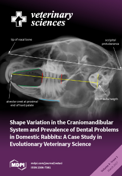
A remarkable peculiarity in veterinary medicine is the prevalence of dental problems among pet rabbits. In contrast to wild lagomorphs, almost 90% of reported pet rabbit patients suffer from malocclusion caused by tooth overgrowth. The present study suggests diet-induced phenotypic plasticity in skull morphology of domestic rabbits affecting mastication performance and, hence, oral health. “The geometric morphometric analysis reveals that the skull shape strongly differs between domestic and wild rabbits” says Dr. Christine Böhmer Since diet is known to influence skull morphology, this suggests that dietary habits are a major factor contributing to acquired malocclusions. “These results strengthen the importance to offer pet rabbits an adequate close-to-nature nutrition throughout the whole life and especially beginning early parallel to weaning (phase of increased phenotypic plasticity)” explains Dr. [...] Read more.
  • Issues are regarded as officially published after their release is announced to the table of contents alert mailing list.
  • You may sign up for e-mail alerts to receive table of contents of newly released issues.
  • PDF is the official format for papers published in both, html and pdf forms. To view the papers in pdf format, click on the "PDF Full-text" link, and use the free Adobe Reader to open them.
Order results
Result details
Section
Select all
Export citation of selected articles as:
294 KiB  
Review
Genomic Insights into Cardiomyopathies: A Comparative Cross-Species Review
by Siobhan Simpson, Paul Rutland and Catrin Sian Rutland
Vet. Sci. 2017, 4(1), 19; https://doi.org/10.3390/vetsci4010019 - 21 Mar 2017
Cited by 51 | Viewed by 8773
Abstract
In the global human population, the leading cause of non-communicable death is cardiovascular disease. It is predicted that by 2030, deaths attributable to cardiovascular disease will have risen to over 20 million per year. This review compares the cardiomyopathies in both human and [...] Read more.
In the global human population, the leading cause of non-communicable death is cardiovascular disease. It is predicted that by 2030, deaths attributable to cardiovascular disease will have risen to over 20 million per year. This review compares the cardiomyopathies in both human and non-human animals and identifies the genetic associations for each disorder in each species/taxonomic group. Despite differences between species, advances in human medicine can be gained by utilising animal models of cardiac disease; likewise, gains can be made in animal medicine from human genomic insights. Advances could include undertaking regular clinical checks in individuals susceptible to cardiomyopathy, genetic testing prior to breeding, and careful administration of breeding programmes (in non-human animals), further development of treatment regimes, and drugs and diagnostic techniques. Full article
(This article belongs to the Special Issue Comparison of Cardiovascular Systems and Diseases Across Species)
441 KiB  
Article
Effect of Oral Alpha Lipoic Acid in Preventing the Genesis of Canine Diabetic Cataract: A Preliminary Study
by David L. Williams
Vet. Sci. 2017, 4(1), 18; https://doi.org/10.3390/vetsci4010018 - 16 Mar 2017
Cited by 20 | Viewed by 7232
Abstract
Blinding cataract is a significant effect of canine diabetes with 75% of animals affected two years after diagnosis. Lens opacification occurs primarily through the generation of sorbitol, a sugar alcohol, through the action of aldose reductase (AR). The osmotic effect of sorbitol draws [...] Read more.
Blinding cataract is a significant effect of canine diabetes with 75% of animals affected two years after diagnosis. Lens opacification occurs primarily through the generation of sorbitol, a sugar alcohol, through the action of aldose reductase (AR). The osmotic effect of sorbitol draws water into the lens, causing opacification. Inhibition of AR should thus prevent the generation of cataracts. A topical AR inhibitor has been shown to have this effect, as has the commercially available neutraceutical OcuGLO, containing the AR inhibitor alpha lipoic acid (ALA) together with other plant-based antioxidants. Here a comparison is made between the number of diabetic dogs developing cataracts when given oral ALA alone and those given a mix containing ascorbic acid and tocopherol. Animals given ALA developed significantly fewer lens opacities than those given conventional antioxidants. Cataracts which formed occurred at a significantly greater duration after the commencement of treatment than those on the antioxidant mix. Although this is a small study conducted over a short period, the significant benefit of ALA in diabetic dogs is a reason to evaluate these effects in larger trials. As AR is involved in diabetic retinopathy and neuropathy, this enzyme inhibitor may be worthy of evaluation in preventing these conditions in human diabetics also. Full article
(This article belongs to the Special Issue Diabetes Mellitus in Companion Animals)
Show Figures

Figure 1

1063 KiB  
Article
Serum Fructosamine Concentration in Uncontrolled Hyperthyroid Diabetic Cats Is within the Population Reference Interval
by Arnon Gal, Brie Trusiano, Adrienne F. French, Nicolas Lopez-Villalobos and Amy L. MacNeill
Vet. Sci. 2017, 4(1), 17; https://doi.org/10.3390/vetsci4010017 - 15 Mar 2017
Cited by 7 | Viewed by 6545
Abstract
Diabetes mellitus is a common endocrinopathy of cats that is characterized by persistent fasting hyperglycemia. However, stress induces substantial hyperglycemia in cats that poses a challenge to the veterinarian who may wrongly interpret the high serum concentration of blood glucose as evidence of [...] Read more.
Diabetes mellitus is a common endocrinopathy of cats that is characterized by persistent fasting hyperglycemia. However, stress induces substantial hyperglycemia in cats that poses a challenge to the veterinarian who may wrongly interpret the high serum concentration of blood glucose as evidence of diabetes mellitus. Fructosamine is a glycated serum protein that serves as an index of glycemic control in cats and is useful because it is not affected by stress hyperglycemia. However, factors such as body weight, hypoproteinemia, and increased serum thyroid hormone concentration can alter fructosamine concentration. The goal of this retrospective study was to compare the fructosamine concentrations in diabetic and nondiabetic cats with and without uncontrolled hyperthyroidism. A secondary goal was to determine the effect of sex, age, different populations of cats, and diabetes on the variability of fructosamine. We found that the mean (±SE) serum fructosamine of hyperthyroid diabetic cats (332 ± 24 µmol/L, 95% CI 291–379 µmol/L) was within the population-based reference interval (200–360 µmol/L) and significantly lower in comparison to euthyroid diabetic cats (527 ± 10 µmol/L, 95% CI 515–553 µmol/L). Additionally, in this study, diabetes accounted only for approximately 50% of the variance in serum fructosamine, while age, sex, and population made a minor contribution to this variance. In conclusion, finding serum fructosamine that is within the population-based reference interval in an uncontrolled diabetic cat should alert the veterinarian to the possibility of concurrent hyperthyroidism. Additionally, the veterinary clinician should consider that serum fructosamine might be substantially affected by factors other than diabetes. Full article
(This article belongs to the Special Issue Diabetes Mellitus in Companion Animals)
Show Figures

Figure 1

502 KiB  
Article
Felis Catus Gammaherpesvirus 1 DNAemia in Whole Blood from Therapeutically Immunosuppressed or Retrovirus-Infected Cats
by Alicia J. McLuckie, Vanessa R. Barrs, Bethany Wilson, Mark E. Westman and Julia A. Beatty
Vet. Sci. 2017, 4(1), 16; https://doi.org/10.3390/vetsci4010016 - 14 Mar 2017
Cited by 44 | Viewed by 5280
Abstract
Gammaherpesviruses are major co-pathogens of human immunodeficiency virus (HIV) infection, making the interactions between feline immunodeficiency virus (FIV) and Felis catus gammaherpesvirus 1 (FcaGHV1) pertinent to both human and veterinary medical research. FIV-infected cats are at increased risk of FcaGHV1 DNAemia and consistently [...] Read more.
Gammaherpesviruses are major co-pathogens of human immunodeficiency virus (HIV) infection, making the interactions between feline immunodeficiency virus (FIV) and Felis catus gammaherpesvirus 1 (FcaGHV1) pertinent to both human and veterinary medical research. FIV-infected cats are at increased risk of FcaGHV1 DNAemia and consistently harbor higher FcaGHV1 loads than FIV-uninfected cats. Whether immune deficiencies unrelated to FIV are associated with similar risks is unknown. Using whole blood FcaGHV1 qPCR, we found no difference in the frequency of DNAemia or DNA load in therapeutically immunosuppressed (P1, n = 18) or feline leukemia virus (FeLV)-infected (P2, n = 57) patients compared with age- and sex-matched controls (C1, n = 58; C2, n = 57). In contrast, FIV/FeLV-co-infected cats (P3, n = 5) were at increased risk of FcaGHV1 DNAemia compared to retrovirus uninfected controls (C3, n = 39; p = 0.0068), and had a higher median FcaGHV1 DNA load, although the latter was not significant. FIV/FeLV-co-infected cats (P3) had a similar frequency of FcaGHV1 DNAemia reported compared to FIV-infected controls (C4). In conclusion, we found no evidence that cats with therapeutic immunosuppression or FeLV infection were at greater risk of FcaGHV1 DNAemia or had higher FcaGHV1 DNA load in whole blood. The risk of DNAemia in FIV/FeLV-co-infected cats was similar to that documented previously in cats infected with FIV alone. Full article
(This article belongs to the Special Issue Comparative Studies on HIV and FIV in Animals and Humans)
Show Figures

Figure 1

1044 KiB  
Article
The Awareness of the International Veterinary Profession of Evidence-Based Veterinary Medicine and Preferred Methods of Training
by Selene J. Huntley, Rachel S. Dean and Marnie L. Brennan
Vet. Sci. 2017, 4(1), 15; https://doi.org/10.3390/vetsci4010015 - 08 Mar 2017
Cited by 12 | Viewed by 6556
Abstract
Evidence-based veterinary medicine (EVM) is an evolving discipline in veterinary medicine so it is important to periodically “benchmark” opinion about EVM across the profession. An international survey to assess veterinarians’ awareness of EVM was conducted. Veterinarians were surveyed via an online questionnaire (all [...] Read more.
Evidence-based veterinary medicine (EVM) is an evolving discipline in veterinary medicine so it is important to periodically “benchmark” opinion about EVM across the profession. An international survey to assess veterinarians’ awareness of EVM was conducted. Veterinarians were surveyed via an online questionnaire (all countries) or a postal questionnaire (UK only). Participants were asked whether they had heard of EVM, where they had first heard the term, and their preferences of method for receiving continuing professional development (CPD). There were 6310 respondents, of which 4579 (72.5%) worked in the UK and 5384 (85.3%) were clinicians. Veterinarians that had heard of EVM (n = 5420, 85.9%) were most likely to be clinicians (OR = 4.00; 95% CI: 3.37, 4.75), respondents working in the UK (OR = 1.32; CI: 1.13, 1.54), or respondents with a postgraduate degree or qualification (OR = 1.77; CI: 1.51, 2.08). The most common sources from which respondents had heard of EVM were at vet school or university (n = 1207, 29.8%), via literature (peer-reviewed papers or other publications) (n = 1074, 26.5%), and via CPD courses (n = 564, 13.9%). Most respondents were interested in finding out more about EVM (n = 4256 of 6173, 69%). The preferred methods of CPD were day or evening seminars (n = 2992 of 6017, 49.7%), conferences (n = 1409, 23.4%), and online courses (n = 524, 8.7%), although the order of preference differed slightly between groups. There appears to be substantial awareness of EVM amongst veterinarians internationally. However, it appears that further training in EVM would be welcomed. Preferences on how CPD in general is received differs between groups, so this should be borne in mind by training providers when formulating a strategy for the dissemination of EVM training across the global profession. Full article
Show Figures

Figure 1

7759 KiB  
Review
Feline Immunodeficiency Virus Neuropathogenesis: A Model for HIV-Induced CNS Inflammation and Neurodegeneration
by Rick B. Meeker and Lola Hudson
Vet. Sci. 2017, 4(1), 14; https://doi.org/10.3390/vetsci4010014 - 06 Mar 2017
Cited by 17 | Viewed by 7962
Abstract
Feline Immunodeficiency virus (FIV), similar to its human analog human immunodeficiency virus (HIV), enters the central nervous system (CNS) soon after infection and establishes a protected viral reservoir. The ensuing inflammation and damage give rise to varying degrees of cognitive decline collectively known [...] Read more.
Feline Immunodeficiency virus (FIV), similar to its human analog human immunodeficiency virus (HIV), enters the central nervous system (CNS) soon after infection and establishes a protected viral reservoir. The ensuing inflammation and damage give rise to varying degrees of cognitive decline collectively known as HIV-associated neurocognitive disorders (HAND). Because of the similarities to HIV infection and disease, FIV has provided a useful model for both in vitro and in vivo studies of CNS infection, inflammation and pathology. This mini review summarizes insights gained from studies of early infection, immune cell trafficking, inflammation and the mechanisms of neuropathogenesis. Advances in our understanding of these processes have contributed to the development of therapeutic interventions designed to protect neurons and regulate inflammatory activity. Full article
(This article belongs to the Special Issue Comparative Studies on HIV and FIV in Animals and Humans)
Show Figures

Figure 1

2856 KiB  
Article
Immuno-Detection of C3a, a C3 Complement Activated Product in Mastitis Milk, a Potential Diagnostic Marker
by Thanislass Jacob, Gangasudan Subramani, Prathiba Sivaprakasam, Antony P. Xavier and Hirak K. Mukhopadhyay
Vet. Sci. 2017, 4(1), 13; https://doi.org/10.3390/vetsci4010013 - 23 Feb 2017
Cited by 61 | Viewed by 4173
Abstract
The sub-clinical form of mastitis is difficult to detect and causes huge economic loss to the dairy industry. It has become a threat to public health at large, thus there is a need for definite diagnosis of the disease. Therefore, this study was [...] Read more.
The sub-clinical form of mastitis is difficult to detect and causes huge economic loss to the dairy industry. It has become a threat to public health at large, thus there is a need for definite diagnosis of the disease. Therefore, this study was undertaken to identify the novel diagnostic marker for the detection of the sub-clinical form of mastitis. Two-dimensional gel analysis of the whey protein fraction of normal and mastitis milk samples revealed the presence of proteose peptone component 3 precursor, Trypsin precursor, complement component-C3, Ig heavy chain precursors and a C-type lectin domain as differentially expressed protein during the early stage of mastitis. Of these proteins identified, complement component-C3 was tested for its diagnostic potential. Western blot analysis of the milk whey of sub-clinical mastitis cases (M+, M++ & M+++) identified the accumulation of C3a, an activated product of complement component-C3. Further, the hemolytic activity of the above milk whey samples positively correlated with the somatic cell count. As C3a is already reported as an anaphylotoxic agent, it chemo tactically attracts lymphocytes at the site of inflammation, the detection of which in the milk whey can be of diagnostic importance for sub-clinical mastitis. Full article
Show Figures

Figure 1

1106 KiB  
Article
Micro-RNA 10a Is Increased in Feline T Regulatory Cells and Increases Foxp3 Protein Expression Following In Vitro Transfection
by Yan Wang, Mukta Nag, Joanne L. Tuohy and Jonathan E. Fogle
Vet. Sci. 2017, 4(1), 12; https://doi.org/10.3390/vetsci4010012 - 21 Feb 2017
Cited by 53 | Viewed by 5073
Abstract
CD4+CD25+Foxp3+ T regulatory (Treg) cells are activated during the course of lentiviral infection and exhibit heightened suppressor function when compared to Treg cells from uninfected controls. Foxp3 is essential to Treg cell function and multiple studies have documented [...] Read more.
CD4+CD25+Foxp3+ T regulatory (Treg) cells are activated during the course of lentiviral infection and exhibit heightened suppressor function when compared to Treg cells from uninfected controls. Foxp3 is essential to Treg cell function and multiple studies have documented that lentivirus-activated Treg cells exhibit heightened Foxp3 expression when compared to Treg cells from uninfected controls. Our hypothesis was that lentivirus-induced micro-RNAs (miRNAs) contribute to heightened Treg cell suppressor function by stabilizing Foxp3 expression. We demonstrated that CD4+CD25+ T cells from both feline immunodeficiency virus infected (FIV+) cats and uninfected control cats exhibit increased miRNA 10a and 21 levels compared to autologous CD4+CD25 T cells but there was no difference in the levels of these miRNAs when Treg cells from FIV+ cats were compared to Treg cells from uninfected controls. Further, there was no increase in Foxp3 mRNA following transfection of miRNA 10a or 21 into a feline cell line. However, transfection with miRNA 10a resulted in increased Foxp3 protein expression. Full article
(This article belongs to the Special Issue Comparative Studies on HIV and FIV in Animals and Humans)
Show Figures

Figure 1

1113 KiB  
Article
Protein Composition of the Bovine Herpesvirus 1.1 Virion
by Kaley A. Barber, Hillary C. Daugherty, Stephanie E. Ander, Victoria A. Jefferson, Leslie A. Shack, Tibor Pechan, Bindu Nanduri and Florencia Meyer
Vet. Sci. 2017, 4(1), 11; https://doi.org/10.3390/vetsci4010011 - 20 Feb 2017
Cited by 22 | Viewed by 6456
Abstract
Bovine herpesvirus (BoHV) type 1 is an important agricultural pathogen that infects cattle and other ruminants worldwide. Acute infection of the oro-respiratory tract leads to immune suppression and allows commensal bacteria to infect an otherwise healthy lower respiratory tract. This condition is known [...] Read more.
Bovine herpesvirus (BoHV) type 1 is an important agricultural pathogen that infects cattle and other ruminants worldwide. Acute infection of the oro-respiratory tract leads to immune suppression and allows commensal bacteria to infect an otherwise healthy lower respiratory tract. This condition is known as the Bovine Respiratory Disease (BRD). BoHV-1 latently infects the host for life and periodical stress events re-initiate BRD, translating into high morbidity and large economic losses. To gain a better understanding of the biology of BoHV-1 and the disease it causes, we elucidated the protein composition of extracellular virions using liquid chromatography-mass spectrometry analysis. We detected 33 viral proteins, including the expected proteins of the nucleocapsid and envelope as well as other regulatory proteins present in the viral tegument. In addition to viral proteins, we have also identified packaged proteins of host origin. This constitutes the first proteomic characterization of the BoHV virion. Full article
Show Figures

Figure 1

7138 KiB  
Case Report
Metastatic Squamous Cell Carcinoma in a Northern Brown Bandicoot (Isoodon macrourus)
by Amanda P. Beck, Amy L. Shima, Mark D. Bennett and Linda K. Johnson
Vet. Sci. 2017, 4(1), 10; https://doi.org/10.3390/vetsci4010010 - 14 Feb 2017
Cited by 16 | Viewed by 9361
Abstract
Aside from a handful of notable exceptions, neoplasia is not reported as a major cause of mortality in wild animal populations and often goes undetected. For northern brown bandicoots specifically, there are few reported tumors in the literature and on file in the [...] Read more.
Aside from a handful of notable exceptions, neoplasia is not reported as a major cause of mortality in wild animal populations and often goes undetected. For northern brown bandicoots specifically, there are few reported tumors in the literature and on file in the Australian Registry of Wildlife Health. This report describes a case of squamous cell carcinoma in a northern brown bandicoot (Isoodon macrourus), with metastases to the draining lymph nodes and lung. This neoplasm consisted predominantly of well-differentiated squamous cells and multifocal keratin pearls, with areas possibly consistent with epithelial to mesenchymal transition, as identified by positive immunohistochemical staining by both pancytokeratin (AE1/AE3) and vimentin. Additional investigations were negative for bandicoot papillomatosis carcinomatosis viruses. Full article
Show Figures

Figure 1

3020 KiB  
Review
Exercise-Induced Cardiac Remodeling: Lessons from Humans, Horses, and Dogs
by Rob Shave, Glyn Howatson, Dave Dickson and Lesley Young
Vet. Sci. 2017, 4(1), 9; https://doi.org/10.3390/vetsci4010009 - 12 Feb 2017
Cited by 46 | Viewed by 12208
Abstract
Physical activity is dependent upon the cardiovascular system adequately delivering blood to meet the metabolic and thermoregulatory demands of exercise. Animals who regularly exercise therefore require a well-adapted heart to support this delivery. The purpose of this review is to examine cardiac structure, [...] Read more.
Physical activity is dependent upon the cardiovascular system adequately delivering blood to meet the metabolic and thermoregulatory demands of exercise. Animals who regularly exercise therefore require a well-adapted heart to support this delivery. The purpose of this review is to examine cardiac structure, and the potential for exercise-induced cardiac remodeling, in animals that regularly engage in strenuous activity. Specifically, we draw upon the literature that has studied the “athlete’s heart” in humans, horses, and dogs, to enable the reader to compare and contrast cardiac remodeling in these three athletic species. The available literature provides compelling evidence for exercise-induced cardiac remodeling in all three species. However, more work is required to understand the influence of species/breed specific genetics and exercise-related hemodynamics, in order to fully understand the impact of exercise on cardiac structure. Full article
(This article belongs to the Special Issue Comparison of Cardiovascular Systems and Diseases Across Species)
Show Figures

Figure 1

6070 KiB  
Case Report
Hyponatremia as the Presenting Feature of a Pituitary Abscess in a Calf
by Jamie L. Stewart, Maria C. Bates, B. Wade Edwards and Brian M. Aldridge
Vet. Sci. 2017, 4(1), 8; https://doi.org/10.3390/vetsci4010008 - 06 Feb 2017
Cited by 4 | Viewed by 5094
Abstract
A 2-month-old Simmental heifer presented for acute onset of neurological behavior. Laboratory tests confirmed the presence of hyponatremia, hypochloremia, and hypokalemia that improved with intravenous fluid therapy. Despite an initial cessation of neurological signs, symptoms re-emerged, and the heifer was euthanized due to [...] Read more.
A 2-month-old Simmental heifer presented for acute onset of neurological behavior. Laboratory tests confirmed the presence of hyponatremia, hypochloremia, and hypokalemia that improved with intravenous fluid therapy. Despite an initial cessation of neurological signs, symptoms re-emerged, and the heifer was euthanized due to poor prognosis. A pituitary abscess (Trueperella pyogenes) was observed on gross necropsy, suggesting that the effects of panhypopituitarism (inappropriate anti-diuretic hormone (ADH), adrenocorticotropic hormone (ACTH), and/or thyroid-stimulating hormone (TSH) secretion) may have resulted in the clinical findings. Pituitary abscess syndrome carries a poor prognosis due to the inability to penetrate the area with systemic antibiotic therapy. These findings highlight the unusual clinical presentations that may occur following pituitary abscess syndrome in cattle that practitioners need to consider when determining prognosis. Full article
(This article belongs to the Special Issue Comparative studies on Endocrine Diseases in Animals and Humans)
Show Figures

Figure 1

855 KiB  
Review
The Comparative Value of Feline Virology Research: Can Findings from the Feline Lentiviral Vaccine Be Translated to Humans?
by Margaret J. Hosie, Navapon Techakriengkrai, Paweł M. Bęczkowski, Matthew Harris, Nicola Logan and Brian J. Willett
Vet. Sci. 2017, 4(1), 7; https://doi.org/10.3390/vetsci4010007 - 28 Jan 2017
Cited by 23 | Viewed by 5209
Abstract
Feline immunodeficiency virus (FIV) is a lentivirus of domestic cats that shares several similarities with its human counterpart, human immunodeficiency virus (HIV). Their analogies include genomic organization, lymphocyte tropism, viral persistence and induction of immunodeficiency. FIV is the only lentivirus for which a [...] Read more.
Feline immunodeficiency virus (FIV) is a lentivirus of domestic cats that shares several similarities with its human counterpart, human immunodeficiency virus (HIV). Their analogies include genomic organization, lymphocyte tropism, viral persistence and induction of immunodeficiency. FIV is the only lentivirus for which a commercial vaccine is registered for prevention in either human or veterinary medicine. This provides a unique opportunity to investigate the mechanisms of protection induced by lentivirus vaccines at the population level and might contribute to the development of efficacious HIV vaccines. As well as having comparative value for vaccine studies, FIV research has shed some light on the relationship between lentiviral tropism and pathogenesis. Recent studies in our laboratory demonstrated that the interaction between FIV and its primary receptor changes as disease progresses, reminiscent of the receptor switch observed as disease progresses in HIV infected individuals. Here we summarise findings illustrating that, in addition to its veterinary significance, FIV has comparative value, providing a useful model to explore lentivirus–host interactions and to examine potential immune correlates of protection against HIV infection. Full article
(This article belongs to the Special Issue Comparative Studies on HIV and FIV in Animals and Humans)
Show Figures

Figure 1

436 KiB  
Review
Review on Usage of Vancomycin in Livestock and Humans: Maintaining Its Efficacy, Prevention of Resistance and Alternative Therapy
by Panditharathnalage Nishantha Kumara Wijesekara, Wikum Widuranga Kumbukgolla, Jayaweera Arachchige Asela Sampath Jayaweera and Diwan Rawat
Vet. Sci. 2017, 4(1), 6; https://doi.org/10.3390/vetsci4010006 - 26 Jan 2017
Cited by 45 | Viewed by 19151
Abstract
Vancomycin is one of the “last-line” classes of antibiotics used in the treatment of life-threatening infections caused by Gram-positive bacteria. Even though vancomycin was discovered in the 1950s, it was widely used after the 1980s for the treatment of infections caused by methicillin-resistant [...] Read more.
Vancomycin is one of the “last-line” classes of antibiotics used in the treatment of life-threatening infections caused by Gram-positive bacteria. Even though vancomycin was discovered in the 1950s, it was widely used after the 1980s for the treatment of infections caused by methicillin-resistant Staphylococci, as the prevalence of these strains were increased. However, it is currently evident that vancomycin-resistant Staphylococcus aureus and vancomycin-resistant Enterococci have developed for various reasons, including the use of avaparcin—an analog of vancomycin—as a feed additive in livestock. Therefore, prophylactic and empiric use of antibiotics and their analogues need to be minimized. Herein we discuss the rational use of vancomycin in treating humans, horses, farm animals, and pet animals such as dogs, cats, and rabbits. In present day context, more attention should be paid to the prevention of the emergence of resistance to antibiotics in order to maintain their efficacy. In order to prevent emergence of resistance, proper guidance for the responsible use of antimicrobials is indispensable. Therefore, almost all stakeholders who use antibiotics should have an in-depth understanding of the antibiotic that they use. As such, it is imperative to be aware of the important aspects of vancomycin. In the present review, efforts have been made to discuss the pharmacokinetics and pharmacodynamics, indications, emergence of resistance, control of resistance, adverse effects, and alternative therapy for vancomycin. Full article
Show Figures

Figure 1

3509 KiB  
Article
Shape Variation in the Craniomandibular System and Prevalence of Dental Problems in Domestic Rabbits: A Case Study in Evolutionary Veterinary Science
by Christine Böhmer and Estella Böhmer
Vet. Sci. 2017, 4(1), 5; https://doi.org/10.3390/vetsci4010005 - 24 Jan 2017
Cited by 24 | Viewed by 15023
Abstract
In contrast to wild lagomorphs, pet rabbits exhibit a noticeably high frequency of dental problems. Although dietary habits are considered as a major factor contributing to acquired malocclusions, the exact causes and interrelationships are still under debate. In this regard, an important aspect [...] Read more.
In contrast to wild lagomorphs, pet rabbits exhibit a noticeably high frequency of dental problems. Although dietary habits are considered as a major factor contributing to acquired malocclusions, the exact causes and interrelationships are still under debate. In this regard, an important aspect that has not been considered thoroughly to date is the effect of diet-induced phenotypic plasticity in skull morphology. Therefore, we conducted a geometric morphometric analysis on skull radiological images of wild and pet rabbits in order to quantify intraspecific variation in craniomandibular morphology. The statistical analyses reveal a significant morphological differentiation of the craniomandibular system between both groups. Furthermore, the analysis of covariance shows that the force-generating modules (cranium and mandible) vary independently from the force-receiving module (hypselodont teeth) in pet rabbits, which is in contrast to their wild relatives. Our findings suggest that the phenotypic changes in domestic rabbits impact mastication performance and, consequently, oral health. An adequate close-to-nature nutrition throughout the whole life and especially beginning early parallel to weaning (phase of increased phenotypic plasticity) is necessary to ensure a normal strain on the teeth by promoting physiological lateral gliding movements and avoiding direct axial loads. Full article
Show Figures

Graphical abstract

362 KiB  
Article
Exposure to Photoperiod-Melatonin-Induced, Sexually-Activated Rams after Weaning Advances the Resumption of Sexual Activity in Post-Partum Mediterranean Ewes Lambing in January
by José A. Abecia, Philippe Chemineau, Andrea Gómez, Carlos Palacios, Matthieu Keller and José A. Delgadillo
Vet. Sci. 2017, 4(1), 4; https://doi.org/10.3390/vetsci4010004 - 21 Jan 2017
Cited by 48 | Viewed by 5177
Abstract
This study was aimed to determine whether the presence of sexually stimulated rams by photoperiodic and melatonin treatments can advance the resumption of post-partum sexual activity in Mediterranean ewes lambing in January and weaned at the end of the breeding season at 41°N, [...] Read more.
This study was aimed to determine whether the presence of sexually stimulated rams by photoperiodic and melatonin treatments can advance the resumption of post-partum sexual activity in Mediterranean ewes lambing in January and weaned at the end of the breeding season at 41°N, in March. Rams were exposed to two months of long days (16 h light/day) and given three melatonin implants at the end of the long days (sexually-activated rams; SAR). Control rams (CR) were exposed to the natural photoperiod. Thirty-six ewes weaned on 25 February were assigned to one of two groups. From 1 March to 30 June, one group was housed with four SAR males (SAR-treated; n = 18), and the other group (CR-treated; n = 18) was housed with four unstimulated rams. Ovulation was assessed once per week based on plasma progesterone concentrations. Estrus was monitored daily by marks left on ewes by rams’ harnesses. SAR-treated ewes had a shorter (p < 0.01) weaning–first estrus interval than CR-treated ewes (61 ± 17 days vs. 102 ± 47 days; mean date of first estrus after weaning on April 26 and June 6, respectively). The proportion of the ewes ovulating in April or May was higher (p < 0.05) in the SAR-treated group than in the CR-treated group. SAR-treated ewes resumed estrous activity sooner than CR-treated ewes such that, in April, May, and June, the proportion of females that exhibited estrus was higher (p < 0.01) in the SAR-treated group (72%, 89%, and 100%, respectively) than in the CR-treated group (17%, 44%, and 61%, respectively). In conclusion, the introduction at weaning of sexually activated rams advanced the resumption of estrous activity in ewes in spring. The practical implications of this work could be important in ewes adapted for intensive production and accelerated lambing systems. Full article
Show Figures

Figure 1

162 KiB  
Editorial
Acknowledgement to Reviewers of Veterinary Sciences in 2016
by Veterinary Sciences Editorial Office
Vet. Sci. 2017, 4(1), 3; https://doi.org/10.3390/vetsci4010003 - 10 Jan 2017
Cited by 23 | Viewed by 2870
Abstract
The editors of Veterinary Sciences would like to express their sincere gratitude to the following reviewers for assessing manuscripts in 2016.[...] Full article
1392 KiB  
Article
Detection of Leptospiral DNA in the Urine of Donkeys on the Caribbean Island of Saint Kitts
by Bernard Grevemeyer, Michel Vandenplas, Brittney Beigel, Ellen Cho, Arve Lee Willingham and Ashutosh Verma
Vet. Sci. 2017, 4(1), 2; https://doi.org/10.3390/vetsci4010002 - 10 Jan 2017
Cited by 25 | Viewed by 4693
Abstract
Leptospirosis is a global zoonosis caused by pathogenic spirochetes classified within the genus Leptospira. Leptospires live in the proximal renal tubules of reservoir or chronic carrier animals, and are shed in the urine. Naïve animals acquire infection either when they come in [...] Read more.
Leptospirosis is a global zoonosis caused by pathogenic spirochetes classified within the genus Leptospira. Leptospires live in the proximal renal tubules of reservoir or chronic carrier animals, and are shed in the urine. Naïve animals acquire infection either when they come in direct contact with a reservoir or infected animals or by exposure to environmental surface water or soil that is contaminated with their urine. In this study, urine samples from a herd of donkeys on the Caribbean island of St. Kitts were screened using a TaqMan-based real-time quantitative polymerase chain reaction (qPCR) targeting a pathogen-specific leptospiral gene, lipl32. Out of 124 clinically normal donkeys, 22 (18%) tested positive for leptospiral DNA in their urine. Water samples from two water troughs used by the donkeys were also tested, but were found to be free from leptospiral contamination. Detection of leptospiral DNA in the urine of clinically healthy donkeys may point to a role that these animals play in the maintenance of the bacteria on St. Kitts. Full article
(This article belongs to the Special Issue Control, Prevention and Elimination of Zoonotic Diseases)
Show Figures

Figure 1

820 KiB  
Review
In Vitro Research Tools in the Field of Human Immediate Drug Hypersensitivity and Their Present Use in Small Animal Veterinary Medicine
by Sidonie N. Lavergne
Vet. Sci. 2017, 4(1), 1; https://doi.org/10.3390/vetsci4010001 - 22 Dec 2016
Cited by 15 | Viewed by 6098
Abstract
Drug hypersensitivity reactions (DHR) are immune-mediated idiosyncratic adverse drug events. Type I DHR are often referred to as “immediate” and involve B lymphocyte-secreted IgE that bind to the membrane of basophils and mast cells, inducing their degranulation. This review presents various in vitro [...] Read more.
Drug hypersensitivity reactions (DHR) are immune-mediated idiosyncratic adverse drug events. Type I DHR are often referred to as “immediate” and involve B lymphocyte-secreted IgE that bind to the membrane of basophils and mast cells, inducing their degranulation. This review presents various in vitro tests that were developed in the field of human type I HS and implemented as clinical diagnostic tools in human cases of immediate DHR. The respective strengths and weaknesses of each test will be discussed in parallel of validation data such as specificity and sensitivity whenever available. Some of them have also been used as diagnostic tools in veterinary medicine, but not in cases of immediate DHR. Most of these diagnostic tools can be categorized into humoral and cellular tests. The former tests measure serum concentrations of factors, such as histamine, tryptase, and drug-specific IgE. The latter assays quantify markers of drug-induced basophil activation or drug-specific lymphocyte proliferation. Pharmacogenetic markers have also been investigated in immediate DHR, but not as extensively as in non-immediate ones. Throughout, practical aspects and limitations of the tests, as well as sensitivity and specificity parameters, will be presented. In addition, the experience of veterinary medicine with these diagnostic tools will be summarized. However, to date, none of them has ever been reported in a veterinary case of type I DHR. Full article
(This article belongs to the Special Issue Allergies in Animals and Humans)
Show Figures

Figure 1

Previous Issue
Next Issue
Back to TopTop