Review

Vertebrate Left-Right Asymmetry: What Can Nodal Cascade Gene Expression Patterns Tell Us?

Institute of Zoology, University of Hohenheim, 70593 Stuttgart, Germany
*
Author to whom correspondence should be addressed.
J. Cardiovasc. Dev. Dis. 2018, 5(1), 1; https://doi.org/10.3390/jcdd5010001
Submission received: 11 December 2017 / Revised: 25 December 2017 / Accepted: 25 December 2017 / Published: 29 December 2017
(This article belongs to the Special Issue Left–Right Asymmetry and Cardiac Morphogenesis)

Abstract

:
Laterality of inner organs is a wide-spread characteristic of vertebrates and beyond. It is ultimately controlled by the left-asymmetric activation of the Nodal signaling cascade in the lateral plate mesoderm of the neurula stage embryo, which results from a cilia-driven leftward flow of extracellular fluids at the left-right organizer. This scenario is widely accepted for laterality determination in wildtype specimens. Deviations from this norm come in different flavors. At the level of organ morphogenesis, laterality may be inverted (situs inversus) or non-concordant with respect to the main body axis (situs ambiguus or heterotaxia). At the level of Nodal cascade gene activation, expression may be inverted, bilaterally induced, or absent. In a given genetic situation, patterns may be randomized or predominantly lacking laterality (absence or bilateral activation). We propose that the distributions of patterns observed may be indicative of the underlying molecular defects, with randomizations being primarily caused by defects in the flow-generating ciliary set-up, and symmetrical patterns being the result of impaired flow sensing, on the left, the right, or both sides. This prediction, the reasoning of which is detailed in this review, pinpoints functions of genes whose role in laterality determination have remained obscure.

1. Introduction

The development of the left-right body axis in an otherwise bilaterally symmetrical organism has drawn the attention of anatomists and developmental biologists alike at all times [1,2,3]. Asymmetric positioning and morphogenesis of many organs (situs solitus), including heart, lung, gut, liver, and stomach, is relevant to humans, as deviations from the norm impact on health or are even incompatible with life. A mirror-imaged organ placement (situs inversus), however, mostly lacks clinical relevance, in stark contrast to the misplacement of single organs (situs ambiguus or heterotaxia) or duplications of sided arrangements (left or right isomerism), which often cause embryonic lethality [4]. The molecular analysis of left-right (LR) development was initiated by the identification of left-asymmetrically expressed genes during neurula stages of chick, mouse, and frog embryos [5,6,7]. The TGFβ-type morphogene Nodal, its secreted feedback inhibitor Lefty, and the homeobox transcription factor Pitx2 are activated in a sided manner in the left lateral plate mesoderm (LPM) and make up the so-called Nodal cascade. Once activated, Nodal signaling directly induces its own transcription, resulting in the fast spreading of nodal mRNA expression in the LPM [8]. In addition, Nodal activates lefty and pitx2 transcription. Nodal expression vanishes after a very short time, due to the feedback inhibition by Lefty, whereas left-sided pitx2 expression is maintained up to organ morphogenesis, which is initiated right after Nodal has disappeared from the LPM [9]. Several mutants affect this narrow timing, such as, for example, dand5 in the mouse [10]. Loss-of-function experiments either targeting Nodal signaling or Pitx2 resulted in aberrant organ situs in all animal model systems analyzed, demonstrating the conserved requirement of the Nodal-Lefty-Pitx2 cassette for LR development [5,11]. The morphogen nodal, known to act as a long range signaling molecule, is further restricted by Lefty expression at the midline. Defective midline Lefty enables Nodal to diffuse to the right LPM and to activate a right-sided cascade, resulting in LR defects as well [12]. At an evolutionary scale, the Nodal cascade governs asymmetries in the entire deuterostome tree of life, from sea urchins to mammals [13,14].
Besides evolutionary implications, the presence of the Nodal cascade at the center of events leading up to asymmetric organ placement and morphogenesis has enabled the analysis of processes upstream and downstream [15,16]. A relatively strong correlation between organ situs and Nodal cascade induction has been observed in many mouse mutants, best exemplified by the classical LR mutant inversus viscerum, or iv [17,18,19]. Nodal cascade induction is completely randomized in iv mutant embryos, with equal 25% frequencies of wildtype, absent, bilateral and right-sided expression patterns each (Figure 1). The organs of homozygous iv mutants are either wt (situs solitus; SS), inverted (situs inversus; SI), or mirror images of the left or right body half (left or right isomerism) [17,18,19], although deviations from this predicted distribution were recorded as well [20]. iv specimens with isomerisms die in utero, while both SS and SI animals are born and vital. This correlation is less clear in other mutants, especially when aberrant organ situs is only encountered in a minority of cases. Compensation of germ line mutations is only beginning to be studied, let alone to be understood, but may contribute in cases when Nodal cascade expression patterns and organ situs correlate less well [21,22]. Lower vertebrates, i.e., fish and frog, for the most parts lack isomerisms at the organ level. Absent or bilateral expression of Nodal cascade genes therefore result in the same set of phenotypes, situs solitus, situs inversus, and situs ambiguus (heterotaxia). This observation suggests that gastrointestinal and heart primordia of lower vertebrates are able to undergo an intrinsic chiral morphogenesis, leading to asymmetric organ placement even when positional information by the Nodal cascade is lacking or present on both sides. Up to now, these differences of asymmetric organ morphogenesis between species have not been systematically addressed. When considering all cases, however, it seems safe to hypothesize that Nodal cascade gene expression patterns in the LPM are indicative of organ situs development. In the context of the present discussion, we shall therefore exclusively consider Nodal cascade patterns as indicators of upstream mechanisms. In the following, we will take what may be considered a somewhat unusual approach of a more theoretical look at concepts of flow generation, sensing and signal transfer. In some cases, this will result in over-simplifications that neglect the complexity of phenotypes encountered in mutants and morphants, for which we apologize, as we do to all colleagues whose work we could not cite due to space restrictions.

2. Cilia-Dependent Symmetry Breaking
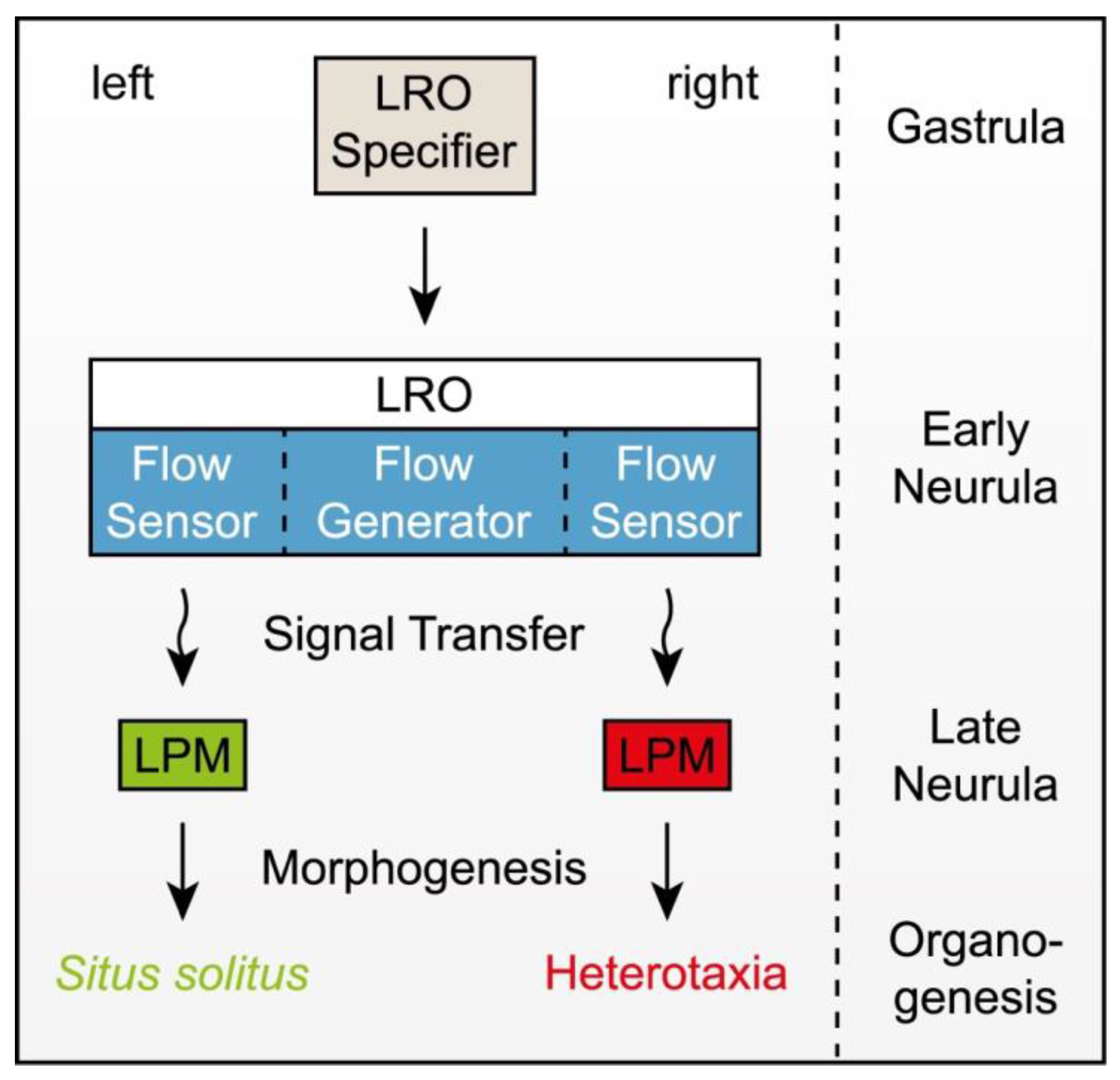
While the mechanisms of asymmetric organ morphogenesis downstream of the Nodal cascade are only slowly beginning to be unraveled (cf. [16] for a recent review), upstream events have received much attention in the various model organisms and a great deal of knowledge has accumulated in recent years [15]. In a sense, the iv mutant again has paved the way: the identification of its target gene as an axonemal dynein motor has introduced cilia as a central player, with a multitude of ciliary mutants being involved in LR development and a great many of ciliopathies displaying LR defects as one of their characteristics [18]. In short, the situation as of today presents as follows (Figure 2): in the perfectly bilateral symmetrical neurula embryo, the ciliated left-right organizer (LRO) forms at the midline. Precursors arise during gastrulation and are patterned by the embryonic organizer (Figure 2). The LRO creates an extracellular fluid flow from the right to the left side. This leftward flow may or may not transport cargo, which should originate from within the LRO. The LRO harbors flow sensors at the margins on both sides, which react to flow and/or to the cargo when it gets delivered to the sensor. The sensor generates a signal that gets transmitted to the LPM, where it induces the Nodal cascade. Any disturbance of this sequence of events, which together rules symmetry breaking in the embryo, inevitably results in alterations of LPM Nodal cascade expression. However, the type of alteration observed does not necessarily pinpoint the underlying defects in symmetry breaking. For example, it is not easily reconciled that in a mutant such as iv, which renders cilia immotile one quarter of specimens develop without any LR defects, although left LPM Nodal in iv is not induced by the exactly same mechanism as in wildtype embryos [23,24]. Such cases have in the past led to the proposal that it is utterly impossible that cilia take center stage in symmetry breaking [25,26,27], and they continue to plague the field, as honestly nobody knows how left-asymmetric Nodal is induced in the absence of ciliary motility or in any of the other mutants that eventually show situs solitus in at least a proportion of homozygous specimens.
We propose that it is profitable to think about disturbances of distinct steps in the chain of events that account for symmetry breaking, and to predict what kind of alterations of Nodal cascade gene expression patterns should result. Some mutants will fall in one or another category, in some other cases combinations of defects may cause a specific pattern.

2.1. LRO Specifier

To start the discussion, we first like to consider global LRO specifiers, i.e., mutants in which an LRO does not form at all. So far, LRO precursors have only been identified in fish and frog with the dorsal forerunner cells and the superficial mesoderm, respectively [28,29]. From the timing and geometry of embryonic development, the generation of these LRO specifiers must be intimately linked to the function of the primary embryonic (or Spemann) organizer. LROs inevitably localize to the posterior end of the notochord, which develops from the organizer [30]. Interestingly, mechanical ablations of frog and fish LROs have not impacted on embryonic development beyond laterality determination, demonstrating that symmetry breaking is a distinct event [31,32,33]. Quite obviously, if no LRO is specified, then the Nodal cascade should not be induced, i.e., LPM gene expression should be absent. Several mutants are known in which LRO formation is distorted, but only one in which this ciliated epithelium does not form at all: brachyury. To no surprise, the LPM Nodal cascade is not induced, and this is true for mouse mutants [34,35,36] as well as frog morphants (SK and MB, unpublished. In zebrafish, although the LRO is missing (with flow generator and sensor being absent, see below), LPM nodal is induced (REF), which remains a mystery. Another prominent gene that affects LRO morphogenesis is noto [32,37]. In contrast to brachyury, LRO remnants form, for example, in zebrafish, where Kupffer’s vesicle is present but much reduced in size [28]. In this case, the Nodal cascade is induced in a bilateral manner, i.e., both in the left and right LPM. Interestingly, LRO specifier genes are generally required for notochord formation, in agreement with the notion that LRO and notochord morphogenesis are intimately linked [30]. The exact pathway how these genes set up this unique tissue during gastrulation has not been elucidated as yet, but mutants that abrogate LRO formation should always result in the absence of Nodal cascade gene induction.

2.2. Flow Generator

We next consider flow generators, i.e., cells that harbor motile cilia. These are found at the center of the LRO and may be present or absent and harbor cilia or not. If cilia are present, then they may be motile or immotile, of correct length, morphology, and posterior polarization, or not. As a result, leftward flow may proceed with normal speed and directionality or deviate from the norm. All of the deviations should result in altered LPM expression patterns. But what is to be expected in every single case? Mispolarized cilia should produce fluid flows that are directed to other dimensions than left-only. In such settings, the Nodal cascade might be induced on the right side. No case is known in which all cilia polarize in the wrong (anterior) direction and therefore invert the cascade, but several genes are known that result in a proportion of anteriorly polarized cilia. In such cases, which comprise some of the planar cell polarity genes, such as vangl1/2 and wnt11, as well as the ion pump ATP4 [38,39,40,41], bilateral induction of the Nodal cascade has been reported (Table 1). Variations in cilia lengths cause altered flow dynamics and LR defects, as recently shown in zebrafish [42]. Deviations in speed of leftward flow have not systematically been studied, beyond reports of aberrant velocities in mutants, such as inv [23]. As flow is only required on the left side of the LRO [43], and a few cilia are sufficient to break symmetry [44], the vigor of flow may not be of too much relevance. However, the case is open; studies that assess physical flow parameters are few, and it may be profitable to re-investigate mutants that otherwise are hard to interpret. Absence and immobility of cilia have, however, been analyzed. Both should result in the absence of Nodal cascade induction, which is, however, not observed. Immotile cilia, such as in iv mutant embryos, cause randomized nodal1 expression, which is counterintuitive, while mutants that lack cilia, such as kif3a [45] and ift88 [46,47,48], display bilateral expression, which asks for a mechanistic explanation as well (and we shall come back to both scenarios below).

2.3. Flow Cargo

The question whether or not there is cargo that flow transports to the left has been controversially discussed in the field for more than a decade. Early evidence for the presence of so-called Nodal vesicular parcels, or NVPs [49,50], have not been followed up until recently, when the prevailing view that flow itself triggers the flow sensor [51,52] it has been challenged [53,54], but published novel evidence on this issue is still missing. For our hypothetical discussion, the presence, nature and mode of action of NVPs are irrelevant, though. If it were and cargo would be traveling along with leftward flow, this cargo could be present or absent, and it could reach the flow sensors in the presence or absence of leftward flow, for example by diffusion. This is when cases of randomized induction of the Nodal cascade become interesting. In the iv mutant, for example, cargo could be released at the LRO and reach the flow sensors on the left and right margin of the LRO in a stochastic manner. This scenario is still not totally satisfying, as the distribution of cases (25% each) asks for a mechanistic explanation. Taking some sort of cargo into account, however, helps the discussion. But, why should absence of cilia, such as in the kif3a or ift88 mutants, be different and result in bilateral induction? In that case, one would need to argue that cargo release and the presence of cilia are coupled. There are several possible scenarios, such as, for example, cargo release from the tip of cilia (in form of exosomes). Care should be applied, however, as cilia mutants impact on very many signaling pathways, some of which may impact on LR patterning downstream of Nodal. Anyways, the cargo option in our opinion should be considered in cases of randomized Nodal expression patterns in the LPM, and the nearer future should tell whether NVPs exist beyond mouse, and when and how they act.

2.4. Flow Sensor

The flow sensor should be intimately linked to flow and cargo. In the absence of its trigger, the sensor should not be activated, i.e., Nodal should not be induced. In order to assure robust induction of the Nodal cascade on the left and not the right side of the LPM, the trigger must be locked in the absence of flow and/or cargo. The sensor could be present or absent, and it could be activated on the left, the right, on both or neither side. There should be an absolute correlation between sensor status (on/off) and Nodal cascade activation in the LPM at a slightly later stage. Conceptually, the sensor is the simplest player in the game, as it is this main gateway that should decide on laterality specification [15]. Experimental manipulations of the sensor should overrule all of the upstream events and should be autonomously capable of inducing the cascade on the side where it is touched off. Central to the sensor function is Nodal and its inhibitor Dand5 (formerly known as Coco in frog, charon in zebrafish, or cerl2 in mouse); both are co-expressed in the sensory cells at the LRO margin [55,56,57,58,59,60], or in neighboring cells in the case of zebrafish, where the transformation of the flat epithelium of primitive fish LRO, such as in the sturgeon [30], into the sphere of Kupffer’s vesicle (KV) probably placed these cells just next to the KV. Experimental manipulations of both factors in these very cells underscore their central role: Nodal is strictly required on the left side of the LRO. Its presence in the sensor on the right is not required for left-sided LPM activation. Dand5, in contrast, has to be repressed on the left in order to de-repress Nodal action. When Dand5 disappears from the right sensor and Nodal is present, the cascade gets activated on both sides. Nodal and Dand5 are able to overrule a loss of the flow generator: flow-independent repression of Dand5 activates Nodal, as does the overexpression of Nodal itself. Dand5 depends on Nodal as the left determinant, as in the absence of Nodal in the sensor, Dand5 manipulations remain without consequences. Artificial flow can trigger the sensor in the absence of cilia or ciliary motility [51,61], demonstrating that flow is the natural activator of the sensor, but whether or not this artificial flow carries along cargo remains to be seen. Sensor manipulations are extremely efficient, at least in the frog Xenopus, where manipulations can be performed in a sided manner, and in the mouse, reaching efficiencies of close to one hundred percent [58], numbers that are never encountered with any other experimental or genetic manipulation. How flow sets off the sensor, i.e., represses Dand5, is a matter of intense research. Genes that fulfill characteristics, such as Nodal and Dand5, qualify for this process, i.e., flow sensing: they should be highly efficient in knockout or knockdown situations and they should act in a strictly sided manner. Screening the literature for factors complying with these criteria might uncover genes that are involved in sensor function; pkd2 and pkd1l1 are certainly good examples [62,63].

2.5. Signal Transfer

Finally, the signal that is generated in the sensor needs to transfer from the LRO to the LPM. The transfer system must be present on either side, otherwise bilateral induction of the cascade would not be possible. In its absence, Nodal should never get activated in the LPM. However, contrary to the sensor, it strictly depends on the upstream chain of events and cannot be activated in the absence of the signal. The conceptually most parsimonious scenario, namely that Nodal itself gets transferred, is fully compatible with the available experimental data. While gdf1/gdf3, which is expressed in the sensor and complex with Nodal, is required for de-repressed Nodal to transfer to the LPM [60,64], more specific transfer mutants are not known, indicating that the mechanism relies on available cell biological characteristics such as the extracellular matrix or coupling of cells by gap junctions [65,66,67]. Mutations in any such functions would likely impact on other processes as well, obscuring an LR-specific function. Contrary to all the other functions that are discussed above, transfer is likely permissive in nature and not instructive, which is why Nodal cascade expression patterns in our opinion do not reveal a lot in that context.

2.6. Precautions

Besides the above-mentioned possibility of full or partial compensation of germline mutations [21], which might blur the function of a given gene when judged by the resulting LPM marker gene expression patterns, genes might act in more than one function, such as Nodal itself, which is required to set up the flow generator [73], in the sensor [74] and in the LPM [75,76]. Some ciliary genes certainly are required in the flow generator and the sensor as well, which has to be taken into account when analyzing the Nodal cascade. Despite these restrictions, we are convinced that a careful re-evaluation of LPM gene expression patterns might be beneficial to place factors in the progress of symmetry breaking, as does the assessment of novel factors that keep to be identified. Such analyses need of course be restricted to model organisms, in which a ciliated LRO has been demonstrated. In vertebrates, this excludes birds and reptiles, which likely have lost these mechanisms and have come up with an alternative strategy of left-asymmetric Nodal cascade induction (cf. [14] for a detailed discussion). Such a loss might even have happened in some mammalian groups, as indicated by the absence of a recently described novel LR determinant of unknown function, the matrix metalloproteinase Mmp21, in the cetartiodactyla, a systematic group of mammals that includes whales and even-toed ungulates [77]. That even-toed ungulates differ from other mammals has been previously suspected, as a ciliated LRO is absent in pig embryos [78].

3. Conclusions

In the more than 20 years that have passed since the first description of asymmetric gene expression in the left LPM of the chicken embryo [6], a great many of factors have been involved in the process of laterality determination. Despite all the progress, large gaps remain in our understanding of the molecular and cellular mechanisms that drive organ asymmetry. The majority of vertebrates use a cilia-dependent mode of symmetry breaking; in these cases, the assessment of Nodal cascade gene expression patterns in the LPM can tell us much about the underlying defects. We have argued here that randomized patterns are the result of defects in flow generation, while symmetric patterns arise from defective flow sensing. These different LPM patterns often are not discussed, or only very low cases numbers are reported when a new factor is presented in the literature; any deviation from the norm is taken as evidence for a general role in LR development. While this is correct, important information may be lost when the differences in efficacies and patterns are not pinpointed, evaluated, and weighed. The basic conceptual framework is there; we need to fill in the gaps. It should be profitable to discuss the options any result gives us, rather than concealing our uneasiness. The Nodal cascade has started the molecular analysis of LR analysis, and it remains at center stage now that we tackle the nature of flow generator and sensor.

Acknowledgments

Work in the Blum lab has been supported by DFG grants BL-285/9 and BL-285/10. S.K. and T.O. have been recipients of fellowships from the Landesgraduiertenförderung Baden-Württemberg. S.K. was also funded by the Federal Ministry of Education and Research (01PL11003), project Humboldt reloaded.

Author Contributions

A.S. had the idea for this review, which M.B. and A.S. wrote with the help of all authors. T.O. and S.K. assembled the Figures and M.T., M.M., F.F., S.K. and T.O. researched the data for the table.

Conflicts of Interest

The authors declare no conflict of interest.

References

  1. Dareste, C. Recherches sur la Production Artificielle des Monstruosités, ou Essais de Tératogénie Expérimentale; C. Reinwald: Paris, France, 1891. [Google Scholar]
  2. Fol, H.; Warynsky, S. Sur la production artificielle de l’inversion viscérale ou hétérotaxie chez des embryons de poulet. CR Acad. Sci. Paris 1883, 96, 1674–1676. [Google Scholar]
  3. Tisler, M.; Schweickert, A.; Blum, M. Xenopus, an ideal model organism to study laterality in conjoined twins. Genesis 2017, 55. [Google Scholar] [CrossRef] [PubMed]
  4. Capdevila, J.; Vogan, K.J.; Tabin, C.J.; Izpisúa Belmonte, J.C. Mechanisms of left-right determination in vertebrates. Cell 2000, 101, 9–21. [Google Scholar] [CrossRef]
  5. Campione, M.; Steinbeisser, H.; Schweickert, A.; Deissler, K.; van Bebber, F.; Lowe, L.A.; Nowotschin, S.; Viebahn, C.; Haffter, P.; Kuehn, M.R.; et al. The homeobox gene Pitx2: Mediator of asymmetric left-right signaling in vertebrate heart and gut looping. Development 1999, 126, 1225–1234. [Google Scholar] [PubMed]
  6. Levin, M.; Johnson, R.L.; Sterna, C.D.; Kuehn, M.; Tabin, C. A molecular pathway determining left-right asymmetry in chick embryogenesis. Cell 1995, 82, 803–814. [Google Scholar] [CrossRef]
  7. Nakamura, T.; Hamada, H. Left-right patterning: Conserved and divergent mechanisms. Development 2012, 139, 3257–3262. [Google Scholar] [CrossRef] [PubMed]
  8. Nakamura, T.; Mine, N.; Nakaguchi, E.; Mochizuki, A.; Yamamoto, M.; Yashiro, K.; Meno, C.; Hamada, H. Generation of robust left-right asymmetry in the mouse embryo requires a self-enhancement and lateral-inhibition system. Dev. Cell 2006, 11, 495–504. [Google Scholar] [CrossRef] [PubMed]
  9. Shiratori, H. Conserved regulation and role of Pitx2 in situs-specific morphogenesis of visceral organs. Development 2006, 133, 3015–3025. [Google Scholar] [CrossRef] [PubMed]
  10. Inácio, J.M.; Marques, S.; Nakamura, T.; Shinohara, K.; Meno, C.; Hamada, H.; Belo, J.A. The dynamic right-to-left translocation of Cerl2 is involved in the regulation and termination of Nodal activity in the mouse node. PLoS ONE 2013, 8, e60406. [Google Scholar] [CrossRef] [PubMed]
  11. Oki, S.; Kitajima, K.; Marques, S.; Belo, J.A.; Yokoyama, T.; Hamada, H.; Meno, C. Reversal of left-right asymmetry induced by aberrant Nodal signaling in the node of mouse embryos. Development 2009, 136, 3917–3925. [Google Scholar] [CrossRef] [PubMed]
  12. Lohr, J.L.; Danos, M.C.; Groth, T.W.; Yost, H.J. Maintenance of asymmetric nodal expression in Xenopus laevis. Dev. Genet. 1998, 23, 194–202. [Google Scholar] [CrossRef]
  13. Tisler, M.; Wetzel, F.; Mantino, S.; Kremnyov, S.; Thumberger, T.; Schweickert, A.; Blum, M.; Vick, P. Cilia are required for asymmetric nodal induction in the sea urchin embryo. BMC Dev. Biol. 2016, 16, 28. [Google Scholar] [CrossRef] [PubMed]
  14. Blum, M.; Feistel, K.; Thumberger, T.; Schweickert, A. The evolution and conservation of left-right patterning mechanisms. Development 2014, 141, 1603–1613. [Google Scholar] [CrossRef] [PubMed]
  15. Blum, M.; Schweickert, A.; Vick, P.; Wright, C.V.E.; Danilchik, M.V. Symmetry breakage in the vertebrate embryo: When does it happen and how does it work? Dev. Biol. 2014, 393, 109–123. [Google Scholar] [CrossRef] [PubMed]
  16. Grimes, D.T.; Burdine, R.D. Left–Right Patterning: Breaking Symmetry to Asymmetric Morphogenesis. Trends Genet. 2017, 33, 616–628. [Google Scholar] [CrossRef] [PubMed]
  17. Supp, D.M.; Brueckner, M.; Kuehn, M.R.; Witte, D.P.; Lowe, L.A.; McGrath, J.; Corrales, J.; Potter, S.S. Targeted deletion of the ATP binding domain of left-right dynein confirms its role in specifying development of left-right asymmetries. Development 1999, 126, 5495–5504. [Google Scholar] [PubMed]
  18. Supp, D.M.; Witte, D.P.; Potter, S.S.; Brueckner, M. Mutation of an axonemal dynein affects left-right asymmetry in inversus viscerum mice. Nature 1997, 389, 963–966. [Google Scholar] [CrossRef] [PubMed]
  19. Lowe, L.A.; Supp, D.M.; Sampath, K.; Yokoyama, T.; Wright, C.V.; Potter, S.S.; Overbeek, P.; Kuehn, M.R. Conserved left-right asymmetry of nodal expression and alterations in murine situs inversus. Nature 1996, 381, 158–161. [Google Scholar] [CrossRef] [PubMed]
  20. Seo, J.W.; Brown, N.A.; Ho, S.Y.; Anderson, R.H. Abnormal laterality and congenital cardiac anomalies. Relations of visceral and cardiac morphologies in the iv/iv mouse. Circulation 1992, 86, 642–650. [Google Scholar] [CrossRef] [PubMed]
  21. Rossi, A.; Kontarakis, Z.; Gerri, C.; Nolte, H.; Hölper, S.; Krüger, M.; Stainier, D.Y. Genetic compensation induced by deleterious mutations but not gene knockdowns. Nature 2015, 524, 230–233. [Google Scholar] [CrossRef] [PubMed]
  22. Blum, M.; De Robertis, E.M.; Wallingford, J.B.; Niehrs, C. Morpholinos: Antisense and Sensibility. Dev. Cell 2015, 35, 145–149. [Google Scholar] [CrossRef] [PubMed]
  23. Okada, Y.; Nonaka, S.; Tanaka, Y.; Saijoh, Y.; Hamada, H.; Hirokawa, N. Abnormal nodal flow precedes situs inversus in iv and inv mice. Mol. Cell 1999, 4, 459–468. [Google Scholar] [CrossRef]
  24. Grimes, D.T.; Keynton, J.L.; Buenavista, M.T.; Jin, X.; Patel, S.H.; Kyosuke, S.; Vibert, J.; Williams, D.J.; Hamada, H.; Hussain, R.; et al. Genetic Analysis Reveals a Hierarchy of Interactions between Polycystin-Encoding Genes and Genes Controlling Cilia Function during Left-Right Determination. PLoS Genet. 2016, 12, e1006070. [Google Scholar] [CrossRef] [PubMed]
  25. Vandenberg, L.N.; Levin, M. Consistent left-right asymmetry cannot be established by late organizers in Xenopus unless the late organizer is a conjoined twin. Development 2010, 137, 1095–1105. [Google Scholar] [CrossRef] [PubMed]
  26. Vandenberg, L.N.; Levin, M. Far from solved: A perspective on what we know about early mechanisms of left-right asymmetry. Dev. Dyn. 2010, 239, 3131–3146. [Google Scholar] [CrossRef] [PubMed]
  27. Vandenberg, L.N.; Levin, M. Perspectives and open problems in the early phases of left-right patterning. Semin. Cell Dev. Biol. 2009, 20, 456–463. [Google Scholar] [CrossRef] [PubMed]
  28. Melby, A.E.; Warga, R.M.; Kimmel, C.B. Specification of cell fates at the dorsal margin of the zebrafish gastrula. Development 1996, 122, 2225–2237. [Google Scholar] [PubMed]
  29. Shook, D.R.; Majer, C.; Keller, R. Pattern and morphogenesis of presumptive superficial mesoderm in two closely related species, Xenopus laevis and Xenopus tropicalis. Dev. Biol. 2004, 270, 163–185. [Google Scholar] [CrossRef] [PubMed]
  30. Blum, M.; Andre, P.; Muders, K.; Schweickert, A.; Fischer, A.; Bitzer, E.; Bogusch, S.; Beyer, T.; van Straaten, H.W.; Viebahn, C. Ciliation and gene expression distinguish between node and posterior notochord in the mammalian embryo. Differentiation 2007, 75, 133–146. [Google Scholar] [CrossRef] [PubMed]
  31. Bajoghli, B.; Aghaallaei, N.; Soroldoni, D.; Czerny, T. The roles of Groucho/Tle in left-right asymmetry and Kupffer’s vesicle organogenesis. Dev. Biol. 2007, 303, 347–361. [Google Scholar] [CrossRef] [PubMed]
  32. Essner, J.J.; Amack, J.D.; Nyholm, M.K.; Harris, E.B.; Yost, H.J. Kupffer’s vesicle is a ciliated organ of asymmetry in the zebrafish embryo that initiates left-right development of the brain, heart and gut. Development 2005, 132, 1247–1260. [Google Scholar] [CrossRef] [PubMed]
  33. Blum, M.; Beyer, T.; Weber, T.; Vick, P.; Andre, P.; Bitzer, E.; Schweickert, A. Xenopus, an ideal model system to study vertebrate left-right asymmetry. Dev. Dyn. 2009, 238, 1215–1225. [Google Scholar] [CrossRef] [PubMed]
  34. King, T.; Beddington, R.S.; Brown, N.A. The role of the brachyury gene in heart development and left-right specification in the mouse. Mech. Dev. 1998, 79, 29–37. [Google Scholar] [CrossRef]
  35. Amack, J.D.; Yost, H.J. The T box transcription factor no tail in ciliated cells controls zebrafish left-right asymmetry. Curr. Biol. 2004, 14, 685–690. [Google Scholar] [CrossRef] [PubMed]
  36. Concepcion, D.; Papaioannou, V.E. Nature and extent of left/right axis defects in TWis/TWis mutant mouse embryos. Dev. Dyn. 2014, 243, 1046–1053. [Google Scholar] [CrossRef] [PubMed]
  37. Beckers, A.; Alten, L.; Viebahn, C.; Andre, P.; Gossler, A. The mouse homeobox gene Noto regulates node morphogenesis, notochordal ciliogenesis, and left right patterning. Proc. Natl. Acad. Sci. USA 2007, 104, 15765–15770. [Google Scholar] [CrossRef] [PubMed]
  38. Walentek, P.; Beyer, T.; Thumberger, T.; Schweickert, A.; Blum, M. ATP4a Is Required for Wnt-Dependent Foxj1 Expression and Leftward Flow in Xenopus Left-Right Development. Cell Rep. 2012, 1, 516–527. [Google Scholar] [CrossRef] [PubMed]
  39. Walentek, P.; Schneider, I.; Schweickert, A.; Blum, M. Wnt11b is involved in cilia-mediated symmetry breakage during Xenopus left-right development. PLoS ONE 2013, 8, e73646. [Google Scholar] [CrossRef] [PubMed]
  40. Song, H.; Hu, J.; Chen, W.; Elliott, G.; Andre, P.; Gao, B.; Yang, Y. Planar cell polarity breaks bilateral symmetry by controlling ciliary positioning. Nature 2010, 466, 378–382. [Google Scholar] [CrossRef] [PubMed]
  41. Antic, D.; Stubbs, J.L.; Suyama, K.; Kintner, C.; Scott, M.P.; Axelrod, J.D. Planar cell polarity enables posterior localization of nodal cilia and left-right axis determination during mouse and Xenopus embryogenesis. PLoS ONE 2010, 5, e8999. [Google Scholar] [CrossRef] [PubMed]
  42. Pintado, P.; Sampaio, P.; Tavares, B.; Montenegro-Johnson, T.D.; Smith, D.J.; Lopes, S.S. Dynamics of cilia length in left-right development. R. Soc. Open Sci. 2017, 4, 161102. [Google Scholar] [CrossRef] [PubMed]
  43. Vick, P.; Schweickert, A.; Weber, T.; Eberhardt, M.; Mencl, S.; Shcherbakov, D.; Beyer, T.; Blum, M. Flow on the right side of the gastrocoel roof plate is dispensable for symmetry breakage in the frog Xenopus laevis. Dev. Biol. 2009, 331, 281–291. [Google Scholar] [CrossRef] [PubMed]
  44. Kawasumi, A.; Takamatsu, A.; Yoshiba, S.; Botilde, Y.; Motoyama, N.; Reith, W.; Durand, B.; Shiratori, H.; Hamada, H. Two rotating cilia in the node cavity are sufficient to break left–right symmetry in the mouse embryo. Nat. Commun. 2011, 3, 622–628. [Google Scholar] [CrossRef]
  45. Takeda, S.; Yonekawa, Y.; Tanaka, Y.; Okada, Y.; Nonaka, S.; Hirokawa, N. Left-right asymmetry and kinesin superfamily protein KIF3A: New insights in determination of laterality and mesoderm induction by kif3A-/- mice analysis. J. Cell Biol. 1999, 145, 825–836. [Google Scholar] [CrossRef] [PubMed]
  46. Bisgrove, B.W.; Snarr, B.S.; Emrazian, A.; Yost, H.J. Polaris and Polycystin-2 in dorsal forerunner cells and Kupffer’s vesicle are required for specification of the zebrafish left-right axis. Dev. Biol. 2005, 287, 274–288. [Google Scholar] [CrossRef] [PubMed]
  47. Taulman, P.D.; Haycraft, C.J.; Balkovetz, D.F.; Yoder, B.K. Polaris, a protein involved in left-right axis patterning, localizes to basal bodies and cilia. Mol. Biol. Cell 2001, 12, 589–599. [Google Scholar] [CrossRef] [PubMed]
  48. Yoder, B.K.; Tousson, A.; Millican, L.; Wu, J.H.; Bugg, C.E.; Schafer, J.A.; Balkovetz, D.F. Polaris, a protein disrupted in orpk mutant mice, is required for assembly of renal cilium. Am. J. Physiol. Renal Physiol. 2002, 282, F541–F552. [Google Scholar] [CrossRef] [PubMed]
  49. Tanaka, Y.; Okada, Y.; Hirokawa, N. FGF-induced vesicular release of Sonic hedgehog and retinoic acid in leftward nodal flow is critical for left-right determination. Nature 2005, 435, 172–177. [Google Scholar] [CrossRef] [PubMed]
  50. Hirokawa, N.; Tanaka, Y.; Okada, Y. Left-Right Determination: Involvement of Molecular Motor KIF3, Cilia, and Nodal Flow. Cold Spring Harb. Perspect. Biol. 2009, 1, a000802. [Google Scholar] [CrossRef] [PubMed]
  51. Nonaka, S.; Shiratori, H.; Saijoh, Y.; Hamada, H. Determination of left-right patterning of the mouse embryo by artificial nodal flow. Nature 2002, 418, 96–99. [Google Scholar] [CrossRef] [PubMed]
  52. Yuan, S.; Zhao, L.; Brueckner, M.; Sun, Z. Intraciliary calcium oscillations initiate vertebrate left-right asymmetry. Curr. Biol. 2015, 25, 556–567. [Google Scholar] [CrossRef] [PubMed]
  53. Norris, D.P.; Jackson, P.K. Cell biology: Calcium contradictions in cilia. Nature 2016, 531, 582–583. [Google Scholar] [CrossRef] [PubMed]
  54. Delling, M.; Indzhykulian, A.A.; Liu, X.; Li, Y.; Xie, T.; Corey, D.P.; Clapham, D.E. Primary cilia are not calcium-responsive mechanosensors. Nature 2016, 531, 656–660. [Google Scholar] [CrossRef] [PubMed]
  55. Brennan, J.; Norris, D.P.; Robertson, E.J. Nodal activity in the node governs left-right asymmetry. Genes Dev. 2002, 16, 2339–2344. [Google Scholar] [CrossRef] [PubMed]
  56. Sampath, K.; Robertson, E.J. Keeping a lid on nodal: Transcriptional and translational repression of nodal signalling. Open Biol. 2016, 6, 150200–150208. [Google Scholar] [CrossRef] [PubMed]
  57. Toyoizumi, R.; Ogasawara, T.; Takeuchi, S.; Mogi, K. Xenopus nodal related-1 is indispensable only for left-right axis determination. Int. J. Dev. Biol. 2005, 49, 923–938. [Google Scholar] [CrossRef] [PubMed]
  58. Schweickert, A.; Vick, P.; Getwan, M.; Weber, T.; Schneider, I.; Eberhardt, M.; Beyer, T.; Pachur, A.; Blum, M. The nodal inhibitor Coco is a critical target of leftward flow in Xenopus. Curr. Biol. 2010, 20, 738–743. [Google Scholar] [CrossRef] [PubMed]
  59. Marques, S.; Borges, A.C.; Silva, A.C.; Freitas, S.; Cordenonsi, M.; Belo, J.A. The activity of the Nodal antagonist Cerl-2 in the mouse node is required for correct L/R body axis. Genes Dev. 2004, 18, 2342–2347. [Google Scholar] [CrossRef] [PubMed]
  60. Vonica, A.; Brivanlou, A.H. The left-right axis is regulated by the interplay of Coco, Xnr1 and derrière in Xenopus embryos. Dev. Biol. 2007, 303, 281–294. [Google Scholar] [CrossRef] [PubMed]
  61. Yoshiba, S.; Shiratori, H.; Kuo, I.Y.; Kawasumi, A.; Shinohara, K.; Nonaka, S.; Asai, Y.; Sasaki, G.; Belo, J.A.; Sasaki, H.; et al. Cilia at the node of mouse embryos sense fluid flow for left-right determination via Pkd2. Science 2012, 338, 226–231. [Google Scholar] [CrossRef] [PubMed]
  62. Pennekamp, P.; Karcher, C.; Fischer, A.; Schweickert, A.; Skryabin, B.; Horst, J.; Blum, M.; Dworniczak, B. The ion channel polycystin-2 is required for left-right axis determination in mice. Curr. Biol. 2002, 12, 938–943. [Google Scholar] [CrossRef]
  63. Field, S.; Riley, K.-L.; Grimes, D.T.; Hilton, H.; Simon, M.; Powles-Glover, N.; Siggers, P.; Bogani, D.; Greenfield, A.; Norris, D.P. Pkd1l1 establishes left-right asymmetry and physically interacts with Pkd2. Development 2011, 138, 1131–1142. [Google Scholar] [CrossRef] [PubMed]
  64. Hanafusa, H.; Masuyama, N.; Kusakabe, M.; Shibuya, H.; Nishida, E. The TGF-β family member derrière is involved in regulation of the establishment of left-right asymmetry. EMBO Rep. 2000, 1, 32–39. [Google Scholar] [CrossRef] [PubMed]
  65. Viotti, M.; Niu, L.; Shi, S.-H.; Hadjantonakis, A.-K. Role of the gut endoderm in relaying left-right patterning in mice. PLoS Biol. 2012, 10, e1001276. [Google Scholar] [CrossRef] [PubMed]
  66. Bessodes, N.; Haillot, E.; Duboc, V.; Röttinger, E.; Lahaye, F.; Lepage, T. Reciprocal signaling between the ectoderm and a mesendodermal left-right organizer directs left-right determination in the sea urchin embryo. PLoS Genet. 2012, 8, e1003121. [Google Scholar] [CrossRef] [PubMed]
  67. Beyer, T.; Thumberger, T.; Schweickert, A.; Blum, M. Connexin26-mediated transfer of laterality cues in Xenopus. Biol. Open 2012, 1, 473–481. [Google Scholar] [CrossRef] [PubMed]
  68. Schweickert, A.; Weber, T.; Beyer, T.; Vick, P.; Bogusch, S.; Feistel, K.; Blum, M. Cilia-driven leftward flow determines laterality in Xenopus. Curr. Biol. 2007, 17, 60–66. [Google Scholar] [CrossRef] [PubMed]
  69. Zhang, M.; Bolfing, M.F.; Knowles, H.J.; Karnes, H.; Hackett, B.P. Foxj1 regulates asymmetric gene expression during left-right axis patterning in mice. Biochem. Biophys. Res. Commun. 2004, 324, 1413–1420. [Google Scholar] [CrossRef] [PubMed]
  70. Yu, X.; Ng, C.P.; Habacher, H.; Roy, S. Foxj1 transcription factors are master regulators of the motile ciliogenic program. Nat. Genet. 2008, 40, 1445–1453. [Google Scholar] [CrossRef] [PubMed]
  71. Chung, M.I.; Peyrot, S.M.; LeBoeuf, S.; Park, T.J.; McGary, K.L.; Marcotte, E.M.; Wallingford, J.B. RFX2 is broadly required for ciliogenesis during vertebrate development. Dev. Biol. 2012, 363, 155–165. [Google Scholar] [CrossRef] [PubMed]
  72. Bisgrove, B.W.; Makova, S.; Yost, H.J.; Brueckner, M. RFX2 is essential in the ciliated organ of asymmetry and an RFX2 transgene identifies a population of ciliated cells sufficient for fluid flow. Dev. Biol. 2012, 363, 166–178. [Google Scholar] [CrossRef] [PubMed]
  73. Tözser, J.; Earwood, R.; Kato, A.; Brown, J.; Tanaka, K.; Didier, R.; Megraw, T.L.; Blum, M.; Kato, Y. TGF-β Signaling Regulates the Differentiation of Motile Cilia. Cell Rep. 2015, 11, 1000–1007. [Google Scholar] [CrossRef] [PubMed]
  74. Norris, D.P.; Robertson, E.J. Asymmetric and node-specific nodal expression patterns are controlled by two distinct cis-acting regulatory elements. Genes Dev. 1999, 13, 1575–1588. [Google Scholar] [CrossRef] [PubMed]
  75. Ohi, Y.; Wright, C.V.E. Anteriorward shifting of asymmetric Xnr1 expression and contralateral communication in left-right specification in Xenopus. Dev. Biol. 2007, 301, 447–463. [Google Scholar] [CrossRef] [PubMed]
  76. Marjoram, L.; Wright, C. Rapid differential transport of Nodal and Lefty on sulfated proteoglycan-rich extracellular matrix regulates left-right asymmetry in Xenopus. Development 2011, 138, 475–485. [Google Scholar] [CrossRef] [PubMed]
  77. Guimier, A.; Gabriel, G.C.; Bajolle, F.; Tsang, M.; Liu, H.; Noll, A.; Schwartz, M.; El Malti, R.; Smith, L.D.; Klena, N.T.; et al. MMP21 is mutated in human heterotaxy and is required for normal left-right asymmetry in vertebrates. Nat. Genet. 2015, 47, 1260–1263. [Google Scholar] [CrossRef] [PubMed]
  78. Gros, J.; Feistel, K.; Viebahn, C.; Blum, M.; Tabin, C.J. Cell movements at Hensen’s node establish left/right asymmetric gene expression in the chick. Science 2009, 324, 941–944. [Google Scholar] [CrossRef] [PubMed]
Figure 1. Expression patterns of Nodal cascade genes. Shown are Pitx2 gene expression patterns in Xenopus tadpoles that can be wildtype (A), inverted (B), bilateral (C), or absent (D) on both sides, such as encountered in mutants and morphants of the dynein motor protein defective in iv mutant mice.
Jcdd 05 00001 g001
Figure 2. Schematic depiction of left-right (LR) axis specification in vertebrate embryos that use a ciliated left-right organizer (LRO) to break the bilateral symmetry of the early embryo. For a detailed discussion, see main text.
Jcdd 05 00001 g002
Table 1. Nodal cascade gene expression patterns in selected LR mutants.
ProcessGene/TreatmentSpeciesMutantMorphantNodal Cascade *References
LRO specifierbrachyurymouseT/T absent[34,36]
TWis/TWis absent
fishntl absent[35]
SK & MB
frogXbraTBMOabsent(unpublished)
LRO flowmethyl celluloseXenopus absent[44,68]
mouse bilateral
flow generatorFOXJ1Zebrafish TBMOrandom[69,70]
mouseFoxj1neo/neo random
absent, bilateral
KIF3Amouse-/- bilateral[45,61]
RFX2Xenopus SBMOabsent, bilateral[71,72]
zebrafish TBMOabsent
cilia motilityDNAH11mouseDnah11iv/iv random[5,19,43]
DNAH9Xenopus TBMOabsent
DNAH5Xenopus TBMOabsent
DYX1C1zebrafish TBMOabsent
ciliapolarityVANGL1mouseVangl1gt/gt bilateral[40,41]
VANGL2 Vangl2−/− bilateral
Xenopus TBMOabsent
LRO sensorNODALmouse absent[55]
zebrafish TBMOrandom[56,57,58]
Xenopus TBMOabsent
DAND5mouse-/- random[58,59,60]
Xenopus TBMObilateral
zebrafish TBMObilateral
GDF1/GDF3mouse-/- absent[60]
zebrafish TBMOabsent
zebrafishmz-/- absent
PKD2mousePkd2lacZ/lacZ absent[62]
zebrafish TBMObilateral
zebrafish SBMOabsent
PKD1L1mousePkd1l1rks/rks absent[63]
medakaabcaA12 absent
* Expression of nodal, pitx2 and lefty2 in the left lateral plate mesoderm.

