Next Issue
Volume 3, December
Previous Issue
Volume 3, June


J. Fungi, Volume 3, Issue 3 (September 2017) – 20 articles

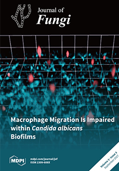
Cover Story (view full-size image): The response of immune cells to fungal biofilms is not well understood. A 3D live cell imaging analysis of macrophage interactions with Candida albicans revealed impaired phagocyte migration towards cells forming biofilms compared to cells in suspension. Alterations in cell wall architecture enhanced macrophage migration towards planktonic fungal cells, but did not affect the response towards C. albicans biofilms. Therefore biofilms are not only recalcitrant to antifungal therapy, but they also limit the effectiveness of the innate immune response. View this paper
  • Issues are regarded as officially published after their release is announced to the table of contents alert mailing list.
  • You may sign up for e-mail alerts to receive table of contents of newly released issues.
  • PDF is the official format for papers published in both, html and pdf forms. To view the papers in pdf format, click on the "PDF Full-text" link, and use the free Adobe Reader to open them.
Order results
Result details
Section
Select all
Export citation of selected articles as:
1401 KiB  
Article
Verapamil Inhibits Aspergillus Biofilm, but Antagonizes Voriconazole
by Hasan Nazik, Varun Choudhary and David A. Stevens
J. Fungi 2017, 3(3), 50; https://doi.org/10.3390/jof3030050 - 20 Sep 2017
Cited by 6 | Viewed by 3722
Abstract
The paucity of effective antifungals against Aspergillus and increasing resistance, the recognition of the importance of Aspergillus biofilm in several clinical settings, and reports of verapamil—a calcium channel blocker—efficacy against Candida biofilm and hyphal growth, and synergy with an azole antifungal in vitro, [...] Read more.
The paucity of effective antifungals against Aspergillus and increasing resistance, the recognition of the importance of Aspergillus biofilm in several clinical settings, and reports of verapamil—a calcium channel blocker—efficacy against Candida biofilm and hyphal growth, and synergy with an azole antifungal in vitro, led to a study of verapamil ± voriconazole against Aspergillus. Broth macrodilution methodology was utilized for MIC (minimum inhibitory concentration) and MFC (minimum fungicidal concentration) determination. The metabolic effects (assessed by XTT [2,3-bis[2-methoxy-4-nitro-5-sulfophenyl]-2H-tetrazolium-5-carboxanilide inner salt]) on biofilm formation by conidia were studied upon exposure to verapamil, verapamil plus voriconazole, or voriconazole alone. For biofilm formation, we found less inhibition from the combinations than with either drug alone, or less inhibition from the combination than that of the more potent drug alone. For preformed biofilm, we found no significant change in activity comparing voriconazole alone compared to added verapamil, and no significant alteration of activity of the more potent voriconazole, at any concentration in the range tested, by addition of a concentration of verapamil that is inhibitory alone. In full checkerboard assays with planktonic fungus, there was no indication of any effect of one drug on the other (indifference). Although verapamil was similarly inactive against planktonic Aspergillus, as with Candida, verapamil was indeed active against Aspergillus biofilm. However, indifference and antagonism was found with voriconazole. Full article
Show Figures

Figure 1

4594 KiB  
Article
Conserved Inhibition of Neutrophil Extracellular Trap Release by Clinical Candida albicans Biofilms
by John F. Kernien, Chad J. Johnson and Jeniel E. Nett
J. Fungi 2017, 3(3), 49; https://doi.org/10.3390/jof3030049 - 06 Sep 2017
Cited by 26 | Viewed by 4773
Abstract
Candida albicans biofilms are difficult to eradicate due to their resistance to host defenses and antifungal drugs. Although neutrophils are the primary responder to C. albicans during invasive candidiasis, biofilms resist killing by neutrophils. Prior investigation, with the commonly used laboratory strain SC5314, [...] Read more.
Candida albicans biofilms are difficult to eradicate due to their resistance to host defenses and antifungal drugs. Although neutrophils are the primary responder to C. albicans during invasive candidiasis, biofilms resist killing by neutrophils. Prior investigation, with the commonly used laboratory strain SC5314, linked this phenotype to the impaired release of neutrophil extracellular traps (NETs), which are structures of DNA, histones, and antimicrobial proteins involved in extracellular microbial killing. Considering the diversity of C. albicans biofilms, we examined the neutrophil response to a subset of clinical isolates forming biofilms with varying depths and architectures. Using fluorescent staining of DNA and scanning electron microscopy, we found that inhibition of NET release was conserved across the clinical isolates. However, the dampening of the production of reactive oxygen species (ROS) by neutrophils was strain-dependent, suggesting an uncoupling of ROS and NET inhibition. Our findings show that biofilms formed by clinical C. albicans isolates uniformly impair the release of NETs. Further investigation of this pathway may reveal novel approaches to augment immunity to C. albicans biofilm infections. Full article
(This article belongs to the Special Issue Host–Fungus Interactions)
Show Figures

Graphical abstract

1610 KiB  
Review
Innate and Adaptive Immunity to Mucorales
by Harlene Ghuman and Kerstin Voelz
J. Fungi 2017, 3(3), 48; https://doi.org/10.3390/jof3030048 - 05 Sep 2017
Cited by 60 | Viewed by 7473
Abstract
Mucormycosis is an invasive fungal infection characterised by rapid filamentous growth, which leads to angioinvasion, thrombosis, and tissue necrosis. The high mortality rates (50–100%) associated with mucormycosis are reflective of not only the aggressive nature of the infection and the poor therapeutics currently [...] Read more.
Mucormycosis is an invasive fungal infection characterised by rapid filamentous growth, which leads to angioinvasion, thrombosis, and tissue necrosis. The high mortality rates (50–100%) associated with mucormycosis are reflective of not only the aggressive nature of the infection and the poor therapeutics currently employed, but also the failure of the human immune system to successfully clear the infection. Immune effector interaction with Mucorales is influenced by the developmental stage of the mucormycete spore. In a healthy immune environment, resting spores are resistant to phagocytic killing. Contrarily, swollen spores and hyphae are susceptible to damage and degradation by macrophages and neutrophils. Under the effects of immune suppression, the recruitment and efficacy of macrophage and neutrophil activity against mucormycetes is considerably reduced. Following penetration of the endothelial lining, Mucorales encounter platelets. Platelets adhere to both mucormycete spores and hyphae, and exhibit germination suppression and hyphal damage capacity in vitro. Dendritic cells are activated in response to Mucorales hyphae only, and induce adaptive immunity. It is crucial to further knowledge regarding our immune system’s failure to eradicate resting spores under intact immunity and inhibit fungal growth under immunocompromised conditions, in order to understand mucormycosis pathogenicity and enhance therapeutic strategies for mucormycosis. Full article
(This article belongs to the Special Issue Mucosal Fungal Infections)
Show Figures

Figure 1

1176 KiB  
Review
Immune Recognition of Fungal Polysaccharides
by Brendan D. Snarr, Salman T. Qureshi and Donald C. Sheppard
J. Fungi 2017, 3(3), 47; https://doi.org/10.3390/jof3030047 - 28 Aug 2017
Cited by 63 | Viewed by 10466
Abstract
The incidence of fungal infections has dramatically increased in recent years, in large part due to increased use of immunosuppressive medications, as well as aggressive medical and surgical interventions that compromise natural skin and mucosal barriers. There are relatively few currently licensed antifungal [...] Read more.
The incidence of fungal infections has dramatically increased in recent years, in large part due to increased use of immunosuppressive medications, as well as aggressive medical and surgical interventions that compromise natural skin and mucosal barriers. There are relatively few currently licensed antifungal drugs, and rising resistance to these agents has led to interest in the development of novel preventative and therapeutic strategies targeting these devastating infections. One approach to combat fungal infections is to augment the host immune response towards these organisms. The polysaccharide-rich cell wall is the initial point of contact between fungi and the host immune system, and therefore, represents an important target for immunotherapeutic approaches. This review highlights the advances made in our understanding of the mechanisms by which the immune system recognizes and interacts with exopolysaccharides produced by four of the most common fungal pathogens: Aspergillus fumigatus, Candida albicans, Cryptococcus neoformans, and Histoplasma capsulatum. Work to date suggests that inner cell wall polysaccharides that play an important structural role are the most conserved across diverse members of the fungal kingdom, and elicit the strongest innate immune responses. The immune system senses these carbohydrates through receptors, such as lectins and complement proteins. In contrast, a greater diversity of polysaccharides is found within the outer cell walls of pathogenic fungi. These glycans play an important role in immune evasion, and can even induce anti-inflammatory host responses. Further study of the complex interactions between the host immune system and the fungal polysaccharides will be necessary to develop more effective therapeutic strategies, as well as to explore the use of immunosuppressive polysaccharides as therapeutic agents to modulate inflammation. Full article
(This article belongs to the Special Issue Host–Fungus Interactions)
Show Figures

Graphical abstract

1356 KiB  
Review
Natural Antimicrobial Peptides as Inspiration for Design of a New Generation Antifungal Compounds
by Małgorzata Bondaryk, Monika Staniszewska, Paulina Zielińska and Zofia Urbańczyk-Lipkowska
J. Fungi 2017, 3(3), 46; https://doi.org/10.3390/jof3030046 - 26 Aug 2017
Cited by 98 | Viewed by 8988
Abstract
Invasive fungal infections are associated with high mortality rates, despite appropriate antifungal therapy. Limited therapeutic options, resistance development and the high mortality of invasive fungal infections brought about more concern triggering the search for new compounds capable of interfering with fungal viability and [...] Read more.
Invasive fungal infections are associated with high mortality rates, despite appropriate antifungal therapy. Limited therapeutic options, resistance development and the high mortality of invasive fungal infections brought about more concern triggering the search for new compounds capable of interfering with fungal viability and virulence. In this context, peptides gained attention as promising candidates for the antimycotics development. Variety of structural and functional characteristics identified for various natural antifungal peptides makes them excellent starting points for design novel drug candidates. Current review provides a brief overview of natural and synthetic antifungal peptides. Full article
(This article belongs to the Special Issue Antifungal Peptides)
Show Figures

Figure 1

194 KiB  
Editorial
Fungal Pigments: Deep into the Rainbow of Colorful Fungi
by Laurent Dufossé, Yanis Caro and Mireille Fouillaud
J. Fungi 2017, 3(3), 45; https://doi.org/10.3390/jof3030045 - 07 Aug 2017
Cited by 2 | Viewed by 4379
Abstract
With the impact of globalization on research trends, the search for healthier life styles, the increasing public demand for natural, organic, and ”clean labelled” products, as well as the growing global market for natural colorants in economically fast-growing countries all over the world, [...] Read more.
With the impact of globalization on research trends, the search for healthier life styles, the increasing public demand for natural, organic, and ”clean labelled” products, as well as the growing global market for natural colorants in economically fast-growing countries all over the world, filamentous fungi started to be investigated as readily available sources of chemically diverse pigments and colorants.[...] Full article
(This article belongs to the Special Issue Fungal Pigments)
625 KiB  
Review
Biosynthesis of Astaxanthin as a Main Carotenoid in the Heterobasidiomycetous Yeast Xanthophyllomyces dendrorhous
by Jose L. Barredo, Carlos García-Estrada, Katarina Kosalkova and Carlos Barreiro
J. Fungi 2017, 3(3), 44; https://doi.org/10.3390/jof3030044 - 30 Jul 2017
Cited by 91 | Viewed by 10222
Abstract
Carotenoids are organic lipophilic yellow to orange and reddish pigments of terpenoid nature that are usually composed of eight isoprene units. This group of secondary metabolites includes carotenes and xanthophylls, which can be naturally obtained from photosynthetic organisms, some fungi, and bacteria. One [...] Read more.
Carotenoids are organic lipophilic yellow to orange and reddish pigments of terpenoid nature that are usually composed of eight isoprene units. This group of secondary metabolites includes carotenes and xanthophylls, which can be naturally obtained from photosynthetic organisms, some fungi, and bacteria. One of the microorganisms able to synthesise carotenoids is the heterobasidiomycetous yeast Xanthophyllomyces dendrorhous, which represents the teleomorphic state of Phaffia rhodozyma, and is mainly used for the production of the xanthophyll astaxanthin. Upgraded knowledge on the biosynthetic pathway of the main carotenoids synthesised by X. dendrorhous, the biotechnology-based improvement of astaxanthin production, as well as the current omics approaches available in this yeast are reviewed in depth. Full article
(This article belongs to the Special Issue Fungal Pigments)
Show Figures

Graphical abstract

825 KiB  
Communication
Utilization of High Performance Liquid Chromatography Coupled to Tandem Mass Spectrometry for Characterization of 8-O-methylbostrycoidin Production by Species of the Fungus Fusarium
by Mark Busman
J. Fungi 2017, 3(3), 43; https://doi.org/10.3390/jof3030043 - 25 Jul 2017
Cited by 10 | Viewed by 3843
Abstract
The pigment 8-O-methylbostrycoidin is a polyketide metabolite produced by multiple species of the fungus Fusarium that infects plant crops, including maize. A technique was developed for the analysis of 8-O-methylbostrycoidin by high performance liquid chromatography coupled to electrospray ionization [...] Read more.
The pigment 8-O-methylbostrycoidin is a polyketide metabolite produced by multiple species of the fungus Fusarium that infects plant crops, including maize. A technique was developed for the analysis of 8-O-methylbostrycoidin by high performance liquid chromatography coupled to electrospray ionization tandem mass spectrometry. The quantitative nature of the LC-MS/MS experiment was demonstrated over a range of concentrations in maize. Limits of detection for the method (10 ng/g from 8-O-methylbostrycoidin spiked into ground maize) were shown, and susceptibility of the method to matrix effects from maize was also evaluated. The method was applied to evaluate the ability of the maize pathogen Fusarium verticillioides to produce 8-O-methylbostrycoidin in developing maize ears grown in an agricultural field. Full article
(This article belongs to the Special Issue Fungal Pigments)
Show Figures

Graphical abstract

4136 KiB  
Article
The Effect of Novel Heterocyclic Compounds on Cryptococcal Biofilm
by Maya Korem, Sarah Kagan and Itzhack Polacheck
J. Fungi 2017, 3(3), 42; https://doi.org/10.3390/jof3030042 - 20 Jul 2017
Cited by 7 | Viewed by 4116
Abstract
Biofilm formation by microorganisms depends on their communication by quorum sensing, which is mediated by small diffusible signaling molecules that accumulate in the extracellular environment. During human infection, the pathogenic yeast Cryptococcus neoformans can form biofilm on medical devices, which protects the organism [...] Read more.
Biofilm formation by microorganisms depends on their communication by quorum sensing, which is mediated by small diffusible signaling molecules that accumulate in the extracellular environment. During human infection, the pathogenic yeast Cryptococcus neoformans can form biofilm on medical devices, which protects the organism and increases its resistance to antifungal agents. The aim of this study was to test two novel heterocyclic compounds, S-8 (thiazolidinedione derivative, TZD) and NA-8 (succinimide derivative, SI), for their anti-biofilm activity against strains of Cryptococcus neoformans and Cryptococcus gattii. Biofilms were formed in a defined medium in 96-well polystyrene plates and 8-well micro-slides. The effect of sub-inhibitory concentrations of S-8 and NA-8 on biofilm formation was measured after 48 h by a metabolic reduction assay and by confocal laser microscopy analysis using fluorescent staining. The formation and development of cryptococcal biofilms was inhibited significantly by these compounds in concentrations below the minimum inhibitory concentration (MIC) values. These compounds may have a potential role in preventing fungal biofilm development on indwelling medical devices or even as a therapeutic measure after the establishment of biofilm. Full article
(This article belongs to the Special Issue Fungal Biofilms)
Show Figures

Graphical abstract

316 KiB  
Review
Strategies to Reduce Mortality in Adult and Neonatal Candidemia in Developing Countries
by Harsimran Kaur and Arunaloke Chakrabarti
J. Fungi 2017, 3(3), 41; https://doi.org/10.3390/jof3030041 - 19 Jul 2017
Cited by 49 | Viewed by 6952
Abstract
Candidemia, the commonest invasive fungal infection, is associated with high morbidity and mortality in developing countries, though the exact prevalence is not known due to lack of systematic epidemiological data from those countries. The limited studies report a very high incidence of candidemia [...] Read more.
Candidemia, the commonest invasive fungal infection, is associated with high morbidity and mortality in developing countries, though the exact prevalence is not known due to lack of systematic epidemiological data from those countries. The limited studies report a very high incidence of candidemia and unique epidemiology with a different spectrum of Candida species. The recent global emergence of multi-drug resistant Candida auris is looming large as an important threat in hospitalized patients of developing countries. While managing candidemia cases in those countries several challenges are faced, which include poor infrastructure; compromised healthcare and infection control practices; misuse and overuse of antibiotics and steroids; lack of awareness in fungal infections; non-availability of advance diagnostic tests and antifungal drugs in many areas; poor compliance to antifungal therapy and stewardship program. Considering the above limitations, innovative strategies are required to reduce mortality due to candidemia in adults and neonates. In the present review, we have unraveled the challenges of candidemia faced by low resource countries and propose a ten part strategy to reduce mortality due candidemia. Full article
(This article belongs to the Special Issue Fungal Infections in the Developing World)
Show Figures

Graphical abstract

1253 KiB  
Review
Metabolic Interactions between Bacteria and Fungi in Commensal Oral Biofilms
by Marloes Lof, Marleen M. Janus and Bastiaan P. Krom
J. Fungi 2017, 3(3), 40; https://doi.org/10.3390/jof3030040 - 14 Jul 2017
Cited by 30 | Viewed by 11176
Abstract
Oral health is more than just the absence of disease. The key to oral health is a diverse microbiome in an ecological balance. The oral microbiota is one of the most complex and diverse microbial communities in the human body. To maintain oral [...] Read more.
Oral health is more than just the absence of disease. The key to oral health is a diverse microbiome in an ecological balance. The oral microbiota is one of the most complex and diverse microbial communities in the human body. To maintain oral health, balance between the human host and the intrinsic microorganisms is essential. The healthy oral cavity is represented by a great microbial diversity, including both bacteria and fungi. The bacterial microbiome is very well studied. In contrast, fungi inhabiting the oral cavity are often overlooked. All microbial species in the oral cavity form communities which establish a variety of micro-niches and inter- and intra-species interactions. These interactions can be classified into three main groups: physical, chemical and metabolic interactions. Different metabolic interactions are reviewed in this report, among which are the metabolism of sugars, carbon, lactate and oxygen. This review set out with the aim of assessing the importance of metabolic interactions between fungi and bacteria in the healthy oral cavity. Full article
(This article belongs to the Special Issue Fungal Biofilms)
Show Figures

Figure 1

3247 KiB  
Review
Carotenoid Biosynthesis in Fusarium
by Javier Avalos, Javier Pardo-Medina, Obdulia Parra-Rivero, Macarena Ruger-Herreros, Roberto Rodríguez-Ortiz, Dámaso Hornero-Méndez and María Carmen Limón
J. Fungi 2017, 3(3), 39; https://doi.org/10.3390/jof3030039 - 07 Jul 2017
Cited by 49 | Viewed by 9046
Abstract
Many fungi of the genus Fusarium stand out for the complexity of their secondary metabolism. Individual species may differ in their metabolic capacities, but they usually share the ability to synthesize carotenoids, a family of hydrophobic terpenoid pigments widely distributed in nature. Early [...] Read more.
Many fungi of the genus Fusarium stand out for the complexity of their secondary metabolism. Individual species may differ in their metabolic capacities, but they usually share the ability to synthesize carotenoids, a family of hydrophobic terpenoid pigments widely distributed in nature. Early studies on carotenoid biosynthesis in Fusarium aquaeductuum have been recently extended in Fusarium fujikuroi and Fusarium oxysporum, well-known biotechnological and phytopathogenic models, respectively. The major Fusarium carotenoid is neurosporaxanthin, a carboxylic xanthophyll synthesized from geranylgeranyl pyrophosphate through the activity of four enzymes, encoded by the genes carRA, carB, carT and carD. These fungi produce also minor amounts of β-carotene, which may be cleaved by the CarX oxygenase to produce retinal, the rhodopsin’s chromophore. The genes needed to produce retinal are organized in a gene cluster with a rhodopsin gene, while other carotenoid genes are not linked. In the investigated Fusarium species, the synthesis of carotenoids is induced by light through the transcriptional induction of the structural genes. In some species, deep-pigmented mutants with up-regulated expression of these genes are affected in the regulatory gene carS. The molecular mechanisms underlying the control by light and by the CarS protein are currently under investigation. Full article
(This article belongs to the Special Issue Fungal Pigments)
Show Figures

Graphical abstract

689 KiB  
Article
Assessment of the Dyeing Properties of the Pigments Produced by Talaromyces spp.
by Lourdes Morales-Oyervides, Jorge Oliveira, Maria Sousa-Gallagher, Alejandro Méndez-Zavala and Julio Cesar Montañez
J. Fungi 2017, 3(3), 38; https://doi.org/10.3390/jof3030038 - 05 Jul 2017
Cited by 28 | Viewed by 4915
Abstract
The high production yields of pigments by Talaromyces spp. and their high thermal stability have implied that industrial application interests may emerge in the food and textile industries, as they both involve subjecting the colourants to high temperatures. The present study aimed to [...] Read more.
The high production yields of pigments by Talaromyces spp. and their high thermal stability have implied that industrial application interests may emerge in the food and textile industries, as they both involve subjecting the colourants to high temperatures. The present study aimed to assess the potential application of the pigments produced by Talaromyces spp. in the textile area by studying their dyeing properties. Dyeing studies were performed on wool. The dyeing process consisted of three stages: scouring, mordanting, and dyeing. Two different mordants (alum, A; ferric chloride, F) were tested at different concentrations on fabric weight (A: 5, 10, 15%; F: 10, 20, 30%). The mordanting process had a significant effect on the final colour of the dyed fabrics obtained. The values of dyeing rate constant (k), half-time of dyeing (t1/2), and sorption kinetics behaviour were evaluated and discussed. The obtained results showed that pigments produced by Talaromyces spp. could serve as a source for the natural dyeing of wool textiles. Full article
(This article belongs to the Special Issue Fungal Pigments)
Show Figures

Graphical abstract

2891 KiB  
Review
Methodological Issues in Antifungal Susceptibility Testing of Malassezia pachydermatis
by Andrea Peano, Mario Pasquetti, Paolo Tizzani, Elisa Chiavassa, Jacques Guillot and Elizabeth Johnson
J. Fungi 2017, 3(3), 37; https://doi.org/10.3390/jof3030037 - 05 Jul 2017
Cited by 19 | Viewed by 7203
Abstract
Reference methods for antifungal susceptibility testing of yeasts have been developed by the Clinical and Laboratory Standards Institute (CLSI) and the European Committee on Antibiotic Susceptibility Testing (EUCAST). These methods are intended to test the main pathogenic yeasts that cause invasive infections, namely [...] Read more.
Reference methods for antifungal susceptibility testing of yeasts have been developed by the Clinical and Laboratory Standards Institute (CLSI) and the European Committee on Antibiotic Susceptibility Testing (EUCAST). These methods are intended to test the main pathogenic yeasts that cause invasive infections, namely Candida spp. and Cryptococcus neoformans, while testing other yeast species introduces several additional problems in standardization not addressed by these reference procedures. As a consequence, a number of procedures have been employed in the literature to test the antifungal susceptibility of Malassezia pachydermatis. This has resulted in conflicting results. The aim of the present study is to review the procedures and the technical parameters (growth media, inoculum preparation, temperature and length of incubation, method of reading) employed for susceptibility testing of M. pachydermatis, and when possible, to propose recommendations for or against their use. Such information may be useful for the future development of a reference assay. Full article
(This article belongs to the Special Issue Antifungal Susceptibility Testing)
Show Figures

Graphical abstract

5250 KiB  
Article
Biodiversity of Pigmented Fungi Isolated from Marine Environment in La Réunion Island, Indian Ocean: New Resources for Colored Metabolites
by Mireille Fouillaud, Mekala Venkatachalam, Melissa Llorente, Helene Magalon, Pascale Cuet and Laurent Dufossé
J. Fungi 2017, 3(3), 36; https://doi.org/10.3390/jof3030036 - 02 Jul 2017
Cited by 37 | Viewed by 9011
Abstract
Marine ecosystems cover about 70% of the planet surface and are still an underexploited source of useful metabolites. Among microbes, filamentous fungi are captivating organisms used for the production of many chemical classes of secondary metabolites bound to be used in various fields [...] Read more.
Marine ecosystems cover about 70% of the planet surface and are still an underexploited source of useful metabolites. Among microbes, filamentous fungi are captivating organisms used for the production of many chemical classes of secondary metabolites bound to be used in various fields of industrial application. The present study was focused on the collection, isolation, screening and genotyping of pigmented filamentous fungi isolated from tropical marine environments around La Réunion Island, Indian Ocean. About 150 micromycetes were revived and isolated from 14 marine samples (sediments, living corals, coral rubble, sea water and hard substrates) collected in four different locations. Forty-two colored fungal isolates belonging to 16 families, 25 genera and 31 species were further studied depending on their ability to produce pigments and thus subjected to molecular identification. From gene sequence analysis, the most frequently identified colored fungi belong to the widespread Penicillium, Talaromyces and Aspergillus genera in the family Trichocomaceae (11 species), then followed by the family Hypocreaceae (three species). This study demonstrates that marine biotopes in La Réunion Island, Indian Ocean, from coral reefs to underwater slopes of this volcanic island, shelter numerous species of micromycetes, from common or uncommon genera. This unstudied biodiversity comes along with the ability for some fungal marine inhabitants, to produce a range of pigments and hues. Full article
(This article belongs to the Special Issue Fungal Pigments)
Show Figures

Graphical abstract

311 KiB  
Review
Innate Immune Responses to Cryptococcus
by Lena J. Heung
J. Fungi 2017, 3(3), 35; https://doi.org/10.3390/jof3030035 - 02 Jul 2017
Cited by 23 | Viewed by 5377
Abstract
Cryptococcus species are encapsulated fungi found in the environment that predominantly cause disease in immunocompromised hosts after inhalation into the lungs. Even with contemporary antifungal regimens, patients with cryptococcosis continue to have high morbidity and mortality rates. The development of more effective therapies [...] Read more.
Cryptococcus species are encapsulated fungi found in the environment that predominantly cause disease in immunocompromised hosts after inhalation into the lungs. Even with contemporary antifungal regimens, patients with cryptococcosis continue to have high morbidity and mortality rates. The development of more effective therapies may depend on our understanding of the cellular and molecular mechanisms by which the host promotes sterilizing immunity against the fungus. This review will highlight our current knowledge of how Cryptococcus, primarily the species C. neoformans, is sensed by the mammalian host and how subsequent signaling pathways direct the anti-cryptococcal response by effector cells of the innate immune system. Full article
(This article belongs to the Special Issue Host–Fungus Interactions)
7653 KiB  
Article
Production and New Extraction Method of Polyketide Red Pigments Produced by Ascomycetous Fungi from Terrestrial and Marine Habitats
by Juliana Lebeau, Mekala Venkatachalam, Mireille Fouillaud, Thomas Petit, Francesco Vinale, Laurent Dufossé and Yanis Caro
J. Fungi 2017, 3(3), 34; https://doi.org/10.3390/jof3030034 - 28 Jun 2017
Cited by 64 | Viewed by 8697
Abstract
The use of ascomycetous fungi as pigment producers opens the way to an alternative to synthetic dyes, especially in the red-dye industries, which have very few natural pigment alternatives. The present paper aimed to bio-prospect and screen out 15 selected ascomycetous fungal strains, [...] Read more.
The use of ascomycetous fungi as pigment producers opens the way to an alternative to synthetic dyes, especially in the red-dye industries, which have very few natural pigment alternatives. The present paper aimed to bio-prospect and screen out 15 selected ascomycetous fungal strains, originating from terrestrial and marine habitats belonging to seven different genera (Penicillium, Talaromyces, Fusarium, Aspergillus, Trichoderma, Dreschlera, and Paecilomyces). We identified four strains, Penicillium purpurogenum rubisclerotium, Fusarium oxysporum, marine strains identified as Talaromyces spp., and Trichoderma atroviride, as potential red pigment producers. The extraction of the pigments is a crucial step, whereby the qualitative and quantitative compositions of each fungal extract need to be respected for reliable identification, as well as preserving bioactivity. Furthermore, there is a growing demand for more sustainable and cost-effective extraction methods. Therefore, a pressurized liquid extraction technique was carried out in this study, allowing a greener and faster extraction step of the pigments, while preserving their chemical structures and bioactivities in comparison to conventional extraction processes. The protocol was illustrated with the production of pigment extracts from P. purpurogenum rubisclerotium and Talaromyces spp. Extracts were analyzed by high-performance liquid-chromatography combined with photodiode array-detection (HPLC-DAD) and high-resolution mass spectrometry (UHPLC-HRMS). The more promising strain was the isolate Talaromyces spp. of marine origin. The main polyketide pigment produced by this strain has been characterized as N-threoninerubropunctamine, a non-toxic red Monascus-like azaphilone pigment. Full article
(This article belongs to the Special Issue Fungal Pigments)
Show Figures

Graphical abstract

2047 KiB  
Article
Perstraction of Intracellular Pigments through Submerged Fermentation of Talaromyces spp. in a Surfactant Rich Media: A Novel Approach for Enhanced Pigment Recovery
by Lourdes Morales-Oyervides, Jorge Oliveira, Maria Sousa-Gallagher, Alejandro Méndez-Zavala and Julio Cesar Montañez
J. Fungi 2017, 3(3), 33; https://doi.org/10.3390/jof3030033 - 27 Jun 2017
Cited by 16 | Viewed by 4830
Abstract
A high percentage of the pigments produced by Talaromyces spp. remains inside the cell, which could lead to a high product concentration inhibition. To overcome this issue an extractive fermentation process, perstraction, was suggested, which involves the extraction of the intracellular products out [...] Read more.
A high percentage of the pigments produced by Talaromyces spp. remains inside the cell, which could lead to a high product concentration inhibition. To overcome this issue an extractive fermentation process, perstraction, was suggested, which involves the extraction of the intracellular products out of the cell by using a two-phase system during the fermentation. The present work studied the effect of various surfactants on secretion of intracellular pigments produced by Talaromyces spp. in submerged fermentation. Surfactants used were: non-ionic surfactants (Tween 80, Span 20 and Triton X-100) and a polyethylene glycerol polymer 8000, at different concentrations (5, 20, 35 g/L). The highest extracellular pigment yield (16 OD500nm) was reached using Triton X-100 (35 g/L), which was 44% higher than the control (no surfactant added). The effect of addition time of the selected surfactant was further studied. The highest extracellular pigment concentration (22 OD500nm) was achieved when the surfactant was added at 120 h of fermentation. Kinetics of extracellular and intracellular pigments were examined. Total pigment at the end of the fermentation using Triton X-100 was 27.7% higher than the control, confirming that the use of surfactants partially alleviated the product inhibition during the pigment production culture. Full article
(This article belongs to the Special Issue Fungal Pigments)
Show Figures

Graphical abstract

1375 KiB  
Article
Role of Virulence Determinants in Candida albicans’ Resistance to Novel 2-bromo-2-chloro-2-(4-chlorophenylsulfonyl)-1-phenylethanone
by Monika Staniszewska, Małgorzata Bondaryk and Zbigniew Ochal
J. Fungi 2017, 3(3), 32; https://doi.org/10.3390/jof3030032 - 24 Jun 2017
Cited by 4 | Viewed by 3551
Abstract
We investigated the role of KEX2, SAP4-6, EFG1, and CPH1 in the virulence of Candida under a novel compound 2-bromo-2-chloro-2-(4-chlorophenylsulfonyl)-1-phenylethanone (Compound 4). We examined whether the exposure of C. albicans cells to Compound 4, non-cytotoxic to mammalian cells, [...] Read more.
We investigated the role of KEX2, SAP4-6, EFG1, and CPH1 in the virulence of Candida under a novel compound 2-bromo-2-chloro-2-(4-chlorophenylsulfonyl)-1-phenylethanone (Compound 4). We examined whether the exposure of C. albicans cells to Compound 4, non-cytotoxic to mammalian cells, reduces their adhesion to the human epithelium. We next assessed whether the exposure of C. albicans cells to Compound 4 modulates the anti-inflammatory response (IL-10) and induces human macrophages to respond to the Candida cells. There was a marked reduction in the growth of the sap4Δsap5Δsap6Δ mutant cells when incubated with Compound 4. Under Compound 4 (minimal fungicidal concentration MFC = 0.5–16 µg/mL): (1) wild type strain SC5314 showed a resistant phenotype with down-regulation of the KEX2 expression; (2) the following mutants of C. albicans: sap4Δ, sap5Δ, sap6Δ, and cph1Δ displayed decreased susceptibility with the paradoxical effect and up-regulation of the KEX2 expression compared to SC5314; (3) the immune recognition of C. albicans by macrophages and (4) the stimulation of IL-10 were not blocked ex vivo. The effect of deleting KEX2 in C. albicans had a minor impact on the direct activation of Compound 4’s antifungal activity. The adhesion of kex2Δ is lower than that of the wild parental strain SC5314, and tends to decrease if grown in the presence of a sub-endpoint concentration of Compound 4. Our results provide evidence that SAP4–6 play a role as regulators of the anti-Candida resistance to Compound 4. Compound 4 constitutes a suitable core to be further exploited for lead optimization to develop potent antimycotics. Full article
Show Figures

Figure 1

3976 KiB  
Article
Macrophage Migration Is Impaired within Candida albicans Biofilms
by Maria F. Alonso, Neil A. R. Gow, Lars P. Erwig and Judith M. Bain
J. Fungi 2017, 3(3), 31; https://doi.org/10.3390/jof3030031 - 22 Jun 2017
Cited by 23 | Viewed by 6001
Abstract
Candida albicans is an opportunistic fungal pathogen that infects immunocompromised patients. Infection control requires phagocytosis by innate immune cells, including macrophages. Migration towards, and subsequent recognition of, C. albicans fungal cell wall components by macrophages is critical for phagocytosis. Using live-cell imaging of [...] Read more.
Candida albicans is an opportunistic fungal pathogen that infects immunocompromised patients. Infection control requires phagocytosis by innate immune cells, including macrophages. Migration towards, and subsequent recognition of, C. albicans fungal cell wall components by macrophages is critical for phagocytosis. Using live-cell imaging of phagocytosis, the macrophage cell line J774.1 showed enhanced movement in response to C. albicans cell wall mutants, particularly during the first 30 min, irrespective of the infection ratio. However, phagocyte migration was reduced up to 2-fold within a C. albicans biofilm compared to planktonic fungal cells. Biofilms formed from C. albicans glycosylation mutant cells also inhibited macrophage migration to a similar extent as wildtype Candida biofilms, suggesting that the physical structure of the biofilm, rather than polysaccharide matrix composition, may hamper phagocyte migration. These data illustrate differential macrophage migratory capacities, dependent upon the form of C. albicans encountered. Impaired migration of macrophages within a C. albicans biofilm may contribute to the recalcitrant nature of clinical infections in which biofilm formation occurs. Full article
(This article belongs to the Special Issue Host–Fungus Interactions)
Show Figures

Graphical abstract

Previous Issue
Next Issue
Back to TopTop