Article

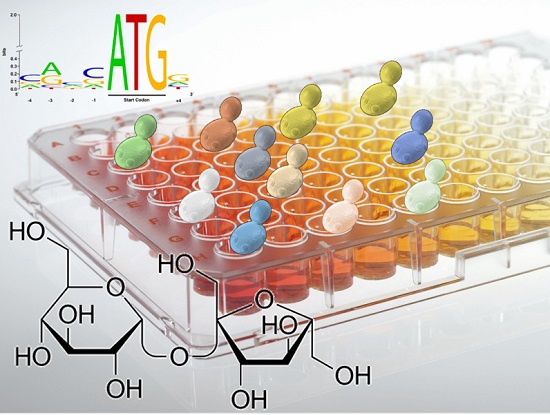
A Microtiter Plate Assay as a Reliable Method to Assure the Identification and Classification of the Veil-Forming Yeasts during Sherry Wines Ageing

by
Marina Ruíz-Muñoz
,
Maria Del Carmen Bernal-Grande
,
Gustavo Cordero-Bueso
*,
Mónica González
,
David Hughes-Herrera
and
Jesús Manuel Cantoral
Department of Biomedicine, Biotechnology and Public Health, University of Cádiz, Cádiz 11510, Spain
*
Author to whom correspondence should be addressed.
Fermentation 2017, 3(4), 58; https://doi.org/10.3390/fermentation3040058
Submission received: 10 October 2017 / Revised: 27 October 2017 / Accepted: 31 October 2017 / Published: 3 November 2017
(This article belongs to the Special Issue Yeast Biotechnology 2.0)

Abstract

:
Yeasts involved in veil formation during biological ageing of Sherry wines are mainly Saccharomyces cerevisiae, and they have traditionally been divided into four races or varieties: beticus, cheresiensis, montuliensis and rouxii. Recent progress in molecular biology has led to the development of several techniques for yeast identification, based on similarity or dissimilarity of DNA, RNA or proteins. In view of the latest yeast taxonomy, there are no more races. However, molecular techniques are not enough to understand the real veil-forming yeast diversity and dynamics in Sherry wines. We propose a reliable method, using a microtiter reader, to evaluate the fermentation and assimilation of carbon and nitrogen sources, the osmotolerance and the antibiotic resistance, using 18 S. cerevisiae and 5 non-Saccharomyces yeast strains, to allow correct identification and classification of the yeast strains present in the velum of flor complex.

Graphical Abstract

1. Introduction

Several types of wines are characterized by growing of a yeast film on its surface, which is known as “veil or velum of flor”. Two of these wines are Fino Sherry and Manzanilla, both produced from Palomino Fino (Vitis vinifera cv. vinifera) in the Jerez-Xérès-Sherry D.O., in Southern Spain [1,2]. The production of these types of wines consists of two successive processes. First, alcoholic fermentation, an anaerobic process in which sugars are converted in energy, produces as a result, ethanol and carbon dioxide gas. After the process is finished, wine is fortified to 15% (v/v) and it is put into oak barrels for storage. The process of biological ageing begins at this moment, using the “Solera” system [3].
Yeasts involved in veil formation during biological ageing are mainly Saccharomyces cerevisiae, and they have traditionally been divided into four races: beticus, cheresiensis, montuliensis and rouxii. The early methods related to taxonomy involved morphological studies and biochemical analyses of differences on fermentation and assimilation tests, where the substrates used were mainly dextrose, lactose, maltose, melibiose, raffinose, galactose and sucrose. These physiological properties were traditionally tested using Durham tubes in a liquid medium, which filled up with gas if fermentation occurred [4]. Nevertheless, both morphological and physiological characteristics may be influenced by culture conditions and spoilages and can provide ambiguous results. Using classical techniques can lead, in some cases, to incorrect classification of species or misidentification of strains. Moreover, the methodology often requires evaluation of different parameters by a complex, laborious and time-consuming process that does not include the most important characteristics of the yeasts from an industrial point of view. On the other hand, sugar fermentation and assimilation tests using Durham tubes are not too reliable because some of the yeast strains do not released CO2 immediately (these are known as slow fermentative yeasts). These yeasts were considered for a long time as non-fermentative, but they have been recently re-classified [5]. In recent years, new methods have been developed, such as rapid kits, for yeast identification (API® strips), analyses of total cell proteins and long-chain fatty acids, using gas chromatography or the determination of chemical compounds formed by the yeasts [6,7], but their cost, reproducibility and suitability to veil-forming yeast strain classification can lead to mistakes [8,9]. Therefore, it is necessary to use other identification methods to analyse them.
Recent progress in molecular biology has led to the development of several techniques for yeast identification based on similarities or dissimilarities of DNA, RNA or proteins. These include electrophoretic karyotyping [10], RFLP (Restriction Fragment Length Polymorphism) of mitochondrial DNA [11], random amplified polymorphic DNA analysis (RAPD), PCR-based techniques such as ribosomal internal transcribed spacers (ITS-PCR) [8], surveys of simple sequences repeats (SSR-Multiplex PCR) [12] or genomic DNA sequencing of the D1/D2 region of 26S rDNA. Some of these methods have been found to be of great interest in enology, due to the high level of resolution in yeast strain characterization, and for making it possible to establish a correlation between the genetic variability and the most important industrial properties of the strains.
A study carried out by [8] showed that S. cerevisiae flor yeasts responsible for the biological ageing of Sherry-type wines present a 24-bp deletion, located in the ITS1 region. These results have extended the analysis of this DNA region to flor yeast isolated in the Montilla-Moriles D.O. region [9]. Both studies corroborated that this deletion is fixed in flor yeast of the species, S. cerevisiae. By using molecular analysis techniques, different properties of industrial interest have been detected, such as physiologic, genetic and metabolic properties in the different identified veil-forming yeast strains. Moreover, in view of the latest yeast taxonomy, it is important to underline that there are no more races. S. cerevisiae (beticus) and S. cerevisiae (cheresiensis) are no longer considered as races or subspecies of S. cerevisiae, according to the last taxonomic study. Indeed, all these previously named races or subspecies are now considered as S. cerevisiae synonyms, based on nuclear DNA relatedness [13]. The former race, S. cerevisiae (rouxii), has been classified as Zygosaccharomyces rouxii and S. cerevisiae (montuliensis) is now considered as Torulaspora delbrueckii [14]. These latter ones are now classified as ethanol-tolerant non-Saccharomyces species and they also should contain the 24-bp deletion in the 131–154 ITS1 region. Other yeast species of genera different to Saccharomyces have been isolated and identified by molecular and/or sequencing methods from Sherry-type wines (from the initial phase of the fermentation until the ageing phase), such as Wickerhamomyces anomalus, Pichia membranaefaciens, Zygosaccharomyces bailii or the undesirable species Dekkera bruxellensis (anamorph Brettanomyces bruxellensis) [3,8,15]. However, the identification strategy should be used more precisely because many database sequences of these species have identical D1/D2 sequences [16]. The comparison of the D1/D2 sequences and ITS sequences between the isolated strains and those of the type strains of the species (most can be found in the CBS database). However, many authors have simply used the first hit in the blast search as the proof of taxonomic affiliation of their isolate. This strategy can be misleading because database curators do not check submissions for the correctness of taxonomic affiliation. In this case, using classical biochemical analyses, in conjunction with molecular methods, can help us to identify and classify the yeast diversity forming the velum of flor in Sherry wines. Therefore, we propose a reliable, reproducible, inexpensive and efficient biochemical test, using polysterene 96-well plates that could reinforce the identification of the different veil-forming yeast strains.

2. Materials and Methods

2.1. Yeast Strains

Six hundred yeast strains were isolated previously from the veil of flor of “Manzanilla” and “Fino” wines, from the Sanlúcar de Barrameda and Jerez wineries (Cádiz, Spain), respectively [2,17]. After a first screening, using a Mitochondrial DNA restriction analysis and electrophoretic karyotyping by pulsed-field-gel electrophoresis (PFGE) and SSR-Multiplex PCR, eighteen different profiles of Saccharomyces strains were shown and then grouped according to their similarities—eight from “Manzanilla”, denoted as MI to MVI, MX and MXVIII, and ten from “Fino”, coded as FI to FX, respectively. Additionally, five potential non-Saccharomyces, isolated from “Fino” veils, which showed different karyotypes to S. cerevisiae, were subjected to identification by ITS-PCR and sequencing of the D1/D2 region of 26S rDNA, as stated in [18]. At least two representative members from each ITS-RFLP genotype group were randomly selected for sequencing of the LSU sRNA gene D1/D2 domain.

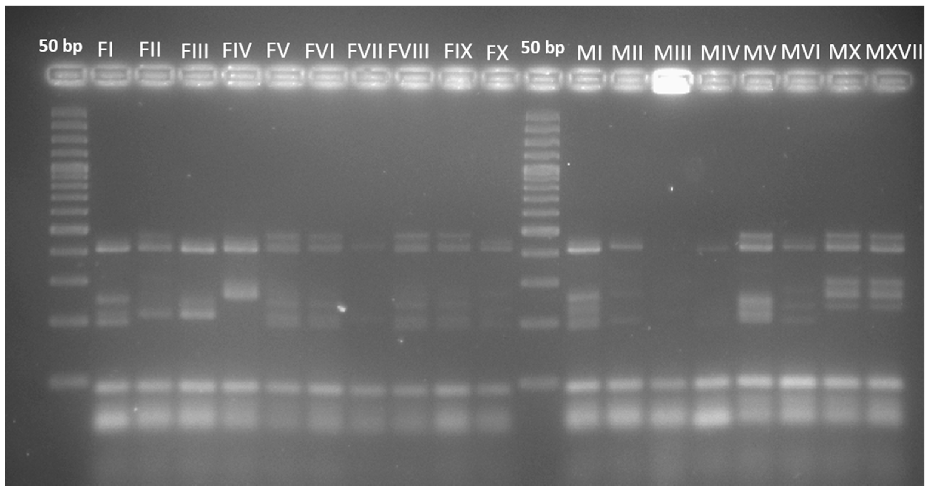
2.2. Mitochondrial DNA (mtDNA) Restriction Analysis

Total DNA extraction and mtDNA restriction analyses of the Saccharomyces yeast strains were performed by the method of [11]. Yeast DNA was digested with a HinfI restriction enzyme, by incubation at 37 °C, for 3 h. Fragments were separated by electrophoresis on a 1% agarose gel in 1× TBE buffer (45 mM tris-borate, 1 mM EDTA) and with 0.5 µg/mL of ethidium bromide. The image of the gel was digitalized in a Molecular Imager apparatus (Gel-Doc XR, Hercules, CA, USA) and analyzed using Quantity One 1-D software (Bio-Rad, Hercules, CA, USA).

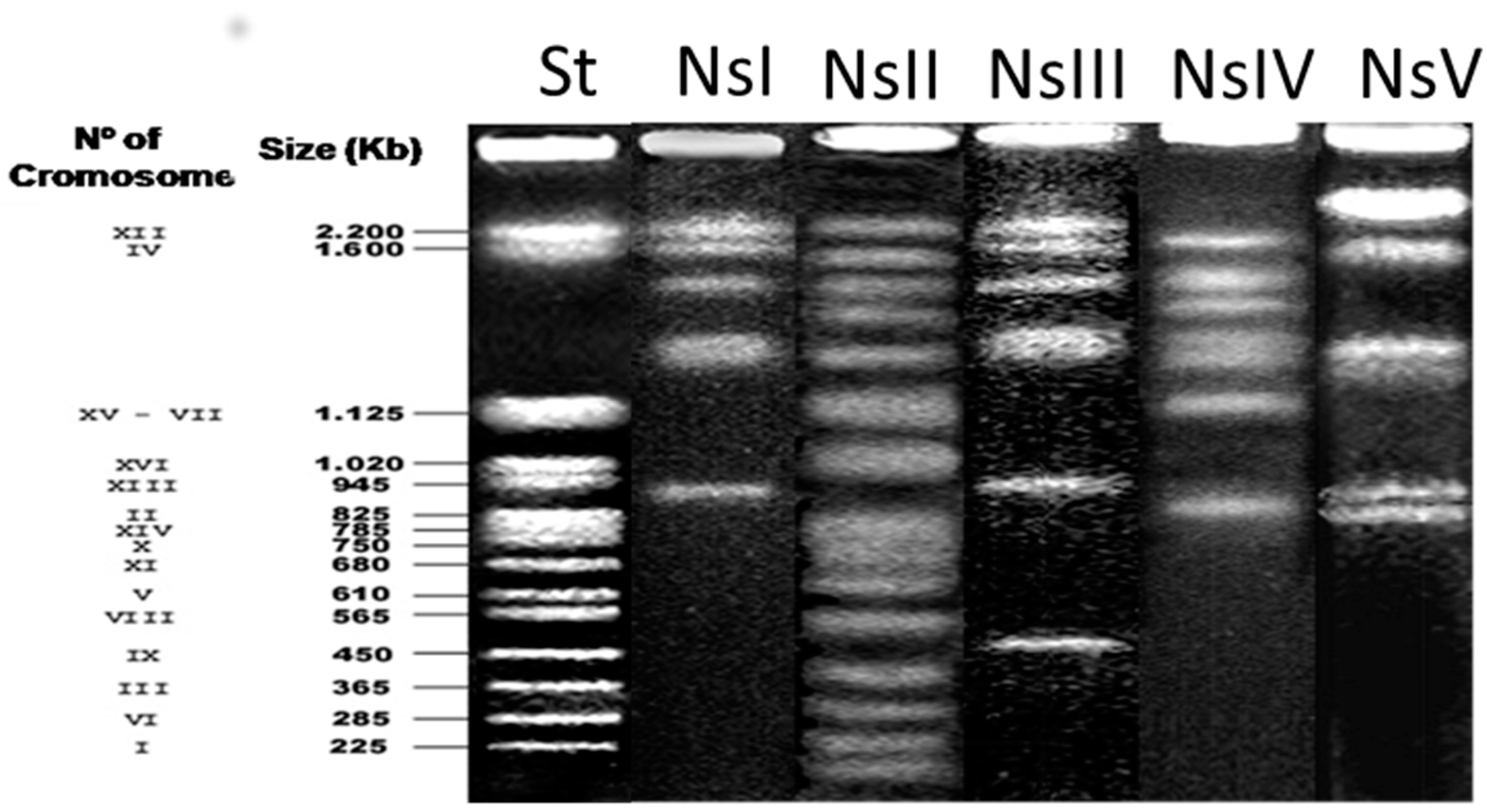
2.3. Electrophoretic Karyotype

Electrophoretic karyotypes of the Saccharomyces and non-Saccharomyces yeast strains were obtained by PFGE. The colonies were embedded in agarose plugs and treated following a procedure based on the protocol of [19], and chromosomes were separated using a CHEF-DR III apparatus (Bio-Rad). Gels were made of 1% pulsed-field certified agarose (Bio-Rad, Hercules, CA, USA) in 0.5× TBE electrophoresis buffer. The electrophoretic conditions were as follows: an initial pulse of 60 s and a final pulse of 120 s, during 22 h, at 14 °C and 6 V/cm, with an angle of 120°. Gels were stained with ethidium bromide solution (0.5 µg/mL ethidium bromide, 0.5× TBE buffer) after electrophoresis, and the image was captured under UV light with a camera (Bio-Rad Imaging System, Hercules, CA, USA). The image of the gel was digitalized in a Molecular Imager apparatus (Gel-Doc) and analyzed using Quantity One-1D software (Bio-Rad). Results were analyzed by comparing the electrophoretic mobility of the chromosomes with a reference strain (Saccharomyces cerevisiae YNN195).

2.4. Multiplex PCR-Microsatellite (SSR) Analysis

To perform multiplex PCR of microsatellite loci, three loci were simultaneously amplified, using the primers SC8132X (Fw: CTGCTCAACTTGTGATGGGTTTTGG; Rv: CCTCGTTACTATCGTCTTCATCTTGC), YOR267C (Fw: GGTGACTCTAACGGCAGAGTGG; Rv: GGATCTACTTGCAGTATACGGG) and SCPTSY7 (Fw: AAAAGCGTAAGCAATGG TGTAGAT; Rv: AAATGATGCCAATATTGAAAAGGT) (MWG Biotech AG, Ebersberg, Germany), proposed by Vaudano and García-Moruno [12]. PCR amplifications were performed in an Applied Biosystems SimplyAmp Termal Cycler in 50 µL volumes, constituted by: 2 µL of DNA solution (30–90 ng of DNA), 2.5 mM MgCl2, 200 µM dNTPs, 10 µL of 5× PCR buffer, 2 U of GoTaq® Flexi DNA Polymerase (Promega) and 10 pmol of primers for loci, SCYOR267C and SC8132X, and 40 pmol of primers for locus SCPTSY7. By using the protocol—4 min at 94 °C, 28 cycles of 30 s at 94 °C, 45 s at 56 °C and 30 s at 72 °C, and 10 min at 72 °C—PCR products were separated on an agarose gel (2.5% w/v) with 5 µg/mL of ethidium bromide in 1× TBE buffer at 115 V for 45 min. DNA fragment sizes were determined by comparison with a molecular ladder marker of 50 bp (Biolab, London, UK). The gel was digitalized in a Molecular Imager apparatus (Gel-Doc XR) and analyzed using Quantity One 1-D software (Bio-Rad).

2.5. Physiological and Biochemical Tests

To perform a quantitative analysis of carbon fermentation and assimilation, nitrogen assimilation, osmotolerance, and resistance to cycloheximide, different solutions, filtered through 0.22-µm Stericup filters (Millipore, Burlington, MA, USA) prior to their addition, were incorporated into the wells of polysterene 96-well microplates (Nunc™ 96-well polystyrene conical bottom Microwell™, Thermofisher, Denmark). The basal medium for carbohydrate fermentation and assimilation tests (BMC) was as follows: sterile deionized water 100 mL, 1 g yeast nitrogen base (YNB, Difco) was prepared with 2 g (w/v) solutions of fructose, glucose, maltose, lactose and sucrose and 4 g (w/v) of raffinose for fermentation tests, and 0.78 g of glucose, galactose, sorbose, glucosamine, ribose, xylose, arabinose, rhamnose, sucrose, maltose, α-threalose, cellobiose, salicin, arbutin, melibiose, lactose, raffinose, melezitose, inulin, starch, erythrytol, ribitol, xylitol, l-arabinitol, d-glucitol, d-mannitol, myo-inositol, d-Glucono-1,5-lactone, 5-Keto-d-gluconate, d-gluconate, d-galacturonate, dl-lactate, succinate, citrate and l-tartaric acid, and 0.78 mL of glycerol, propane 1,2-diol, butane 2,3-diol, Tween 20, Tween 80 for assimilation tests. Nitrogen sources for assimilation were prepared by supplementing 100 mL of an autoclaved basal assimilation medium yeast carbon base (YNB, DIfco) (0.17 g YNB without amino acids, 0.5 g ammonium sulphate, 2 g glucose) with filter-sterilized solutions containing 0.5 g of ammonium sulphate, 0.18 g of ammonium citrate, 0.11 g of nitrate, 0.04 g of nitrite, 0.1 g of ethylamine and creatine, 0.087 g of creatinine, 0.11 g of glucosamine, 0.1 g of urea, and d-proline. Then, 180 µL of each substrate was dropped into the wells of the trays. Also, 180 µL of 2%, 20%, 30%, 40% and 50% of a 1:1 mixture of glucose and fructose solution were incorporated into the wells as an osmotolerance test. Wells for testing the resistance to cycloheximide were prepared by adding 180 µL of a sterilized 0.1% w/v cycloheximide solution. The medium in each well can be dehydrated by drying the plate with the lid in a Speed-Vac vacuum oven (Eppendorf), which forms a small visible residue at the base of the well. After drying, individual trays can be sealed by heat with a polypropylene-aluminium sealing sheet and can be stored at 4 °C for six months, or at −20 °C for one year until subsequent use, rehydrating them with the corresponding basal medium.
The selected yeast strains were previously grown for 24–48 h in 15 mL of YPD (1% yeast extract, 2% peptone, 2% glucose) at 28 °C overnight; when achieved, these were washed twice with sterile, distilled-water. Twenty microliters of inoculum (McFarland standard 2, diluted by a factor of 10) were introduced into each well using a multi-channel pipette. McFarland standards are used as a reference to adjust the turbidity of yeast suspensions, so the number of yeast will be within a given range, to standardize biochemical tests. A 0.5 McFarland standard was prepared by mixing 0.05 mL of 1.175% barium chloride dehydrate with 9.95 mL of 1% sulfuric acid. The standards can be compared by eye to a suspension of yeasts in sterile water. If the yeast suspension is too turbid, it can be diluted. If the suspension is not turbid enough, more yeasts can be added (Table 1).
Microplates were sealed with Breathe-Easy membranes (Sigma, Sant Louis, MI, USA) and placed into an automatic microplate reader (Multiskan go reader, Thermo Fischer Scientific, Waltham, MA, USA). The optical density of the mix was read at 610 nm every hour (one minute of shaking just before automatic reading by the microplate reader) at 25 °C over 72 h. Data were collected and processed with the software, SkanIt RE for Multiskan GO 3.2. The absorbance data were exported to MS Excel for processing. A reading of optical density (OD610) = 1, measured by the microtiter reader, corresponds to 6–7 × 10⁷ cells/mL. Absorbance values at 610 nm were transformed into negative values when OD < 0.4 and positive when OD > 0.4, after 72 h.

2.6. Validation and Reproducibility of the Tests in Microtiter Plates

The yeast strains used in this study were also subjected to a standard test in Durham tubes, following the protocol proposed by [20] and under the same conditions of temperature and time. These tests were repeated with the microplate tests six times with each yeast strain. The stability of the carbon and nitrogen medium of the microtiter plates in storage was also examined. The same yeast species were analyzed for their reactions in microtitre trays that had been freshly prepared and in those that had been stored for six months at 4 °C and for one year at −20 °C.

3. Results

A total of 18 strains belonging to Saccharomyces cerevisiae species were characterized using the pulsed-field-gel electrophoresis (PFGE) technique and mitochondrial DNA restriction analysis. Moreover, analyses of polymorphic microsatellite loci and the proposed biochemical test, using 96-well plates, were used. Chromosomal profiles, obtained from the studied strains, showed fourteen different patterns. Specifically, strains FII and FIII, FV and FVI, FVIII and FIX, MX and MXVIII showed common patterns. In regard to the SSR analysis, fourteen different patterns were also obtained, which complied with the results obtained in the PFGE (Figure 1).
On the other hand, using mitochondrial DNA restriction analyses with the enzyme HinfI, three different patterns were obtained. The most common pattern was H1 (corresponding to FIV, FV, FIX, MI, MII, MIII, MVIII and MX), the second was H2 (corresponding to FI, FII, MIV and MV) and the pattern H3 only corresponded to one strain (MVI).
Fifteen different strains were determined by establishing the correspondence between the three patterns obtained from the analysis of mtRFLP with HinfI—coded as H1, H2 and H3—and fourteen patterns were obtained from the polymorphism study of the tested karyotypes and microsatellite loci. These fifteen different strains are FI, FII, FIII, FIV, FV, FVI, FIX, FX, MI, MII, MIII, MIV, MV, MVI and MX, according to the different combinations observed. The potential five non-Saccharomycces species, analyzed by the molecular method mentioned in the material and methods section (Figure 2) and after sequencing of the D1/D2 region, have been identified as Zygosaccharomyces bailii (NsI), Torulaspora delbrueckii (NsII), Zygosaccharomyces rouxii (NsIII), Rhodotorula mucilaginosa (NsIV) and Rhodotorula minuta (NsV). However, the species, Z. bailii and Z. rouxii, and R. mucilaginosa and R. minuta showed very similar (less than 1%), or identical, D1/D2 sequences to those found in the databases.
The selected strains were also analyzed to determine their fermentation and assimilation abilities, to evaluate if they shared physiological characteristics. Table 2 and Table 3, show the results of those which reached a positive result, among all tested carbon and nitrogen sources.
These tests, repeated six times, were also carried out in Durham tubes, obtaining the same results in all cases, with the exception of a couple of cases (fermentation of raffinose by yeast strain FII and fermentation of sucrose by yeast strain MVI), in which Durham tubes showed questionable results. However, these results obtained by the standard method, had positive results in the microtiter-read assay (OD > 0.4). Although molecular analyses have shown similarities between some strains, the diversity of the fermentation and assimilation abilities were significantly higher (Table 2 and Table 3). In the case of the sequenced non-Saccharomyces yeast strains, Z. bailii did not grow on maltose, which is usually assimilated by Z. rouxii and R. mucilaginosa isolates grew on maltose, sucrose, lactose, which are not assimilated by R. minuta. These results are in accordance with those published in [14,21]. Thus, by applying the biochemical tests, we have ensured that these yeast strains have been satisfactorily identified (Table 4).
Regarding the results obtained for osmotolerance, only two isolates grew in the media supplemented with 50% of glucose:fructose, but neither belonged to the Saccharomyces species (Table 5). The most osmotolerant species was Torulaspora delbrueckii. The tests for cycloheximide resistance gave the same results when tested by either the standard or microtitre method (Table 5).
The reproducibility of the tests in microtiter plates was assessed, using the selected Saccharomyces and non-Saccharomyces identified in this study. They were examined six times in microtiter plates. Responses to the identification tests for each species were the same in all replications. Moreover, the stored 96-well plates gave the same reaction responses, indicating that the multiplates could be stored for at least one year at −20 °C without affecting identification results.

4. Discussion

In recent years, multiplex microsatellite analysis has been implemented as one of the most decisive techniques in determining polymorphisms within the same species, but it has not often been applied to flor yeast analyses, since it could show few different patterns. However, in our study microsatellite analysis have shown fourteen different patterns, obtaining the same results as electrophoretic karyotyping. In any case, by combining multiplex microsatellite and RFLP of mtDNA analyses, when required, it is possible to obtain the same number of patterns as using electrophoretic karyotyping; these results are in agreement with those found in [9].
Pulsed-field gel electrophoresis (PFGE) has a good reproducibility and high discriminatory power, but it is an expensive and time-consuming technique. On the other hand, multiplex PCR of microsatellite loci [12] also has a high discriminatory power, sample preparation is easier and it is faster than PFGE.
Results obtained in the characterization of flor strains are highly variable, since some authors [8] have identified a higher degree of polymorphism between flor strains in mtDNA restriction than karyotyping. However, our study revealed the opposite: mtDNA restriction showed only three different patterns and PFGE showed fourteen different patterns. These results are similar to [9]. Mitochondrial DNA restriction analysis showed less polymorphisms due to the conditions of the wineries, since these DNA could be subjected to positive selection pressure based on their ethanol tolerance (15% v/v) [22] and other environmental factors, such as nutrient composition, growth temperature, and presence of toxic drugs, heavy metals, oxidizing agents, and osmotic/ionic stresses [23]. This might be the reason why the mtDNA restriction analysis showed only three different patterns of all the strains that we analyzed.
Biochemical tests offer supplementary information for characterizing flor strains. Traditionally, veil-forming yeast strains have been classified into four races belonging to the S. cerevisiae species, currently considered synonyms of the S. cerevisiae species [14]; another race, previously referred to as S. cerevisiae var. montuliensis, that has recently been reclassified as Torulaspora delbrueckii [14,24], and a last race named S. cerevisiae rouxii, is now Zygosaccharomyces rouxii [14]. This classification was based on morphological and metabolic studies of fermentation and assimilation of different carbon and nitrogen sources that differentiate these four main breeds [25]. The studies of assimilation and fermentation of sugars that we have carried out in this work have shown a high variability, so we were not able to differentiate races within S. cerevisiae strains. We can conclude that biochemical tests are necessary for identifying and classifying the different yeast strains that form the veil of flor of Sherry wine, because we believe that each yeast strain brings to the Sherry wines a different organoleptical characteristic. Thus, the application of this simplified method, using polysterene 96-well plates, is an inexpensive, low-time consuming and efficient technique, that can be used in combination with the molecular methods described above, to ensure veil-forming yeast diversity in Sherry wines.

Acknowledgments

This work was financed by the project UCA18DGUEII02, and by the European Union’s Seventh Framework Programme via the Marie Curie Action, “Co-funding of Regional, National and International Programs” to stimulate research activities without mobility restrictions, co-financed by the ‘Junta de Andalucía’ and the European Commission under grant agreement No. 291780.

Author Contributions

Jesús Manuel Cantoral and Gustavo Cordero-Bueso contributed to the conception and design of experiments; Marina Ruíz-Muñoz, Gustavo Cordero-Bueso, Mónica González, David Hughes-Herrera and Maria del Carmen Bernal-Grande performed the experiments; Marina Ruíz-Muñoz, Gustavo Cordero-Bueso and Maria del Carmen Bernal-Grande analyzed the data; Marina Ruíz-Muñoz and Gustavo Cordero-Bueso contributed to writing the paper.

Conflicts of Interest

The authors declare no conflict of interest. The founding sponsors had no role in the design of the study; in the collection, analyses, or interpretation of data; in the writing of the manuscript, and in the decision to publish the results.

References

  1. Rodríguez, M.E.; Infante, J.J.; Mesa, J.J.; Rebordinos, L.; Cantoral, J.M. Enological behavior of biofilms formed by genetically-characterized strains of sherry flor yeast. Open Biotechnol. J. 2013, 7, 23–29. [Google Scholar] [CrossRef]
  2. Mesa, J.J.; Infante, J.J.; Rebordinos, L.; Cantoral, J.M. Characterisation of yeasts involved in the biological ageing of sherry wines. LWT-Food Sci. Technol. 1999, 32, 114–120. [Google Scholar] [CrossRef]
  3. Esteve-Zarzoso, B.; Peris-Torán, M.J.; García-Maiquez, E.; Uruburu, F.; Querol, A. Yeast population dynamics during the fermentation and biological aging of sherry wines. Appl. Environ. Microbiol. 2001, 67, 2056–2061. [Google Scholar] [CrossRef] [PubMed]
  4. Martínez, P.; Codon, A.C.; Pérez, L.; Benítez, T. Physiological and molecular characterization of flor yeasts: Polymorphism of flor yeast population. Yeast 1995, 11, 1399–1411. [Google Scholar] [CrossRef] [PubMed]
  5. Boekhout, T.; Kurtzman, C.P. Principles and methods used in yeast classification, and an overview of currently accepted yeast genera. In Nonconventional Yeasts in Biotechnology; Springer: Berlin/Heidelberg, Germany, 1996; pp. 1–81. ISBN 978-3-642-79856-6. [Google Scholar]
  6. Vancanneyt, B.P.; Hennebert, G.; Kersters, K. Differentiation of yeast species based on electrophoretic whole-cell protein patters. Syst. Appl. Microbiol. 1991, 14, 23–32. [Google Scholar] [CrossRef]
  7. Silva, M.M.D.; Malfeito-Ferreira, M.; Loureiro, V.; Aubyn, A.S. Long-chain fatty acid composition as a criterion for yeast distinction in the brewing industry. J. Inst. Brew. 1994, 100, 17–22. [Google Scholar] [CrossRef]
  8. Esteve-Zarzoso, B.; Fernández-Espinar, M.T.; Querol, A. Authentication and identification of Saccharomyces cerevisiae “flor” yeast races involved in sherry ageing. Antonie van Leewenhoek 2004, 85, 151–158. [Google Scholar] [CrossRef] [PubMed]
  9. Marin-Menguiano, M.; Romero-Sánchez, S.; Barrales, R.R.; Ibeas, J.I. Population analysis of biofilm yeasts during fino sherry wine aging in the Montilla Moriles D.O. region. Int. J. Food Microbiol. 2017, 244, 67–73. [Google Scholar] [CrossRef] [PubMed]
  10. Rodríguez, M.E.; Infante, J.J.; Molina, M.; Rebordinos, L.; Cantoral, J.M. Using RFLP-mtDNA for the rapid monitoring of the dominant inoculated yeast strains in industrial wine fermentation. Int. J. Food Microbiol. 2011, 145, 331–335. [Google Scholar] [CrossRef] [PubMed]
  11. Querol, A.; Barrio, E.; Huerta, T.; Ramón, D. Molecular monitoring of wine fermentatios conducted by active dry yeast strains. Appl. Environ. Microbiol. 1992, 58, 2948–2953. [Google Scholar] [PubMed]
  12. Vaudano, E.; García-Moruno, E. Discrimination of Saccharomyces cerevisiae wine strains using microsatellite multiplex PCR and band pattern analysis. Food Microbiol. 2008, 25, 56–64. [Google Scholar] [CrossRef] [PubMed]
  13. Legras, J.C.; Moreno-García, J.; Zara, S.; Zara, G.; García-Martínez, T.; Mauricio, J.C.; Mannazzu, I.; Coi, A.L.; Zeidan, M.B.; Dequin, S.; et al. Flor yeast: New perspectives beyond wine aging. Front. Microbiol. 2016, 7, 503. [Google Scholar] [CrossRef] [PubMed]
  14. Kurtzman, C.; Fell, J.W.; Boekhout, T. The Yeasts: A Taxonomic Study; Elvesier: London, UK, 2011; p. 2363. [Google Scholar]
  15. Fernández-Espinar, M.T.; Esteve-Zarzoso, B.; Querol, A.; Barrio, E. RFLP analysis of the ribosomal internal transcribed spacers and the 5.8 S rRNA gene region of the genus Saccharomyces: A fast method for species identification and the differentiation of flor yeasts. Antonie van Leeuwenhoek 2000, 78, 87–97. [Google Scholar] [CrossRef]
  16. Lopandic, K.; Pfliegler, W.P.; Tiefenbrunner, W.; Gangl, H.; Sipiczki, M.; Sterlinger, K. Genotypic and phenotypic evolution of yeast interspecies hybrids during high-sugar fermentation. Appl. Microbiol. Biotechnol. 2016, 100, 6331–6343. [Google Scholar] [CrossRef] [PubMed]
  17. Espinazo-Romeu, M.; Cantoral, J.M.; Matallana, E.; Aranda, A. Btn2p is involved in ethanol and biofilm formation in flor yeast. FEMS Yeast Res. 2008, 8, 1127–1136. [Google Scholar] [CrossRef] [PubMed]
  18. Cordero-Bueso, G.; Rodríguez, M.E.; Garrido, C.; Cantoral, J.M. Rapid and not culture-dependent assay based on multiplex PCR-SSR analysis for monitoring inoculated yeast strains in industrial wine fermentations. Arch. Microbiol. 2017, 199, 135–143. [Google Scholar] [CrossRef] [PubMed]
  19. Rodríguez, M.E.; Infante, J.J.; Molina, M.; Domínguez, M.; Rebordinos, L.; Cantoral, J.M. Genomic characterization and selection of wine yeast to conduct industrial fermentations of a white wine produced in a SW Spain winery. J. Appl. Microbiol. 2010, 108, 1292–1302. [Google Scholar] [CrossRef] [PubMed]
  20. Van Der Walt, J.P.; Yarrow, D. Methods for the isolation, maintenance, classification and identification of yeasts. In The Yeasts: A Taxonomic Study, 3rd ed.; Kreger-van Rij, N.J.W., Ed.; Elvesier Science Publishers: Amsterdan, The Netherlands, 1984; pp. 45–104. [Google Scholar]
  21. Sipiczki, M. Overwintering of vineyard yeasts: Survival of interacting yeast communities in grapes mummified on vines. Front. Microbiol. 2016, 7, 212. [Google Scholar] [CrossRef] [PubMed]
  22. Ibeas, J.I.; Jiménez, J. Mitochondrial DNA loss caused by etanol in Saccharomyces flor yeasts. Appl. Environ. Microbiol. 1997, 63, 7–12. [Google Scholar] [PubMed]
  23. Kvitek, D.J.; Will, J.L.; Gasch, A.P. Variations in Stress Sensitivity and Genomic Expression in Diverse S. cerevisiae Isolates. PLoS Genet. 2008, 4, e1000223. [Google Scholar] [CrossRef] [PubMed]
  24. Alexandre, H. Flor yeasts of Saccharomyces cerevisiae—Their ecology, genetics and metabolism. Int. J. Food Microbiol. 2013, 167, 269–275. [Google Scholar] [CrossRef] [PubMed]
  25. Barnett, J.A.; Payne, R.W.; Yarrow, D. Yeasts: Characterisation and Identification, 3rd ed.; Cambridge University Press: Cambridge, UK, 2000; p. 1139. [Google Scholar]
Figure 1. Patterns obtained by the simple sequences repeats (SSR)-Multiplex PCR of the S. cerevisiae flor yeasts, selected after the first screening. Strains FII and FIII, FV and FVI, FVIII and FIX, MX and MXVIII showed common patterns.
Fermentation 03 00058 g001
Figure 2. Karyotypes obtained by pulsed-field-gel electrophoresis (PFGE) of the non-Saccharoymyces veil-forming yeasts isolated in “Fino” Sherry wines. The species were identified as Zygosaccharomyces bailii (NsI), Torulaspora delbrueckii (NsII), Zygosaccharomyces rouxii (NsIII), Rhodotorula mucilaginosa (NsIV) and Rhodotorula minuta (NsV).
Fermentation 03 00058 g002
Table 1. McFarland standard values used as a reference to adjust the turbidity of yeast suspensions.
McFarland Standard0.51234
1% Barium chloride (mL)0.050.10.20.30.4
1% Sulfuric acid (mL)9.959.99.89.79.6
Cell density ± 108 cells/mL1.536912
Absorbance (610 nn)0.10.250.450.600.67
Table 2. Fermentation of different carbohydrates by selected S. cerevisiae flor yeasts, analyzed with microtiter technology. Values are expressed as (+) if yeast strains were able to ferment the carbon/nitrogen source and (−) if yeast strains were not able to ferment them.
Strain DesignationSugar Fermentation
FructoseGlucoseLactoseMaltoseRafinoseSucrose
FI+
FII++++
FIII++
FIV+++
FV+++
FVI++++
FIX+++
FX++
MI++
MII+++++
MIII+
MIV++++
MV++
MVI++++
MX++++
Table 3. Assimilation of different carbon/nitrogen compounds by selected S. cerevisiae flor yeasts, analyzed with microtiter technology. Values are expressed as (+) if yeast strains were able to assimilate the carbon/nitrogen source and (−) if yeast strains were not able to assimilate them.
Strain DesignationCarbon/Nitrogen Compounds
Ammonium CitrateGlucoseInulinMelibioseNitriteStarchUrea
FI+
FII+.
FIII+
FIV+
FV+
FVI
FIX++
FX
MI+
MII+++
MIII+++
MIV+
MV+++
MVI++
MX++
Table 4. Assimilation of different carbon/nitrogen compounds by non-Saccharomyces species, isolated from “Fino” Sherry wine, analyzed with microtiter technology. Values are expressed as (+) if yeast strains were able to assimilate the carbon/nitrogen source and (−) if yeast strains were not able to assimilate them.
Strain DesignationCarbon/Nitrogen Compounds
RaffinoseGalactoseInulinMelibioseMaltoseLactosel-SorboseEthanol
NsI++
NsII+++++
NsIII+++
NsIV++++
NsV+++
Table 5. Osmotolerance and cycloheximide resistance of the veil-forming yeasts used in this study and analyzed by microtiter technology. Values are expressed as (+) if yeast strains were able to growth in a particular concentration of glucose/fructose solution and (−) if yeast strains were not able to growth.
Strain DesignationOsmotolerance: Growth on Media Supplemented with 1:1 (Glucose:Fructose) and Cycloheximide Resistance
2%20%30%40%50%Cycloheximide 0.01%Cycloheximide 0.1%
FI+++
FII++
FIII+++
FIV+++
FV+++
FVI+++
FVII++
FVIII++
FIX+++
FX+++
MI+++
MII+++
MIII+++
MIV+++
MV+++
MVI++++
MX++++
MXVIII+++
NsI++
NsII+++++
NsIII+++++
NsIV+++
NsV+++

