Fermentation Assisted by Pulsed Electric Field and Ultrasound: A Review


1
ISA, Univ. Lille 1, INRA, Univ. Artois, Univ. Littoral Côte d’Opale, EA 7394—ICV—Institut Charles Viollette, 48 Boulevard Vauban, F-59000 Lille, France
2
Faculty of Engineering, Universidad Nacional del Centro de la Provincia de Buenos Aires, Av. del Valle, 5737 Olavarría, Argentina
*
Author to whom correspondence should be addressed.
Fermentation 2018, 4(1), 1; https://doi.org/10.3390/fermentation4010001
Submission received: 15 October 2017
/
Revised: 29 November 2017
/
Accepted: 22 December 2017
/
Published: 4 January 2018
(This article belongs to the Special Issue Fermentation and Bioactive Metabolites)
Abstract
:Various novel techniques are proposed to improve process efficiency, quality, and safety of fermented food products. Ultrasound and pulsed electric field (PEF) are versatile technologies that can be employed in conjunction with fermentation processes to enhance process efficiency and production rates by improving mass transfer and cell permeability. The aim of this review is to highlight current and potential applications of ultrasound and PEF techniques in food fermentation processes. Their effects on microbial enzymes, along with mechanisms of action, are also discussed.

1. Introduction
Food fermentation basically involves the chemical transformation of complex organic compounds into simpler compounds by the action of enzymes and microorganisms, which has been reported since ancient times as a means of food preservation [1]. A more recent application of the fermentation technique has been applied to the production and extraction of bioactive compounds in the food, chemical, and pharmaceutical industries. The secondary metabolites produced during fermentation processes range from antibiotics to peptides and are referred to as bioactive compounds due to their numerous biological activities and potential prevention of several chronic diseases [2].
Traditionally, thermal treatment has been used to eliminate or reduce the microbial load and inactivate deleterious enzymes, which otherwise might hinder the process and induce changes in the quality of the fermented food. However, thermal processing can also alter sensorial and nutritional qualities, including reduction of some bioactive compounds [3]. For these reasons and to minimize quality losses, the heat treatment should be carried out under moderate conditions, but at a level sufficient to successfully inactivate detrimental enzymes.
Ultrasound and pulsed electric field (PEF) technologies are relatively novel and have the potential to fully or partially replace the conventional heating processes because they can inactivate enzymes and microorganisms without any, or very little, increase in temperature. However, one of the most exciting new areas of research in the field of fermentation is the application of low-intensity ultrasound and PEF at sublethal levels due to their positive effects on living cells. Depending on the processing conditions, improvements in process efficiency and high production rates have been observed. Thus, these techniques have been gaining interest for industrial applications. In addition, the literature suggests that these technologies can assist food processors in meeting the demands of both the consumer and the industry by producing safer and higher quality products with energy-efficient processes [4].
This review aims to provide an overview of current and potential applications regarding the effect and action mechanisms of ultrasound and PEF technologies on food fermentation processes, mainly focusing on microbials, enzymes, and bioactive compounds.
2. Ultrasound
2.1. Basic Concepts of Ultrasound
Ultrasonic waves are sound waves having frequencies above the human hearing range (>18 kHz). This form of vibrational energy is produced by ultrasonic transducers that convert electrical energy into vibrational sound energy. The frequency of the acoustic waves generated (the number of waves per second) is determined by the frequency of the electric field applied, and this is usually fixed by the instrument provider. Based on frequency, the ultrasonic spectrum can be divided into two zones. A frequency range from 20 kHz to around 1 MHz (even though frequencies between 20 and 40 kHz are commonly used for food processing applications) is known as power ultrasound because the sound waves generated are relatively powerful and can generate significant shear fields within the surrounding fluid. Alternatively, diagnostic ultrasound has a frequency in excess of 1 MHz and is mainly used for medical and industrial imaging purposes.
From an application perspective, ultrasound can also be broadly classified as low intensity and high intensity ultrasound. Applications using low power and high ultrasound frequencies (>1 MHz) are considered as low-intensity ultrasound (also called high-frequency ultrasound). Alternatively, processing technologies using high power and frequencies between 20 kHz and 1 MHz (more commonly ranging between 20 kHz and 100 kHz) fall within the range of high-intensity ultrasound (also named low-frequency ultrasound or power ultrasound) [5].
In the food industry, low-intensity ultrasound exerts no or minimal physical and chemical alterations in the material through which the waves pass; hence, it serves as a physical tool to analyze the physiochemical properties of food and monitor changes during food production. On the other hand, high-intensity ultrasound can induce desirable physical and chemical modifications for various bioprocessing applications [6]. Because high-intensity ultrasound has shown versatility and effectiveness to enhance the productivity and process efficiency in the food industry, only power ultrasound applications in food fermentation will be considered in this section of the review.
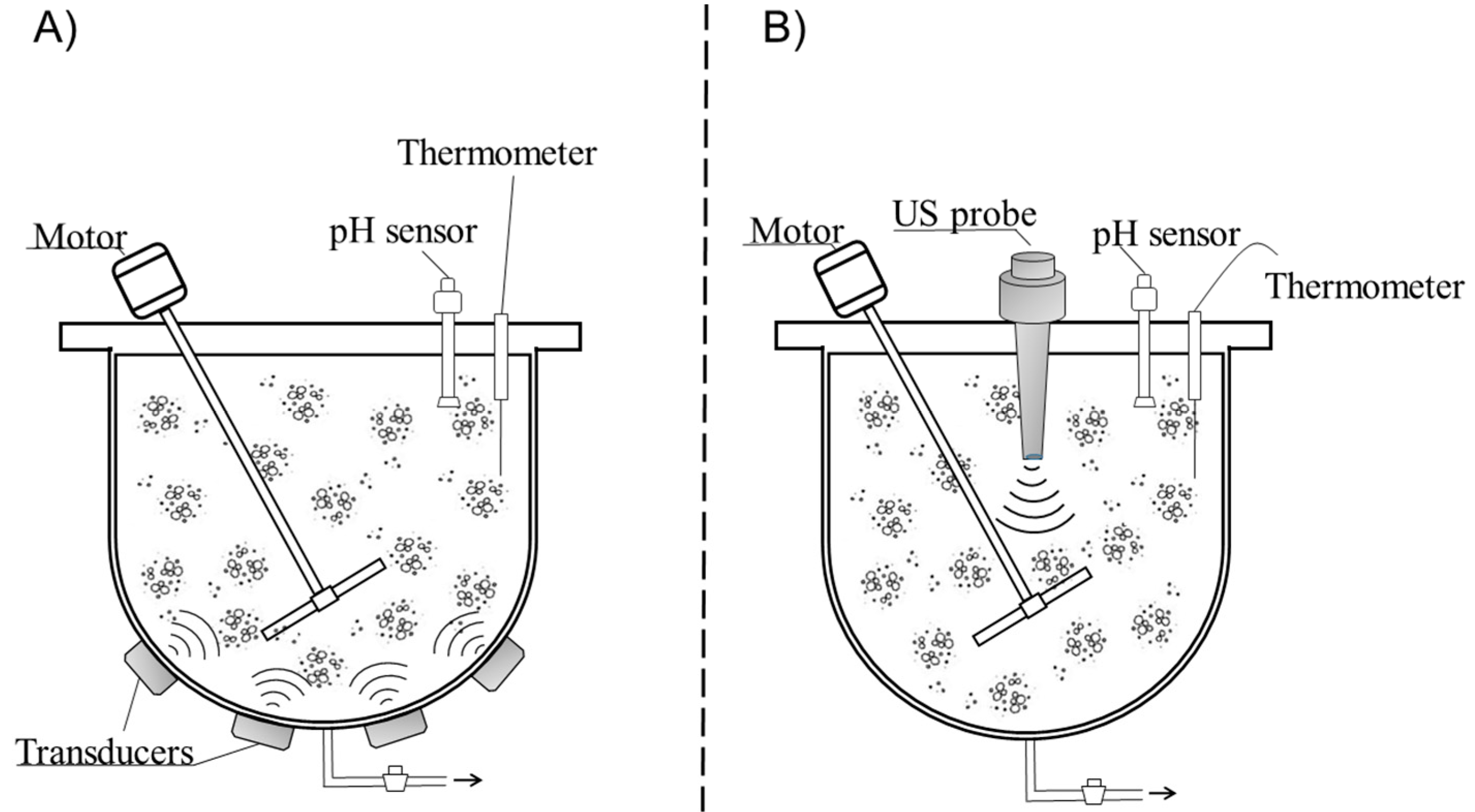
In liquid fermentation, ultrasound generated using a transducer is transmitted into the medium using either an ultrasonic bath or probe-based system. Figure 1 illustrates these two types of ultrasound systems, which are commonly employed in fermentation reactors. When the waves pass through the liquid medium, they induce a longitudinal displacement of particles, whereas the source of the sound wave acts as a piston, resulting in a succession of compression and rarefaction phases in the medium [7]. If the rarefaction cycle is strong enough, the distance between contiguous molecules of the liquid can reach or even exceed the critical molecular distance. The voids created in the medium are cavitation bubbles. These incipient bubbles are able to grow during rarefaction phases and decrease in size during compression cycles until a critical point is reached, after which ultimately results in the bubble’s collapse. Depending on the frequency of the ultrasound processing used, the liquid medium can be exposed to transient or stable cavitation. When the process uses lower frequency ultrasound (20–100 kHz), the nature of the cavitation is transient and the bubble collapse occurs very violently within a few acoustic cycles as the bubble reaches resonance size rapidly. Ultrasound processing at frequencies over 200 kHz, however, can normally result in stable cavitation in which the oscillation in bubble size following the cycles of high and low acoustic pressure can continue for thousands of cycles until the bubble reaches resonance size range [8]. In the megahertz frequency range, no cavitation is observed.
2.2. Physical and Chemical Effects of Ultrasound
The collapsing bubbles are capable of generating extremely high pressures and temperatures, suggested to be up to 50 MPa and up to 5000 K, respectively [7]. The main mechanical effects induced during cavitation of the collapsing bubbles in the solid–liquid interface of a heterogeneous medium include shockwave-induced damage and microjet impacts [9]. In the latter case, an asymmetrical collapse takes place in which the potential energy of the expanded bubble is converted into kinetic energy of a liquid microjet that extends inside the bubble and penetrates the opposite bubble wall, reaching high velocities. Other physical phenomena of ultrasound that are associated with ultrasound frequencies include agitation, turbulence, vibration, pressure, shear forces, and acoustic streaming. In addition to all the physical effects of cavitation, there are also chemical effects. One of the most important is the sonolysis of water (H2O→H+ + OH−) from the effects of temperature and pressure, which leads to the production of free radicals [10]. Because water is one of the most abundant substances in fermented food, these radicals can combine to form other molecules such as hydrogen peroxide and can also react with other components in the surrounding medium to potentially affect microbial enzyme activation.
According to Ashokkumar et al. [11], the physical effects of ultrasound are dominant in the lower frequency range of 20–100 kHz with a higher level of transient cavitation, except for acoustic streaming (i.e., the physical force of the sound capable of displacing ions and small molecules due to a pressure gradient) that is dominant at frequencies above 1 MHz with less physical and chemical effects associated with cavitation. On the other hand, chemical effects are dominant at frequencies >200 kHz with larger amounts of free radicals being produced due to the generation of a large number of active bubbles compared to those produced by lower frequency ultrasound.
The combination of all these effects is the basis of the different uses that ultrasound technology has in the food sector; as such, physical effects have found application in operations such as homogenization, emulsification, extraction, pasteurization, and degassing or deaeration of alcoholic beverages. Particularly, low-frequency ultrasound has been applied to improve many food processing operations toward the enhancement of mass transfer, for enhancing fermentation rates, and other specialized processing applications including wine maturation and aging [5,12].
2.3. Production of Biomolecules from Fermented Foods
The production of biomolecules in fermented foods will depend largely on the effect of ultrasound on both the microorganisms used and the enzymes they produce. This topic will be discussed below with an emphasis on improvements in the fermentation process and the production of active metabolites that are of interest in the food industry.
The lethal effects of ultrasound on microorganisms have been extensively investigated since the late 1990s, but the use of ultrasound to promote or control their activity is much more recent. For instance, the effectiveness of low-frequency ultrasound has been demonstrated in the stimulation of probiotics in milk [13,14]. The authors have reported an improvement in lactose hydrolysis and transgalactosylation by bifidobacteria, as well as a reduction in fermentation time by up to 30 min depending on the probiotic strain. In particular, sonicated samples have shown lower counts of viable probiotic cells at the beginning of fermentation compared to control, whereas no significant changes were observed in the final counts at the end of fermentation [13]. In another study, an increase in the viability of probiotics (Lactobacillus sp. and Bifidobacterium sp.) has been reported compared to the control in the case of fermented soy milk [15]. Activation of a mixed culture of Streptococcus thermophilus and Lactobacillus delbrueckii subsp. bulgaricus at 84 W power ultrasound for 150 s was investigated by Barukčić et al. [16] who reported an approximately 1 log cycle higher count compared to untreated inoculums, resulting in a decreased fermentation time of up to 30 min.
The beneficial effects of sonication have been attributed to the formation of pores or temporary holes in the microbial cell membranes, thus improving their permeability in a process known as sonoporation [17]. Although these pores lead to sublethal injury to the microbial cell, they provide a channel for transport of essential nutrients and removal of toxic substances across these membranes [18,19,20]. Thus, the abovementioned results show that depending on the type of microorganism and the ultrasound processing conditions, treated cells with minor physical damage can recover from injury and subsequently increase in number during fermentation. However, an increase in ultrasonic power or exposure time could lead to inactivation or cell death due to leakage of cellular content.
It is important to highlight that inactivation of microorganisms occurs at low frequencies, whereas when microbial cells are exposed to a higher frequency range, microbial cells are minimally affected with no significant effect on cell viability [5]. Thus, the effect of sonication depends on the processing conditions employed but it is also culture specific since differences in several characteristics—such as size and shape of the cell, and thickness and composition of the cell wall—will determine the resistance of the microorganism towards ultrasound. For example, a greater resistance to ultrasound has been demonstrated for Gram-positive bacteria than Gram-negative bacteria, possibly due to the presence of a thicker and more robust cell wall as a result of the cross-linking of peptidoglycan [21]. In addition, larger cells are more sensitive to ultrasound because of the larger surface that is in contact with cavitation. As to shape, cocci are more resistant than bacilli because of the ratio of cell surface to volume [7].
In the case of enzymes, it is known that power ultrasound is able to break various large biopolymers, including enzymes, thus affecting their functionality. The effect of ultrasound on the inactivation of enzymes of industrial importance was comprehensively reviewed by O’Donnell et al. [10]. Changes in enzyme biological activity are due to modifications in protein structure and also changes in the folding of the proteins (secondary and tertiary structures of the enzyme). The rapid formation and collapse of cavitation bubbles change an enzyme’s environment (e.g., temperature, pressure, shear stress, and pH), which can cause partial or total inactivation of enzyme activity [22]. Additionally, free radicals from sonolysis of water can react with the amino acids of an enzyme’s structure, subsequently affecting the enzyme’s activity and its catalytic function [23]. However, it has been mentioned that ultrasound application at room temperature has minor effects on enzyme activity [10]. Therefore, for effective inactivation of deteriorative enzymes, ultrasound is usually employed in combination with additional factors, such as temperature (thermosonication), pressure (manosonication), or a combination of both (manothermosonication).
Conversely, in some cases, ultrasound processing can increase the activity of certain enzymes, depending on the treatment intensity and processing time [24]. Thus, a positive effect of ultrasound in the production of bioactive molecules takes place. For instance, the application of ultrasound during fermentation has been shown to improve the β-galactosidase activity of probiotics, resulting in the production of health-promoting oligosaccharides in fermented milk [14] and an improvement in isoflavone bioconversion activities in soymilk [15]. Although stimulation of enzymatic activity due to sonication is a process not well understood, an increase in the mass transfer rate of the reagents to the active site seems to be the most important factor. In the case of bioactive peptide generation, it has been suggested that the use of high-power ultrasound in pretreatment and during the hydrolysis process of proteins can modify protein conformation by affecting hydrogen bonds and hydrophobic interactions, while acoustic cavitation acts by disrupting the quaternary and/or tertiary structure of proteins. Therefore, these structural modifications may expose more hydrolysis sites to the enzyme and consequently increase the degree of hydrolysis and bioactivity [25].
Thus, the effect of ultrasound seems to be enzyme specific, and stimulation and/or retardation of enzymatic activity due to sonication will also depend on some other factors, such as ultrasonic intensity (acoustic energy/power), duration of the process, temperature, pressure, pH, and ionic strength of the medium.
Phenolic compounds, which are interesting bioactive compounds with potential health-promoting properties, can be released by enzymatic activity [26]. For instance, depending on acoustic energy density and temperature, a significant increase in total phenolic content in model wine was reported during ultrasound treatment at 25 kHz for 150 min [27]. This is in accordance with the use of ultrasound as an extraction technology in food processing since it enables an increase in both the extraction yield and the extraction rate. In this sense, several works have demonstrated that fermentation has a positive influence on total phenolic content in some cereals, pseudocereals, and legumes [28,29,30]. A large proportion of phenolic compounds in these grains is bound to cell wall components. Hence, microbial enzymes derived from fermentation processes might induce structural breakdown of the plant cell walls and/or hydrolyze the esterified and insoluble bound phenolics, facilitating their liberation. Ultrasound technology could be applied here to enhance the production and/or bioavailability of these compounds.
3. Pulsed Electric Fields
3.1. Basic Concepts of PEF on Fermentation Process
The application of pulsed electric field in fermentation comprises basically the application of direct current electric pulses on the sample source, which is placed between two electrodes in a treatment chamber. The source is exposed to a pulsed voltage (up to 40 kV, even though typically 0.1–5 kV/cm with pulses of 50–1000 µs are used). In this sense, the cell membrane of the microorganisms can be regarded as a capacitor filled with a low dielectric constant material. When cell suspensions are exposed to electric fields, the ions inside the cell move along the field until there is an accumulation of the free charges at both membrane surfaces, which plays an important role in the signal transduction process [31]. When the intensity of an applied electric field increases, the potential difference across a cell membrane also increases. If this transmembrane potential exceeds a stated threshold value (typically 0.2–1 V), a temporary loss of membrane semipermeability occurs. This phenomenon of cell damage is called electroporation (or electropermeabilization) and, depending on the given electric field, leads to the formation of temporary (reversible) or permanent (irreversible) pores [32,33,34]. Thus, the process assists the electrophoretic movement of the intracellular charged compounds between the cellular compartments and the release of them without any significant increase in temperature.
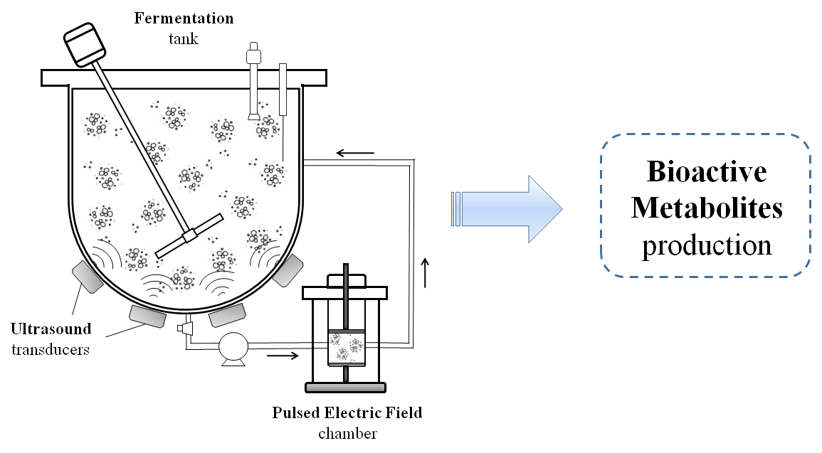
One of the most important components in the PEF system is the treatment chamber. This dispositive can be designed to work in two different modes: static or continuous. In the static mode, food is held between two parallel electrodes, whereas in the continuous mode, food is moved between the electrodes inside the treatment chamber assisted by a pumping system. A typical system to perform PEF treatments in continuous mode is shown in Figure 2.
3.2. Factors Affecting Pulsed Electric Field Treatments
There are tremendous interdependencies of the PEF processing parameters that are related to the pulse generation system, the treatment chamber, and the treatment medium; any alteration in one parameter affects the whole system [35]. Thus, the effects and efficiency of PEF on biosuspensions are related to the process parameters and the physicochemical properties of the treated matrix [34,36]. This section will give a brief overview of the main parameters to consider in PEF experiments.
Regarding the process parameters, an important factor to control in the PEF system is the electric field intensity, which will finally determine the permeability level of the cell membrane. The intensity of the electric field applied to the material being processed (usually up to 10 kV/cm) is indeed the result of the gap fixed between the electrodes, the delivered voltage, the electrode geometry, and their disposition in the reactor. Besides electric field strength, other process parameters should be controlled, such as the number of pulses, pulse width and waveform, treatment time, and total specific energy (kJ/kg), which is generally below 20 kJ/kg [36].
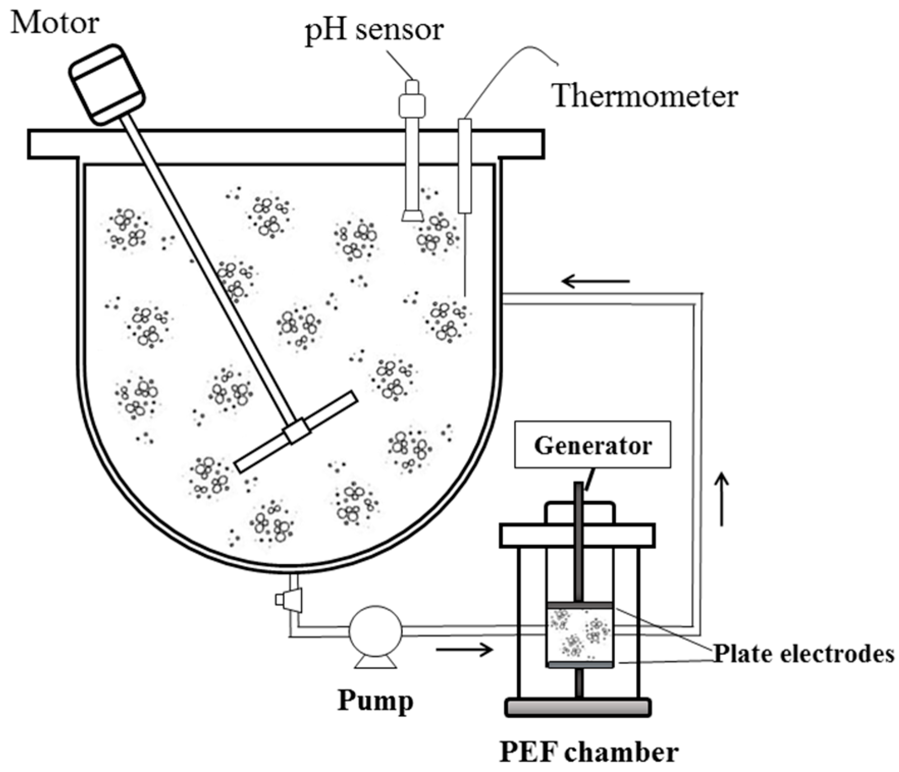
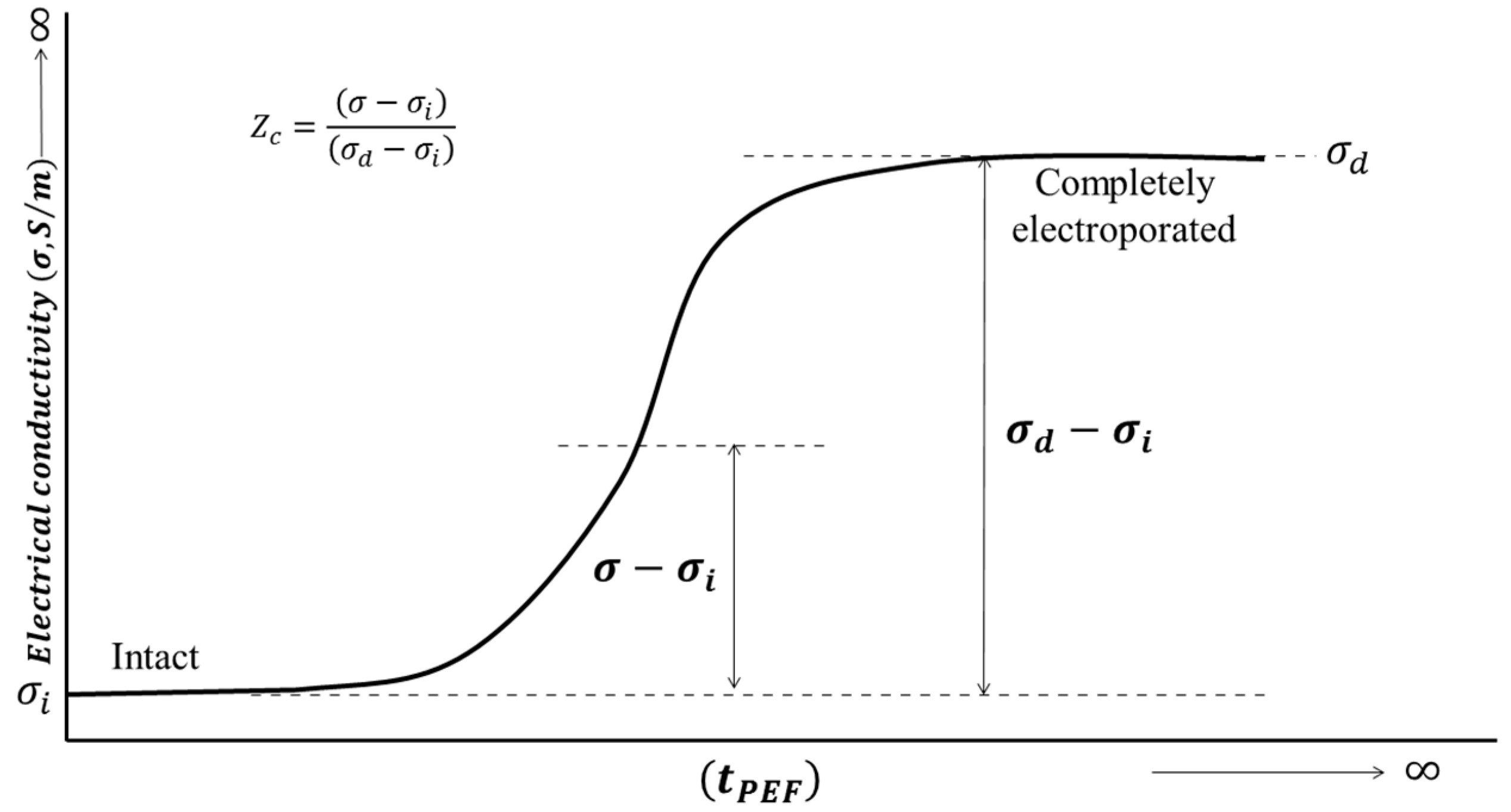
The effects of PEF on the food system are also related to the properties of the fermentation medium. The electrical properties of samples such as the electrical conductivity and their electric strength are key factors in the PEF treatment because they determine the maximum electric field intensity and applicable current flow. Biosuspensions with high electrical conductivities are not ideal for PEF treatments because they display a smaller electric field across the treatment chamber. Therefore, a difference in the electrical conductivity between the microbial cytoplasm and the surrounding medium is desired because a greater flow of ionic substances through the membrane occurs, which causes weakening in its inner structure [37]. Thus, the conductivity is a useful parameter for the characterization of electroporation in cellular tissues [38]. For instance, disintegration induced by PEF can be quantified by the method of estimation of the cell damage index Z [39], based on a simple measurement of electrical conductivity in the low-frequency region (1–10 kHz). The electrical conductivity average of cellular tissue increases with the development of electroporation during the PEF treatment time, as it is shown in Figure 3. To quantify the cell disintegration index (Zc), a simple equation was proposed by Lebovka et al. [39]:
where σ is the electrical conductivity value measured at low frequency (1–10 kHz) and indexes “i” and “d” refer to the conductivities of intact and completely damaged tissues, respectively.
Besides the electrical properties of the fermentation medium, the susceptibility of the microorganisms to PEF treatment is strongly related to the morphological and physiological characteristics of the treated microorganisms, such as cell type, size and shape, cell density, and growth stage of the microorganisms. For instance, Gram-positive vegetative cells are more resistant to PEF treatment than Gram-negative cells, possibly because Gram-positive bacteria possess a thick and more robust cell wall due to cross-linking of peptidoglycan. It is also interesting to note that large cells are more sensitive to lower field strengths than small cells, because of the larger surface that is in contact with the PEF [40]. Yeasts are more sensitive to electric fields than Gram-positive bacteria due to their larger size, but they may be more resistant than Gram-negative bacteria. Here, S-S bonds in the yeast walls seem to stabilize yeast cells against PEF alterations [41].
The efficiency of PEF is also dependent on cell density. The concentration of microorganisms during fermentation may or may not have an effect on their inactivation, depending on the specific conditions of the treatment process. For instance, a study reported that inactivation of E. coli in simulated ultra-filtrated milk was not affected when the concentration of the microorganisms varied from 103 to 108 cfu/mL after being subjected to 70 kV/cm, 16 pulses, and a pulse width of 2 µs [42]. Another study has shown that microbial inactivation in apple juice was slightly lower when the number of S. cerevisiae increased, possibly due to the formation of a cluster of microbial cells or concealed microorganisms in low electric field regions [43].
In addition, the growth stage of the microorganisms can also influence the effectiveness of PEF treatment. In general, cells in logarithmic phase are more sensitive to stress than cells in the lag and stationary phases. The reason is that microbial growth in the logarithmic phase is characterized by a high proportion of cells undergoing division, during which the cell membrane is more sensitive to the applied electric field [44]. For example, PEF treatment of 200 pulses at 19 kV/cm of electric field intensity applied to Salmonella senftenberg cells at different growth stages resulted in a decrease of 1.5 log cycles for the logarithmic phase compared to the lag and stationary phases [45].
3.3. Production of Biomolecules from Fermented Foods
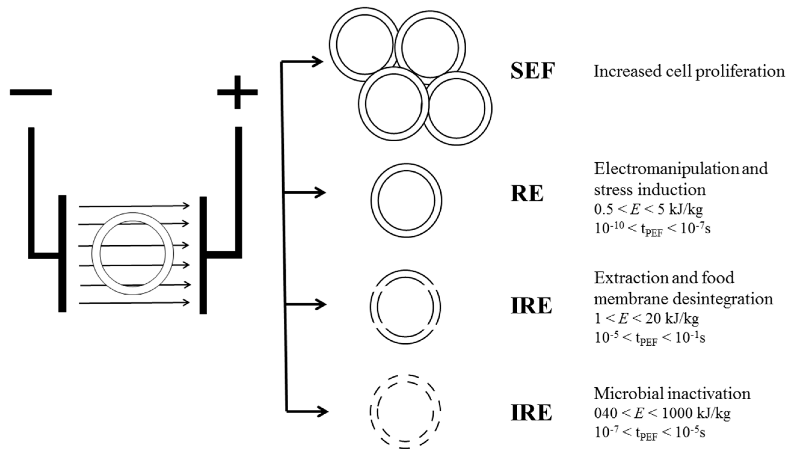
Similar to the abovementioned ultrasound applications, the production of biomolecules from fermented foods treated with PEF technology will depend largely on the effects produced on microbial cells. Thus, depending on the specific energy and treatment time, different situations can occur: stimulating electric fields, reversible electroporation, and irreversible electroporation (Figure 4).
Successful inactivation of microorganisms by pulsed electric field treatment has been reported by several researchers [46,47,48]. One of the most attractive attributes of this technology is that samples can be treated at, or roughly above, ambient temperature for only a few microseconds, thus minimizing the energy loss caused by heating [49]. However, Frey et al. [50] demonstrated that with a decreasing treatment energy—by reducing the field strength and pulse duration to sublethal values—a stimulatory effect on the growth of cells takes place. Another study reported a positive effect of PEF on S. cerevisiae growth using, as the main parameter, frequencies between 5 to 150 Hz and intensities of 0 to 1.625 kV/cm [51]. The reason behind electrostimulation is mostly unknown, but several hypotheses to explain it have been suggested by different researchers. Enzymatic systems or even metabolic pathways related to some processes occurring at the membrane level could be electroactivated, leading to changes in cell proliferation and differentiation, which can be initiated, promoted, or co-promoted [52]. For example, S. cerevisiae cells cultivated under electrical stimulation have shown to alter the culture cycle and promote synchrony in cell division [53]. Synchrony suggests that the applied potential is able to trigger on or off cell cycle activity. It is possible that also, along with this, a reduction in maturation time and cell volume occurs. The same author also proposed that an acceleration of signal transduction paths may be possible, which leads the cell to a “start point” in cell cycle. Another hypothesis is that PEF induces changes in the cell cycle of microorganisms, diminishing the G1 phase. The early passage of the yeast from the G1 phase to the S phase means that the cellular cycle duration would be smaller than the time necessary for the cellular size duplication, thus producing a higher number of smaller cells that would persist until the third generation [54].
Induction of stress in cells (reversible electroporation) has long been applied in biology, biotechnology, and medicine, and yet is fairly new to the food industry [52,55]. Distinct effects in metabolic processes may occur depending on the parameters of the external electric field, such as its strength, cell shape, and membrane properties. The application of a low intensity treatment at low electric field strength and/or pulse number, through initiating a conductive channel across the membrane, does not cause irreversible cell rupture. The variation in the endogenous membrane potential by an external field impacts the structure and function of the membrane compartments, proteins, and lipid bilayer, which allows the extraction of intracellular compounds [56].
Finally, the irreversible permeabilization of cell membranes offers a wide range of applications, including the extraction or diffusion of bioactive metabolites [57,58,59]. It was shown that PEF treatment of the aqueous suspension of wine yeasts (S. cerevisiae bayanus, strain DV10) at E = 10 kV/cm allowed high extraction of ionic components and low extraction of high molecular weight components [57]. Another study has shown that PEF reduced the lag phase of L. acidophilus and increased bacteriocin production [58]. PEF treatment at E = 40 kV/cm to the aqueous suspension of the same wine yeasts allowed the extraction of 70% of ionic substances, 1% of proteins, and 16% of nucleic acids [59], as a result of the enhancement of the mass transfer. In wine production, combining PEF with subsequent fermentation on grape skins produced a more intense color, while combining PEF with subsequent maceration yields an increased content of polyphenolic compounds in the wine [60].
4. Conclusions
The application of high-intensity ultrasound and PEF in fermented foods has shown promising results, enhancing the process and improving the content of some bioactive compounds. All results are associated with the physical and chemical effects generated at the microscopic and structural levels of the food matrix. The effectiveness of both technologies on the fermentation process is strongly influenced by various factors including microbial characteristics (e.g., type of microorganism, medium type, and composition), process parameters (e.g., power, frequency, electric field intensity), treatment time, pH, and temperature. Due to the fact that several variables coexist, the effects of both techniques are complex to predict and cannot be simply generalized to meet the demands of different applications. Therefore, further studies under different operating conditions are needed to verify the potential impact of ultrasound and PEF on the production of bioactive metabolites with the aim of optimizing the process to provide the optimum results for each application.
Acknowledgments
The authors thank the Consejo Nacional de Investigaciones Científicas y Técnicas (CONICET, Argentina) and the Alibiotech project for partial support of this work.
Author Contributions
Leandro Galván-D’Alessandro performed the literature review on pulsed electric field and Ramiro Ariel Carciochi took care of the ultrasound-assisted fermentation process.
Conflicts of Interest
The authors declare no conflict of interest.
References
- Cutzu, R.; Bardi, L. Production of bioethanol from agricultural wastes using residual thermal energy of a cogeneration plant in the distillation phase. Fermentation 2017, 3, 24. [Google Scholar] [CrossRef]
- Limón, R.I.; Peñas, E.; Torino, M.I.; Martínez-Villaluenga, C.; Dueñas, M.; Frias, J. Fermentation enhances the content of bioactive compounds in kidney bean extracts. Food Chem. 2015, 172, 343–352. [Google Scholar] [CrossRef] [PubMed]
- Mañas, P.; Pagán, R. Microbial inactivation by new technologies of food preservation. J. Appl. Microbiol. 2005, 98, 1387–1399. [Google Scholar] [CrossRef] [PubMed]
- Pereira, R.N.; Vicente, A.A. Environmental impact of novel thermal and non-thermal technologies in food processing. Food Res. Int. 2010, 43, 1936–1943. [Google Scholar] [CrossRef] [Green Version]
- Ojha, K.S.; Mason, T.J.; O’Donnell, C.P.; Kerry, J.P.; Tiwari, B.K. Ultrasound technology for food fermentation applications. Ultrason. Sonochem. 2017, 34, 410–417. [Google Scholar] [CrossRef] [PubMed]
- Nowacka, M.; Wedzik, M. Effect of ultrasound treatment on microstructure, colour and carotenoid content in fresh and dried carrot tissue. Appl. Acoust. 2016, 103, 163–171. [Google Scholar] [CrossRef]
- Chemat, F.; Khan, M. K. Applications of ultrasound in food technology: Processing, preservation and extraction. Ultrason. Sonochem. 2011, 18, 813–835. [Google Scholar] [CrossRef] [PubMed]
- Mason, T.J.; Riera, E.; Vercet, A.; Lopez-Buesa, P. Application of ultrasound. In Emerging Technologies for Food Processing, 1st ed.; Sun, D.W., Ed.; Elsevier Academic Press: London, UK, 2005; pp. 323–351. ISBN 0-12-676757-2. [Google Scholar]
- Esclapez, M.D.; Garcia-Perez, J.V.; Mulet, A.; Cárcel, J.A. Ultrasound-assisted extraction of natural products. Food Eng. Rev. 2011, 3, 108–120. [Google Scholar] [CrossRef]
- O’Donnell, C.P.; Tiwari, B.K.; Bourke, P.; Cullen, P.J. Effect of ultrasonic processing on food enzymes of industrial importance. Trends Food Sci. Technol. 2010, 21, 358–367. [Google Scholar] [CrossRef]
- Ashokkumar, M.; Bhaskaracharya, R.; Kentish, S.; Lee, J.; Palmer, M.; Zisu, B. The ultrasonic processing of dairy products—An overview. Dairy Sci. Technol. 2010, 90, 147–168. [Google Scholar] [CrossRef]
- Awad, T.S.; Moharram, H.A.; Shaltout, O.E.; Asker, D.; Youssef, M.M. Applications of ultrasound in analysis, processing and quality control of food: A review. Food Res. Int. 2012, 48, 410–427. [Google Scholar] [CrossRef]
- Nguyen, T.M.P.; Lee, Y.K.; Zhou, W. Stimulating fermentative activities of bifidobacteria in milk by high intensity ultrasound. Int. Dairy J. 2009, 19, 410–416. [Google Scholar] [CrossRef]
- Nguyen, T.M.P.; Lee, Y.K.; Zhou, W. Effect of high intensity ultrasound on carbohydrate metabolism of bifidobacteria in milk fermentation. Food Chem. 2012, 130, 866–874. [Google Scholar] [CrossRef]
- Yeo, S.-K.; Liong, M.-T. Effect of ultrasound on the growth of probiotics and bioconversion of isoflvones in prebiotic-supplemented soymilk. J. Agric. Food Chem. 2011, 59, 885–897. [Google Scholar] [CrossRef] [PubMed]
- Barukčić, I.; Jakopović, K.L.; Herceg, Z.; Karlović, S.; Božanić, R. Influence of high intensity ultrasound on microbial reduction, physico-chemical characteristics and fermentation of sweet whey. Innov. Food Sci. Emerg. Technol. 2015, 27, 94–101. [Google Scholar] [CrossRef]
- Lentacker, I.; De Cock, I.; Deckers, R.; De Smedt, S.C.; Moonen, C.T. Understanding ultrasound induced sonoporation: Definitions and underlying mechanisms. Adv. Drug Deliv. Rev. 2014, 72, 49–64. [Google Scholar] [CrossRef] [PubMed] [Green Version]
- Yang, F.; Gu, N.; Chen, D.; Xi, X.; Zhang, D.; Li, Y.; Wu, J. Experimental study on cell self-sealing during sonoporation. J. Control. Release 2008, 131, 205–210. [Google Scholar] [CrossRef] [PubMed]
- Yeo, S.-K.; Liong, M.-T. Effects and applications of sub-lethal ultrasound, electroporation and UV radiations in bioprocessing. Ann. Microbiol. 2012, 63, 813–824. [Google Scholar] [CrossRef]
- Yeo, S.-K.; Liong, M.-T. Effect of ultrasound on bioconversion of isoflavones and probiotic properties of parent organisms and subsequent passages of Lactobacillus. LWT—Food Sci. Technol. 2013, 51, 289–295. [Google Scholar] [CrossRef]
- Guzel, B.H.; Arroyo, C.; Condón, S.; Pagán, R.; Bayindirli, A.; Alpas, H. Inactivation of Escherichia coli and Listeria monocytogenes by ultrasonic waves under pressure at nonlethal (manosonication) and lethal temperatures (manothermosonication) in acidic fruit juices. Food Bioprocess Technol. 2014, 7, 1701–1712. [Google Scholar] [CrossRef]
- Terefe, N.S.; Buckow, R.; Versteeg, C. Quality-related enzymes in plant-based products: Effects of novel food-processing technologies, Part 3: Ultrasonic processing. Crit. Rev. Food Sci. Nutr. 2015, 55, 147–158. [Google Scholar] [CrossRef] [PubMed]
- Barteri, M.; Diociaiuti, M.; Pala, A.; Rotella, S. Low frequency ultrasound induces aggregation of porcine fumarase by free radicals production. Biophys. Chem. 2004, 111, 35–42. [Google Scholar] [CrossRef] [PubMed]
- Guiseppi-Elie, A.; Choi, S.H.; Geckeler, K.E. Ultrasonic processing of enzymes: Effect on enzymatic activity of glucose oxidase. J. Mol. Catal. B Enzym. 2009, 58, 118–123. [Google Scholar] [CrossRef]
- Ozuna, C.; Paniagua-Martínez, I.; Castaño-Tostado, E.; Ozimek, L.; Amaya-Llano, S.L. Innovative applications of high-intensity ultrasound in the development of functional food ingredients: Production of protein hydrolysates and bioactive peptides. Food Res. Int. 2015, 77, 685–696. [Google Scholar] [CrossRef]
- Rice, S.; Koziel, J.A.; Dharmadhikari, M.; Fennell, A. Evaluation of tannins and anthocyanins in marquette, frontenac, and St. croix cold-hardy grape cultivars. Fermentation 2017, 3, 47. [Google Scholar] [CrossRef]
- Tao, Y.; Zhang, Z.; Sun, D.-W. Experimental and modeling studies of ultrasound-assisted release of phenolics from oak chips into model wine. Ultrason. Sonochem. 2014, 21, 1839–1848. [Google Scholar] [CrossRef] [PubMed]
- Katina, K.; Liukkonen, K.H.; Kaukovirta-Norja, A.; Adlercreutz, H.; Heinonen, S.M.; Lampi, A.M.; Pihlava, J.M.; Poutanen, K. Fermentation-induced changes in the nutritional value of native or germinated rye. J. Cereal Sci. 2007, 46, 348–355. [Google Scholar] [CrossRef]
- Fernández-Orozco, R.; Frías, J.; Zielinski, H.; Muñoz, R.; Piskula, M.K.; Kozlowska, H.; Vidal-Valverde, C. Evaluation of bioprocesses to improve the antioxidant properties of chickpeas. LWT–Food Sci. Technol. 2009, 42, 885–892. [Google Scholar] [CrossRef]
- Carciochi, R.A.; Galván-D’Alessandro, L.; Vandendriessche, P.; Chollet, S. Effect of germination and fermentation process on the antioxidant compounds of quinoa seeds. Plant Foods Hum. Nutr. 2016, 71, 361–367. [Google Scholar] [CrossRef] [PubMed]
- Panagopoulos, D.J.; Karabarbounis, A.; Margaritis, L.H. Mechanism for action of electromagnetic fields on cells. Biochem. Biophys. Res. Commun. 2002, 298, 95–102. [Google Scholar] [CrossRef]
- Lebovka, N.; Vorobiev, E. Techniques and procedures to detect electroporation in food cellular tissues. In Proceedings of the School on Applications of Pulsed Electric Fields for Food Processing, Zaragoza, Spain, 20–23 January 2014; pp. 46–47. [Google Scholar]
- Koubaa, M.; Roselló-Soto, E.; Šic Žlabur, J.; Režek Jambrak, A.; Brnčić, M.; Grimi, N.; Boussetta, N.; Barba, F.J. Current and new insights in the sustainable and green recovery of nutritionally valuable compounds from Stevia rebaudiana bertoni. J. Agric. Food Chem. 2015, 63, 6835–6846. [Google Scholar] [CrossRef] [PubMed]
- Vorobiev, E.; Lebovka, N. Enhanced extraction from solid foods and biosuspensions by pulsed electrical energy. Food Eng. Rev. 2010, 2, 95–108. [Google Scholar] [CrossRef]
- Jaeger, H. PEF Process Performance Analysis. In Proceedings of the School on Applications of Pulsed Electric Fields for Food Processing, Zaragoza, Spain, 20–23 January 2014; pp. 87–93. [Google Scholar]
- Puértolas, E.; Luengo, E.; Alvarez, I.; Raso, J. Improving mass transfer to soften tissues by pulsed electric fields: Fundamentals and applications. Annu. Rev. Food Sci. Technol. 2012, 3, 263–282. [Google Scholar] [CrossRef] [PubMed]
- Jayaram, S.; Castle, G.S.P.; Margaritis, A. Effects of high electric field pulses on Lactobacillus brevis at elevated temperatures. In Proceedings of the Conference on Electrical Insulation and Dielectric Phenomena, Dearborn, MI, USA, 18–21 October 1992; pp. 674–681. [Google Scholar] [CrossRef]
- Pliquett, U. Bioimpedance: A Review for Food Processing. Food Eng. Rev. 2010, 2, 74–94. [Google Scholar] [CrossRef]
- Lebovka, N.I.; Bazhal, M.I.; Vorobiev, E. Estimation of characteristic damage time of food materials in pulsed electric fields. J. Food Eng. 2002, 4, 337–346. [Google Scholar] [CrossRef]
- Ben Ammar, J.; Lanoisellée, J.L.; Lebovka, N.I.; Van Hecke, E.; Vorobiev, E. Impact of a pulsed electric field on damage of plant tissues: Effects of cell size and tissue electrical conductivity. J. Food Sci. 2011, 76, 90–97. [Google Scholar] [CrossRef] [PubMed]
- Shamtsyan, M. The influence of electric field on microbial growth. J. Eur. Hyg. Eng. Des. Group 2012, 1, 88–92. [Google Scholar]
- Zhang, Q.H.; Qin, B.L.; Barbosa-Cánovas, G.V.; Swanson, B.G. Inactivation of Escherichia coli for food pasteurization by highstrength pulsed electric fields. J. Food Process. Preserv. 1995, 19, 103–118. [Google Scholar] [CrossRef]
- Qin, B.L.; Pothakamury, U.R.; Barbosa-Cánovas, G.V.; Swanson, B.G. Nonthermal pasteurization of liquid foods using high-intensity pulsed electric field. Crit. Rev. Food Sci. Nutr. 1996, 36, 603–627. [Google Scholar] [CrossRef] [PubMed]
- Pothakamury, U.R.; Vega, H.; Zhang, Q.; Barbosa-Canovas, G.V.; Swanson, B.G. Effect of growth stage and processing temperature on the inactivation of E. coli by pulsed electric fields. J. Food Prot. 1996, 59, 1167–1171. [Google Scholar] [CrossRef]
- Alvarez, I.; Palop, J.R.A.; Sala, F.J. Influence of different factors on the inactivation of Salmonella senftenberg by pulsed electric fields. Int. J. Food Microbiol. 2000, 55, 143–146. [Google Scholar] [CrossRef]
- Harrison, S.L.; Barbosa-Cánovas, G.V.; Swanson, B.G. Saccharomyces cerevisiae structural changes induced by pulsed electric field treatment. LWT—Food Sci. Technol. 1997, 30, 236–240. [Google Scholar] [CrossRef]
- Heinz, V.; Toepfl, S.; Knorr, D. Impact of temperature on lethality and energy efficiency of apple juice pasteurization by pulsed electric fields treatment. Innov. Food Sci. Emerg. 2003, 4, 167–175. [Google Scholar] [CrossRef]
- Toepfl, S.; Heinz, V.; Knorr, D. High intensity pulsed electric fields applied for food preservation. Chem. Eng. Process. 2007, 46, 537–546. [Google Scholar] [CrossRef]
- Barbosa-Cánovas, G.V.; Gongora-Nieto, M.M.; Pothakamury, U.R.; Swanson, B.G. Preservation of Foods with Pulsed Electric Fields, 1st ed.; Academic Press: London, UK, 1999; ISBN 0-12-078149-2. [Google Scholar]
- Frey, W.; Flickinger, B.; Berghoefer, T.; Eing, C.; Liu, Q.; Nick, P. Electropermeabilization versus nsPEF-stimulation—Pulsed electric fields can stimulate the growth of plants and fungi. In Proceedings of the 10th International Conference of the European Bioelectromagnetic Association, Roma, Italy, 21–24 February 2011. [Google Scholar]
- Fiedler, U.; Gröbner, U.; Berg, H. Electrostimulation of yeast proliferation. Bioelectrochem. Bioenerg. 1995, 38, 423–425. [Google Scholar] [CrossRef]
- Hunt, R.W.; Zavalin, A.; Bhatnagar, A.; Chinnasamy, S.; Das, K.C. Electromagnetic biostimulation of living cultures for biotechnology, biofuel and bioenergy applications. Int. J. Mol. Sci. 2009, 10, 4515–4558. [Google Scholar] [CrossRef] [PubMed]
- Araujo, O.Q.F.; Coelho, M.A.Z.; Margarit, I.C.P.; Vaz-Junior, C.A.; Rocha-Leão, M.H.M. Electrical stimulation of Saccharomyces cerevisiae cultures. Braz. J. Microbiol. 2004, 35, 97–103. [Google Scholar] [CrossRef]
- Rocha-Leão, M.H.; Coelho, M.A.Z.; Margarit, I.P.; Catarino, A.A.; Gandelman, R.A.; Vaz, C.A.; Araújo, O.Q.F. Biochemical cell responses to electrical stress stimulation. In Proceedings of the 2nd Mercosur Congress on Chemical Engineering 4th Mercosur Congress on Process Systems Engineering, Rio de Janeiro, Brazil, 14–18 August 2005. [Google Scholar]
- Tanino, T.; Sato, S.; Oshige, M.; Ohshima, T. Analysis of the stress response of yeast Saccharomyces cerevisiae toward pulsed electric field. J. Electrostat. 2012, 70, 212–216. [Google Scholar] [CrossRef]
- Fologea, D.; Vassu-Dimovb, T.; Stoicab, I.; Csutakb, O.; Radua, M. Increase of Saccharomyces cerevisiae plating efficiency after treatment with bipolar electric pulses. Bioelectrochem. Bioenerg. 1998, 46, 285–287. [Google Scholar] [CrossRef]
- Shynkaryk, M.V.; Lebovka, N.I.; Lanoisellé, J.-L.; Nonus, M.; Bedel-Clotour, C.; Vorobiev, E. Electrically-assisted extraction of bio-products using high pressure disruption of yeast cells (Saccharomyces cerevisiae). J. Food Eng. 2009, 92, 189–195. [Google Scholar] [CrossRef]
- Loghavi, L.; Sastry, S.K.; Yousef, A.E. Effect of moderate electric field frequency on growth kinetics and metabolic activity of Lactobacillus acidophilus. Biotechnol. Progr. 2008, 24, 148–153. [Google Scholar] [CrossRef] [PubMed]
- Liu, D.; Lebovka, N.I.; Vorobiev, E. Impact of electric pulse treatment on selective extraction of intracellular compounds from Saccharomyces cerevisiae yeasts. Food Bioprocess Technol. 2013, 6, 576–584. [Google Scholar] [CrossRef]
- Mahnic-Kalamiza, S.; Vorobiev, E.; Miklavcic, D. Electroporation in food processing and biorefinery. J. Membr. Biol. 2014, 247, 1279–1304. [Google Scholar] [CrossRef] [PubMed]
Figure 1.
Ultrasound-assisted bioreactors: (A) Sonication produced with transducers placed on the outside of the fermentation tank; (B) Ultrasound provided with an ultrasound (US) probe.
Figure 1.
Ultrasound-assisted bioreactors: (A) Sonication produced with transducers placed on the outside of the fermentation tank; (B) Ultrasound provided with an ultrasound (US) probe.

Figure 2.
Schema of pulsed electric field fermentation in a continuous mode.

Figure 3.
Determination of the electrical conductivity disintegration index Z: electrical conductivity versus treatment time of pulsed electric field [32].
Figure 3.
Determination of the electrical conductivity disintegration index Z: electrical conductivity versus treatment time of pulsed electric field [32].

Figure 4.
Schematic presentation of the exposure of a biological cell to pulsed electric field and the applications depending on energy and treatment time: stimulating electric fields (SEF), reversible electroporation (RE), and irreversible electroporation (IRE).
Figure 4.
Schematic presentation of the exposure of a biological cell to pulsed electric field and the applications depending on energy and treatment time: stimulating electric fields (SEF), reversible electroporation (RE), and irreversible electroporation (IRE).
© 2018 by the authors. Licensee MDPI, Basel, Switzerland. This article is an open access article distributed under the terms and conditions of the Creative Commons Attribution (CC BY) license (http://creativecommons.org/licenses/by/4.0/).
Share and Cite
MDPI and ACS Style
Galván-D’Alessandro, L.; Carciochi, R.A. Fermentation Assisted by Pulsed Electric Field and Ultrasound: A Review. Fermentation 2018, 4, 1. https://doi.org/10.3390/fermentation4010001
AMA Style
Galván-D’Alessandro L, Carciochi RA. Fermentation Assisted by Pulsed Electric Field and Ultrasound: A Review. Fermentation. 2018; 4(1):1. https://doi.org/10.3390/fermentation4010001
Chicago/Turabian StyleGalván-D’Alessandro, Leandro, and Ramiro Ariel Carciochi. 2018. "Fermentation Assisted by Pulsed Electric Field and Ultrasound: A Review" Fermentation 4, no. 1: 1. https://doi.org/10.3390/fermentation4010001
Note that from the first issue of 2016, this journal uses article numbers instead of page numbers. See further details here.