Article

Antiviral and Antioxidant Potential of Fungal Endophytes of Egyptian Medicinal Plants

by
Khaled A. Selim
1,2,*,
Waill A. Elkhateeb
2,
Ahmed M. Tawila
3,
Ahmed A. El-Beih
2,*,
Tahany M. Abdel-Rahman
4,
Ahmed I. El-Diwany
2 and
Eman F. Ahmed
2
1
Interfaculty Institute of Microbiology and Infection Medicine, Eberhard Karls Universität Tübingen, 72076 Tübingen, Germany
2
Chemistry of Natural and Microbial Products Department, National Research Center, Dokki, Cairo 12622, Egypt
3
Division of Natural Drug Discovery, Institute of Natural Medicine, University of Toyama, 2630 Sugitani, Toyama 930-0194, Japan
4
Botany Department, Faculty of Science, Cairo University, P.O. Box 12613, Giza, Egypt
*
Authors to whom correspondence should be addressed.
Fermentation 2018, 4(3), 49; https://doi.org/10.3390/fermentation4030049
Submission received: 23 April 2018 / Revised: 10 June 2018 / Accepted: 14 June 2018 / Published: 25 June 2018
(This article belongs to the Special Issue Fermentation and Bioactive Metabolites)

Abstract

:
This study aimed to explore the antioxidant potential and antiviral activity of endophytic fungi which were isolated from healthy living tissues of medicinal plants. Endophytic strains (29 different taxa) were isolated from 18 Egyptian medicinal plants collected from Saint Katherine Protectorate, Egypt. The fungal endophytes were identified based on morphological characters. All isolates were identified as ascomycetes, except two Zygomycetes strains (Absidia corymbifera and Mucor fuscus). Isolated endophytes were cultivated on potato dextrose media. The fungal metabolites were extracted by ethyl acetate and examined for their biological activities. Among 99 total extracts, only Chaetomium globosum, which was isolated from Adiantum capillus, showed a promising DPPH (1,1-diphenyl-2-picrylhydrazyl) scavenging activity (99% at 100 µg/mL). Fifteen extracts prohibited the reproduction of HSV-2 virus. On the other hand, the reproduction of VSV-virus was inhibited by sixteen endophytic extracts. The promising anti-(HSV-2 and VSV) extract of endophytic Pleospora tarda strain; that was originally isolated from the medicinal plant Ephedra aphylla, showed viral inhibitory activity of 40.7% and 15.2%, respectively. Two compounds, for which antiviral activates could be attributed, were isolated and identified as alternariol and alternariol-(9)-methyl ether using different NMR techniques from P. tarda extract. For the first time, we report here the ability of the endophytic fungus P. tarda to produce alternariol and alternariol-(9)-methyl ether. The results indicate that the endophytic fungi from medicinal plants are promising sources of bioactive compounds.

Graphical Abstract

1. Introduction

There is a global need for new antiviral compounds to solve drug resistance problems. The resistance of human disease to well-known (commercial) antibiotics is increasing rapidly nowadays, so discovering new alternative agents is indispensable required for management those maladies. Endophytes are relatively not well-studied microorganisms that recently gained importance due to their different biological activities and bioactive compounds with a high level of structural diversity [1,2,3,4]. They are considered as a rich source of novel natural metabolites for exploitation in medicine, industry and agriculture [1,2,3,4,5].
The medicinal plants are considered as one of the major reservoirs of endophytic bioactive metabolites for the use in the medicinal applications [2,3,4,6]. In the present surveys, we evaluated the antioxidant potential and the antiviral effect of the total metabolites produced by some endophytic fungi that inhabited Egyptian medicinal plants. Egyptian medicinal plants are well known for their applications in traditional medicine to cure many infectious diseases [7,8]. Saint Katherine Protectorate is an area rich with medicinal plants, but the biodiversity of microbial communities in Sinai is poorly studied. Nowadays, this area attracts more attention for studying the diversity of endophytic mycobiota from medicinal plants [8,9]. The importance of compounds with antioxidant activity is their protective effect against the oxidative stress caused by oxygen-derived free radicals [10]. The culture filtrate of Pestalotiopsis microspora, which was isolated from combretaceaous plant Terminalia morobensis, showed promising antioxidant activity [11]. Similarly, graphislactone A, a potent antioxidant agent, was identified as a phenolic metabolite from the endophytic fungus Cephalosporium sp., that resided in Trachelospermum jasminoides [12]. Moreover, many endophytes were reported as potent viral inhibitors. Recently, it was reported that an endophytic Streptomyces sp. strain isolated from the mangrove plant Bruguiera gymnorrhiza, produced xiamycin, which exhibits selective anti-HIV activity [13]. Additionally, the endophytic Emericella sp. (HK-ZJ) isolated from another mangrove plant Aegiceras corniculatum, produces several bioactive isoindolone compounds, for which two of them showed moderate activity against influenza A virus (H1N1) [14].
In this current report, we set to exploit the Egyptian medicinal plants in an important region of Sinai to explore the biodiversity and the bioactivity of endophytic metabolites from these medicinal plants.

2. Results and Discussion

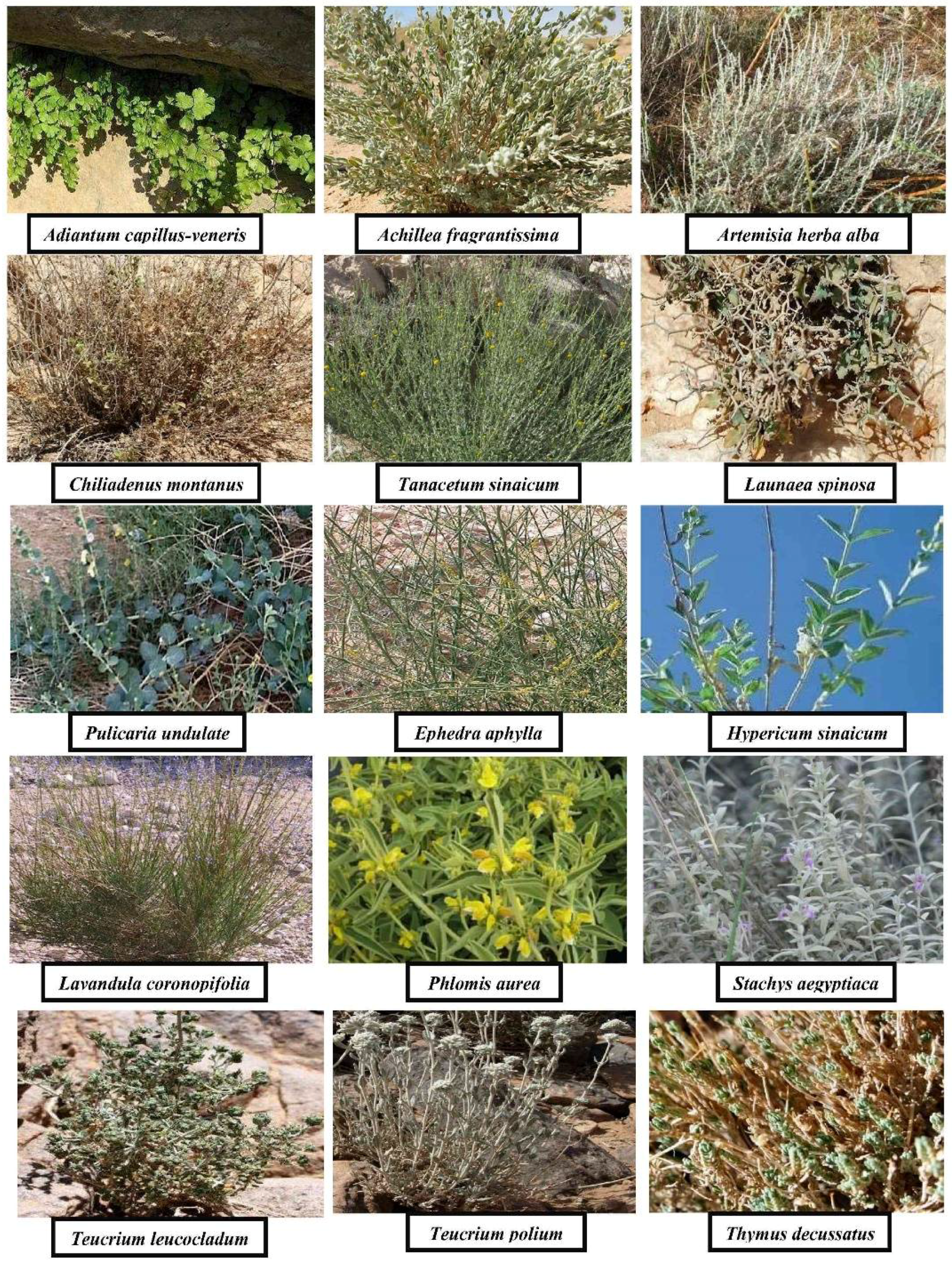
2.1. Collected Medicinal Plants (Sampling)

The Egyptian medicinal plants were collected and identified morphologically as shown in Table 1. The healthy and disease-free samples were classified into seven families (Adiantaceae, Compositae, Ephedraceae, Euphorbiaceae, Hypericaceae, Labiatae, and Rubiaceae). Figure 1 shows the selected medicinal plants.

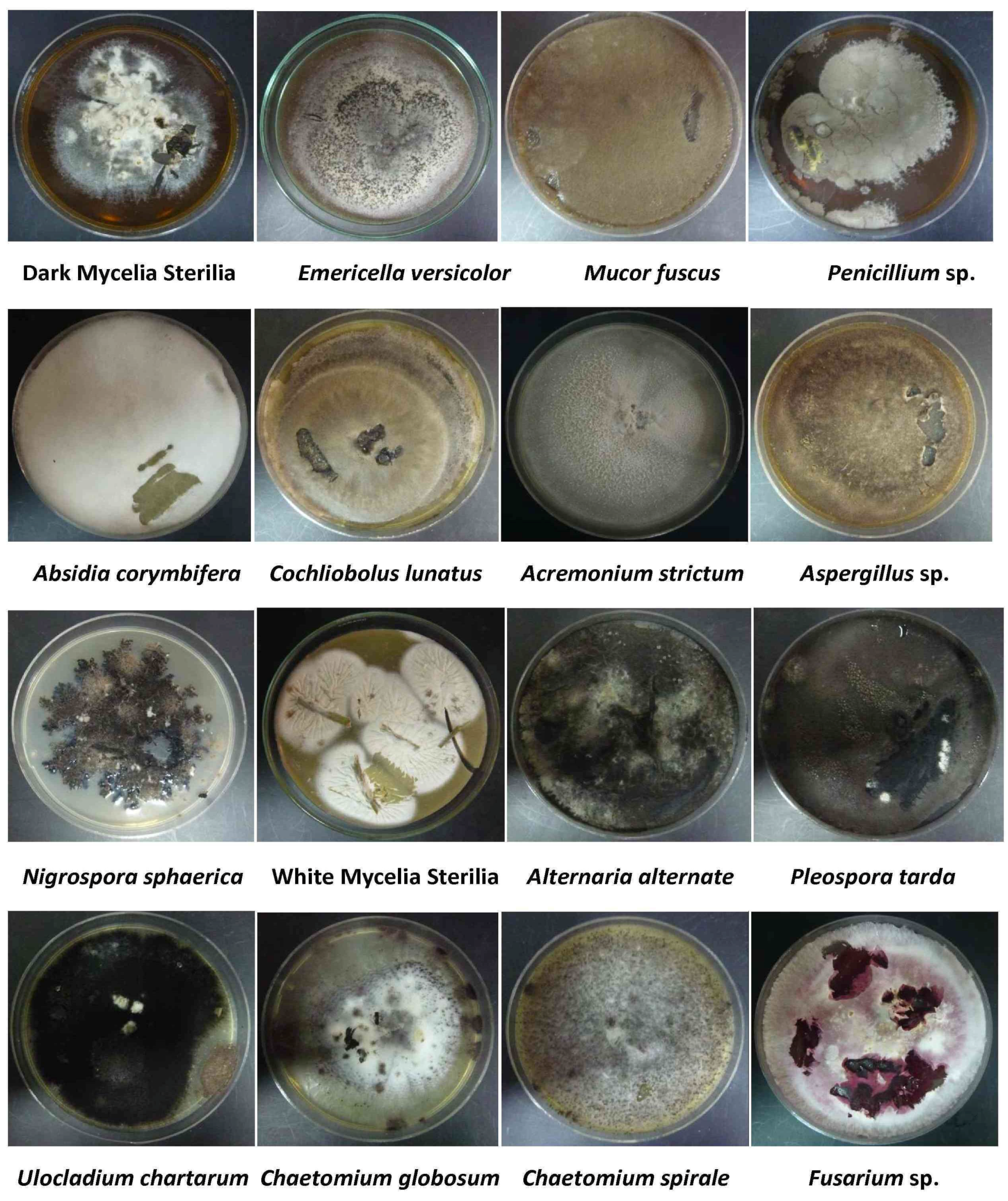
2.2. Isolation and Identification of Endophytic Fungi

Plant samples were used to isolate one hundred and thirty-two endophytic strains, which were purified from the leaves and stems of 18 medicinal plants (Supplementary Tables S1 and S2). The isolated fungal endophytes were identified according to Moubasher (1993) [15]. Isolated fungal strains were cultivated on potato dextrose agar media, as presented in Figure 2.
A collection of 10 isolates from the genus Aspergillus was demonstrated. Aspergillus was represented by four species, belonging to three groups. Aspergillus flavus (three isolates), Aspergillus niger (three isolates), Aspergillus sydowii (two isolates), Aspergillus versicolor (one isolate) and one unidentified Aspergillus species, respectively, as in Supplementary Table S1. Furthermore, nine isolates from genus Penicillium were purified from the studied desert plants, Penicillium was represented by two species, Penicillium chrysogenum and Penicillium corylophilum, which belonged to one section: Biverticillata-Asymmetrica (two species belonged to subsection Velutina. Penicillium chrysogenum (three isolates), Penicillium corylophilum (two isolates) and unidentified Penicillium species (four isolates), respectively, as revealed in Supplementary Table S1.
The remaining isolates of different genera and species in the current study, other than Aspergillus and Penicillium, were listed in Supplementary Table S2. Absidia corymbifora (1 isolates), Acremonium strictum (two isolates), and one unidentified Acremonium sp., Alternaria alternate (one isolate), Chaetomium globosum (two isolates), C. spirale (one isolate), Cochliobolus lunatus (one isolate), Fusarium oxysporum (two isolates) and unidentified Fusarium sp. (one isolate), Mucor fuscus (one isolate), Nigrospora sphaerica (three isolate), Phoma leveillei (one isolate), Scopulariopsis sp. (one isolate), Ulocladium chartarum (three isolate), Ulocladium atrum (one species), Pleospora tarda (five isolates) and yeast (one isolate), and fungi with sterile mycelia (white, (25 isolates) and dark, (26 isolates)). All endophytic isolates belonged to ascomycetes, except two zygomycetes strains (Absidia corymbifera and Mucor fuscus). It was obvious that fungi with sterile mycelia represented 51% from the total number of isolated endophytic strains. Sterile mycelia were common endophytes, 19.69% for dark sterile mycelia, and 18.93% for white sterile mycelia, and these percentages came in accordance with many endophytic studies [16,17].

2.3. Antioxidant and Antiviral Activities

More than one hundred endophytic fungi were isolated and purified from Saint Katharine medicinal plants, and extracted with ethyl acetate. The total extracts at concentration of 50 µg/mL were investigated for their antioxidant and antiviral activities. Of all the 99 crude extracts, only Chaetomium globosum isolated from the internal tissues of Adiantum capillus-veneris revealed a promising antioxidant activity (99%) DPPH scavenging activity at the level 100 μg/mL of total extract (Table 2). Genetic identification (18S rDNA) of the active antioxidant isolate was achieved in our previous report [10]. It showed a high similarity to Chaetomium globosum. The sequence of the fungus nucleotides was submitted to GenBank to estimate DNA similarities, and was assigned the accession number JN711454 [10]. In our previous study [10], we attributed the antioxidant potential of C. globosum to the phenolic compounds. The phenolic hydroxyl groups are considered as functional and characteristic groups responsible for the main antioxidant activity. Our results were in accordance with those of Ravindran and Naveenan (2011) [18], who investigated the bioactivity of the aqueous extracts of C. globosum which exhibited antioxidant activity at different concentrations using different techniques of DPPH assay, hydroxyl radical scavenging assay, and metal chelating assay. On the other hand, the moderately antioxidant property of the endophyte, Penicillium sp., was elucidated, which revealed (28%) DPPH scavenging activity. It is interesting that this strain was also isolated from the same plant (Adiantum capillus-veneris). Accordingly, we concluded that the host–microbe interaction is one of the driving forces for endophytic secondary metabolites production [3,4,5,8]. The previous results confirmed that C. globosum was considered as a promising source of bioactive metabolites which could be used in drug industry [10].
The fungal endophytic extracts which showed, in our previous report [8], promising antimicrobial activities, were selected for investigating their antiviral activities against representative DNA and RNA viruses. The results of antiviral activity were expressed in form of inhibition percent of cytopathogenic effects (% of CPE inhibition), as previously described [10]. The results of antiviral activity in the form of percent of CPE inhibition is shown in Table 2. For simplicity, the VERO cell line was treated with 50 µg/mL of selected endophytic extracts for 24 h before the viral infection, to test the potency of fungal endophytic extracts to stop reproduction of the viruses. The negative signs and zero of inhibition (% of CPE) refer to increase in virus titer in treated cells compared to control (non-treated cells); these signs mean that the viruses adhered to cells, replicated, and the extract was inactive. The positive signs indicate that the extracts are active, where they stopped the viral reproduction machinery, and decreased the viruses’ titer compared to the titer of control [10,18]. Fifteen extracts inhibited viral reproduction (positive signs), and the remaining tested extracts were inactive. On the other hand, the coherence and reproduction of VSV virus was inhibited by sixteen endophytic extracts. The endophytic fungus, Pleospora tarda, associated with medicinal plant Ephedra aphylla, and sterile mycelia (w9) from host plant Ephedra alata, were the most potent candidates against HSV-2, with inhibitory activity of 40.7% for both. On the other hand, different endophytic metabolites interrupted the attack of VSV to Vero cells, for example, Acremonium strictum from Launea spinos, Penicillium sp. from Phlomis aurea, and Mucor fuscus from Stachys aegyptiaca. The most active strain to stop the reproduction of the VSV was Aspergillus sp. from Galium sinaicum.
The endophytic fungus Chaetomium globosum, which was isolated from host plant Hypericum sinaicum, showed a weak antioxidant and anti-VSV activities, while the same isolate from another host plant, Adiantum capillus-veneris, showed only promising antioxidant activity (99% of DPPH scavenging activity). The same trend was observed also in the activities of Ulocladium chartarum, which was isolated from Pulicaria undulate, and from Hypericum sinaicum. The first isolate exhibited a weak antioxidant activity, stopped the contact between the virus and the host cells and prevented VSV reproduction, while the second isolated strain showed a higher antioxidant, but did not influence the HSV reproduction.
Moreover, Acremonium strictum, which was isolated from different hosts, showed significant difference in biological activity, as shown in Table 2. From previous results, it was clear that the same isolates which inhabited different hosts exhibited different and varied biological activities. These results confirmed that in any ecosystem, the production of secondary metabolites is based on the mutualistic relationship of the endophyte and its host.

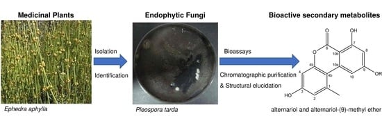
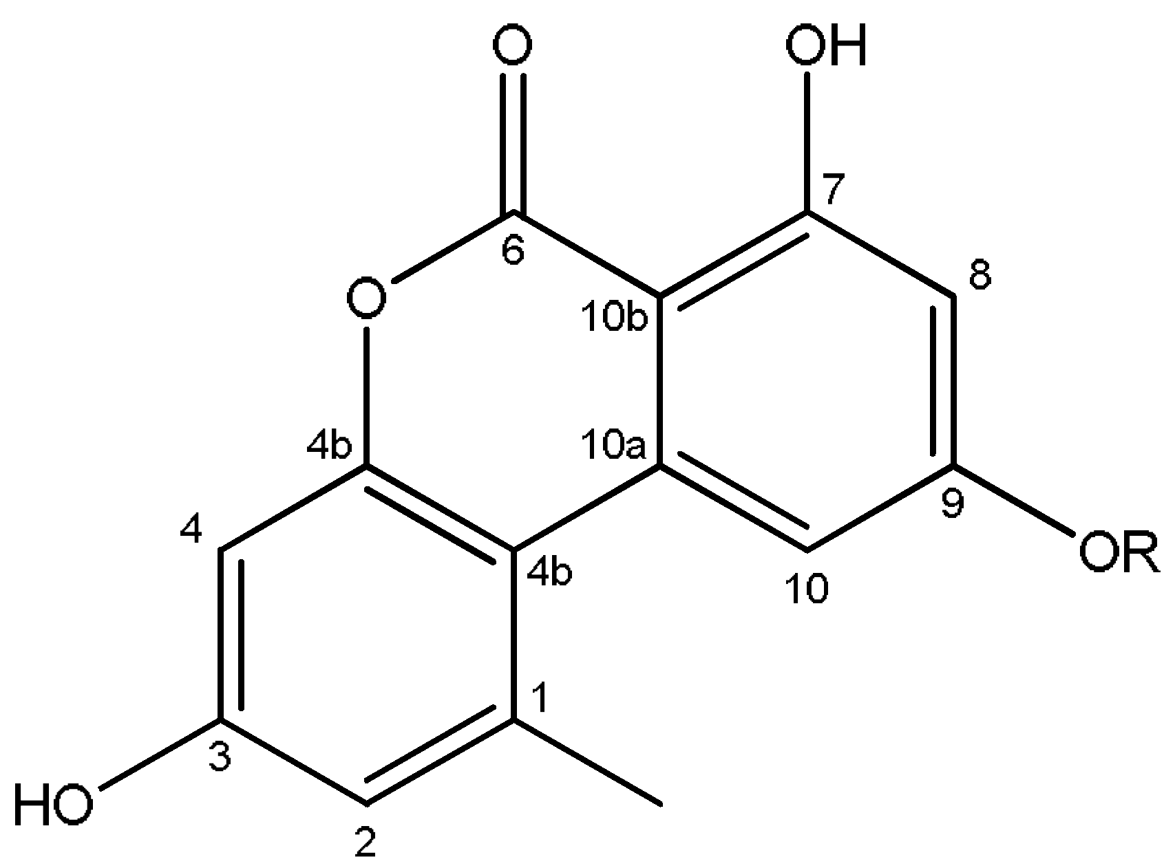
2.4. Identification of the Secondary Metabolites Compounds of Pleospora tarda

Due to the potential activity of the endophytic Pleospora tarda strain, isolated from the medicinal plant Ephedra aphylla, against HSV-2 and VSV viruses, it was selected for further chemical investigation to identify the major secondary metabolites produced by that strain. It was cultivated on potato dextrose agar (PDA) media, and the ethyl acetate extract was subjected to extensive purification using silica gel chromatography followed by purification on HPLC using C-18 column. Two compounds were obtained and identified as alternariol and alternariol-(9)-methyl ether (Figure 3).
The compound (1) was identified based on 1H NMR and 13C NMR, and 2D NMR correlation HMBC (Supplementary Figures S1–S3). 1H NMR (400 MHz, (CD3)2SO): δ 7.19 (1 H, d, J = 2.4 Hz, H10), 6.66 (1 H, d, J = 2.0 Hz, H2), 6.58 (1 H, d, J = 2.4 Hz, H4), 6.32 (1 H, d, J = 2.0 Hz, H8), 2.65 (3 H, s, CH3 at C1) ppm. 13C NMR (100 MHz, (CD3)2SO): δ 166.0 (C6), 165.2 (C7), 164.5 (C9), 158.9 (C3), 153.1 (C4a), 138.8 (C10a), 138.6 (C1), 118.0 (C2), 109.4 (C4b), 104.8 (C10), 102.1 (C4), 101.4 (C8), 97.8 (C10b), 25.7 (CH3 at C1) ppm. Based on HMBC correlation, the compound (1) was identified as alternariol [19].
Compound (2) was identified as a derivative of compound 1 with methoxy group attached to (C9) based on 1H NMR (Supplementary Figure S4). 1H NMR (400 MHz, (CD3)2SO): δ 7.20 (1 H, d, J = 2.4 Hz, H10), 6.69 (1 H, d, J = 2.0 Hz, H2), 6.61 (1 H, d, J = 2.4 Hz, H4), 6.59 (1 H, d, J = 2.0 Hz, H8), 3.88 (3 H, s, OCH3 at C9), 2.71 (3 H, s, CH3 at C1) ppm. Hence, compound (2) was identified as alternariol-(9)-methyl ether [19,20].
To the best of our knowledge, this is the first time the isolation of alternariol (compound 1) and alternariol-(9)-methyl ether (compound 2) from the endophytic fungus Pleospora tarda has been reported. It was demonstrated before that heptaketide compounds isolated from three endolichenic fungal strains had antiviral activity [21]. The potency of alternariol and alternariol-(9)-methyl ether to inhibit herpes simplex virus (HSV), in vitro, has been previously reported. They showed an inhibition with IC50 values of 13.5 and 21.3 μM with selective index of 26.5 and 17.1, respectively. Thus, we referred the antiviral activity of endophytic P. tarda extract to these compounds. The isolated compounds inhibited, previously, the viral replication, either by acting as a polymerase inhibitor, or via inhibiting pre-integration steps. However, it was reported previously that the alternariol and alternariol-9-methyl ether are the main mycotoxins of fungi of the genus Alternaria, which limit the usage of alternariol based compounds in pharmaceutical industry. Several research studies indicated that Alternaria mycotoxins are directly related to the increased incidence of esophageal cancer. Besides, alternariol and alternariol-9-methyl ether were able to induce gene mutations and DNA damage in animal cell lines, and to inhibit topoisomerase I and IIα [22].
From the previous, it was obvious that endophytes represented an abundant and dependable source of bioactive agents, which could be explored for exploitation in pharmaceutical applications. In this current study, light was shed on the bioactivity of some important metabolites from endophytes which were isolated from representative medicinal plants of the Sinai region. We also elucidated the chemical purification and identification of some compounds from these active strains. These results indicated the importance of Egyptian medicinal plants as a new possible source of active compounds which can be used in pharmaceutical drug industries.

3. Conclusions

The total metabolites of a large number of endophytic fungi exhibited different bioactivities. The Adiantum capillus-veneris isolate, Chaetomium globosum, was the most potent antioxidant strain, while Pleospora tarda, from the medicinal plant Ephedra aphylla, showed promising antiviral activity. However, other isolates of the endophytic fungi stopped reproduction of HSV-2and VSV viruses in cells. The great biodiversity and promising bioactivities of endophytic isolates from medicinal plants indicated the importance and requirement for further investigations of pure compounds from the active strains. Therefore, the usage of the endophytes would be crucial approach to search for novel natural products and will help in conservation of medicinal plants and maintenance of environmental biodiversity. Interestingly, it seems that the host–endophytic interaction plays a crucial role in the direction of the endophytes secondary metabolites production.

4. Materials and Methods

4.1. Plant Material

The medicinal plants were collected from Saint Katherine Protectorate, Sinai, Egypt, in September 2010. Only apparently healthy and disease-free plants were collected, to minimize the presence of pathogenic and saprophytic microbes. The plants were stored in separate plastic bags at 4 °C in an ice box until isolation procedures were conducted [8].

4.2. Isolation of Endophytic Fungi

The endophytic fungi were isolated from fresh medicinal plants leaves by the method described by Selim and coworkers in 2011 [8].

4.3. Identification of Endophytic Fungi

Taxonomic identification of endophytic isolates was performed on the basis of morphological characters of fungal culture, colony, hyphae, and the characters of reproductive structure of endophytic fungus, according to identification keys [15].

4.4. Fermentation and Extraction of Fungal Secondary Metabolites

The isolated strains were cultivated over rich standard potato dextrose agar (PDA) medium, (peeled potato 200 g, dextrose 20 g, agar 20 g, and 1 L distilled water). The media were statically incubated at 30 °C, for 3 weeks. Then, the mycelia mat and the media were extracted with ethyl acetate by using an ultrasonic bath for 30 min. The extraction procedures were performed in triplicate. The extract was evaporated to dryness under reduced vacuum [10].

4.5. Identification of Secondary Metabolites

The total ethyl acetate extract of active Pleospora tarda strain grown on PDA media, was subjected to extensive purification using silica gel chromatography, followed by purification on HPLC using C-18 column. The pure compounds were identified on the basis of 1H NMR, 13C NMR, and 2D NMR correlation HMBC.

4.6. Antioxidant Potential of Fungal Extracts

The antioxidant activities of the extracts were assessed using free radical scavenging (FRS) model [23]. Concentrations of fungal extract in DMSO (1000 µg/mL) and DPPH (0.004%) in methanol were prepared. Different concentrations (5–25 µg) of (vitamin C and quercetin) were used as standard controls. In a 96-well plate, 20 µL of (samples or standard) solutions and 180 µL of (0.004%) DPPH were added in each well. The reaction mixture was incubated for 30 min, the assay was run in triplicate, and repeated at least once for active extracts. The absorption was measured at 540 nm by microplate reader. The percentage of scavenging activity was calculated from the following equation:
% of Scavenging Activity = [(A Blank − A Sample)/A blank] × 100
where A Blank is the absorbance of DPPH only, and A Sample is the absorbance of reaction mixture in presence of test samples.

4.7. In Vitro Antiviral Assay of Endophytes Total Metabolites Using EPTT

The end point titration technique (EPTT) bioassay was used to evaluate the antiviral potential of the ethyl acetate extracts of endophytic fungi [24,25]. Two different viruses were chosen for this technique: herpes simplex virus type-2 (HSV-2) as a DNA virus, and vesicular stomatitis virus (VSV) as an example of an RNA virus. Both viruses were obtained from the Holding Company for Biological Products and Vaccines (VACSERA), Egypt. Investigation of the antiviral activity was accomplished according to the previously fully described method [10]. The antiviral activity was calculated based on the ability of the extracts to inhibit the viral cytopathogenic effects (% of CPE inhibition) [10,17,18], according to the following calculation:
% of antiviral activity in form of CPE inhibition % = [1 − (CPE test/CPE control)] × 100

Supplementary Materials

Supplementary materials can be found at https://www.mdpi.com/2311-5637/4/3/49/s1.

Author Contributions

K.A.S. isolated the endophytic fungi, preformed bioassays, and isolated the fungal secondary metabolites. W.A.E. identified the endophytic fungi. K.A.S., A.M.T. and A.A.E.-B. purified and elucidated the structural of fungal secondary metabolites. K.A.S., W.A.E. and E.F.A. wrote the manuscript. A.A.E.-B., T.M.A.-R. and A.I.E.-D. supervised the study.

Funding

The study was financially supported by the National Research Centre, Egypt.

Acknowledgments

The authors would like to thank the national research Centre, Egypt for financial support, and Mohamed Ismael, Katerina Peros, and Eva Bok for the continuous support and the critical reading of the manuscript. Also, we would like to express our appreciation to VACSERA, Egypt for fruitful collaboration.

Conflicts of Interest

The authors declare no conflict of interest.

References

  1. Tan, R.; Zou, W. Endophytes: A rich source of functional metabolites. Nat. Prod. Rep. 2001, 18, 448–459. [Google Scholar] [CrossRef] [PubMed]
  2. Strobel, G.; Daisy, B.; Castillo, U.; Harper, J. Natural products from endophytic microorganisms. J. Nat. Prod. 2004, 67, 257–268. [Google Scholar] [CrossRef] [PubMed]
  3. Selim, K.A.; El-Beih, A.A.; AbdEl-Rahman, T.M.; El-Diwany, A.I. Biology of Endophytic Fungi. Curr. Res. Environ. Appl. Mycol. 2012, 2, 31–82. [Google Scholar] [CrossRef]
  4. Selim, K.A.; Nagia, M.; El-Ghwas, D.E. Endophytic Fungi are Multifunctional Biosynthesizers: Ecological Role and Chemical Diversity. In Endophytic Fungi: Diversity, Characterization and Biocontrol, 1st ed.; NOVA Science Publishers: Hauppauge, NY, USA, 2016. [Google Scholar]
  5. Selim, K.A.; El-Ghwas, D.E.; Selim, R.M.; Hassan, M.I.A. Microbial volatile in defense. In Volatiles and Food Security: Role of Volatiles in Agro-Ecosystems, 1st ed.; Springer: Singapore, 2017. [Google Scholar]
  6. Bacon, C.; White, J. Microbial Endophytes; Marcel Dekker: New York, NY, USA, 2000. [Google Scholar]
  7. Soltan, M.M.; Zaki, A.K. Antiviral screening of forty-two Egyptian medicinal plants. J. Ethnopharmacol. 2009, 126, 102–107. [Google Scholar] [CrossRef] [PubMed]
  8. Selim, K.A.; El-Beih, A.A.; AbdEl-Rahman, T.M.; El-Diwany, A.I. Biodiversity and antimicrobial activity of endophytes associated with Egyptian medicinal plants. Mycosphere 2011, 2, 669–678. [Google Scholar] [CrossRef]
  9. Abdel-Azeem, A.M.; Zaki, S.M.; Khalil, W.F.; Makhlouf, N.A.; Farghaly, L.M. Anti-rheumatoid Activity of Secondary Metabolites Produced by Endophytic Chaetomium globosum. Front. Microbiol. 2016, 7, 1477. [Google Scholar] [CrossRef] [PubMed]
  10. Selim, K.A.; El-Beih, A.A.; Abdel-Rahman, T.M.; El-Diwany, A.I. Biological evaluation of endophytic fungus, Chaetomium globosum JN711454, as potential candidate for improving drug discovery. Cell Biochem. Biophys. 2014, 68, 67–82. [Google Scholar] [CrossRef] [PubMed]
  11. Harper, J.K.E.J.; Ford, G.A.; Strobel, A.; Arif, D.M.; Grant, J.; Porco, D.P.; Tomer-Oneill, K. Pestacin: A 1,3-dihydro isobenzofuran from Pestalotiopsis microspora possessing antioxidant and antimycotic activities. Tetrahedron 2003, 59, 2471–2476. [Google Scholar] [CrossRef]
  12. Song, Y.C.; Huang, W.Y.; Sun, C.; Wang, F.W.; Tan, R.X. Characterization of graphislactone A as the antioxidant and free radical-scavenging substance from the culture of Cephalosporium sp. IFB-E001, an endophytic fungus in Trachelospermum jasminoides. Biol. Pharm. Bull. 2005, 28, 506–509. [Google Scholar] [CrossRef] [PubMed]
  13. Ding, L.; Münch, J.; Goerls, M.; Fiebig, H.H.; Lin, W.H.; Hertweck, C. Xiamycin, a pentacyclic indolosesquiterpene with selective anti-HIV activity from a bacterial mangrove endophyte. Bioorg. Med. Chem. Lett. 2010, 20, 6685–668. [Google Scholar] [CrossRef] [PubMed]
  14. Zhang, G.; Sun, S.; Zhu, T.; Lin, Z.; Gu, J.; Li, D.; Gu, Q. Antiviral isoindolone derivatives from an endophytic fungus Emericella sp. associated with Aegiceras corniculatum. Phytochemistry 2011, 72, 1436–1442. [Google Scholar] [CrossRef] [PubMed]
  15. Moubasher, A.H. Soil Fungi in Qatar and Other Arab Countries; Moreno, G., Diez, J., Manjon, J.L., Eds.; University of Qatar Press: Doha, Qatar, 1993; p. 566. [Google Scholar]
  16. Fröhlich, J.; Hyde, K.D.; Petrini, O. Endophytic fungi associated with palm. Mycol. Res. 2000, 104, 1202–1212. [Google Scholar] [CrossRef]
  17. Promputtha, I.; Jeewon, R.; Lumyong, S.; McKenzie, E.H.C.; Hyde, K.D. Ribosomal DNA fingerprinting in the identification of non sporulating endophytes from Magnolia liliifera (Magnoliaceae). Fungal Divers. 2005, 20, 167–186. [Google Scholar]
  18. Ravindran, C.; Naveenan, T. Adaptation of marine derived fungus Chaetomium globosum (NIOCC 36) to alkaline stress using antioxidant properties. Process Biochem. 2011, 46, 847–857. [Google Scholar] [CrossRef]
  19. Gu, W. Bioactive metabolites from Alternaria brassicicola ML-P08, an endophytic fungus residing in Malus halliana. World J. Microbiol. Biotechnol. 2009, 25, 1677–1683. [Google Scholar] [CrossRef]
  20. Lou, J.; Yu, R.; Wang, X.; Mao, Z.; Fu, L.; Liu, Y.; Zhou, L. Alternariol 9-methyl ether from the endophytic fungus Alternaria sp. Samif01 and its bioactivities. Braz. J. Microbiol. 2016, 47, 96–101. [Google Scholar] [CrossRef] [PubMed]
  21. He, J.W.; Chen, G.D.; Gao, H.; Yang, F.; Li, X.X.; Peng, T.; Guo, L.D.; Yao, X.S. Heptaketides with antiviral activity from three endolichenic fungal strain Nigrospora sp., Alternaria sp. and Phialophora sp. Fitoterapia 2012, 83, 1087–1091. [Google Scholar] [CrossRef] [PubMed]
  22. Burkhardt, B.; Pfeiffer, E.; Metzler, M. Absorption and metabolism of the mycotoxins alternariol and alternariol-9-methyl ether in Caco-2 cells in vitro. Mycotoxin Res. 2009, 25, 149–157. [Google Scholar] [CrossRef] [PubMed]
  23. Selim, K.A.; El-Beih, A.A.; Abdel-Rahman, T.M.; El-Diwany, A.I. High expression level of antioxidants and pharmaceutical bioactivities of endophytic fungus Chaetomium globosum JN711454. Prep. Biochem. Biotechnol. 2016, 46, 131–40. [Google Scholar] [CrossRef] [PubMed]
  24. Vlietinck, A.J.; Van, H.L.; Totte, J.; Lasure, A.; Berghe, D.V.; Rawangabo, P.C. Screening of hundred Rwandese medicinal plants for antimicrobial and antiviral properties. J. Ethnopharmacol. 1995, 46, 31–47. [Google Scholar] [CrossRef]
  25. El-Sayed, R.A. In Vitro Study on Cell–Virus Interaction under the Effect of Selected Ions Concentrations. Ph.D. Thesis, Medical Biophysics, Cairo University Egypt, Giza, Egypt, 2011. [Google Scholar]
Figure 1. Some of the Saint Katherine medicinal plants which used for isolation of endophytic fungi.
Fermentation 04 00049 g001
Figure 2. Some of endophytic strains isolated from Saint Katherine medicinal plants.
Fermentation 04 00049 g002
Figure 3. Compounds isolated from the endophytic fungus P. tarda, alternariol (compound 1) and alternariol-(9)-methyl ether (compound 2). 1: R = H, 2: R = CH3.
Fermentation 04 00049 g003
Table 1. Host plants used for isolation of endophytic fungi.
Plant FamilyHost Medicinal Plants
AdiantaceaeAdiantum capillus-veneris
AsteraceaeAchillea fragrantissima
Artemisia herba alba
Chiliadenus montanus
Launaea spinosa
Pulicaria undulate
Tanacetum sinaicum
EphedraceaeEphedra alata
Ephedra aphylla
EuphorbiaceaeEuphorbia sanctae-catherine
HypericaceaeHypericum sinaicum
LamiaceaeLavandula coronopifolia
Phlomis aurea
Stachys aegyptiaca
Teucrium leucocladum
Teucrium polium
Thymus decussatus
RubiaceaeGalium sinaicum
Table 2. Antioxidant and antiviral activities of different fungal endophytes extracts.
Plant FamilyHost PlantEndophyte% of Antioxidant Activity% of CPE Inhibition of HSV% of CPE Inhibition of VSVPlant FamilyHost PlantEndophyte% of Antioxidant Activity% of CPE Inhibition of HSV% of CPE Inhibition of VSV
AdiantaceaeAdiantum capillus-venerisChaetomium globosum99%0%−15.2EuphorbiaceaeEuphorbia sancte catherineWhite sterile mycelia 323%NDND
Penicillium sp.28%NDNDWhite sterile mycelia 49%NDND
AsteraceaeAchillea fragrantissimaDark sterile mycelia 14%14.8%−6.8Dark sterile mycelia 38%NDND
Dark sterile mycelia 215%0%NDAspergillus sydowii----NDND
White sterile mycelia 11%0%0Acremonium strictum1.5%2.2%−6.8
White sterile mycelia 26%0%−6.8HypericaceaeHypericum sinaicumUlocladium chartarum19%0%ND
Penicillium corylophilum----NDNDDark sterile mycelia14%NDND
Artimisia herba albaNigrospora sphaerica14%NDNDPleospora tarda16%0%8.4
White sterile mycelia 115%0%−6.8Chaetomium globosum12%−14.8%6.8
Dark sterile mycelia14%0%−15.2White sterile mycelia 112%NDND
White sterile mycelia 215%22.2%0Yeast7%NDND
Aspergillus flavus8%NDNDWhite sterile mycelia 211%0%8.3
Chiliadenus montanusDark sterile mycelia10%14.8%0LamiaceaeLavandula coronopifoliaDark sterile mycelia 19%17.0%ND
Nigrospora sphaerica13%14.8%−15.2Dark sterile mycelia 221%NDND
Launea spinosaAcremonium strictum6%14.8%0Dark sterile mycelia 35%NDND
Acremonium sp.7%NDNDScopulariopsis sp.----NDND
Penicillium chrysogenoum0.5%−21.5%−22.13Phlomis aureaDark sterile mycelia 12%NDND
Aspergillus niger0.5%0%−19.03White sterile mycelia 8%0%ND
Pulicaria undulateDark sterile mycelia 121%NDNDDark sterile mycelia 2----−18.5%0
Dark sterile mycelia 215%0%8.4Chaetomium spirale2%0%−15.26
Ulocladium chartarum8%0%8.4Penicillium sp.----−18.5%0
Penicillium sp.24%0%−22.12Aspergillus flavus5.5%NDND
Dark sterile mycelia 314%26%8.4Stachys aegyptiacaMucor fuscus----14.8%0%
Ulocladium atrum14%NDNDWhite sterile mycelia 12%NDND
White sterile mycelia16%NDNDPleospora tarda1%NDND
Tanacetum sinaicumWhite sterile mycelia 16%−14.8%8.39White sterile mycelia 2----NDND
White sterile mycelia 22%NDNDAspergillus flavus0.2%14.8%−9.9%
White sterile mycelia 31%NDNDTeucrium leucocladumWhite sterile mycelia 115%18.5%ND
Penicillium chrysogenoum----NDNDWhite sterile mycelia 215%0%0
Penicillium sp.6%0%15.2Dark sterile mycelia 116%−11.1%−6.8
Aspergillus sydowii2%NDNDDark sterile mycelia 217%NDND
EphedraceaeEphedra alataWhite sterile mycelia 19%40.7%15.2Dark sterile mycelia 314%NDND
White sterile mycelia 215%0%8.3White sterile mycelia 315%NDND
Dark sterile mycelia 12%NDNDTeucrium poliumAlternaria alternata11%0%15.2
Dark sterile mycelia 27.5%NDNDNigrospora sphaerica1%0%−15.4
Dark sterile mycelia 31%−14.8%0White sterile mycelia 110%0%8.4
White sterile mycelia 3----−11.1%−6.8White sterile mycelia 218%NDND
Dark sterile mycelia 422%NDNDPenicillium corylophilum----0%−15.2
Dark sterile mycelia54%NDNDPenicillium chrysogenoum----NDND
Unidentified bacteria----NDNDAspergillus niger0.5%0%0
Ephedra aphyllaPleospora tarda17%40.7%15.2%Thymus decussatesUnidentified bacteria4%NDND
Dark sterile mycelia 1----0%NDRubiaceaeGalium sinaicumPleospora tarda 100%−15.2%
Aspergillus versicolor11%NDNDFusarium oxysporum----NDND
Dark sterile mycelia 26%NDNDUlocladium chartarum----NDND
Aspergillus niger----NDNDAspergillus sp.----−3.7%23.66%
EuphorbiaceaeEuphorbia sancte catherineDark sterile mycelia 121%0%0Cochliobolus lunatus5%NDND
White sterile mycelia 117%0%NDPleospora tarda 2----NAND
Phoma leveillei19%14.8%15.2Absidia corymbifora----NDND
White sterile mycelia 26%NDNDFusarium sp.1%11.1%ND
Dark sterile mycelia 28%NDND
ND: Not determined.

