Next Issue
Volume 4, April
Previous Issue
Volume 4, February


J. Imaging, Volume 4, Issue 3 (March 2018) – 6 articles

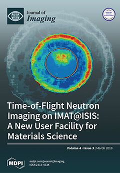
Cover Story (view full-size image): This paper introduces a new neutron imaging facility, called IMAT, which is now well into its commissioning phase and is being prepared for user operation. One of the benefits of operating on a pulsed neutron source like ISIS is that neutron energies are determined via time of flight measurements, thus enabling energy-dispersive neutron imaging for maximizing image contrasts and for mapping microstructure properties of materials. We report the basic performance parameters and survey the infrastructure efforts that have been made to prepare the facility for a diverse range of disciplines such as engineering material science, battery research, earth science and cultural heritage. View this paper
  • Issues are regarded as officially published after their release is announced to the table of contents alert mailing list.
  • You may sign up for e-mail alerts to receive table of contents of newly released issues.
  • PDF is the official format for papers published in both, html and pdf forms. To view the papers in pdf format, click on the "PDF Full-text" link, and use the free Adobe Reader to open them.
Order results
Result details
Section
Select all
Export citation of selected articles as:
22 pages, 2664 KiB  
Technical Note
Python Non-Uniform Fast Fourier Transform (PyNUFFT): An Accelerated Non-Cartesian MRI Package on a Heterogeneous Platform (CPU/GPU)
by Jyh-Miin Lin
J. Imaging 2018, 4(3), 51; https://doi.org/10.3390/jimaging4030051 - 08 Mar 2018
Cited by 29 | Viewed by 9263
Abstract
A Python non-uniform fast Fourier transform (PyNUFFT) package has been developed to accelerate multidimensional non-Cartesian image reconstruction on heterogeneous platforms. Since scientific computing with Python encompasses a mature and integrated environment, the time efficiency of the NUFFT algorithm has been a major obstacle [...] Read more.
A Python non-uniform fast Fourier transform (PyNUFFT) package has been developed to accelerate multidimensional non-Cartesian image reconstruction on heterogeneous platforms. Since scientific computing with Python encompasses a mature and integrated environment, the time efficiency of the NUFFT algorithm has been a major obstacle to real-time non-Cartesian image reconstruction with Python. The current PyNUFFT software enables multi-dimensional NUFFT accelerated on a heterogeneous platform, which yields an efficient solution to many non-Cartesian imaging problems. The PyNUFFT also provides several solvers, including the conjugate gradient method, 1 total variation regularized ordinary least square (L1TV-OLS), and 1 total variation regularized least absolute deviation (L1TV-LAD). Metaprogramming libraries have been employed to accelerate PyNUFFT. The PyNUFFT package has been tested on multi-core central processing units (CPUs) and graphic processing units (GPUs), with acceleration factors of 6.3–9.5× on a 32-thread CPU platform and 5.4–13× on a GPU. Full article
Show Figures

Figure 1

9 pages, 3096 KiB  
Article
Demonstration of Focusing Wolter Mirrors for Neutron Phase and Magnetic Imaging
by Daniel S. Hussey, Han Wen, Huarui Wu, Thomas R. Gentile, Wangchun Chen, David L. Jacobson, Jacob M. LaManna and Boris Khaykovich
J. Imaging 2018, 4(3), 50; https://doi.org/10.3390/jimaging4030050 - 06 Mar 2018
Cited by 11 | Viewed by 5629
Abstract
Image-forming focusing mirrors were employed to demonstrate their applicability to two different modalities of neutron imaging, phase imaging with a far-field interferometer, and magnetic-field imaging through the manipulation of the neutron beam polarization. For the magnetic imaging, the rotation of the neutron polarization [...] Read more.
Image-forming focusing mirrors were employed to demonstrate their applicability to two different modalities of neutron imaging, phase imaging with a far-field interferometer, and magnetic-field imaging through the manipulation of the neutron beam polarization. For the magnetic imaging, the rotation of the neutron polarization in the magnetic field was measured by placing a solenoid at the focus of the mirrors. The beam was polarized upstream of the solenoid, while the spin analyzer was situated between the solenoid and the mirrors. Such a polarized neutron microscope provides a path toward considerably improved spatial resolution in neutron imaging of magnetic materials. For the phase imaging, we show that the focusing mirrors preserve the beam coherence and the path-length differences that give rise to the far-field moiré pattern. We demonstrated that the visibility of the moiré pattern is modified by small angle scattering from a highly porous foam. This experiment demonstrates the feasibility of using Wolter optics to significantly improve the spatial resolution of the far-field interferometer. Full article
(This article belongs to the Special Issue Neutron Imaging)
Show Figures

Figure 1

15 pages, 6013 KiB  
Article
CT-Based Micro-Mechanical Approach to Predict Response of Closed-Cell Porous Biomaterials to Low-Velocity Impact
by Mehrdad Koloushani, Reza Hedayati, Mojtaba Sadighi and Mohammad Mohammadi-Aghdam
J. Imaging 2018, 4(3), 49; https://doi.org/10.3390/jimaging4030049 - 04 Mar 2018
Cited by 8 | Viewed by 4821
Abstract
In this study, a new numerical approach based on CT-scan images and finite element (FE) method has been used to predict the mechanical behavior of closed-cell foams under impact loading. Micro-structural FE models based on CT-scan images of foam specimens (elastic-plastic material model [...] Read more.
In this study, a new numerical approach based on CT-scan images and finite element (FE) method has been used to predict the mechanical behavior of closed-cell foams under impact loading. Micro-structural FE models based on CT-scan images of foam specimens (elastic-plastic material model with material constants of bulk aluminum) and macro-mechanical FE models (with crushable foam material model with material constants of foams) were constructed. Several experimental tests were also conducted to see which of the two noted (micro- or macro-) mechanical FE models can better predict the deformation and force-displacement curves of foams. Compared to the macro-structural models, the results of the micro-structural models were much closer to the corresponding experimental results. This can be explained by the fact that the micro-structural models are able to take into account the interaction of stress waves with cell walls and the complex pathways the stress waves have to go through, while the macro-structural models do not have such capabilities. Despite their high demand for computational resources, using micro-scale FE models is very beneficial when one needs to understand the failure mechanisms acting in the micro-structure of a foam in order to modify or diminish them. Full article
Show Figures

Graphical abstract

12 pages, 5923 KiB  
Article
Energy-Resolved Neutron Imaging for Reconstruction of Strain Introduced by Cold Working
by Anton S. Tremsin, Winfried Kockelmann, Joe F. Kelleher, Anna M. Paradowska, Ranggi S. Ramadhan and Michael E. Fitzpatrick
J. Imaging 2018, 4(3), 48; https://doi.org/10.3390/jimaging4030048 - 28 Feb 2018
Cited by 7 | Viewed by 4693
Abstract
Energy-resolved neutron transmission imaging is used to reconstruct maps of residual strains in drilled and cold-expanded holes in 5-mm and 6.4-mm-thick aluminum plates. The possibility of measuring the positions of Bragg edges in the transmission spectrum in each 55 × 55 µm2 [...] Read more.
Energy-resolved neutron transmission imaging is used to reconstruct maps of residual strains in drilled and cold-expanded holes in 5-mm and 6.4-mm-thick aluminum plates. The possibility of measuring the positions of Bragg edges in the transmission spectrum in each 55 × 55 µm2 pixel is utilized in the reconstruction of the strain distribution within the entire imaged area of the sample, all from a single measurement. Although the reconstructed strain is averaged through the sample thickness, this technique reveals strain asymmetries within the sample and thus provides information complementary to other well-established non-destructive testing methods. Full article
(This article belongs to the Special Issue Neutron Imaging)
Show Figures

Figure 1

22 pages, 12666 KiB  
Article
Time-of-Flight Neutron Imaging on IMAT@ISIS: A New User Facility for Materials Science
by Winfried Kockelmann, Triestino Minniti, Daniel E. Pooley, Genoveva Burca, Ranggi Ramadhan, Freddie A. Akeroyd, Gareth D. Howells, Chris Moreton-Smith, David P. Keymer, Joe Kelleher, Saurabh Kabra, Tung Lik Lee, Ralf Ziesche, Anthony Reid, Giuseppe Vitucci, Giuseppe Gorini, Davide Micieli, Raffaele G. Agostino, Vincenzo Formoso, Francesco Aliotta, Rosa Ponterio, Sebastiano Trusso, Gabriele Salvato, Cirino Vasi, Francesco Grazzi, Kenichi Watanabe, Jason W. L. Lee, Anton S. Tremsin, Jason B. McPhate, Daniel Nixon, Nick Draper, William Halcrow and Jim Nightingaleadd Show full author list remove Hide full author list
J. Imaging 2018, 4(3), 47; https://doi.org/10.3390/jimaging4030047 - 28 Feb 2018
Cited by 49 | Viewed by 10392
Abstract
The cold neutron imaging and diffraction instrument IMAT at the second target station of the pulsed neutron source ISIS is currently being commissioned and prepared for user operation. IMAT will enable white-beam neutron radiography and tomography. One of the benefits of operating on [...] Read more.
The cold neutron imaging and diffraction instrument IMAT at the second target station of the pulsed neutron source ISIS is currently being commissioned and prepared for user operation. IMAT will enable white-beam neutron radiography and tomography. One of the benefits of operating on a pulsed source is to determine the neutron energy via a time of flight measurement, thus enabling energy-selective and energy-dispersive neutron imaging, for maximizing image contrasts between given materials and for mapping structure and microstructure properties. We survey the hardware and software components for data collection and image analysis on IMAT, and provide a step-by-step procedure for operating the instrument for energy-dispersive imaging using a two-phase metal test object as an example. Full article
(This article belongs to the Special Issue Neutron Imaging)
Show Figures

Figure 1

24 pages, 1285 KiB  
Article
Feature Importance for Human Epithelial (HEp-2) Cell Image Classification
by Vibha Gupta and Arnav Bhavsar
J. Imaging 2018, 4(3), 46; https://doi.org/10.3390/jimaging4030046 - 26 Feb 2018
Cited by 7 | Viewed by 5823
Abstract
Indirect Immuno-Fluorescence (IIF) microscopy imaging of human epithelial (HEp-2) cells is a popular method for diagnosing autoimmune diseases. Considering large data volumes, computer-aided diagnosis (CAD) systems, based on image-based classification, can help in terms of time, effort, and reliability of diagnosis. Such approaches [...] Read more.
Indirect Immuno-Fluorescence (IIF) microscopy imaging of human epithelial (HEp-2) cells is a popular method for diagnosing autoimmune diseases. Considering large data volumes, computer-aided diagnosis (CAD) systems, based on image-based classification, can help in terms of time, effort, and reliability of diagnosis. Such approaches are based on extracting some representative features from the images. This work explores the selection of the most distinctive features for HEp-2 cell images using various feature selection (FS) methods. Considering that there is no single universally optimal feature selection technique, we also propose hybridization of one class of FS methods (filter methods). Furthermore, the notion of variable importance for ranking features, provided by another type of approaches (embedded methods such as Random forest, Random uniform forest) is exploited to select a good subset of features from a large set, such that addition of new features does not increase classification accuracy. In this work, we have also, with great consideration, designed class-specific features to capture morphological visual traits of the cell patterns. We perform various experiments and discussions to demonstrate the effectiveness of FS methods along with proposed and a standard feature set. We achieve state-of-the-art performance even with small number of features, obtained after the feature selection. Full article
(This article belongs to the Special Issue Selected Papers from “MIUA 2017”)
Show Figures

Figure 1

Previous Issue
Next Issue
Back to TopTop