
Neuroimaging through Sonolucent Cranioplasty: A Systematic Scoping Review Protocol
by
 ,
,
Christina P. Rossitto
*, 
Alex Devarajan
,
Gabrielle Price
,
Muhammad Ali
and
Christopher P. Kellner
Department of Neurosurgery, Icahn School of Medicine, 1468 Madison Avenue, Annenberg Building, 8th Floor, New York, NY 10029, USA
*
Author to whom correspondence should be addressed.
Methods Protoc. 2022, 5(5), 80; https://doi.org/10.3390/mps5050080
Submission received: 12 August 2022
/
Revised: 22 September 2022
/
Accepted: 30 September 2022
/
Published: 9 October 2022
(This article belongs to the Section Biomedical Sciences and Physiology)
Abstract
:Cranioplasty is a neurosurgical procedure in which the skull bone is repaired after craniectomy. Recently, studies have suggested that sonolucent synthetic materials are safe and useful for cranioplasty. Sonolucent cranioplasty (SC) implants provide unprecedented opportunity in adult neurosurgery to monitor neuroanatomy, assess hemodynamics, view devices located within the implant, and conduct focused ultrasound treatments. Current research on SC includes proof-of-concept cadaveric studies, patient-related safety and feasibility studies, and case series demonstrating transcranioplasty ultrasonography (TCUS). The purpose of this protocol is to investigate the current literature on SC use and outcomes in TCUS. We will perform a systematic literature search following PRISMA-ScR guidelines. The search will be conducted using Ovid Embase, Ovid Medline, and Web of Science Core Collection databases. Titles, abstracts, and full texts will be screened. Joanna Briggs Institute critical appraisal tools will be utilized. Data extraction points will include subject characteristics, SC implant characteristics, ultrasound characteristics, and sonographic findings. These findings will provide a comprehensive review of the literature on sonolucent cranioplasty and directions for future research.
1. Introduction
Cranioplasty is a neurosurgical procedure in which the skull bone is repaired after craniectomy. Cranioplasty provides cosmetic benefits, protection from external atmospheric pressure distortion, and restoration of several critical physiologic processes. The ideal construct to correct cranial defects has evolved over time as several materials have been developed to replace the original skull bone [1].
In pediatric neurosurgery, open fontanelles provide a naturally occurring acoustic window for diagnostic ultrasound. However, fontanelle closure makes it impossible to leverage this method as a diagnostic tool in adult neurosurgery [2]. Sonolucent cranioplasty (SC) implants provide an unprecedented opportunity in adult neurosurgery to conduct neuromonitoring, evaluate hemodynamics, visually track devices located within the implant, and perform focused ultrasound treatments [2,3,4,5,6]. Current research on SC has utilized polymethylmethacrylate (PMMA), polyetheretherketone (PEEK), and polyolefin as sonolucent materials [7,8,9]. The first publication detailing diagnostic imaging through cranioplasty was published by Mursch et al. in 2018 using PEEK cranioplasty implants [9]. The term transcranioplasty ultrasound was first coined at Johns Hopkins University by Belzberg et al. in a proof-of-concept publication demonstrating TCUS through clear PMMA in a living patient and comparing clear PMMA, opaque PMMA, and PEEK in a cadaveric model [2]. This paper was quickly followed up by a second comparative study assessing transcranioplasty imaging through clear PMMA in a brain phantom [10]. Despite a large amount of clinical evidence supporting the use of cranioplasty and the frequency that these surgical procedures are performed, there is a dearth of knowledge about outcomes and risk factors associated with SC. Previous research on SC has been largely limited to proof-of-concept studies and case series [3,7,8].
Accordingly, this scoping review aims to analyze the trends, outcomes, risk factors, and characteristics of preceding investigations on transcranioplasty ultrasonography (TCUS) through SC in current neurosurgical practice.
2. Methods
2.1. Study Design
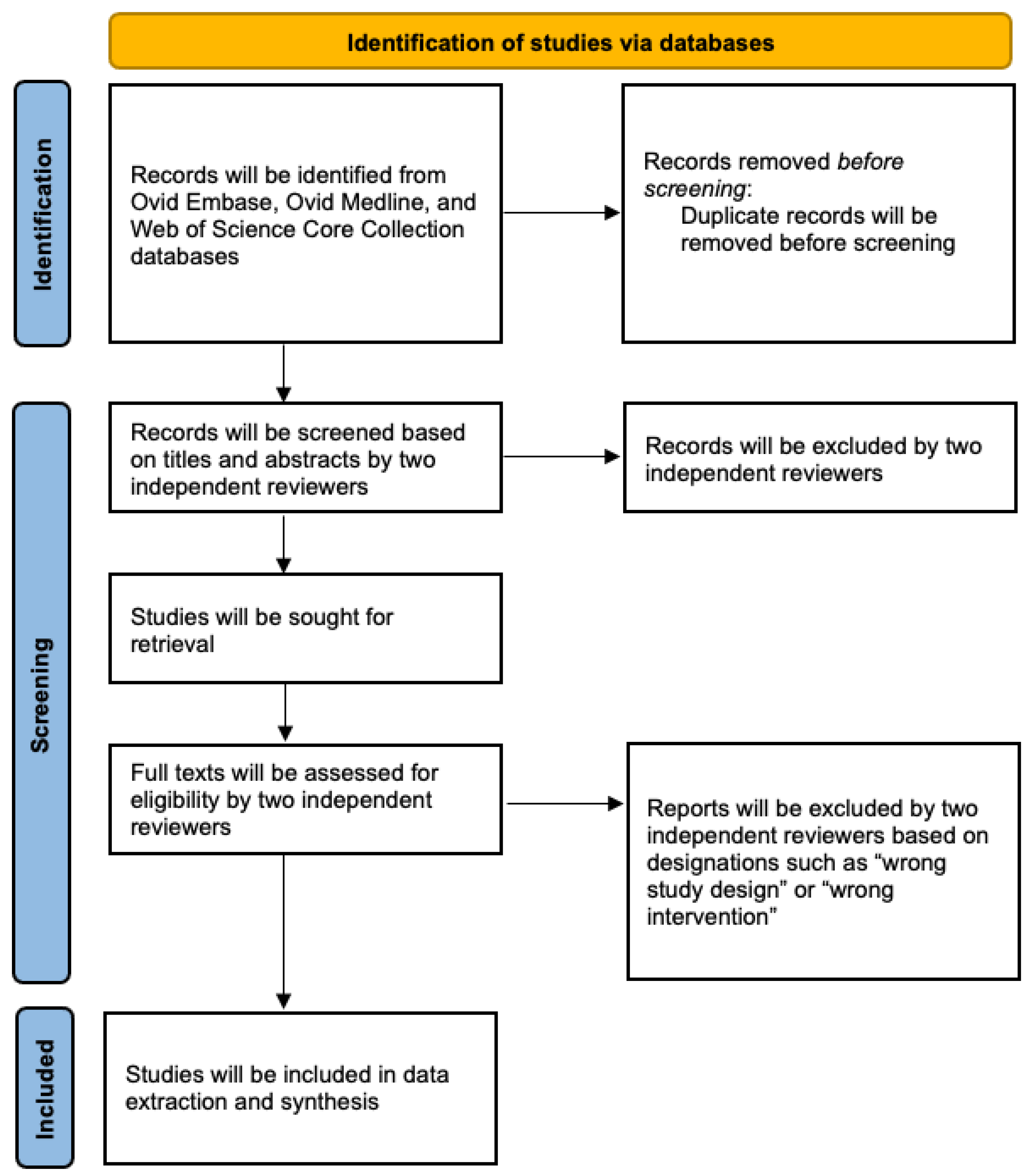
The study selection and screening procedures are shown in Figure 1 [11]. The scope of this review will examine literature regarding the use of SC, among humans and bench top models, for the use of neuroimaging. Published full text articles, inclusive of case reports and series, which detail the new use of SC for diagnostic imaging will be considered. Manuscripts exploring the in vitro and in vivo characterization of sonolucent materials will be included. Studies detailing the use of SC for purposes other than neuroimaging and neuromonitoring will not be included for the review. Publications that are not original research articles (i.e., abstracts, review articles, and commentaries) will not be included. This systematic scoping review will adhere to PRISMA-ScR guidelines [12].
2.2. Participants
This scoping review will include manuscripts that involve neurosurgery patients who undergo TCUS through SC. There are no limitations on the types or number of participants included. Studies on bench top mdoels and animal models are also included.
2.3. Search Strategy
Ovid Embase, Ovid Medline, and Web of Science Core Collection databases will be searched to identify peer-reviewed articles characterizing the use of SC for preclinical in vitro and in vivo models and clinical patient studies (Table 1). Particularly, databases will be queried using search terms “clear”, “sonolucent”, “translucent”, “polymethyl methacrylate”, “polyolefin”, or “polyetheretherketone”, and “cranioplasty”. With this combination of broad search terms, we hope to encapsulate a vast array of manuscripts for our analysis.
2.4. Screening
Studies will be uploaded into Covidence for review and duplicates will be removed. Inclusion criteria will focus on (1) published full text articles, (2) which detailed new use of SC for the purpose of neuroimaging. Two reviewers will independently perform title and abstract screening of all studies. A full-text review will be performed by two reviewers. Any questions or disagreements during this process will be discussed and submitted to an additional supervising reviewer. The Covidence systematic review software will be used to evaluate inter-rater reliability.
2.5. Joanna Briggs Institute Critical Appraisal Tools
Article quality will be assessed using the Joanna Briggs Institute (JBI) critical appraisal tool for case reviews, case series, and quasi-experimental studies. An example of the JBI critical appraisal tool for case reviews is shown in ref [13]. JBI has published theories, protocols, and methodical processes for the critical appraisal and synthesis of several forms of peer-reviewed literature. The JBI critical appraisal tool was developed with the impetus to help improve clinical decision making in healthcare. Consequently, this tool will be used to critically appraise each article and evaluate its methodological quality. The purpose of this appraisal is to determine risk of bias, guarantee proper reporting and statistical analysis, and to assess the extent to which an investigation has addressed the potential for bias in its design, conduct, and analysis.
2.6. Data Extraction and Management
Two reviewers will independently extract data from included studies as follows: methods, characteristics of participants, interventions, and primary and secondary outcomes. Any disagreements on data extraction will be adjudicated through discussion with a third reviewer. The data items extracted by two independent investigators will include authors, date published, study design, prosthesis material, prosthesis brand, ultrasound transducer used, confirmation imaging, imaging artifact, imaging findings. For preclinical studies, the vessel used for experimental design will also be extracted. For clinical studies, sample size, sex, clinical indication for cranioplasty, prosthesis size and thickness, prosthesis location, dural substitute, and clinical complications will be extracted.
3. Discussion
Cranioplasty is a critical neurosurgical procedure that provides cosmetic benefits, protection from external atmospheric pressure distortion, and restoration of several key physiologic processes such as glymphatic circulation, cerebral hemodynamics, and cellular mechanisms [14,15]. Harnessing the features of cranioplasty to create a sonolucent window offers several advantages over radiographic imaging, including reduced cost burden, no radiation exposure, ease of use, interactive image acquisition, and sequential monitoring. SC can be used for preventative and observational purposes which include but are not limited to sonographic detection of brain tumor recurrence, monitoring of cerebral blood flow and Doppler imaging of vessel collateralization, and measuring ventricular size for hydrocephalus [8]. Although our review focuses on the diagnostic imaging capabilities of TCUS, SC also can be used to perform several therapeutic techniques, including delineation of microbubbles with blood–brain barrier disruption to improve drug delivery and focused ultrasound ablation of brain malformations [7,16].
This review will discuss the most up to date literature on TCUS through sonolucent prosthesis. To our knowledge, this is the first systematic scoping review to assess the safety and effectiveness of TCUS using SC. This review will describe the landscape of SC experimental investigations and help to guide future studies. We will add to the literature by highlighting seminal studies that enhance our understanding of SC utilization and outcomes.
This assessment may pose some limitations that must be taken into consideration. Because the literature applicable to this topic is sparse and largely includes case reports and case studies, these articles may lack detailed findings and consist of conclusions that are limited. The heterogeneous nature of the literature may also challenge data reporting. However, rigor will be maintained by utilizing PRISMA-SR guidelines, Covidence, and JBI critical appraisal.
Altogether, this review will identify current evidence and gaps in knowledge on SC to motivate future research. Larger clinical trials are currently ongoing to evaluate TCUS in larger patient populations following extracranial–intracranial bypass, stroke, tumor, and external ventricular drain placement [3,17]. The hope is for this review to motivate controlled, quantitative data-focused clinical trials, which would enhance the likelihood that clinicians consider using sonolucent materials to perform cranioplasty. As these trials begin design, enrollment, and data analysis, this review will guide key variables in SC evaluation. By developing a framework for investigating SC, this investigation should serve as a gold standard reference describing the impact of SC in the field of neurosurgery.
Author Contributions
Conceptualization, C.P.R. and C.P.K.; methodology, C.P.R., A.D. and C.P.K.; writing—original draft preparation, C.P.R.; writing—review and editing, G.P., A.D., M.A. and C.P.K.; supervision, C.P.K. All authors have read and agreed to the published version of the manuscript.
Funding
This research received no external funding.
Institutional Review Board Statement
Not applicable.
Informed Consent Statement
Not applicable.
Data Availability Statement
Not applicable.
Acknowledgments
We would like to acknowledge the work of Samantha Walsh in developing the search strategy used in this scoping review.
Conflicts of Interest
The authors declare no conflict of interest.
References
- Shah, A.M.; Jung, H.; Skirboll, S. Materials used in cranioplasty: A history and analysis. Neurosurg. Focus 2014, 36, E19. [Google Scholar] [CrossRef] [PubMed] [Green Version]
- Belzberg, M.; Ben Shalom, N.; Yuhanna, E.; Manbachi, A.; Tekes, A.; Huang, J.; Brem, H.; Gordon, C.R. Sonolucent Cranial Implants. J. Craniofacial Surg. 2019, 30, 1456–1461. [Google Scholar] [CrossRef] [PubMed]
- Flores, A.R.; Srinivasan, V.M.; Seeley, J.; Huggins, C.; Kan, P.; Burkhardt, J.-K. Safety, Feasibility, and Patient-Rated Outcome of Sonolucent Cranioplasty in Extracranial-Intracranial Bypass Surgery to Allow for Transcranioplasty Ultrasound Assessment. World Neurosurg. 2020, 144, e277–e284. [Google Scholar] [CrossRef] [PubMed]
- Gordon, C.R.; Santiago, G.F.; Huang, J.; Bergey, G.K.; Liu, S.; Armand, M.; Brem, H.; Anderson, W.S. First In-Human Experience With Complete Integration of Neuromodulation Device Within a Customized Cranial Implant. Oper. Neurosurg. 2018, 15, 39–45. [Google Scholar] [CrossRef] [PubMed]
- Gordon, C.R.; Wolff, A.; Santiago, G.F.; Liebman, K.; Veznedaroglu, E.; Vrionis, F.D.; Huang, J.; Brem, H.; Luciano, M. First-in-Human Experience With Integration of a Hydrocephalus Shunt Device Within a Customized Cranial Implant. Oper. Neurosurg. 2019, 17, 608–615. [Google Scholar] [CrossRef] [PubMed] [Green Version]
- Mitchell, K.S.; Anderson, W.; Shay, T.; Huang, J.; Luciano, M.; Suarez, J.I.; Manson, P.; Brem, H.; Gordon, C.R. First-In-Human Experience With Integration of Wireless Intracranial Pressure Monitoring Device Within a Customized Cranial Implant. Oper. Neurosurg. 2020, 19, 341–350. [Google Scholar] [CrossRef] [PubMed] [Green Version]
- Del Bene, M.; Raspagliesi, L.; Carone, G.; Gaviani, P.; Silvani, A.; Solbiati, L.; Prada, F.; DiMeco, F. Cranial sonolucent prosthesis: A window of opportunity for neuro-oncology (and neuro-surgery). J. Neurooncol. 2022, 156, 529–540. [Google Scholar] [CrossRef] [PubMed]
- Shay, T.; Mitchell, K.-A.; Belzberg, M.; Zelko, I.; Mahapatra, S.; Qian, J.; Mendoza, L.; Huang, J.; Brem, H.; Gordon, C. Translucent Customized Cranial Implants Made of Clear Polymethylmethacrylate. Ann. Plast. Surg. 2020, 85, e27–e36. [Google Scholar] [CrossRef] [PubMed]
- Mursch, K.; Behnke-Mursch, J. Polyether Ether Ketone Cranioplasties Are Permeable to Diagnostic Ultrasound. World Neurosurg. 2018, 117, 142–143. [Google Scholar] [CrossRef] [PubMed]
- Belzberg, M.; Ben Shalom, N.; Lu, A.; Yuhanna, E.; Manbachi, A.; Tekes, A.; Huang, J.; Brem, H.; Gordon, C. Transcranioplasty Ultrasound Through a Sonolucent Cranial Implant Made of Polymethyl Methacrylate. J. Craniofacial Surg. 2019, 30, e626–e629. [Google Scholar] [CrossRef] [PubMed]
- Page, M.J.; McKenzie, J.E.; Bossuyt, P.M.; Boutron, I.; Hoffmann, T.C.; Mulrow, C.D.; Shamseer, L.; Tetzlaff, J.M.; Akl, E.A.; Brennan, S.E.; et al. The PRISMA 2020 statement: An updated guideline for reporting systematic reviews. BMJ 2021, 372, n71. [Google Scholar] [CrossRef] [PubMed]
- Tricco, A.C.; Lillie, E.; Zarin, W.; O’Brien, K.K.; Colquhoun, H.; Levac, D.; Moher, D.; Peters, M.D.J.; Horsley, T.; Weeks, L.; et al. PRISMA Extension for Scoping Reviews (PRISMA-ScR): Checklist and Explanation. Ann. Intern. Med. 2018, 169, 467–473. [Google Scholar] [CrossRef] [PubMed] [Green Version]
- Munn, Z.; Barker, T.H.; Moola, S.; Tufanaru, C.; Stern, C.; McArthur, A.; Stephenson, M.; Aromataris, E. Methodological quality of case series studies: An introduction to the JBI critical appraisal tool. JBI Evid. Synth. 2020, 18, 2127–2133. [Google Scholar] [CrossRef] [PubMed]
- Plog, B.A.; Lou, N.; Pierre, C.A.; Cove, A.; Kenney, H.M.; Hitomi, E.; Kang, H.; Iliff, J.J.; Zeppenfeld, D.M.; Nedergaard, M.; et al. When the air hits your brain: Decreased arterial pulsatility after craniectomy leading to impaired glymphatic flow. J. Neurosurg. 2020, 133, 210–223. [Google Scholar] [CrossRef] [PubMed]
- Lilja-Cyron, A.; Andresen, M.; Kelsen, J.; Andreasen, T.H.; Petersen, L.G.; Fugleholm, K.; Juhler, M. Intracranial pressure before and after cranioplasty: Insights into intracranial physiology. J. Neurosurg. 2019, 133, 1548–1558. [Google Scholar] [CrossRef] [PubMed]
- Nicodemus, N.E.; Becerra, S.; Kuhn, T.P.; Packham, H.R.; Duncan, J.; Mahdavi, K.; Iovine, J.; Kesari, S.; Pereles, S.; Whitney, M.; et al. Focused transcranial ultrasound for treatment of neurodegenerative dementia. Alzheimer’s Dementia: Transl. Res. Clin. Interv. 2019, 5, 374–381. [Google Scholar] [CrossRef] [PubMed]
- Evaluation of Cranioplasty Using Native Bone Autograft Versus Synthetic Bone Allograft. Available online: https://clinicaltrials.gov/ct2/show/NCT04855175 (accessed on 3 October 2022).
Figure 1.
Flow diagram outlining the study search and selection.


Table 1.
Electronic search plan for Ovid Embase, Ovid Medline, and Web of Science Core Collection.
Embase Classic + Embase <1947 to date of search>
|
Ovid MEDLINE(R) and Epub Ahead of Print, In-Process, In-Data-Review & Other Non-Indexed Citations and Daily <1946 to date of search>
|
| Web of Science Core Collection TS = ((cranioplast*) NEAR/3 (clear or translucent or Sonolucent or PMMA or polymethyl or polyolefin or polyether or polyetheretherketone or polymethylmethacrylate)) OR TS = ((transcranioplast*) NEAR/3 (clear or translucent or Sonolucent or PMMA or polymethyl or polyolefin or polyether or polyetheretherketone or polymethylmethacrylate)) OR TS = (((implant) NEAR/3 (clear or translucent or Sonolucent or PMMA or polymethyl or polyolefin or polyether or polyetheretherketone or polymethylmethacrylate)) AND (brain OR crani* OR neuro*)) OR TS = ((prosthesis) NEAR/3 (clear or translucent or Sonolucent or PMMA or polymethyl or polyolefin or polyether or polyetheretherketone or polymethylmethacrylate)) OR TS = ((clear) NEAR/2 (polyether or polyolefin or PMMA or polymethyl)) OR TS = ((translucent) NEAR/2 (polyether or polyolefin or PMMA or polymethyl)) OR TS = ((Sonolucent) NEAR/2 (polyether or polyolefin or PMMA or polymethyl)) |
Publisher’s Note: MDPI stays neutral with regard to jurisdictional claims in published maps and institutional affiliations. |
© 2022 by the authors. Licensee MDPI, Basel, Switzerland. This article is an open access article distributed under the terms and conditions of the Creative Commons Attribution (CC BY) license (https://creativecommons.org/licenses/by/4.0/).
Share and Cite
MDPI and ACS Style
Rossitto, C.P.; Devarajan, A.; Price, G.; Ali, M.; Kellner, C.P. Neuroimaging through Sonolucent Cranioplasty: A Systematic Scoping Review Protocol. Methods Protoc. 2022, 5, 80. https://doi.org/10.3390/mps5050080
AMA Style
Rossitto CP, Devarajan A, Price G, Ali M, Kellner CP. Neuroimaging through Sonolucent Cranioplasty: A Systematic Scoping Review Protocol. Methods and Protocols. 2022; 5(5):80. https://doi.org/10.3390/mps5050080
Chicago/Turabian StyleRossitto, Christina P., Alex Devarajan, Gabrielle Price, Muhammad Ali, and Christopher P. Kellner. 2022. "Neuroimaging through Sonolucent Cranioplasty: A Systematic Scoping Review Protocol" Methods and Protocols 5, no. 5: 80. https://doi.org/10.3390/mps5050080