Article

Neutron Study of Multilevel Structures of Diamond Gels

1
Petersburg Nuclear Physics Institute, NRC Kurchatov Institute, 188300 Gatchina, Russia
2
Joint Institute for Nuclear Research, Joliot-Curie 6, 141980 Dubna, Russia
3
Ioffe Institute, 194021 St. Petersburg, Russia
*
Authors to whom correspondence should be addressed.
Condens. Matter 2016, 1(1), 10; https://doi.org/10.3390/condmat1010010
Submission received: 9 November 2016 / Revised: 5 December 2016 / Accepted: 12 December 2016 / Published: 14 December 2016

Abstract

:
The structure of a hydrogel consisting of diamond nanoparticles formed by the explosion method has been studied. Small angle neutron scattering has been used as a method for characterization of the gel. Joint approaches for data analysis in reciprocal and direct space have been developed to restore a multilevel structure. The pristine hydrogel of positively charged diamond particles (~5 nm in size, concentration ~5 wt %), even by four-fold dilution below its formation critical point, (C* ~ 4 wt %) retains practically the original structure where single particles are joined into small groups integrated into chain fractal-type aggregates creating a network. This indicates a local stability of the gel and means a transformation of continuous gel into a system of micro-domains suspended in water. A perfection of the diamond crystals’ facets was revealed that is of principal importance for the configuration of potentials, inducing the diamonds’ electrostatic attraction due to different electric charges of facets. It is distinguished from the results for the suspensions of diamonds in graphene shells that showed a deviation of scattering from Porod’s law.

1. Introduction

The detonation synthesis of diamonds [1,2,3] is associated with a problem of their removal from aggregated carbon products, because milling yields the damaged diamonds with graphene-like coatings [4,5]. Recently the method of non-destructive separation of diamonds was developed to produce desirable crystals (polyhedrons, size of 4–5 nm) [3,6,7] for modern technologies, especially for medical and pharmaceutical applications (targeted delivery of preparations, luminescent markers, isotope carriers) [8,9,10], which need stable aqueous systems with diamonds. Meanwhile, in neutron scattering experiments the trends of diamonds to aggregate into mass fractals at the scales of tens of nanometers were found [11,12,13]. In previous papers [11,12,13] devoted to the studies of diamonds’ suspensions, the analysis was based mainly on modeling the q-dependencies of scattering intensities (cross sections) by some fractal laws which described the data in the corresponding intervals of momentum transfers. This approach gives a set of structural parameters resulting from the analysis in the frameworks of the chosen models. Meanwhile, such a general description cannot resolve the subtle features of particle correlations at different spatial scales. Therefore, in our work we have applied the formalism of correlation functions to restore in detail the specific features of particles’ arrangements to find the characteristics of their self-assembly in hydrogels where different structural levels exist.
A transformation of suspensions into the gels is considered as a way to produce stable and controlled structuring via a connection of particles in the networks. Their stability should be provided by a fine balance of attractive and repulsive forces between the particles when they are covered by aqueous layers preventing irreversible aggregation. Authors [7,14] have realized that the conditions of diamond hydration using a chemical treatment of diamonds’ surfaces in an atmosphere of hydrogen was where the facets of crystals obtained the charges of different signs and their hydrated surfaces were protected from direct contacts. These systems show a gel formation above the critical concentration (С*) which depends on the sign of the particles’ potential (С* ~ 4 wt % and 7 wt % for ζ > 0 and ζ < 0). On the other hand, for the application of gels it is necessary to save their local structure even when water content may be varied.
The present work is aimed at searching the subtle features of gels’ local structure by the dilution of gel below its critical point.

2. Results

2.1. Samples and Scattering Experiments

The hydrosols of detonation nanodiamonds (DND) with positive potential (ζ+) were obtained using the following method [7]. The industrial DND-powder was annealed in hydrogen flux at 500 °C, mixed with deionized water, and sonicated. The particles of small-sized fraction were separated by centrifugation and their size distribution was controlled by dynamic light scattering (DLS). Hydrosol (concentration C ~ 1 wt %) in the process water evaporation in a vacuum rotary evaporator at temperature 50–60 °C was transformed into the gel with a diamond content of С1 = 5.05 wt % when the critical concentration С ~ C* ~ 4.2 wt % was exceeded. A detail description of the synthesis and characteristics of gels are given in previous articles [14,15]. This gel was used to obtain the systems with the lower concentrations of diamonds, С2 = 2.25 wt %, С3 = 1.13 wt %, by water addition (Table 1).
Neutron scattering experiments have been carried out on the Diffractometer “YuMO” (IBR-2 reactor, JINR, Dubna, Russia) in the range of momentum transfer q = 0.06–5 nm−1 for the samples with diamond concentrations С1, С2, and С3 at ambient temperature and atmospheric pressure. A thin layer of gel or diluted systems (1 mm) was used to minimize the multiple scattering effects. The data were normalized to the samples’ thickness and the intensities measured for the Vanadium-standard of incoherent scattering. These data corrected for the solvent contribution gave the desirable coherent cross sections of the samples per unit solid angle and cm3 of the volume.

2.2. Discussion

2.2.1. Dependencies of Cross Sections on Momentum Transfer

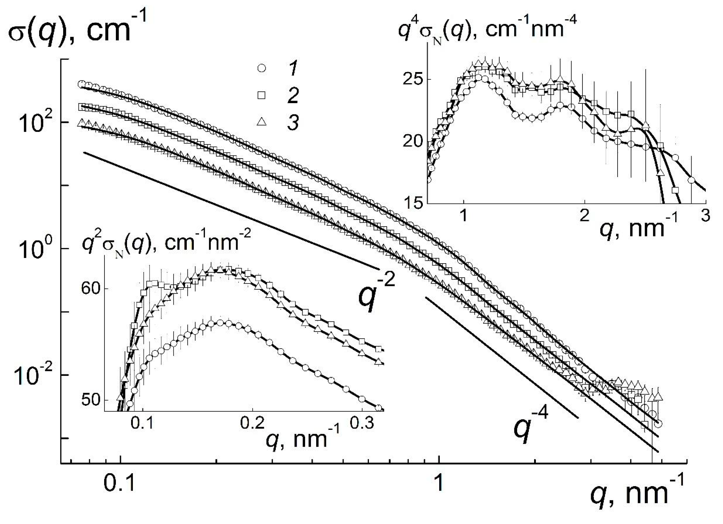
The analysis of neutron data combines the structural models’ application in reciprocal space and rebuilding of the correlation functions to determine more exactly the structures of the original gel and its derivatives. As seen in Figure 1, the scattering cross sections σ(q) for pristine gel and diluted systems grow by four orders in magnitude at low momentum transfers, that indicates an assembly of particles at the scales of R ~ π/qε ≥ 101 nm. At the same time, the dilution leads to the decrease of cross sections σ(q) according to the decrement of concentration, while at first glance, the behavior of σ(q) does not change. Hence, the gel’s ordering achieved above the critical point (С1 > C* ~ 4.2 wt %) is rather stable and exists after two- and four-fold dilution.
The retention of stable local structures below the critical point C* can be treated as a disintegration of the original gel network into large domains in water. Their size exceeds the value ~2π/qmin ~ 102 nm that is defined by the minimal momentum transfer qmin. Such a micro-gel exists in the range of concentrations from C*/4 ≤ СС*. It is remarkable that all the curves of σ(q) demonstrate a kink at the momentum transfer q ~ q* ~ 1 nm−1 (Figure 1). Obviously, at qq* the scattering on single particles dominate, whereas at qq* the interference is observed in scattering at the distances 2π/qds exceeding the particles’ diameter ds ~ 2π/q* ~ 6 nm. In these regions the cross sections show the exponential behaviors, σ(q) ~ 1/qD, with the parameters D = D1 ~ 4 and D = D2 ~ 2 where the first one corresponds to Porod’s law observed for the particles with sharp borders and the second one indicates chain-like aggregates of particles (Figure 1).
The existence of low-dimensional structures, i.e., linear (chain-like) aggregates is clearly visible from the q-dependence of the cross section, σ(q) ~ 1/q2, since this scattering law is established from the statistics of flexible chains (polymers) [16]. At the same time, except for linear fragments, the observed structures may include some branched (e.g., dendrimer-like) fragments characterized by the exponent D2 > 2 [16,17].
The logarithmic presentation (Figure 1) has revealed the basic motives of gel structuring. The subtle features of the systems are visible by a compensation of the exponential dependencies when we used the Kratky- and Porod-plots at qq* ~ 1 nm−1 and q < q* for the cross sections σN(q) = σ(q)/C normalized onto the diamonds’ concentrations, given in Figure 1 (inserts).
The right inset in Figure 1 illustrates the spacing (dint) between the centers of neighboring particles coupled in network structure of gel. The spacing dint defines the position of the peak, qint ≈ 1.1 nm−1 = X/dint where the coefficient X corresponds to the maximum X = qintdint ≈ 7.72 of Debye scattering functions sin ( q d i n t ) q d i n t for a pair of particles at the distance dint. The value of dint ≈ 7 nm exceeds a characteristic diameter of a diamond particle, dintdS ≈ 1 nm, i.e., the particles in the network structure of gel contact via water shells with a thickness of two molecular layers. The first (qint) and second maxima (~2qint) on the curves for different concentrations confirm a short range order of particles in gels.
On the other hand, the data at low momentum transfers (Figure 1, left insert) testify that the structuring at higher distance Dw = X/qw ≈ 40 nm where qw ≈ 0.18 nm−1 is the maximum position for a broad peak at q ~ 0.1–0.3 nm−1. This spacing should be attributed already to the size of the cells in gels’ multilevel structures, which are discussed below.
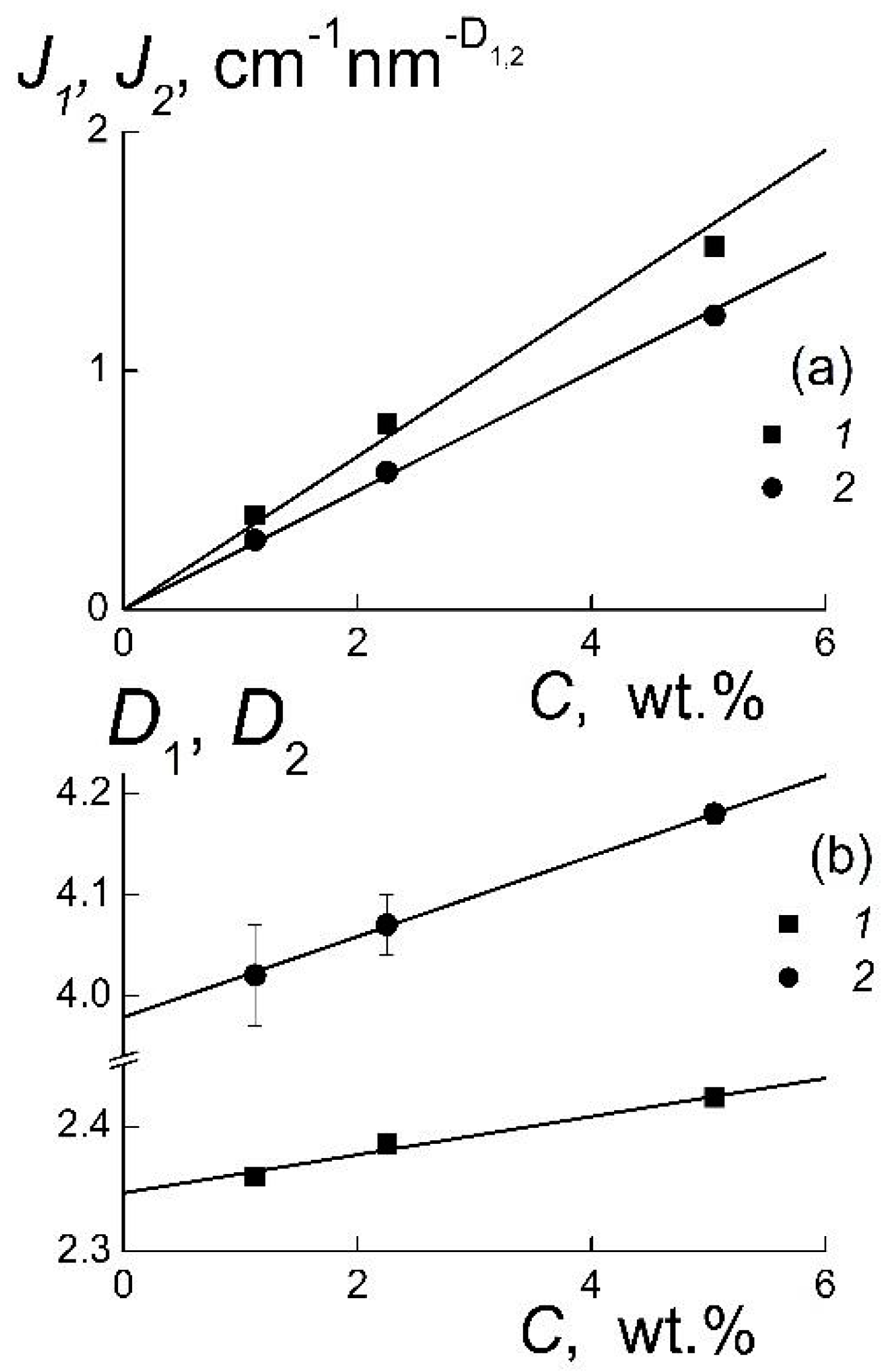
This analysis was extended to obtain more detailed information on the quality of the particles’ surface and the features of their association. The approximations of the data (Figure 1) with the exponential law
σ(q) = Ji/qDi, i = 1, 2
have given the coefficients Ji showing the scattering ability of the observed objects and the parameters Di which characterize their geometry (Figure 2).
A linear behavior of the coefficients J1, J2 indicates a change of the amount of particles’ or aggregates by the variation of concentration, when their geometry does not undergo any substantial transformation (Figure 2a). Only a weak linear decrease of the geometric parameters D1, D2 is observed (Figure 2b). The D2 reflects the geometry of particles’ surface. This parameter declines by dilution, and in the limit, C → 0, the value of D2 = D02 = 3.98 ± 0.04 is very close to the exponent in Porod’s law describing the scattering for the particles with sharp borders [18]. It confirms a perfection of the diamond crystals’ facets that is of principal importance for the configuration of potentials inducing the diamonds’ electrostatic attraction due to different electric charges of the facets [19,20]. Meanwhile, even at a moderate concentration (С3 = 5.05 wt %) the scattering from the particles’ surface is disturbed due to the interference in scattering from neighboring particles that gives D02 > 4 (Figure 2b). This must be distinguished from the results [5,12] for the suspensions of diamonds in graphene shells, which showed a deviation of scattering from Porod’s law.
The sharp surfaces of diamonds facilitate their association into branched structures evidenced from the values of D1 ~ 2.3–2.4 > 2 (Figure 2b). Such a magnitude in the range 2 < D1 < 3 is inherent in the structures formed via a diffusion limited aggregation (DLA) which creates mass fractals [17]. For pristine gel the fractal dimension D1 ~ 2.4 exceeds the parameter for a linear Gaussian polymer chain DG = 2 [16], that confirms the existence of branched aggregates forming a network of gel. A dilution of gel retains mostly the exponent D1 decreasing linearly with concentration (Figure 2b). In the limit С → 0, the extrapolated D1 = D01 = 2.35 ± 0.01 is substantially greater than the DG = 2 for linear chains. This shows a good stability of local structure of gel grown at the volume fraction of diamonds φ1 = 1.44% keeping in its integrity at their low content, φ3 = 0.22%. It is noticeable that this ordering is realized in the ensembles of particles with relatively broad size-distributions. For a comprehensive analysis of these structures, it is necessary to rebuild the size-distributions of diamonds.

2.2.2. Size-Distributions of Diamonds

To get the size distributions of the diamonds, the data (Figure 1) were approximated by the sum of the scattering function for spheres [18]. Therefore, the volume fractions Φ(R) of objects in the range of radii R = 0–30 nm were evaluated (Figure 3). The spectra Φ(R) demonstrate a broad peak with the maximum at R = R1m ~ 2.4–2.6 nm that agrees well with the DLS data for the diamonds prepared by the described method [14].
Except for the main peak for single particles (R1m ~ 2.6 nm), in the spectrum of the gel (Figure 3) the additional peak is seen at R2m ~ 5.1 nm ~ 2R1m. Its position lies in the range of radii comparable to the particles’ diameter. This indicates the particles’ association within the first coordination sphere. The dilution of the gel leads to disappearance of the secondary peak which overlaps with the first one that makes it complicated to distinguish single particles and small aggregates. Besides, all the samples show an aggregation at distances of double or triple the diameter of particle, R ~ 10–15 nm (Figure 3). To describe the ordering of the particles in gel, this model may serve as the first approximation which considers only spherical objects which scatter independently. However, various spatial correlations of particles and their aggregates should be taken into account.

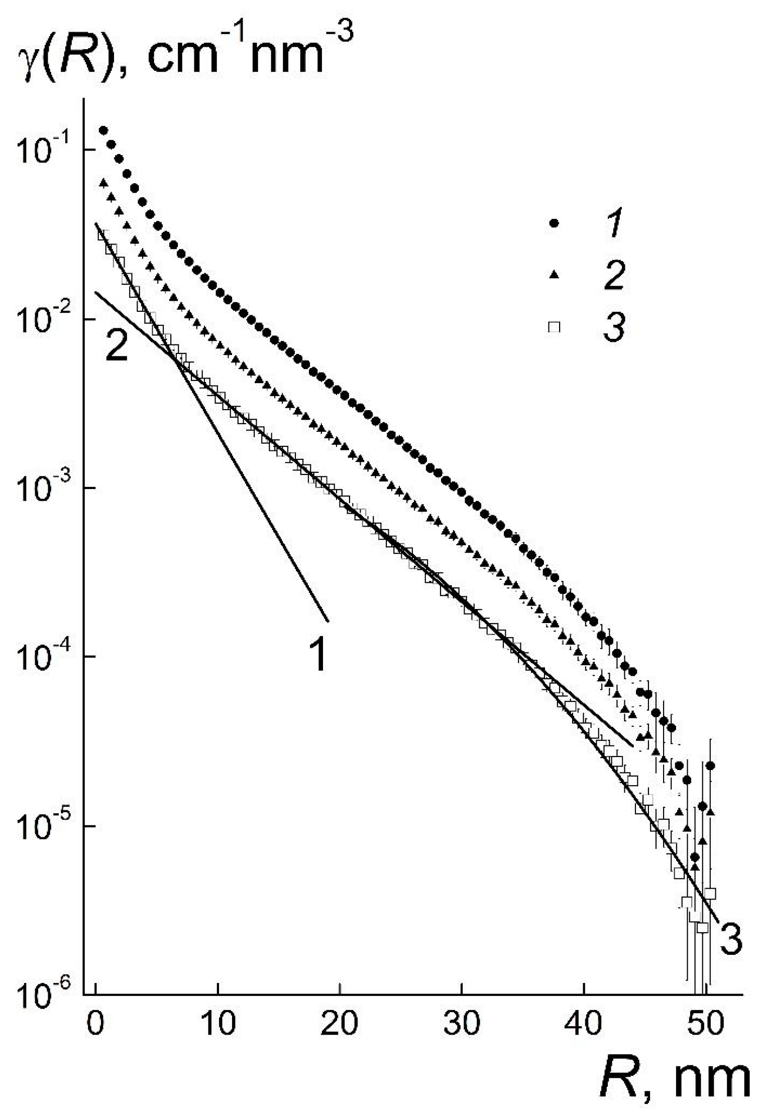
2.2.3. Correlation Functions

The ensembles of diamond particles are characterized by the functions γ(R) showing the correlations between various scattering centers (atomic nuclei, particles, aggregates). The function γ(R) = <δρ(r)·δρ(r + R)> is the averaged product of the deviations, δρ(r) = ρ(r) − <ρ>, δρ(r + R) = ρ(r + R) − <ρ>, in scattering length densities from the mean value <ρ> in the sample. A pair correlation is considered for two points at the distance R. The magnitude of <ρ> = Σ(biNi) is defined by the contributions of nuclei with scattering lengths bi and concentrations Ni. For isotropic samples, the functions γ(R) being the Fourier-transforms of scattering data [18] depend on the modulus of vector R,
γ(R) = (1/2π)3∫σ(q)[sin(qR)/(qR)]4πq2dq.
The γ(R) for the samples are similar and demonstrate three regions of behavior (Figure 4).
When the radii, 0 ≤ R ≤ 6 nm, are less than the diameter of the particles, the functions γ(R) obey the exponential law γ1(R) = g1exp(−R/r1). It includes the correlation length r1 comparable to the radius of particles and the coefficient g1 proportional to the concentration of diamonds (Table 1). Since the functions γ1(R) describe the atomic correlations inside the particles, the parameter r1 is the averaged correlation radius of particles r1 ~ R1m which is slightly larger than their geometric radius (Figure 3). This follows from the comparison of γ1(R) and the correlation function γS ~ [1 − (3/4)(R/rS) + (1/16)(R/rS)3] of a sphere [18] with the radius rS = R1m.
At low radii, Rr1, the γ1(R) is approximately a linear function, γ1 ~ [1 − R/r1]. At the same time, the linear term γS ~ [1 − (3/4)(R/rS)] dominates in the correlation function γS(R) at RrS. The identity γ1(R) ≡ γS(R) is fulfilled if rS = (3/4)r1. Thus, in gel the particles with a correlation radius r1 = 3.40 ± 0.01 nm have the geometric radius rS = (3/4)r1 ≈ 2.6 nm ≈ R1m in accordance with experimental data (Figure 3). In addition, the γ1(R) yields the information on the contrast factor of diamonds ΔρD relative to the surrounding medium. The ΔρD depends on the scattering lengths and the concentrations of chemical elements in the particles and surroundings. This gives the indication of how tightly packed the particles are. In pristine gel with the volume fraction of diamonds φ = 1.44%, the parameter g1 = (ΔρD)2φ (Table 1) affords the estimate ΔρD = (10.36 ± 0.01) × 1010 cm−2. The ΔρD= ρD − <ρ> = (1 − φ)(ρDρW) is the difference between the scattering length densities ρD of the carbon material and a similar parameter <ρ> for the whole volume of the sample <ρ> = φρD + (1 − φ)ρW, where ρW = −0.56 × 1010 cm−1 is the scattering length density for light water.
The obtained scattering length density of particles ρD = ΔρD/(1 − φ) + ρW = (9.95 ± 0.01) × 1010 cm−2 is 15% lower than a similar parameter, ρDI = 11.7 × 1010 cm−2, for the crystals of diamonds with a characteristic density of 3.5 g/cm3. The deficit can be explained by the presence of volume (surface) defects (vacancies) in diamonds. On the other hand, we neglected the possible influence of the gel’s structure factor, taken as S(q → 0) = 1. Even at a low volume content of particles, their coordination may cause a deviation of this factor from unity, S(q) < 1.
In these systems the structuring is revealed at the radii, 6 ≤ R ≤ 20 nm, comparable to the particles’ diameter (first coordination sphere) that is described by the exponential function, γ2(R) = g2exp(−R/r2). The coefficient g2 is proportional to the particles’ concentration but the correlation radius r2 ~ 7 nm remains almost constant (Table 1). This confirms a stability of the local ordering even by a substantial dilution of gel.
For the functions γ1(R), γ2(R) describing the correlations inside the particles and in the first coordination sphere, one can compare the correlation volumes, V1 = 4π∫exp(−R/r1)R2dR = 8πr13 ≈ 990 nm3 and V2 = 8πr23 ≈ 9.3 × 10³ nm3. The volume V2 exceeds V1 by an order of magnitude but the first coordination sphere is not completely filled with particles. This is evident from a comparison of the forward cross sections σ2 and σ1 calculated in the limit q → 0 using the functions γ1, γ2.
The cross section σ1 = (ΔρD)2φV1 = g1V1 is defined by the parameters of single particles while the magnitude of σ2 = (ΔρD)2φ(mV1) = g2V2 is proportional to the number of particles (m) within the correlation volume V2. The ratio of cross sections gives the value m = (g2VС)/(g1V1) = 3.6 ± 0.1. Hence, a particle in the gel is connected with two to three neighboring particles. This short linear or branched fragment (aggregation number m) is really stable even in the case of a four-fold dilution of gel where only a slight reduction in the aggregation number is observed (~10%).
The structure of gel outside the first coordination sphere is seen in the γ(R) behaviors which obey the law γ3(R) = g3exp[−(R/r3)2] at R ≥ 20 nm. It corresponds to Guinier’s function exp[−(qRG)2/3] with the gyration radius RG = (3/4)1/2r3 being proportional to the correlation length r3. In gel and diluted systems, the lengths r3 ~ 20 nm are the same (Table 1), as well as the gyration radii RG = (3/4)1/2r3 ~ 17 nm. One may imagine the structure of these samples being composed of spherical domains having the diameter of ~2RG ~ 40 nm.
This approximation is appropriated at q ≤ 0.1 nm−1 (Figure 1) while in the extended interval, q ≤ 1 nm−1, these domains are characterized as chain fractal aggregates (Table 1). The distribution of distances between the particles inside them is described by the function G(R) = γ3(R)R2 having the maximum at R = r3 as the most probable distance between the units in aggregates. For a Gaussian chain connecting two particles at the distance r3 ~ 17 nm, the number of units is equal to n = (r32/dS2) + 1 ≈ 16, and the contour length LC = ndS ≈ 80 nm. The latter should be compared to the spacing between the chains in the gel that is set by their numerical concentration, NL = φ/(nπdS3/6) ≈ 1.2 × 1016 cm−3 for the particles’ volume fraction, φ = 1.44%. For uniform distribution of the chains in the gel, the average distance between them, DwNL−1/3 ≈ 40 nm ~ 2r3, is approximately half of the contour length. The chains principally overlap and form the interconnected structure of the gel with cells’ diameter ~
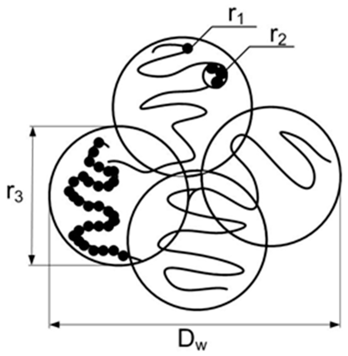
Dw ~ 40 nm. This is illustrated in Figure 5 where three levels of gel structure are shown. Single particles (size r1 ~ 3 nm, the first level) create small linear (branched) fragments (correlation radii r2 ~ 7 nm, the second level) associated into chain structures (size r3 ~ 20 nm) forming the cells (diameter Dw ~ 2r3 ~ 40 nm) which are integrated into a gel network.
In the network, the junctions’ functionality fC = mL/n can be found using the amount of particles (mL) per a cell. This value is included in the forward cross section σ3 = (ΔρD)2φdS3/6)mLSL = g3VL which is defined by the parameters of the correlation function (g3, r3) where VL = 4π∫γ3(R)R2dR = π3/2r33 is the correlation volume and SL(q → 0) is the structure factor of the systems of the cells. The model of random contacts of spherical objects in the sample volume [18] was used to evaluate the SL = 1 − 8VL/V1L, where V1L = fC/NL is the volume per cell that is determined by the numerical concentration of the cells (NL). The parameter VL = fCVn/β includes the volume of the chain’s region, Vn = πr33/6, and the coefficient β characterizing the density of the chains’ package in a cell. If the regions of chains fill the total volume, β = 1. If between the regions any cavities exist, β < 1, e.g., in the case of spheres, the most dense packing β = βS ≈ 0.74. Further we used the average βa = (1 + βS)/2 ≈ 0.87 to estimate the SL = 1 − 8(φ/nβa)(r3/dS)3 ≈ 0.54. Under these assumptions the amount of particles in a cell and the functionality of the network junctions is determined, mL= (g3VL)/[(ΔρD)2φdS3/6)SL] ≈ 70 and fC = mL/n ≈ 4.These parameters are attributed to the original and diluted systems, saving almost their initial structural features at different scales (from one to tens of diameter of particle) characterizing the multilevel order detected in gels.

3. Conclusions

Combined neutron scattering treatment in direct and reciprocal space has allowed us to discover the subtle structural features of diamonds’ hydrogels at three levels. The formation of short range order in the ensembles of diamonds was established, primarily the aggregation of particles within the first coordination sphere around a particle and secondarily the association of tiny fragments into extended chain aggregates creating a network of gel. At the macroscopic scale, the formation of gel was detected at the critical concentration when a giant increase of suspension viscosity was detected.
It would be useful to mention that increasing the viscosity of hydrosol of the nanodiamond particles was already previously detected [4]. However that experiments used nanodiamond particles obtained by the milling method [6] having a sp2 phase on the surface of the diamond particles. In the case of hydrosol produced by another method [14], we have observed a giant increase of viscosity at a concentration of diamond particles more than 4–5 wt % [21].
In neutron experiments, it was found that a dilution of gel below the critical point does not destroy the local structure of the gel. This phenomenon can be treated as a disintegration of a continuous gel into microscopic domains, which save their intrinsic structure despite the water surrounding. These results confirm the concept of the main role of electrostatic attraction of hydrated diamonds with different charges on their facets, predicted by [19,20] in the formation of gel consisting of nanodiamond particles.

Acknowledgments

The work is supported by the Russian Foundation for Basic Research (grant No. 14-23-01015 ofi-m) in part for neutron scattering studies. Alexander Vul thanks the support of the Russian Science Foundation (project 14-13-00795) in part for preparation and research of the properties of nanodiamond gel.

Author Contributions

Vasily Lebedev and Alexander Vul promoted the main idea of the manuscript, wrote the main manuscript text, and prepared all figures. Yury Kulvelis and Alexander Kuklin provided measurements, analysis, and discussion of small angle scattering data. All authors discussed the results and reviewed the manuscript. All authors have read and approved the final manuscript.

Conflicts of Interest

The authors declare no conflict of interest.

References

  1. Lymkin, A.I.; Petrov, E.A.; Ershov, A.P.; Sakovitch, G.V.; Staver, A.M.; Titov, V.M. Production of diamonds from explosives. Sov. Phys. Dokl. 1988, 33, 705–708. [Google Scholar]
  2. Greiner, N.R.; Philips, D.S.; Johnson, J.D.; Volk, F. Diamonds in detonation soot. Nature 1988, 333, 440–442. [Google Scholar] [CrossRef]
  3. Vul, A.Y.; Dideikin, A.T.; Aleksenskii, A.E.; Baidakova, M.V. Detonation nanodiamond: synthesis, properties and applications. In Nanodiamond; Williams, O., Ed.; Royal Society of Chemistry: Cambridge, UK, 2014; pp. 27–48. [Google Scholar]
  4. Vul, A.Y.; Eydelman, E.D.; Inakuma, M.; Ōsawa, E. Correlation between Viscosity and Absorption of Electromagnetic Waves in an Aqueous UNCD Suspension. Diam. Relat. Mater. 2007, 16, 2035–2038. [Google Scholar] [CrossRef]
  5. Avdeev, M.V.; Aksenov, V.L.; Tomchuk, O.V.; Bulavin, L.A.; Garamus, V.M.; Osawa, E. The spatial diamond-graphite transition in detonation nanodiamond as revealed by small-angle neutron scattering. J. Phys. Condens. Matter 2013, 25, 445001. [Google Scholar] [CrossRef] [PubMed]
  6. Osawa, E. Monodisperse single nanodiamond particulates. Pure Appl. Chem. 2008, 80, 1356–1379. [Google Scholar] [CrossRef]
  7. Williams, A.; Hees, J.; Dieker, C.; Jager, W.; Kirste, L.; Nebel, C.E. Size-Dependent Reactivity of Diamond Nanoparticles. ACS Nano 2010, 4, 4824–4830. [Google Scholar] [CrossRef] [PubMed]
  8. Mochalin, V.N.; Shenderova, O.; Ho, D.; Gogotsi, Y. The properties and applications of nanodiamonds. Nat. Nanotechnol. 2012, 7, 11–23. [Google Scholar] [CrossRef] [PubMed]
  9. Ho, D. (Ed.) Nanodiamonds: Applications in Biology and Nanoscale Medicine; Springer: New York, NY, USA, 2010.
  10. Vul, A.; Shenderova, O. (Eds.) Detonation Nanodiamonds: Science and Applications; Pan Stanford: Singapore, 2014.
  11. Avdeev, M.V.; Rozhkova, N.N.; Aksenov, V.L.; Garamus, V.M.; Willumeit, R.; Osawa, E.J. Aggregate Structure in Concentrated Liquid Dispersions of Ultrananocrystalline Diamond by Small-Angle Neutron Scattering. J. Phys. Chem. C 2009, 113, 9473–9479. [Google Scholar] [CrossRef]
  12. Tomchuk, O.V.; Bulavin, L.A.; Aksenov, V.L.; Garamus, V.M.; Ivankov, O.I.; Vul, A.Y.; Dideikin, A.T.; Avdeev, M.V. Small-angle scattering from polydisperse particles with a diffusive surface. J. Appl. Cryst. 2014, 47, 642–653. [Google Scholar] [CrossRef]
  13. Tomchuk, O.V.; Volkov, D.S.; Bulavin, L.A.; Rogachev, A.V.; Proskurnin, M.A.; Korobov, M.V.; Avdeev, M.V. Structural Characteristics of Aqueous Dispersions of Detonation Nanodiamond and Their Aggregate Fractions as Revealed by Small-Angle Neutron Scattering. J. Phys. Chem. C 2015, 119, 794–802. [Google Scholar] [CrossRef]
  14. Aleksenskiy, A.E.; Eydelman, E.D.; Vul, A.Y. Deagglomeration of Detonation Nanodimonds. Nanotechnol. Lett. 2011, 3, 68–74. [Google Scholar] [CrossRef]
  15. Shvidchenko, A.V.; Zhukov, A.N.; Dideikin, A.T.; Baidakova, M.V.; Shestakov, M.S.; Shnitov, V.V.; Vul, A.Y. Electrosurface properties of single-crystalline detonation nanodiamond particles obtained by air annealing of their agglomerates. Colloid J. 2016, 78, 235–241. [Google Scholar] [CrossRef]
  16. De Gennes, P.-G. Scaling Concepts in Polymer Physics; Cornell University Press: Ithaca, NY, USA, 1979. [Google Scholar]
  17. Witten, T.A.; Sander, L.M. Diffusion-limited aggregation. Phys. Rev. B 1983, 27, 5686–5697. [Google Scholar] [CrossRef]
  18. Feigin, L.A.; Svergun, D.I. Structure Analysis by Small-Angle X-ray and Neutron Scattering; Plenum Press: New York, NY, USA, 1987; p. 323. [Google Scholar]
  19. Barnard, A.S.; Sternberg, M. Crystallinity and surface electrostatics of diamond nanocrystals. J. Mater. Chem. 2007, 17, 4811–4819. [Google Scholar] [CrossRef]
  20. Barnard, A.S. Modeling polydispersive ensembles of diamond nanoparticles. Nanotechnology 2013, 24, 085703. [Google Scholar] [CrossRef] [PubMed]
  21. Vul, A.Y.; Eidelman, E.D.; Aleksenskiy, A.E.; Shvidchenko, A.V.; Dideikin, A.T.; Yuferev, V.S.; Lebedev, V.T.; Kul’velis, Y.V. Transition sol-gel in nanodiamond hydrosols. Carbon 2016, in press. [Google Scholar] [CrossRef]
Figure 1. Cross sections σ(q) for the samples with diamond contents of 5.05, 2.25, and 1.13 wt % (1–3) vs. momentum transfer. Straight lines show characteristic slopes for cross sections’ q-dependencies. The fitting curves for the model of spherical particles are plotted also. Inserts show the Kratky- and Porod-plots (left, right) for the cross sections σN(q) normalized to the diamonds’ concentrations.
Condensedmatter 01 00010 g001
Figure 2. Parameters of function (1): (a) J1, J2—the characteristics of the scattering ability of fractal structures and single particles; (b) D1—the fractal dimension of aggregates, D2—the exponent describing the geometry of particles’ surface.
Condensedmatter 01 00010 g002
Figure 3. Volume fractions Φ(R) of scattering objects vs. their radii: diamonds’ content of 5.05, 2.25, and 1.13 wt % (1–3). Arrows indicate the main (R1m) and secondary maxima (R2m).
Condensedmatter 01 00010 g003
Figure 4. Functions γ(R) for the samples with 5.05, 2.25 and 1.13 wt % of diamonds (1–3). The lines show the fitting functions γ1, γ2, γ3 (1–3) for the lowest concentration (see text).
Condensedmatter 01 00010 g004
Figure 5. Structural levels in gel: the single particles (correlation radius r1), the first coordination sphere (radius r2), and the chains (size r3) associated into the cells (diameter Dw).
Condensedmatter 01 00010 g005
Table 1. Parameters of partial correlation functions γi(R) of the samples with different contents of diamond (C): coefficients gi and correlation radii ri (i = 1, 2, 3).
С, wt %1.132.255.05
g1·103, cm−1·nm−336.4 ± 0.174.5 ± 0.2154.8 ± 0.3
g2·103, cm−1·nm−314.3 ± 0.127.9 ± 0.259.0 ± 0.5
g3·104, cm−1·nm−322.8 ± 0.445.1 ± 0.894.6 ± 1.8
r1, nm3.51 ± 0.013.48 ± 0.013.40 ± 0.01
r2, nm7.12 ± 0.037.31 ± 0.037.19 ± 0.03
r3, nm19.7 ± 0.120.2 ± 0.119.9 ± 0.1

