Article

Linking the Physicochemical Properties of Calcined Titania Nanoparticles with Their Biocidal Activity

1
Department of Biomedical, Chemical and Environmental Engineering, University of Cincinnati, Cincinnati, OH 45221-0012, USA
2
National Risk Management Research Laboratory, U.S. Environmental Protection Agency, Research Triangle Park, NC 27711, USA
3
Department of Civil Engineering, The University of Texas at Arlington, Arlington, TX 76019-0308, USA
4
National Health and Environmental Effects Research Laboratory, U.S. Environmental Protection Agency, Research Triangle Park, NC 27711, USA
*
Authors to whom correspondence should be addressed.
Inventions 2016, 1(4), 26; https://doi.org/10.3390/inventions1040026
Submission received: 27 October 2016 / Revised: 9 December 2016 / Accepted: 13 December 2016 / Published: 20 December 2016
(This article belongs to the Special Issue Nanomaterials and Nanocomposites for Environmental Applications)

Abstract

:
Titanium dioxide nanoparticles (nTiO2) show biocidal activity when exposed to UV illumination. Modification of their physical properties can expand their photoresponse region toward visible light. In this study, such modification was made through a sol-gel synthesis followed by calcination at a range of temperatures (250–900 °C), generating a series of nTiO2 particles with different crystal phases, sizes, porosities, zeta potentials, and BET surface areas. The unique properties of nTiO2 were linked to their toxicity to the marine bacterium, Vibrio fischeri. A modified “Flash” high-through put assay was used to test the viability of these marine organisms after short term (15–60 min) exposure under visible light only to the individual groups of nTiO2 (500–2000 μg/mL). Linear regression analysis indicated that across all concentrations and time points, high biocidal activity correlated with the amorphous and anatase crystal phases, high BET surface area, high pore volume and small crystal size. The linkage between physicochemistry and nanotoxicity would be helpful for future design of more efficient and sustainable nTiO2.

Graphical Abstract

1. Introduction

Nanosize titanium dioxide particles (nTiO2) are used in environmental remediation (water purification), consumer supplies (paints, cosmetics, sunscreens) and medical devices [1,2,3,4,5,6,7,8]. The biocidal properties of nTiO2 are based on its photoactivation by UV radiation (<380 nm for anatase and <400 nm for rutile) which generates a variety of reactive oxygen species (ROS) (e.g., superoxide anion, hydroxyl radical, and singlet oxygen) [9,10]. ROS is effective against Gram-positive and Gram-negative bacteria, filamentous and unicellular fungi, algae, protozoa and viruses [3,7,10,11,12] and toxicity studies on nTiO2 have been conducted in various species [13,14,15,16,17,18,19,20,21,22].
Applications using conventional nTiO2 are restricted by its photosensitivity to UV wavelengths. Various methods (e.g., doping, surface coating, calcination) are used to decrease the band-gap energy of nTiO2 which increases its photosensitivity to visible/near visible radiation wavelengths [23,24,25,26,27]. Although these procedures often create more sustainable and effective materials, their toxic threat to the environment and “at-risk” species must be addressed.
Recent studies link the physicochemical properties (e.g., crystal size, Brunauer, Emmett and Teller (BET) surface area, and pore volume) of commercial nTiO2 to its toxicity. To better understand how subtle changes in these properties modify biocidal activity, a series of lab-synthesized nTiO2 particles with different properties, calcined at different temperatures, was tested for their relative toxicity to the marine bacteria, Vibrio fischeri, a Gram negative, bioluminescent micro-organism [28]. The Vibrio fischeri transforms a fraction of its metabolic energy into bioluminescence. The organisms’ auto-lumination reflects their viability, which can be measured spectrophotometrically in the absence of any reagents. Because of this, the Vibrio Fischeri micro-organisms has been used to assess the potential toxicity of ecotoxic substances [29,30,31,32,33,34]. In this study, a high-through-put (HTP) “Flash” assay (EN ISO 11348-1) was used to measure the toxicity of the modified nTiO2 to Vibrio Fischeri micro-organism. This commercially available assay has been used to test the micro-biocidal effects of a variety of environmental and industrial samples [29,35,36,37], including nanoparticles [30,31,32,38,39]. Use of the Vibrio fischeri in this assay is especially valuable in view of the need to examine the potential toxicity of the increasing number of materials entering aquatic marine environments [14,19,40].
The in vitro screening of particles and chemicals, using commercially available, rapid and cost-effective cell assays, provides valuable “first-tier” level data on their potential toxicity. This study uses such a “screen” and the luminating marine bacteria Vibrio fischeri to demonstrate that modified nTiO2 particles are biocidal in the absence of UV illumination and that their relative toxicity can be associated with several physicochemical characteristics. Such data are essential to the design of safer, effective and more sustainable nanomaterials in environmental remediation.

2. Experimental Section

2.1. Synthesis and Physicochemical Characterization

The procedures to synthesize and characterize the nTiO2 in this study have been published previously [41]. Briefly, titanium tetraisopropoxide (TTIP, Sigma-Aldrich, St. Louis, MI, USA) as a TiO2 molecular precursor was added to isopropanol (i-PrOH, Fisher Inc, Waltham, MA, USA) and water. The molar ratio of the H2O/i-PrOH/TTIP sol was 100/30/1. After hydrolysis and condensation reactions under vigorous stirring, TiO2 particles were formed. They were recovered by filtration and purified by refluxing with acetonitrile (Fisher Inc) several times. Finally, they were dried at room temperature for 24 h. The resulting “as synthesized” (as-syn) nTiO2 particles were calcined at temperatures of 250–900 °C in a furnace (Paragon HT-22-D) to transform them to crystal phase TiO2 particles.
The crystal size of TiO2 was determined by using the Scherrer equation from the XRD peak broadening analysis at (101) obtained using a Kristalloflex D500 diffractometer (Siemens, Munich, Germany) with Cu Kα (λ = 1.5406 Å) radiation. Its BET surface area and pore volume were measured based on nitrogen adsorption and desorption isotherms using Tristar 3000 analyzer (Micromeritics, Norcross, GA, USA). The zeta potential of nTiO2 was immediately measured using a ZetaSizer nano ZS (Malvern Inc., Southborough, MA, USA) in 10–20 μg/mL suspensions of photobacterium marine broth (Carolina Biological Supply, Burlington, NC, USA) after ultra-sonication (see below). The data (4 measures) were collected and calculated into zeta potentials using the Smoluchowski equation [42,43]. Averages of zeta potentials were analyzed using a one-way ANOVA followed by a Tukey’s post-analysis and graphed using Graph Pad Prism 5. An attempt was made to measure particle size distribution using dynamic light scattering (DLS). This technique measures the diffusion of particles moving under Brownian motion, and converts this to size and size distribution using the Stokes-Einstein relationship. However, a stable solution of suspended particles is necessary for valid measurement of Z-averages (d. nm), (i.e., the average diameter of the particles). Because of the high osmolarity of the marine broth, the individual samples were settled out of suspension within a few minutes (below the instruments minimal time for the initial cuvette readings), preventing DLS particle size measurements.

2.2. Storage and Light Exposure of Modified nTiO2 Samples

After the calcination process, synthesized nTiO2 samples were stored in the dark upon receipt until used. In preparation for the plate exposures, stock suspensions of each sample were ultra-sonicated using a Daiger Ultrasonic processor (Model GE 750 watt, Sonics®, Newtown, CT, USA) at a reduced amplitude of 20% for 5–7 min under dim fluorescence lighting. Stock concentrations (10-fold) were prepared and exposed under low fluorescent lighting to the 96 well plates containing the population-he adjusted bacteria plate (see below). The 96 well microplate, containing bacteria exposed to the various samples of modified nTiO2 was automatically transferred into the luminometer and the chamber door closed from all visible light. The microplate remained in the dark for the remainder of the 60 min exposure.

2.3. Microorganisms and Viability Assay

Vibrio fischeri (ATCC 49387) were grown in photobacterium broth (chemical composition listed in reference [44]) on agar slants in test tubes. Their log growth was empirically determined to be 48–72 h after the initial inoculation. Photo-bacterium marine broth (10–20 mL) was added to each inoculated agar tube without agitation to make suspensions of the micro-organism. The micro-organisms were held in complete darkness until the time of their exposure (see below) which was done under a dimmed visible (fluorescence) lighting. Suspensions of bacteria (200 μL) were transferred to a 96 well, white culture plate (Costar Inc., Washington, DC, USA). Their luminescence signal of these untreated suspensions was first adjusted with additional broth to produce a uniform baseline emission of 800–1000 chemiluminescent units. Control readings were taken of wells containing: (broth +/− organisms; broth +/− nTiO2 samples; broth +/− organisms +/− nTiO2 samples). None of control treatments generated significant signal and were discounted in the calculations. For the viability testing, aliquots of the individual groups of as-syn-nTiO2 (500–2000 μg/mL) were administered to wells containing population-corrected Vibrio fischeri at 10-fold concentrations (n = 6 well/concentration). Readings were taken <30 s after exposure and used as the (t = 0) baseline value. Reduction of luminescence was recorded at three post-exposure time intervals (t = 15, 30, and 60 min) using a LMAX II384 luminescent plate reader (Molecular Device, Sunnyvale, CA, USA) and the results were used to measure loss of viability. Each assay was replicated 4–6 times and representative data presented. Data were collected using Softpro5 (SoftPro, Raleigh, NC, USA) and transferred to Excel software where they were normalized to their individual baseline (t = 0). Linear correlation R2 values were calculated and plotted against individual physicochemical characteristics using Excel software.

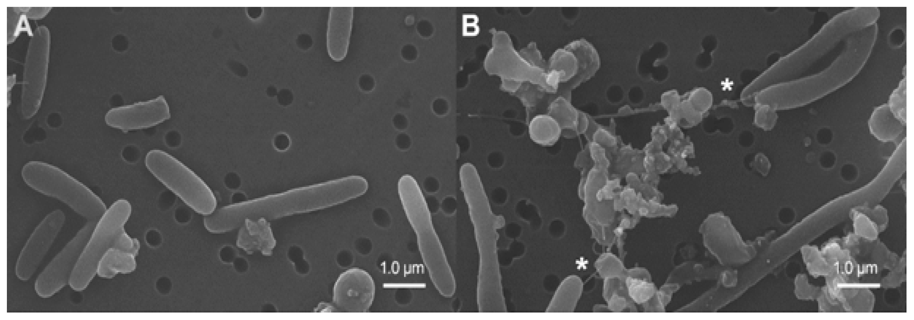
2.4. Transmission and Scanning Electron Microscopy

For transmission electron microscopy (TEM) examination of nTiO2, samples were sonicated (2510R-DH, Branson Ultrasonics, Danbury, CT, USA) in HPLC grade methanol (Pharmco, Brookfield, CT, USA) for 5 min, transferred to a carbon coated 200 mesh, copper grid (EMS), and photographed using a JEM-2010F (JEOL) high resolution TEM (HR-TEM) with field emission of 200 kV. The diameter of particles was measured using ImageJ software (National Institute of Health, New York, NY, USA). For biological samples, the bacteria were exposed in Eppendorf 5 mL tubes for 1 h, pelleted in marine broth, and washed several times in warm marine broth to remove all non-internalized particles. The pellets were fixed overnight in cold cacodylate-buffered 4% glutaraldehyde (Poly Scientific, Bayside, NY, USA) and processed using standard procedures. For scanning electron microscopy (SEM), samples were exposed to the individual as-syn-nTiO2 samples, pelleted and fixed for 2 h in cold cacodylate-buffered 4% glutaraldehyde (Poly Scientific, Bayside, NY, USA). They were then transferred to a 0.4 μm nuclear pore filter and sputter-coated with 50 Angstrom gold–palladium before the examination with a JEOL SEM (JSM-5900 LV).

3. Results

3.1. Physicochemical Properties

Table 1 summarizes the characterization of nTiO2, as reported elsewhere [41]. The as-syn-nTiO2-100 sample had the largest BET surface area of 482 m2/g and pore volume of 0.659 cm3/g. Increasing the calcination temperatures decreased the BET surface area and pore volume as expected [45,46]. Increasing the calcination temperature increased the crystal size and shifted amorphous, non-photoactive nTiO2 to anatase phase and subsequently to rutile phase. The zeta potential values of as-syn-nTiO2 measured in 500 μg/mL marine broth suspensions were all negatively charged (−24 to −28 mV) at these experimental conditions. The measurement of aggregate sizes by DLS was prevented due to rapid precipitation of each nTiO2 suspension in the highly osmolar photobacterium, dehydrated marine broth.

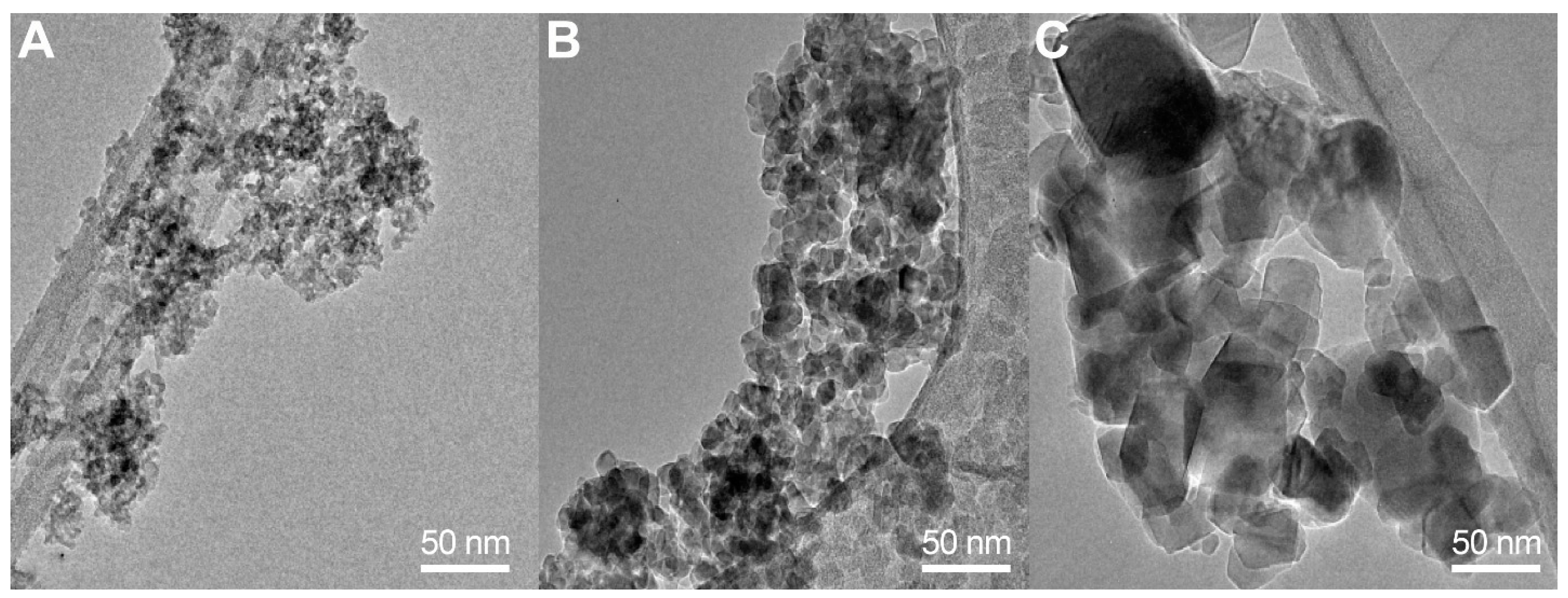
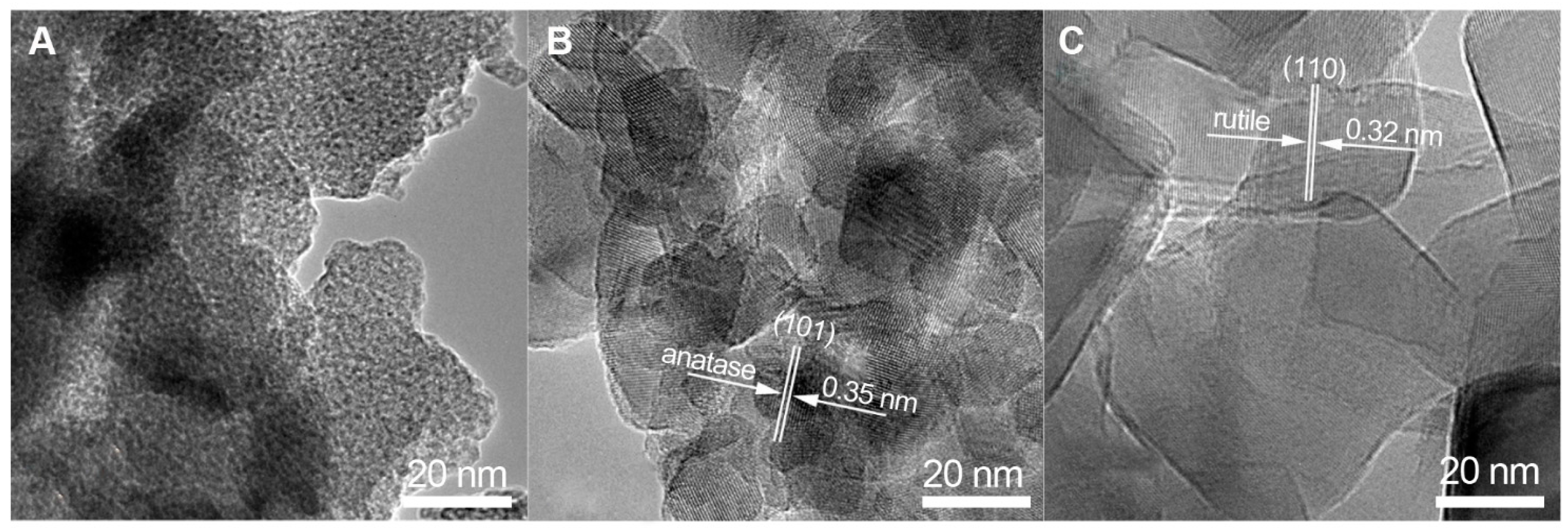
3.2. Transmission Electron Microscopy

TEM was used to examine the effects of calcination temperatures on particle size and crystal phase as shown in Figure 1. The crystal size increased when as-syn-nTiO2 was calcined at higher temperatures. ImageJ software determined the sizes of the particles, calcined at 250, 500 and 700 °C to be 5.7 ± 1.5 nm, 14.0 ± 2.9 nm and 47.5 ± 21.9 nm, respectively.
HR-TEM was also used to examine the effect of calcination temperature on crystal size and crystal phase of the samples as shown in Figure 2. The as-syn-nTiO2-100 sample (Figure 2A) exhibited an amorphous structure while the sample calcined at 500 °C (Figure 2B) showed crystals of 15–20 nm sizes and lattice spacing of 0.35 nm which corresponds to the (101) plane of anatase phase. As-syn-nTiO2 calcined at 700 °C (Figure 2C) showed well-defined rutile crystals with sizes of 40–50 nm. The measured lattice spacing was 0.32 nm, corresponding to the (110) plane of rutile phase.
SEM was used to examine the control and treated microorganisms as presented in Figure 3. The control Vibrio fischeri (Figure 3A) appeared spherical and elongated with a thick outer membrane and an occasional single flagellum projecting from one pole. In contrast, nTiO2-treated organisms (Figure 3B) were more variable in appearance, with normal bacteria interspersed with rounded or overtly disrupted organisms. The seemingly normal organisms projected multiple filamentous extensions to each other and to the damaged Vibrio fischeri. Cell walls of the affected bacterium appeared damaged in spots which was followed by a breakdown of the cell membrane, cell rounding and the eventual leakage of their contents.

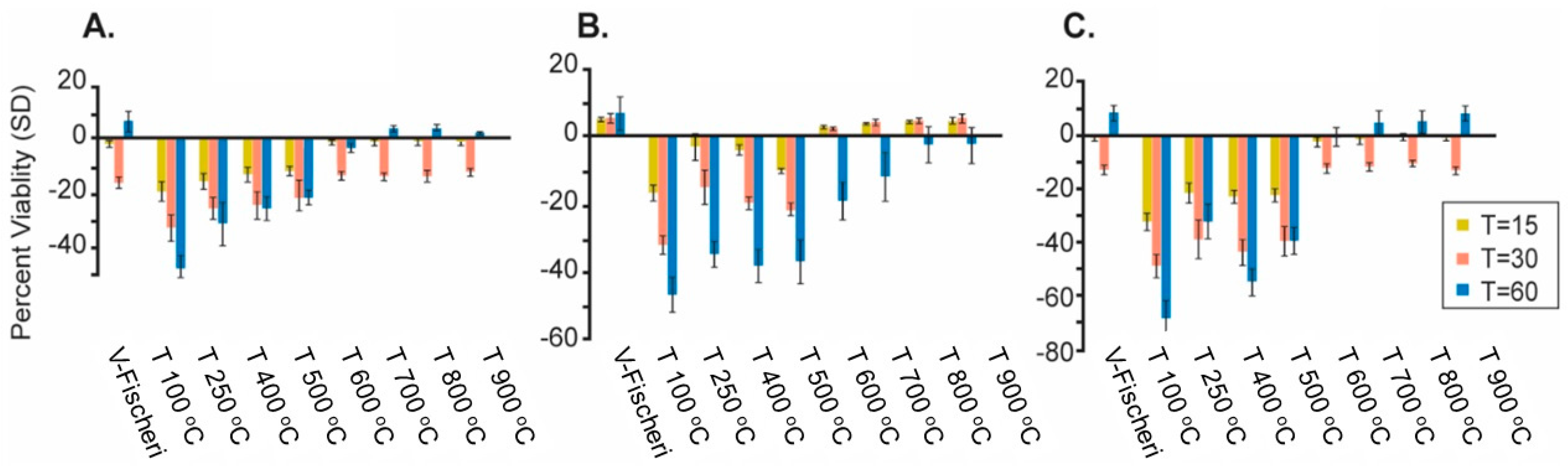
3.3. Viability Studies

Reduction of luminescence was used as an index of viability and recorded in a dose-response fashion as early as 15 min post-exposure as shown in Figure 4. Data were normalized to their individual post-exposure values at t = 0. Viability data indicated that the toxicity of the nTiO2 series corresponded to their calcination temperatures and a simple rank order indicated that high biocidal toxicity related inversely to higher calcination temperatures (i.e., small crystals demonstrate high biocidal toxicity) (data not shown).

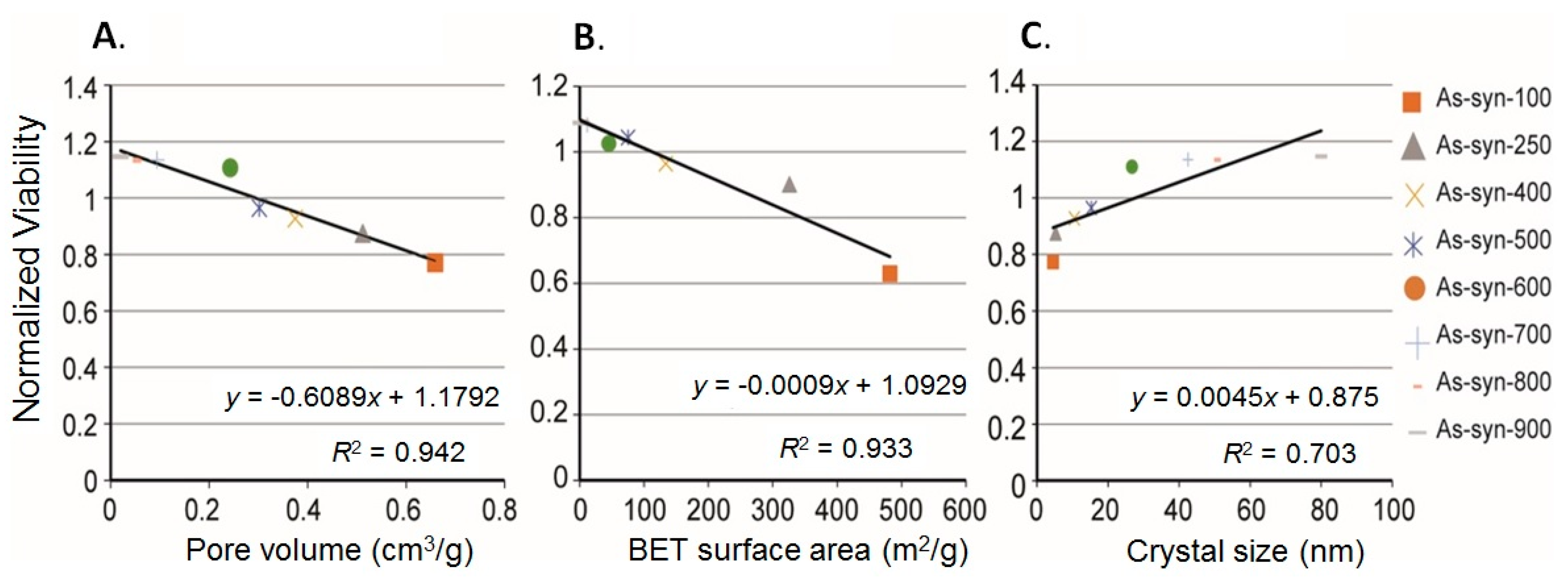
3.4. Linear Correlation

R2 values were calculated on the viability studies and plotted using Excel software. Similar ranking based on R2 values were seen at all time-points and concentrations and indicated that high toxicity was associated with large BET surface area and pore volume, and small crystal size. The highest R2 values, correlating lethality with pore volume, BET surface area and crystal size appeared highest in the groups exposed to 500 μg/mL for 30 min and are graphically presented in Figure 5. These data indicated that high biocidal activity correlated with large BET surface area (R2 = 0.93, −slope) and pore volume (R2 = 0.94, −slope), and small crystal size (R2 = 0.70, +slope). The values of R2 for pore volume, BET surface area and crystal size after 30 min exposure to 1000 and 2000 μg/mL were calculated to be (0.84, 0.79, 0.55 and 0.66, 0.64, 0.49), respectively.

4. Discussion

The primary aim of these experiments was to test a set of closely related nTiO2 for their biocidal activity and relate this toxicity to their specific physicochemical properties. The marine bacterium Vibrio fischeri was chosen as the target micro-organism since nTiO2 affects a range of organisms in marine ecosystems [9,47,48,49,50,51,52].

4.1. Physicochemical Effects of Calcination Temperatures

The different calcination temperatures (250–900 °C) used in the synthesis produced a series of modified materials with subtle differences in their physicochemical properties. An overview of these properties (Table 1) indicated that low calcination temperatures related to large BET surface area, large pore volume and small crystal size. HR-TEM showed that increasing the calcination temperature from 500 to 700 °C shifted the crystal phase of nTiO2 from anatase towards rutile. Data from related studies [53,54,55] also showed that altering the calcination temperature changed the crystallographic properties of as-syn-nTiO2. Increasing the calcination temperatures also increased the crystal size due to sintering and growth of particles. The crystal sizes measured from the HR-TEM micrographs using ImageJ software and those given in Table 1 which were calculated from XRD data [41], differed slightly. However, both calculations indicated that larger nTiO2 crystals were formed at higher calcination temperatures.

4.2. Biocidal Effects

The SEM micrograph shown in Figure 3B was representative of the exposed Vibrio fischeri. It suggests membrane damage and rounding of the micro-organism as its membranous network broke-down. Other studies have used TEM and biochemical analysis to describe similar effects of UV and visible light activated TiO2 and have documented a leakage of ions and decreases in membrane fluidity in Escherichia coli (E. coli) and Staphylococcus aureus [56,57]. The explanation for the differences in vulnerability of micro-organisms to nTiO2 is unclear but in part reflects the different thickness of the cell walls of gram+/gram bacteria [58,59].
A number of research studies have reported on the toxicity of nanoTiO2 in various micro-organisms (e.g., E. coli, Staphylococcus aureus, Enterococcus faecalis, coliform) and fungi (Candida albicans, Aspergillus niger). These studies have primarily used UV- or visible (“doped”) light-activated materials and a variety of endpoints and exposure time-points, making a comparison among studies difficult. In general, bulk and even nanoTiO2 in most forms, show low-toxicity to organisms and even in the case of UV-activated TiO2, concentrations of 800 μg/mL are required to kill 100% of the organism (coliform) in polluted waters [4]. This value might be compared with the biocidal effects (Figure 5A) associated with exposures to 500 μg/mL calcined TiO2 samples.
Several research studies have examined the effects of visible light-activated, nanostructured TiO2 (VLA nano-TiO2) on Vibrio fischeri, using surface coatings [60,61]; or different “doping” agents [62]. One study [63] examined the relative phenol degradation activity in a series of nTiO2 samples generated after calcination at 350–750 °C. Similar to our findings, they found that the materials calcined at lower temperatures showed the highest phenol degradation activity and were most sensitive to visible light. Given this, the present study represents a unique contribution to the field as it examines the biocidal activity of nine molecularly related VLA nano-TiO2 materials.

4.3. Physicochemical Properties Influence Toxicity

The viability data demonstrated a linkage between the viability of Vibrio fischeri and those modified nTiO2 groups with large BET surface area and pore volume, small crystal size, and amorphous-anatase crystal phases. The physicochemical properties of particles, especially nanoparticles, are fundamental to their toxicity. Among the properties thought to influence a particle’s biological activation include the size, shape or topography, surface area, crystal structure, surface charge, porosity and the tendency to aggregate [64,65,66,67,68,69]. Although many studies associate surface area and crystalline size of nTiO2 with its toxicity, others relate toxicity more to the crystal structure and particle shape/topography. Numerous studies have linked small particle size with higher nTiO2 toxicity in rodents, eco-species, cell culture, and microorganisms [13,18,64,70,71,72,73,74,75]. Relationships between particle toxicity, and the particle’s size and BET surface area have been repeatedly demonstrated in inhalation toxicology studies [70,75,76]. It has also been reported that nanosize nTiO2 can damage human bronchial epithelial cells (BEAS-2B) in the absence of photoactivation [77]. On a cellular lever, an ultrafine particle size is thought to facilitate movement through cellular barriers and give the particle closer proximity to vulnerable subcellular systems (e.g., nucleic acids, enzymes) [65,78,79,80].
The crystal phase of nTiO2 also seems to influence toxicity with anatase being more toxic to a variety of species [15,64,81,82] while other studies associate a higher toxicity and nucleic acid damage with a mixture of rutile/anatase phases [77,83].
Aggregation is also considered to be a factor in nTiO2 toxicity since it affects particle shape, size and bioavailability [20,74,76,84]. Aggregation is affected by pH, ionic strength, and ionic identity (inorganic and organic) of aqueous suspensions. The relatively low toxicity associated with rutile phase particles has been attributed to their tendency to form larger aggregates in physiological exposure vehicles [15]. However, data from our study and others [76] failed to support a role for particle aggregation in nTiO2 toxicity. In the current study, exposure of our particles in the highly osmolar marine broth resulted in an immediate agglomeration and sedimentation. Yet, the biocidal activity of these materials showed a high correlation with the particle’s size and surface properties as measured on dry particles or in distilled water. One explanation for this could be that the nTiO2 particles, although aggregated, still showed a sufficiently high fractal surface for the formation of ROS and resulting toxicity. It is possible that studying the response to aggregated nanomaterials could be as relevant as that of highly dispersed material if the aggregates retain the characteristics of material (e.g., a large specific surface area, fractal surface, and nanosize dimensions) after agglomeration. In all likelihood the biological activity of nanoparticles relates to physicochemical parameters yet to be rigorously tested in current toxicity screening studies such as redox activity, surface affinity or combinations of more conventional physicochemical properties.
A (primary) mechanism underlying the toxicity of both UV and VLA nTiO2 centers on ROS formation [9,10]. Studies have demonstrated a linear correlation between the amount of ROS generated by TiO2 and biocidal activity. The larger surface areas of those modified TiO2 calcined at lower temperatures might provide more absorption sites for UV and visible light and result in higher ROS formation. The larger surface area leads to high reactivity [85,86]. It is reasonable to assume that the higher surface area produced by the lower temperature modified TiO2 in our study would allow for a greater ROS generation and higher toxicity of these damaging free radicals. ROS formation is influenced by both the BET area and particle size [9,23,51,87,88,89,90]. Although this study did not directly address the formation of ROS, an association with ROS and the loss of viability of the Vibrio fischeri is suggested. At no time in the study were the bacteria nor the nTiO2 materials exposed to UV lighting. The modified particles were exposed to dim fluorescent lighting only during their sonication, as were the Vibrio fischeri during their dosing. We are confident that the biocidal activity recorded in these micro-organisms resulted from their exposure to VLA nTiO2 and did not result from exposure to UV light. A number of studies have addressed this phenomenon and indicate that visible light is able to photo-activate nTiO2 and generate ROS [23,88,89,91].

5. Conclusions

Data generated in this study show an association between the physicochemical properties of nTiO2, calcined at a range of temperatures, and its biocidal activity to marine bacteria. Findings indicate that lower calcination temperatures produced larger BET surface areas and smaller crystal sizes which increased the toxicity of nTiO2 and suggests that calcination temperature should be considered an important parameter on the modification of nTiO2 biocidal toxicity. The commercially available, inexpensive, rapid “screen” assay used in this study can be useful to test groups of physicochemically related nanomaterials for their potential ecotoxicity risk. Altering a nanomaterials’ toxicity with a relatively cost-effective technique promises to reduce its environmental burden and to develop a more sustainable nanomaterial for environmental remediation.

Acknowledgments

Acknowledgements are given to Valerie K. Lapham (Center for Electron Microscopy, North Carolina State University, Raleigh, NC, USA) for the preparation of SEM samples and to Molly Windsor (Systems Research and Applications (SRA) International graphics, Research Triangle Park, NC, USA) for the preparation of graphics and the Table of Contents illustration. In addition, Veronesi is now enjoying retirement after a 30 year career in (neuro) toxicology.

Author Contributions

Performing the experiment: Doris Betancourt and Bellina Veronesi; Synthesizing and Characterizing materials: Hyeok Choi, Miguel Pelaez, Changseok Han; Drafting manuscript: Changseok Han, Bellina Veronesi; Critical revision: Dionysios D. Dionysiou, Bellina Veronesi, Changseok Han; Planning and supervision of the research: Bellina Veronesi, Dionysiou D. Dionysiou.

Conflicts of Interest

The authors declare no conflict of interest.

Disclaimer

The contents of this manuscript do not necessarily reflect EPA policy. Mention of trade names or commercial products does not constitute endorsement or recommendation for use.

References

  1. Jacobs, J.F.; van de Poel, I.; Osseweijer, P. Sunscreens with titanium dioxide (TiO2) nano-particles: A societal experiment. Nanoethics 2010, 4, 103–113. [Google Scholar] [CrossRef] [PubMed]
  2. Kongsong, P.; Sikong, L.; Niyomwas, S.; Rachpech, V. Photocatalytic antibacterial performance of glass fibers thin film coated with n-doped SnO2/TiO2. Sci. World J. 2014, 2014, 869706. [Google Scholar] [CrossRef] [PubMed]
  3. Pelgrift, R.Y.; Friedman, A.J. Nanotechnology as a therapeutic tool to combat microbial resistance. Adv. Drug Deliv. Rev. 2013, 65, 1803–1815. [Google Scholar] [CrossRef] [PubMed]
  4. Rahmani, A.; Samarghandi, M.; Samadi, M.; Nazemi, F. Photocatalytic disinfection of coliform bacteria using UV/TiO2. J. Res. Health Sci. 2009, 9, 1–6. [Google Scholar] [PubMed]
  5. Schilling, K.; Bradford, B.; Castelli, D.; Dufour, E.; Nash, J.F.; Pape, W.; Schulte, S.; Tooley, I.; van den Bosch, J.; Schellauf, F. Human safety review of “nano” titanium dioxide and zinc oxide. Photochem. Photobiol. Sci. 2010, 9, 495–509. [Google Scholar] [CrossRef] [PubMed]
  6. Sethi, D.; Pal, A.; Sakthivel, R.; Pandey, S.; Dash, T.; Das, T.; Kumar, R. Water disinfection through photoactive modified Titania. J. Photochem. Photobiol. B Biol. 2014, 130, 310–317. [Google Scholar] [CrossRef] [PubMed]
  7. Tsuang, Y.H.; Sun, J.S.; Huang, Y.C.; Lu, C.H.; Chang, W.H.S.; Wang, C.C. Studies of photokilling of bacteria using titanium dioxide nanoparticles. Artif. Organs 2008, 32, 167–174. [Google Scholar] [CrossRef] [PubMed]
  8. Wang, S.Q.; Tooley, I.R. Photoprotection in the Era of Nanotechnology, Seminars in Cutaneous Medicine and Surgery; Frontline Medical Communications: Parsippany, NJ, USA, 2011; pp. 210–213. [Google Scholar]
  9. Li, F.; Liang, Z.; Zheng, X.; Zhao, W.; Wu, M.; Wang, Z. Toxicity of nano-TiO2 on algae and the site of reactive oxygen species production. Aquat. Toxicol. 2015, 158, 1–13. [Google Scholar] [CrossRef] [PubMed]
  10. Li, Y.; Zhang, W.; Niu, J.; Chen, Y. Mechanism of photogenerated reactive oxygen species and correlation with the antibacterial properties of engineered metal-oxide nanoparticles. ACS Nano 2012, 6, 5164–5173. [Google Scholar] [CrossRef] [PubMed]
  11. Allahverdiyev, A.M.; Abamor, E.S.; Bagirova, M.; Rafailovich, M. Antimicrobial effects of TiO2 and Ag2O nanoparticles against drug-resistant bacteria and leishmania parasites. Future Microbiol. 2011, 6, 933–940. [Google Scholar] [CrossRef] [PubMed]
  12. Maness, P.-C.; Smolinski, S.; Blake, D.M.; Huang, Z.; Wolfrum, E.J.; Jacoby, W.A. Bactericidal activity of photocatalytic TiO2 reaction: Toward an understanding of its killing mechanism. Appl. Environ. Microbiol. 1999, 65, 4094–4098. [Google Scholar] [PubMed]
  13. Angelstorf, J.S.; Ahlf, W.; von der Kammer, F.; Heise, S. Impact of particle size and light exposure on the effects of TiO2 nanoparticles on caenorhabditis elegans. Environ. Toxicol. Chem. 2014, 33, 2288–2296. [Google Scholar] [CrossRef] [PubMed]
  14. Blaise, C.; Gagné, F.; Férard, J.; Eullaffroy, P. Ecotoxicity of selected nano-materials to aquatic organisms. Environ. Toxicol. 2008, 23, 591–598. [Google Scholar] [CrossRef] [PubMed]
  15. Clément, L.; Hurel, C.; Marmier, N. Toxicity of TiO2 nanoparticles to cladocerans, algae, rotifers and plants–effects of size and crystalline structure. Chemosphere 2013, 90, 1083–1090. [Google Scholar] [CrossRef] [PubMed]
  16. Hall, S.; Bradley, T.; Moore, J.T.; Kuykindall, T.; Minella, L. Acute and chronic toxicity of nano-scale TiO2 particles to freshwater fish, cladocerans, and green algae, and effects of organic and inorganic substrate on TiO2 toxicity. Nanotoxicology 2009, 3, 91–97. [Google Scholar] [CrossRef]
  17. Hu, C.; Li, M.; Cui, Y.; Li, D.; Chen, J.; Yang, L. Toxicological effects of TiO2 and ZnO nanoparticles in soil on earthworm Eisenia fetida. Soil Biol. Biochem. 2010, 42, 586–591. [Google Scholar] [CrossRef]
  18. Menard, A.; Drobne, D.; Jemec, A. Ecotoxicity of nanosized TiO2. Review of in vivo data. Environ. Pollut. 2011, 159, 677–684. [Google Scholar] [CrossRef] [PubMed]
  19. Minetto, D.; Libralato, G.; Ghirardini, A.V. Ecotoxicity of engineered TiO2 nanoparticles to saltwater organisms: An overview. Environ. Int. 2014, 66, 18–27. [Google Scholar] [CrossRef] [PubMed]
  20. Sharma, V.K. Aggregation and toxicity of titanium dioxide nanoparticles in aquatic environment—A review. J. Environ. Sci. Health Part A 2009, 44, 1485–1495. [Google Scholar] [CrossRef] [PubMed]
  21. Sheng, L.; Wang, L.; Su, M.; Zhao, X.; Hu, R.; Yu, X.; Hong, J.; Liu, D.; Xu, B.; Zhu, Y. Mechanism of TiO2 nanoparticle-induced neurotoxicity in zebrafish (Danio rerio). Environ. Toxicol. 2014, 31, 163–175. [Google Scholar] [CrossRef] [PubMed]
  22. Tourinho, P.S.; Van Gestel, C.A.; Lofts, S.; Svendsen, C.; Soares, A.M.; Loureiro, S. Metal-based nanoparticles in soil: Fate, behavior, and effects on soil invertebrates. Environ. Toxicol. Chem. 2012, 31, 1679–1692. [Google Scholar] [CrossRef] [PubMed]
  23. Banerjee, S.; Pillai, S.C.; Falaras, P.; O’shea, K.E.; Byrne, J.A.; Dionysiou, D.D. New insights into the mechanism of visible light photocatalysis. J. Phys. Chem. Lett. 2014, 5, 2543–2554. [Google Scholar] [CrossRef] [PubMed] [Green Version]
  24. Chen, X.; Burda, C. Photoelectron spectroscopic investigation of nitrogen-doped Titania nanoparticles. J. Phys. Chem. B 2004, 108, 15446–15449. [Google Scholar] [CrossRef]
  25. Liu, G.; Wang, X.; Chen, Z.; Cheng, H.-M.; Lu, G.Q.M. The role of crystal phase in determining photocatalytic activity of nitrogen doped TiO2. J. Colloid Interface Sci. 2009, 329, 331–338. [Google Scholar] [CrossRef] [PubMed]
  26. Reddy, K.M.; Baruwati, B.; Jayalakshmi, M.; Rao, M.M.; Manorama, S.V. S-, N- and C-doped titanium dioxide nanoparticles: Synthesis, characterization and redox charge transfer study. J. Solid State Chem. 2005, 178, 3352–3358. [Google Scholar] [CrossRef]
  27. Virkutyte, J.; Baruwati, B.; Varma, R.S. Visible light induced photobleaching of methylene blue over melamine-doped TiO2 nanocatalyst. Nanoscale 2010, 2, 1109–1111. [Google Scholar] [CrossRef] [PubMed]
  28. Dunn, A.K. Vibrio fischeri metabolism: Symbiosis and beyond. Adv. Microb. Physiol. 2012, 61, 37. [Google Scholar] [PubMed]
  29. Lopez-Roldan, R.; Kazlauskaite, L.; Ribo, J.; Riva, M.C.; González, S.; Cortina, J.L. Evaluation of an automated luminescent bacteria assay for in situ aquatic toxicity determination. Sci. Total Environ. 2012, 440, 307–313. [Google Scholar] [CrossRef] [PubMed]
  30. Menz, J.; Schneider, M.; Kümmerer, K. Toxicity testing with luminescent bacteria-characterization of an automated method for the combined assessment of acute and chronic effects. Chemosphere 2013, 93, 990–996. [Google Scholar] [CrossRef] [PubMed]
  31. Mortimer, M.; Kasemets, K.; Heinlaan, M.; Kurvet, I.; Kahru, A. High throughput kinetic Vibrio fischeri bioluminescence inhibition assay for study of toxic effects of nanoparticles. Toxicol. In Vitro 2008, 22, 1412–1417. [Google Scholar] [CrossRef] [PubMed]
  32. Parvez, S.; Venkataraman, C.; Mukherji, S. A review on advantages of implementing luminescence inhibition test (Vibrio fischeri) for acute toxicity prediction of chemicals. Environ. Int. 2006, 32, 265–268. [Google Scholar] [CrossRef] [PubMed]
  33. Pinto, P.C.; Costa, S.P.; Lima, J.L.; Saraiva, M.L.M. Automated high-throughput Vibrio fischeri assay for (eco) toxicity screening: Application to ionic liquids. Ecotoxicol. Environ. Saf. 2012, 80, 97–102. [Google Scholar] [CrossRef] [PubMed]
  34. Stolper, P.; Fabel, S.; Weller, M.G.; Knopp, D.; Niessner, R. Whole-cell luminescence-based flow-through biodetector for toxicity testing. Anal. Bioanal. Chem. 2008, 390, 1181–1187. [Google Scholar] [CrossRef] [PubMed]
  35. Heinlaan, M.; Kahru, A.; Kasemets, K.; Kurvet, I.; Waterlot, C.; Sepp, K.; Dubourguier, H.-C.; Douay, F. Rapid screening for soil ecotoxicity with a battery of luminescent bacteria tests. Altern. Lab. Anim.: ATLA 2007, 35, 101–110. [Google Scholar] [PubMed]
  36. Paulovits, G.; Kováts, N.; Ács, A.; Ferincz, Á.; Kovacs, A.; Kakasi, B.; Nagy, S.; Kiss, G. Ecotoxicological characterisation of sedimentation in the kis-balaton water protection system. Acta Biol. Hung. 2012, 63, 268–276. [Google Scholar] [CrossRef] [PubMed]
  37. Villa, S.; Migliorati, S.; Monti, G.S.; Vighi, M. Toxicity on the luminescent bacterium Vibrio fischeri (Beijerinck). II: Response to complex mixtures of heterogeneous chemicals at low levels of individual components. Ecotoxicol. Environ. Saf. 2012, 86, 93–100. [Google Scholar] [CrossRef] [PubMed]
  38. Binaeian, E.; Attar, H.; Safekordi, A.A.; Saber, R.; Chaichi, M.J. Study on toxicity of seven manufactured nano particles and two phenolic compounds to bacteria Vibrio fischeri using homemade luminometer. Asian J. Chem. 2012, 24, 5211. [Google Scholar]
  39. Garcia, A.; Recillas, S.; Sánchez, A.; Font, X. The luminescent bacteria test to determine the acute toxicity of nanoparticle suspensions. Nanotoxic. Methods Protoc. 2012, 255–259. [Google Scholar]
  40. Baun, A.; Hartmann, N.B.; Grieger, K.; Kusk, K.O. Ecotoxicity of engineered nanoparticles to aquatic invertebrates: A brief review and recommendations for future toxicity testing. Ecotoxicology 2008, 17, 387–395. [Google Scholar] [CrossRef] [PubMed]
  41. Choi, H.; Kim, Y.J.; Varma, R.S.; Dionysiou, D.D. Thermally stable nanocrystalline TiO2 photocatalysts synthesized via sol-gel methods modified with ionic liquid and surfactant molecules. Chem. Mater. 2006, 18, 5377–5384. [Google Scholar] [CrossRef]
  42. Diehl, A.; Levin, Y. Smoluchowski equation and the colloidal charge reversal. J. Chem. Phys. 2006, 125, 54902. [Google Scholar] [CrossRef] [PubMed]
  43. Doane, T.L.; Chuang, C.-H.; Hill, R.J.; Burda, C. Nanoparticle ζ-potentials. Acc. Chem. Res. 2011, 45, 317–326. [Google Scholar] [CrossRef] [PubMed]
  44. Safety Data Sheet. Available online: http://www.carolina.com/pdf/msds/photobrothdeghs.pdf (accessed on 19 December 2016).
  45. Adam, F.; Appaturi, J.N.; Thankappan, R.; Nawi, M.A.M. Silica-tin nanotubes prepared from rice husk ash by sol–gel method: Characterization and its photocatalytic activity. Appl. Surf. Sci. 2010, 257, 811–816. [Google Scholar] [CrossRef]
  46. Gaber, A.; Abdel-Rahim, M.; Abdel-Latief, A.; Abdel-Salam, M.N. Influence of calcination temperature on the structure and porosity of nanocrystalline SnO2 synthesized by a conventional precipitation method. Int. J. Electrochem. Sci. 2014, 9, 81–95. [Google Scholar]
  47. Barmo, C.; Ciacci, C.; Canonico, B.; Fabbri, R.; Cortese, K.; Balbi, T.; Marcomini, A.; Pojana, G.; Gallo, G.; Canesi, L. In vivo effects of N-TiO2 on digestive gland and immune function of the marine bivalve Mytilus galloprovincialis. Aquat. Toxicol. 2013, 132, 9–18. [Google Scholar] [CrossRef] [PubMed]
  48. Della Torre, C.; Buonocore, F.; Frenzilli, G.; Corsolini, S.; Brunelli, A.; Guidi, P.; Kocan, A.; Mariottini, M.; Mottola, F.; Nigro, M. Influence of titanium dioxide nanoparticles on 2, 3, 7, 8-tetrachlorodibenzo-p-dioxin bioconcentration and toxicity in the marine fish european sea bass (Dicentrarchus labrax). Environ. Pollut. 2015, 196, 185–193. [Google Scholar] [CrossRef] [PubMed]
  49. Dineshram, R.; Subasri, R.; Somaraju, K.; Jayaraj, K.; Vedaprakash, L.; Ratnam, K.; Joshi, S.; Venkatesan, R. Biofouling studies on nanoparticle-based metal oxide coatings on glass coupons exposed to marine environment. Colloids Surf. B Biointerfaces 2009, 74, 75–83. [Google Scholar] [CrossRef] [PubMed]
  50. Galloway, T.; Lewis, C.; Dolciotti, I.; Johnston, B.D.; Moger, J.; Regoli, F. Sublethal toxicity of nano-titanium dioxide and carbon nanotubes in a sediment dwelling marine polychaete. Environ. Pollut. 2010, 158, 1748–1755. [Google Scholar] [CrossRef] [PubMed]
  51. Xia, B.; Chen, B.; Sun, X.; Qu, K.; Ma, F.; Du, M. Interaction of TiO2 nanoparticles with the marine microalga Nitzschia closterium: Growth inhibition, oxidative stress and internalization. Sci. Total Environ. 2015, 508, 525–533. [Google Scholar] [CrossRef] [PubMed]
  52. Zhu, X.; Zhou, J.; Cai, Z. The toxicity and oxidative stress of TiO2 nanoparticles in marine abalone (Haliotis diversicolor supertexta). Mar. Pollut. Bull. 2011, 63, 334–338. [Google Scholar] [CrossRef] [PubMed]
  53. Chen, T.; Yan, J.; Li, Y. Genotoxicity of titanium dioxide nanoparticles. J. Food Drug Anal. 2014, 22, 95–104. [Google Scholar] [CrossRef] [PubMed]
  54. Han, C.; Pelaez, M.; Likodimos, V.; Kontos, A.G.; Falaras, P.; O’Shea, K.; Dionysiou, D.D. Innovative visible light-activated sulfur doped TiO2 films for water treatment. Appl. Catal. B Environ. 2011, 107, 77–87. [Google Scholar] [CrossRef]
  55. Puddu, V.; Choi, H.; Dionysiou, D.D.; Puma, G.L. TiO2 photocatalyst for indoor air remediation: Influence of crystallinity, crystal phase, and UV radiation intensity on trichloroethylene degradation. Appl. Catal. B Environ. 2010, 94, 211–218. [Google Scholar] [CrossRef]
  56. Liou, J.-W.; Chang, H.-H. Bactericidal effects and mechanisms of visible light-responsive titanium dioxide photocatalysts on pathogenic bacteria. Arch. Immunol. Ther. Exp. 2012, 60, 267–275. [Google Scholar] [CrossRef] [PubMed]
  57. Liu, P.; Duan, W.; Wang, Q.; Li, X. The damage of outer membrane of Escherichia coli in the presence of TiO2 combined with UV light. Colloids Surf. B Biointerfaces 2010, 78, 171–176. [Google Scholar] [CrossRef] [PubMed]
  58. Swetha, S.; Kumari Singh, M.; Minchitha, K.; Geetha Balakrishna, R. Elucidation of cell killing mechanism by comparative analysis of photoreactions on different types of bacteria. Photochem. Photobiol. 2012, 88, 414–422. [Google Scholar] [CrossRef] [PubMed]
  59. Zhang, B.; Cui, D.; Liu, M.; Gong, H.; Huang, Y.; Han, F. Corn porous starch: Preparation, characterization and adsorption property. Int. J. Biol. Macromol. 2012, 50, 250–256. [Google Scholar] [CrossRef] [PubMed]
  60. Sadowski, R.; Strus, M.; Buchalska, M.; Heczko, P.B.; Macyk, W. Visible light induced photocatalytic inactivation of bacteria by modified titanium dioxide films on organic polymers. Photochem. Photobiol. Sci. 2015, 14, 514–519. [Google Scholar] [CrossRef] [PubMed] [Green Version]
  61. Tallósy, S.P.; Janovák, L.; Ménesi, J.; Nagy, E.; Juhász, Á.; Balázs, L.; Deme, I.; Buzás, N.; Dékány, I. Investigation of the antibacterial effects of silver-modified TiO2 and ZnO plasmonic photocatalysts embedded in polymer thin films. Environ. Sci. Pollut. Res. 2014, 21, 11155–11167. [Google Scholar] [CrossRef] [PubMed]
  62. Wong, M.-S.; Chu, W.-C.; Sun, D.-S.; Huang, H.-S.; Chen, J.-H.; Tsai, P.-J.; Lin, N.-T.; Yu, M.-S.; Hsu, S.-F.; Wang, S.-L. Visible-light-induced bactericidal activity of a nitrogen-doped titanium photocatalyst against human pathogens. Appl. Environ. Microbiol. 2006, 72, 6111–6116. [Google Scholar] [CrossRef] [PubMed]
  63. Górska, P.; Zaleska, A.; Kowalska, E.; Klimczuk, T.; Sobczak, J.W.; Skwarek, E.; Janusz, W.; Hupka, J. TiO2 photoactivity in vis and UV light: The influence of calcination temperature and surface properties. Appl. Catal. B Environ. 2008, 84, 440–447. [Google Scholar] [CrossRef]
  64. Johnston, H.J.; Hutchison, G.R.; Christensen, F.M.; Peters, S.; Hankin, S.; Stone, V. Identification of the mechanisms that drive the toxicity of TiO2 particulates: The contribution of physicochemical characteristics. Part. Fibre Toxicol. 2009, 6, 33. [Google Scholar] [CrossRef] [PubMed]
  65. Kreyling, W.G.; Semmler-Behnke, M.; Seitz, J.; Scymczak, W.; Wenk, A.; Mayer, P.; Takenaka, S.; Oberdörster, G. Size dependence of the translocation of inhaled iridium and carbon nanoparticle aggregates from the lung of rats to the blood and secondary target organs. Inhal. Toxicol. 2009, 21, 55–60. [Google Scholar] [CrossRef] [PubMed]
  66. Oberdörster, G.; Maynard, A.; Donaldson, K.; Castranova, V.; Fitzpatrick, J.; Ausman, K.; Carter, J.; Karn, B.; Kreyling, W.; Lai, D. Principles for characterizing the potential human health effects from exposure to nanomaterials: Elements of a screening strategy. Part. Fibre Toxicol. 2005, 2, 8. [Google Scholar] [CrossRef] [PubMed]
  67. Pal, S.; Tak, Y.K.; Song, J.M. Does the antibacterial activity of silver nanoparticles depend on the shape of the nanoparticle? A study of the gram-negative bacterium Escherichia coli. Appl. Environ. Microbiol. 2007, 73, 1712–1720. [Google Scholar] [CrossRef] [PubMed]
  68. Rabolli, V.; Thomassen, L.C.; Princen, C.; Napierska, D.; Gonzalez, L.; Kirsch-Volders, M.; Hoet, P.H.; Huaux, F.; Kirschhock, C.E.; Martens, J.A. Influence of size, surface area and microporosity on the in vitro cytotoxic activity of amorphous silica nanoparticles in different cell types. Nanotoxicology 2010, 4, 307–318. [Google Scholar] [CrossRef] [PubMed]
  69. Wiesenthal, A.; Hunter, L.; Wang, S.; Wickliffe, J.; Wilkerson, M. Nanoparticles: Small and mighty. Int. J. Dermatol. 2011, 50, 247–254. [Google Scholar] [CrossRef] [PubMed]
  70. Grassian, V.H.; O’Shaughnessy, P.T.; Adamcakova-Dodd, A.; Pettibone, J.M.; Thorne, P.S. Inhalation exposure study of titanium dioxide nanoparticles with a primary particle size of 2 to 5 nm. Environ. Health Perspect. 2007, 397–402. [Google Scholar] [CrossRef] [PubMed]
  71. Hamzeh, M.; Sunahara, G.I. In vitro cytotoxicity and genotoxicity studies of titanium dioxide (TiO2) nanoparticles in Chinese hamster lung fibroblast cells. Toxicol. In Vitro 2013, 27, 864–873. [Google Scholar] [CrossRef] [PubMed]
  72. Iavicoli, I.; Leso, V.; Fontana, L.; Bergamaschi, A. Toxicological effects of titanium dioxide nanoparticles: A review of in vitro mammalian studies. Eur. Rev. Med. Pharmacol. Sci. 2011, 15, 481–508. [Google Scholar] [PubMed]
  73. Musee, N.; Thwala, M.; Nota, N. The antibacterial effects of engineered nanomaterials: Implications for wastewater treatment plants. J. Environ. Monit. 2011, 13, 1164–1183. [Google Scholar] [CrossRef] [PubMed]
  74. Noël, A.; Charbonneau, M.; Cloutier, Y.; Tardif, R.; Truchon, G. Rat pulmonary responses to inhaled nano-TiO2: Effect of primary particle size and agglomeration state. Part. Fibre Toxicol. 2013, 10, 48. [Google Scholar] [CrossRef] [PubMed]
  75. Oberdorster, G. Significance of particle parameters in the evaluation of exposure-dose-response relationships of inhaled particles. Inhal. Toxicol. 1995, 8, 73–89. [Google Scholar] [CrossRef]
  76. Rabolli, V.; Thomassen, L.C.; Uwambayinema, F.; Martens, J.A.; Lison, D. The cytotoxic activity of amorphous silica nanoparticles is mainly influenced by surface area and not by aggregation. Toxicol. Lett. 2011, 206, 197–203. [Google Scholar] [CrossRef] [PubMed]
  77. Gurr, J.-R.; Wang, A.S.; Chen, C.-H.; Jan, K.-Y. Ultrafine titanium dioxide particles in the absence of photoactivation can induce oxidative damage to human bronchial epithelial cells. Toxicology 2005, 213, 66–73. [Google Scholar] [CrossRef] [PubMed]
  78. Kreyling, W.G.; Semmler, M.; Möller, W. Dosimetry and toxicology of ultrafine particles. J. Aerosol Med. 2004, 17, 140–152. [Google Scholar] [CrossRef] [PubMed]
  79. Nel, A. Air pollution-related illness: Effects of particles. Science 2005, 308, 804–806. [Google Scholar] [CrossRef] [PubMed]
  80. Oberdörster, G.; Finkelstein, J.; Johnston, C.; Gelein, R.; Cox, C.; Baggs, R.; Elder, A. Ultrafine particles as inducers of acute lung injury: Mechanisms and correlation with age and disease. Health Effects Inst. 2000, 96, 1–88. [Google Scholar]
  81. Jin, C.; Tang, Y.; Yang, F.G.; Li, X.L.; Xu, S.; Fan, X.Y.; Huang, Y.Y.; Yang, Y.J. Cellular toxicity of TiO2 nanoparticles in anatase and rutile crystal phase. Biol. Trace Elem. Res. 2011, 141, 3–15. [Google Scholar] [CrossRef] [PubMed]
  82. Silva, R.M.; TeeSy, C.; Franzi, L.; Weir, A.; Westerhoff, P.; Evans, J.E.; Pinkerton, K.E. Biological response to nano-scale titanium dioxide (TiO2): Role of particle dose, shape, and retention. J. Toxicol. Environ. Health Part A 2013, 76, 953–972. [Google Scholar] [CrossRef] [PubMed]
  83. Gerloff, K.; Fenoglio, I.; Carella, E.; Kolling, J.; Albrecht, C.; Boots, A.W.; Förster, I.; Schins, R.P. Distinctive toxicity of TiO2 rutile/anatase mixed phase nanoparticles on Caco-2 cells. Chem. Res. Toxicol. 2012, 25, 646–655. [Google Scholar] [CrossRef] [PubMed]
  84. Maurer-Jones, M.A.; Lin, Y.-S.; Haynes, C.L. Functional assessment of metal oxide nanoparticle toxicity in immune cells. ACS Nano 2010, 4, 3363–3373. [Google Scholar] [CrossRef] [PubMed]
  85. Buzea, C.; Pacheco, I.I.; Robbie, K. Nanomaterials and nanoparticles: Sources and toxicity. Biointerphases 2007, 2, MR17–MR71. [Google Scholar] [CrossRef] [PubMed]
  86. Roduner, E. Size matters: Why nanomaterials are different. Chem. Soc. Rev. 2006, 35, 583–592. [Google Scholar] [CrossRef] [PubMed]
  87. Erdem, A.; Metzler, D.; Cha, D.K.; Huang, C. The short-term toxic effects of TiO2 nanoparticles toward bacteria through viability, cellular respiration, and lipid peroxidation. Environ. Sci. Pollut. Res. 2015, 22, 17917–17924. [Google Scholar] [CrossRef] [PubMed]
  88. Fotiou, T.; Triantis, T.M.; Kaloudis, T.; O’Shea, K.E.; Dionysiou, D.D.; Hiskia, A. Assessment of the roles of reactive oxygen species in the UV and visible light photocatalytic degradation of cyanotoxins and water taste and odor compounds using C-TiO2. Water Res. 2016, 90, 52–61. [Google Scholar] [CrossRef] [PubMed]
  89. Han, C.; Likodimos, V.; Khan, J.A.; Nadagouda, M.N.; Andersen, J.; Falaras, P.; Rosales-Lombardi, P.; Dionysiou, D.D. UV-visible light-activated Ag-decorated, monodisperse TiO2 aggregates for treatment of the pharmaceutical oxytetracycline. Environ. Sci. Pollut. Res. 2014, 21, 11781–11793. [Google Scholar] [CrossRef] [PubMed]
  90. Yin, J.-J.; Liu, J.; Ehrenshaft, M.; Roberts, J.E.; Fu, P.P.; Mason, R.P.; Zhao, B. Phototoxicity of nano titanium dioxides in HaCaT keratinocytes-generation of reactive oxygen species and cell damage. Toxicol. Appl. Pharmacol. 2012, 263, 81–88. [Google Scholar] [CrossRef] [PubMed]
  91. Pathakoti, K.; Morrow, S.; Han, C.; Pelaez, M.; He, X.; Dionysiou, D.D.; Hwang, H.-M. Photoinactivation of Escherichia coli by sulfur-doped and nitrogen–fluorine-codoped TiO2 nanoparticles under solar simulated light and visible light irradiation. Environ. Sci. Technol. 2013, 47, 9988–9996. [Google Scholar] [CrossRef] [PubMed]
Figure 1. (AC): TEM of the as-syn-nTiO2 particles indicated that increasing the calcination temperatures (A = 250 °C; B = 500 °C; C = 700 °C) increased particle size. Scale bars = 50 nm.
Inventions 01 00026 g001
Figure 2. Non-calcined, as-syn-TiO2 dried at 100 °C (A), and materials calcined at 500 °C (B) and 700 °C (C) were photographed using HR-TEM. Scale bars = 20 nm.
Inventions 01 00026 g002
Figure 3. SEM images of control (A) and as-syn-nTiO2-treated (B) Vibrio fischeri. Multiple filamentous projections (*) are seen connecting seemingly unaffected bacteria and rounded, disrupted ones. Bacteria were exposed for 30 min to 500 μg/mL of nTiO2 calcined at 250 °C.
Inventions 01 00026 g003
Figure 4. Reduction of luminescence was measured at 15–60 min in response to the series of as-syn-nTiO2 at 500–2000 μg/mL exposures. Higher toxicity corresponded to those nTiO2 materials dried/calcined at the lower temperatures. (A) Toxicity of nTiO2 with 500 μg/mL exposure, (B) Toxicity of nTiO2 with 1000 μg/mL exposure, and (C) Toxicity of nTiO2 with 2000 μg/mL exposure.
Inventions 01 00026 g004
Figure 5. R2 values, calculated from the 500 μg/mL, 30 min exposure, are shown. These values are representative of treatments at other times and exposure concentrations. The numbers next to “as-syn” sample represent its individual calcination temperature (°C). (A) Viability for pore volumes, (B) Viability for BET surface areas, and (C) Viability for crystal sizes.
Inventions 01 00026 g005
Table 1. All materials were previously synthesized and characterized [41].
TiO2 SampleBET Area (m2/g)Pore Volume (cm3/g)Zeta Potential (mV)Crystal Size (nm) aCrystal Phase b
As-syn-1004820.659−28.74.5Amp
As-syn-2503260.512−27.85.2Amp
As-syn-4001340.375−22.810.5A
As-syn-50075.40.302−27.915.3A
As-syn-60045.40.243−25.526.7A
As-syn-70011.80.094−26.942.5A:R = 4:6
As-syn-8003.450.046−28.350.0R
As-syn-9000.950.02−24.780.0R
Amp = amorphous, A = anatase phase, R = rutile; a Calculated as the apparent size along the anatase (101) using the Scherrer equation (D = 0.9 λ/B·cosθ) where D is the average crystalline size, λ is the wavelength of X-ray radiation (0.154 nm), B is the full width at half maxima (FWHM) of the main peak of each phase, and θ is the diffraction angle; b Calculated as the ratio of anatase and rutile using X (%) = (1/[1 + 1.26 (IR/IA)]) × 100 (IA: Integrated intensity of anatase (101) and IR: Integrated intensity of rutile (110)).

