Article

Development of Capillary Loop Convective Polymerase Chain Reaction Platform with Real-Time Fluorescence Detection

1
Graduate Institute of Biochemical and Biomedical Engineering, Chang Gung University, Taoyuan 33302, Taiwan
2
Department of Mechanical Engineering, National Taiwan University, Taipei 10617, Taiwan
*
Author to whom correspondence should be addressed.
Inventions 2017, 2(1), 3; https://doi.org/10.3390/inventions2010003
Submission received: 2 December 2016 / Revised: 25 January 2017 / Accepted: 3 February 2017 / Published: 8 February 2017
(This article belongs to the Collection Feature Innovation Papers)

Abstract

:
Polymerase chain reaction (PCR) has been one of the principal techniques of molecular biology and diagnosis for decades. Conventional PCR platforms, which work by rapidly heating and cooling the whole vessel, need complicated hardware designs, and cause energy waste and high cost. On the other hand, partial heating on the various locations of vessels to induce convective solution flows by buoyancy have been used for DNA amplification in recent years. In this research, we develop a new convective PCR platform, capillary loop convective polymerase chain reaction (clcPCR), which can generate one direction flow and make the PCR reaction more stable. The U-shaped loop capillaries with 1.6 mm inner diameter are designed as PCR reagent containers. The clcPCR platform utilizes one isothermal heater for heating the bottom of the loop capillary and a CCD device for detecting real-time amplifying fluorescence signals. The stable flow was generated in the U-shaped container and the amplification process could be finished in 25 min. Our experiments with different initial concentrations of DNA templates demonstrate that clcPCR can be applied for precise quantification. Multiple sample testing and real-time quantification will be achieved in future studies.

1. Introduction

Polymerase chain reaction (PCR) is a basal and general technique in the field of molecular diagnosis and biological research [1]. The main principle is based on repeating and cycling of three temperatures for performing the three major steps of DNA amplification: denaturation, annealing and extension. Based on this principle, the PCR machine can amplify millions of DNA copy numbers in vitro via digital temperature control. This conventional PCR platform generally applies heat conduction with metal blocks or forced convection by air to heat the whole tube containing the PCR buffer mixture [2]. However, the dedicated heating component and insulating structure design are indispensable for the purpose of rapid and precise temperature control. This implies high cost when manufacturing and energy waste during high speed heating and cooling. In this decade, microfluidics technology has developed rapidly and has been applied to the field of PCR [3,4,5,6,7,8]. This platform pumps a low volume of PCR buffer to pass through two or three constant-temperature zones repeatedly. It can shorten the whole reaction time dramatically, but using an external syringe pump and the complex and delicate fabrication of the pipeline are issues for commercial usage.
Recently, the concept of natural convection has been utilized for DNA amplification. The first research group used one plastic hollow cylinder as a reagent container and clamped the two opening ends of the cylinder by metal blocks [9,10]. The temperature gradient was created when heating metal blocks with different temperatures. The phenomenon of Rayleigh–Bénard convection occurs in the cavity of the container, and soon formed the steady flow and temperature field. Therefore, without heating and cooling the container repeatedly, reagents circulate spontaneously and then experience three amplification processes at corresponding temperature zones to amply millions of target DNA copy numbers within one hour. Afterward, our research group established a convective capillary PCR (ccPCR) technique [11]. For the purpose of the convenience of liquid handling, the glass capillary tube as a reagent container was used in this platform. Another feature of ccPCR utilizes only one isothermal heater at the bottom of a capillary tube. When the system reaches thermal equilibrium, the steady flow fields are developed and the reagents are ready to amplify DNA fragments in the tube. Although ccPCR is simple and easy-to-use, the efficiency of amplification could be varied because solutions could circulate around either the inner or outer paths [12,13,14,15]. This consequence is unfavorable for the quantification of copy numbers.
Therefore, we proposed a new PCR platform, capillary loop convective polymerase chain reaction, clcPCR, to improve the amplification efficiency. It can generate one direction flow and make the PCR reaction more stable. The U-shaped loop glass capillaries with 1.6 mm inner diameter are designed as PCR reagent containers. The clcPCR platform also utilizes one isothermal heater for heating the bottom of the loop capillary and detects the amplifying fluorescent signals in real-time with a charge-coupled device (CCD). We also amplified three (high/medium/low) different initial concentrations of DNA templates and analyze individual fluorescent curves. The plot of DNA copy numbers and fluorescent thresholds represent the result of R2 = 0.977, which means clcPCR can offer the ability of precise quantification. The whole reaction can be completed in 25 min. Multiple sample testing and real-time quantification will be studied in the future.

2. Materials and Methods

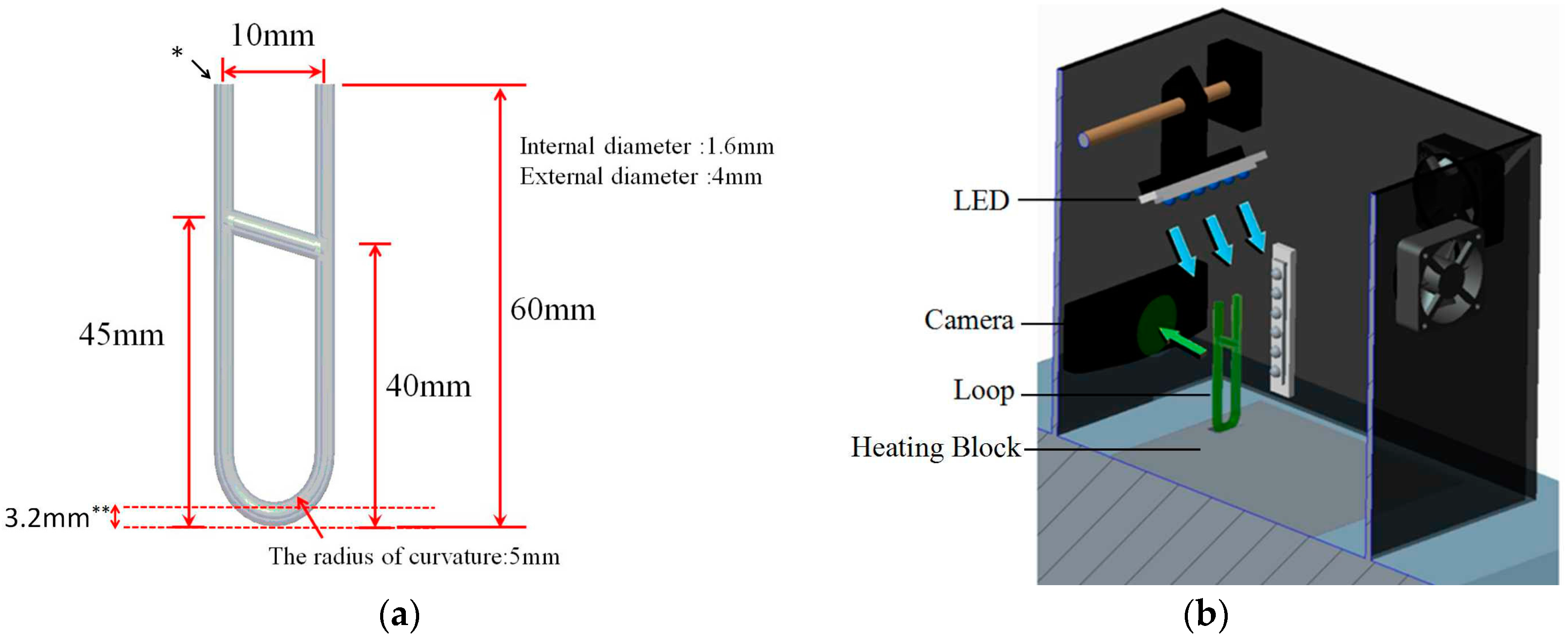
2.1. Experimental Equipment

2.1.1. U-Shaped Loop Capillaries

In this research, the U-shaped loop glass capillaries are used to test the PCR. The physical dimension of the reactor is shown in Figure 1a. The outside diameter, inner diameter, and radius of curvature of the U-shaped loop capillaries are 4, 1.6 and 5 mm, respectively. To produce a closed loop, two positions of the tube are burnt though and connected with a short capillary. As shown in Figure 1a, the height of the higher position of connection is 45 mm and the height of the lower position of connection is 40 mm. Such an inclination is designed for maintaining the one-directional flow in the U-shaped loop capillaries. The width and length of the U-shaped loop are 10 and 60 mm, respectively. The convective reactor is made with lampworking techniques and the volume of it is 240 μL. The properties of glass provide a smooth surface and hydrophilicity, greatly decreasing the formation of bubbles in loop capillaries. Furthermore, the high optical transparency of the glass is suitable for observing the flow field and fluorescent signals.

2.1.2. Heating System

A dry bath incubator with a temperature range setting of 0–180 °C is used as the single heater to maintain the required temperature gradient in the loop for clcPCR. An aluminum heating block with the top surface machined with a rectangular-shaped seat (15.4 mm × 6 mm × 3 mm) is placed in the dry bath incubator. In the PCR process, the bottom of the U-shaped loop capillary is heated with the aluminum heating block where the 70 μL mineral oil (M5904, Sigma-Aldrich, Saint Louis, MO, USA) is injected in order to obtain uniform and stable heating.

2.1.3. Fluorescence Detection

The optical detection system, as shown in Figure 1b, is constructed to observe the fluorescence generated by SYBR Green fluorescent dye in the U-shaped loop capillaries, and an acrylic cabinet is placed on top of the dry bath incubator to fulfill fluorescence detection and decrease interference of light from the environment. The loop capillaries with SYBR Green reagent are illuminated by the row of blue LEDs (5 mm diameter, 470 nm wavelength, 2130 mcd luminous intensity, 30 degree viewing angle, produced by Cree company, Durham, NC, USA), and the fluorescence image is recorded by a CCD device (C920, Logitech company, Lausanne, Switzerland). The position of CCD enables us to detect the front view of the U-shaped loop capillaries, and the blue LED is tilted down at an angle of approximately 30° away from the CCD detector to avoid direct exposure.

2.1.4. Reagent

Each PCR reaction contained 10 μL DNA template, 24 μL of LightCycler® FastStart DNA Master PLUS SYBR Green I Mixture (Taq DNA polymerase, PCR reaction buffer, 10 mM MgCl2, and dNTP mixture; Roche company, Penzberg, Upper Bavaria, Germany), 28.8 μL of 25 mM MgCl2, 4.8 μL of 10 mM each primer, and 167.6 mL of double-distilled water. The cycling conditions for the traditional thermal cycler (TGradient, Biometra, Göttingen, Germany) were: 95 °C for 10 min (one cycle); 95 °C for 15 s, 65 °C for 30 s, and 72 °C for 30 s (40 cycles). Two microliters of PCR or clcPCR products were run on a 2.5% agarose electrophoresis gel (MJ-105, Shorter Mini Gel System, Blossom Biotechnologies Inc., Taipei, Taiwan) containing 1 mg/mL ethidium bromide submerged in 1XTris-acetate-EDTA(Ethylenediaminetetraacetic acid) (TAE) buffer at 100 V for 40 min.

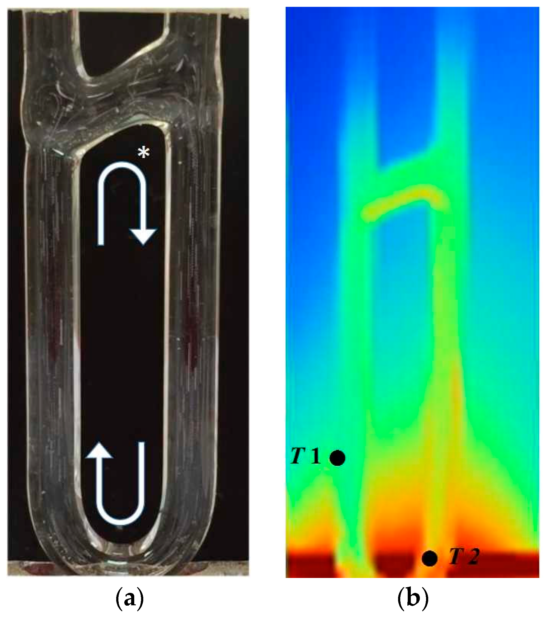
2.2. The Observation of Flow and Temperature Field

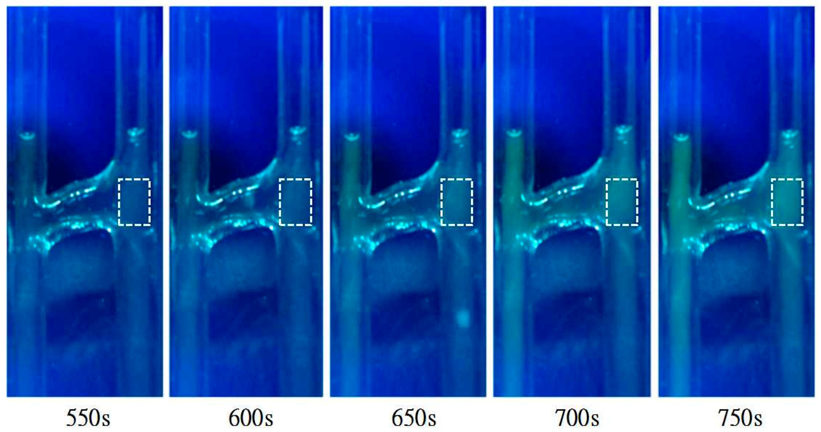
2.2.1. The Internal Flow Field

To obtain the verification of convective flow in the U-shaped loop, tracer particles are used to visualize the flow field. A camera is used to record the front view of the U-shaped loop, and then the video is analyzed with software (Photoimpact 10, Ulead System Inc., Taipei, Taiwan). The diameter of tracer particles (DANTEC, polyamide seeding particles) are 50 μm, and the density is 1.03 g/cm3 on average. It is similar to water so the path line of particles can imitate the flow field of reagents during the PCR process. The captured images from the recorded video, the visualization of the flow field, as shown in Figure 2a, is presented through accumulating 20 images of transient PSP particles (polyamid seeding particles, PSP-50, DANTEC company, Skovlunde, Denmark) in one second. Under the condition of setting the temperature to 135 °C for the dry bath incubator and ambient temperature of 25–26 °C, the image results clearly show the flow circulation in the U-shaped loop. From observation of the particles in the central f loop from the video, the tracer particles take around 11–12 s to circulate a run.

2.2.2. Temperature Measurement

The maximum and minimum temperatures are the key points of the experiments because they have an influence on denaturing and annealing during the PCR process. This means that the temperature distribution should be confirmed in the capillary tube before applying DNA amplification. With using an infrared sensor (EasIR-4, Wuhan Guide Infrared Co., Ltd., Wuhan, China), a record of the temperature distribution was made after heating the loop capillary tube for 10 min. Under the bottom temperature being 135 °C and the ambient temperature being 25–26 °C, the maximum temperature is located at the right side bottom and the minimum temperature is at the left side of the middle of the loop containers (Figure 2b). The t-type thermocouple measurement is also operated to ensure the temperature distribution is in the range of PCR reaction. The heating time is 1500 s and the stable range (500–1500 s) is recorded and analyzed. It shows that the average of high temperature is 94.6 °C, with a maximum value of 96.0 °C and a minimum value of 93.3 °C (standard deviation 0.57 °C). The average low temperature is 66.7 °C, with the maximum value being 68.7 °C, and the minimum value being 63.8 °C (standard deviation = 1.32 °C). According to this result of the thermal profile, the temperature distribution meets the requirements for the PCR process of denaturing (90–95 °C), annealing, and extension (55–70 °C).

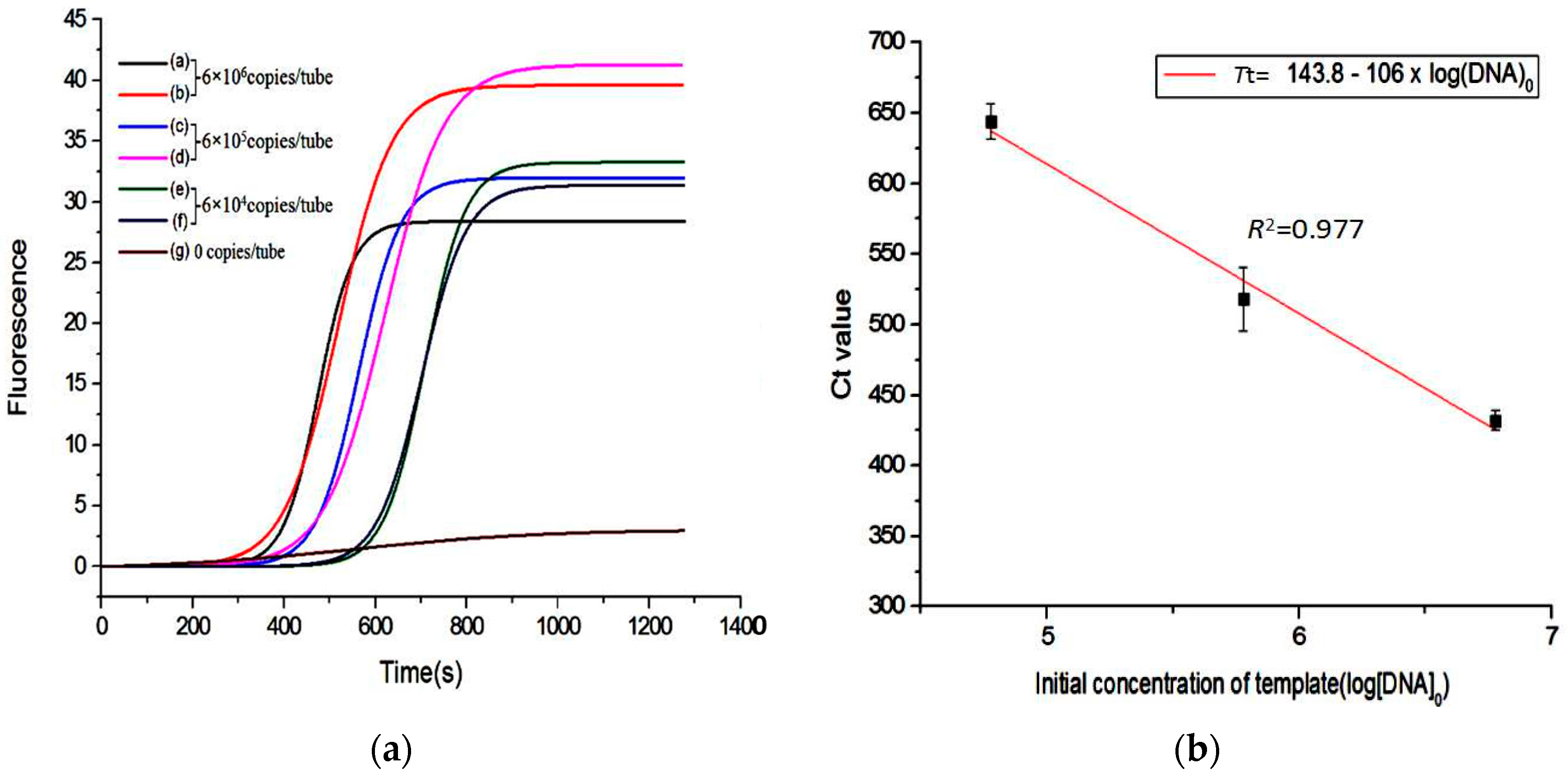
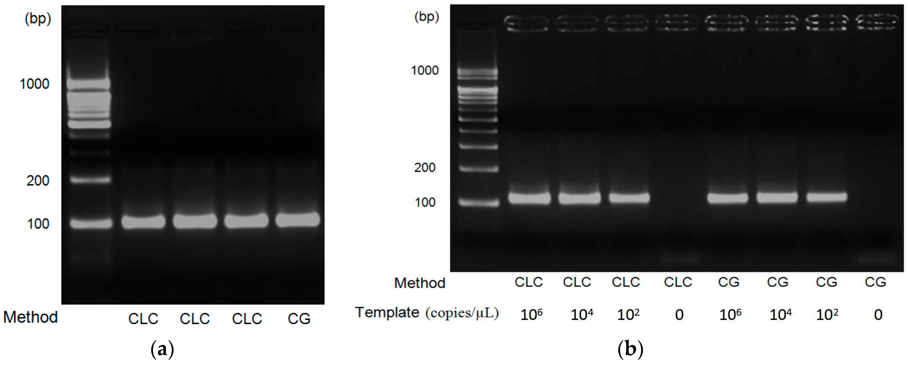
3. Results and Discussion

Firstly, the study conducts qualitative experiments to check the reproducibility and sensitivity of the amplification. Secondly, we work on the quantitative experiment to verify the fluorescent PCR signal and distinguish the different initial concentrations of DNA templates through clcPCR. Hepatitis B Virus (HBV) DNA is amplified by clcPCR and the size of the amplicon is 96 base pairs (bps). The primer sequence is shown in Table 1. Every test was triplicated to verify the stability of the clcPCR platform. The initial concentration of the template is 6 × 10⁶ copies/tube, and the traditional PCR machine was also implemented as the control group with the condition of 95 °C for 10 min and forty cycles repeated (one cycle with 95 °C denaturing for 5 s, 72 °C annealing for 10 s, and 72 °C extension for 10 s). The electrophoresis data shows clcPCR amplified the correct size of DNA fragment, showed in Figure 3a. To verify the linear range of detection, different initial concentrations of HBV DNA were tested with 6 × 10⁶ copies/tube, 6 × 10⁴ copies/tube, and 6 × 10² copies/tube. The same samples were also amplified by the traditional PCR machine as the control group. According to the amplification results of different initial concentrations (Figure 3b), clcPCR can amplify HBV DNA from high to low concentrations within 25 min.
Furthermore, the optical system was constructed for studying the real-time process of amplification and carried out the experiment with HBV DNA (6 × 10⁶ copies/tube) as the initial concentration of the template for recording the fluorescent variations during the amplification. In this experiment, we used HBV 96 bp primer pair, SYBR Green I as the fluorescent dye, and 1500 s reaction time. The CCD camera took pictures every 0.5 s. Pictures captured from 550 to 750 s were showed significant changes in the fluorescence intensity during this time period, demonstrating that the system can clearly distinguish the change of the fluorescence intensity during the amplification process (Figure 4). As a consequence, we could capture the trend of fluorescence changes and plot the fluorescence curves after digitizing the light intensity. In Figure 4b, there are three kinds of different initial template concentration DNA for quantitative testing in this study, and experimented with the same concentration of reagents to test the stability of clcPCR. The initial DNA copy number were 6 × 10⁶ copies/tube, 6 × 10⁵ copies/tube, and 6 × 10⁴ copies/tube, respectively. Additionally, a set of negative controls were also tested. The quantitated results of original curves fitted by a sigmoid model show the significant differences of the threshold time in the different initial DNA concentrations, and the higher the initial concentration of DNA, the sooner it will climb (Figure 5a). We defined a threshold time as the Tt value, the maximum value of the second derivative of the fluorescence curve, and it is used to compare the repeatability of the same initial DNA concentration amplification results. After collecting data, the Tt value versus the corresponding initial template concentration can be plotted as a standard curve, which represents the basis for quantitative analysis.
This plot shows data as follows: when the initial DNA concentration was 6 × 10⁶ copies/tube, the Tt values were 423 and 441 s, and the average of the two results was 432 s. When the initial DNA concentration was 6 × 10⁵ copies/tube, the Tt values were 502 and 534 s, and the average of the two results was 518 s. When the initial DNA concentration was 6 × 10⁴ copies/tube, the Tt values were 649 and 638 s, and the average of the two results was 644 s. Then, the average Tt value against the logarithm of the corresponding three different initial DNA concentrations is plotted (Figure 5b). The experimental results show that the Tt value is linear with the logarithm of the initial DNA templates. The correlation coefficient of the standard curve is 0.977, which means clcPCR can offer the ability of precise quantification.

4. Conclusions

This work presented a new PCR platform, capillary loop convective polymerase chain reaction (clcPCR), which can make convective PCR more stable. The results of clcPCR showed that the polymerase chain reaction could be completed in 25 min. In addition, the fluorescent intensity of clcPCR could be observed and the quantitative results were plotted. The plot of DNA copy numbers and the fluorescent threshold shows that the result of R2 = 0.977. The multiple sample testing and real-time quantification will be investigated in the future.

Acknowledgments

The work was supported by the Ministry of Science and Technology, Taiwan, Republic of China (R.O.C.) MOST 105-2218-E-002-019.

Author Contributions

Wen-Pin Chou and Ping-Hei Chen made substantial contributions to the conception, design, analysis, and interpretation of data, and wrote the paper; Chien Lee, Zong-Jyun Hsu, Mei-Hui Lai, and Long-Sheng Kuo performed the experiments, wrote the paper, discussed the results and implications, and commented on the manuscript at all stage.

Conflicts of Interest

The authors declare no conflict of interest.

References

  1. Espinosa de los Monteros, L.E. Polymerase chain reaction (PCR) in clinical diagnosis. Review of the use of the technique. Rev. Latinoam. Microbiol. 1993, 35, 225–230. [Google Scholar] [PubMed]
  2. Wittbrodt, J.; Erhardt, W. An inexpensive and versatile computer-controlled PCR machine using a peltier element as a thermoelectric heat pump. Trends Genet. 1989, 5, 202–203. [Google Scholar] [CrossRef]
  3. Fang, T.H.; Ramalingam, N.; Xian-Dui, D.; Ngin, T.S.; Xianting, Z.; Lai Kuan, A.T.; Peng Huat, E.Y.; Hai-Qing, G. Real-time PCR microfluidic devices with concurrent electrochemical detection. Biosens. Bioelectron. 2009, 24, 2131–2136. [Google Scholar] [CrossRef] [PubMed]
  4. Khandurina, J.; McKnight, T.E.; Jacobson, S.C.; Waters, L.C.; Foote, R.S.; Ramsey, J.M. Integrated system for rapid PCR-based DNA analysis in microfluidic devices. Anal. Chem. 2000, 72, 2995–3000. [Google Scholar] [CrossRef] [PubMed]
  5. Nakayama, T.; Kurosawa, Y.; Furui, S.; Kerman, K.; Kobayashi, M.; Rao, S.R.; Yonezawa, Y.; Nakano, K.; Hino, A.; Yamamura, S.; et al. Circumventing air bubbles in microfluidic systems and quantitative continuous-flow PCR applications. Anal. Bioanal. Chem. 2006, 386, 1327–1333. [Google Scholar] [CrossRef] [PubMed]
  6. Zhang, C.; Xu, J.; Ma, W.; Zheng, W. PCR microfluidic devices for DNA amplification. Biotechnol. Adv. 2006, 24, 243–284. [Google Scholar] [CrossRef] [PubMed]
  7. Ottesen, E.A.; Hong, J.W.; Quake, S.R.; Leadbetter, J.R. Microfluidic digital PCR enables multigene analysis of individual environmental bacteria. Science 2006, 314, 1464–1467. [Google Scholar] [CrossRef] [PubMed]
  8. Schell, W.A.; Benton, J.L.; Smith, P.B.; Poore, M.; Rouse, J.L.; Boles, D.J.; Johnson, M.D.; Alexander, B.D.; Pamula, V.K.; Eckhardt, A.E.; et al. Evaluation of a digital microfluidic real-time PCR platform to detect DNA of candida albicans in blood. Eur. J. Clin. Microbiol. Infect. Dis. 2012, 31, 2237–2245. [Google Scholar] [CrossRef] [PubMed]
  9. Krishnan, M.; Ugaz, V.M.; Burns, M.A. PCR in a rayleigh-benard convection cell. Science 2002, 298, 793. [Google Scholar] [CrossRef] [PubMed]
  10. Agrawal, N.; Hassan, Y.A.; Ugaz, V.M. A pocket-sized convective PCR thermocycler. Angew. Chem. Int. Ed. Engl. 2007, 46, 4316–4319. [Google Scholar] [CrossRef] [PubMed]
  11. Chou, W.P.; Chen, P.H.; Miao, M., Jr.; Kuo, L.S.; Yeh, S.H.; Chen, P.J. Rapid DNA amplification in a capillary tube by natural convection with a single isothermal heater. Biotechniques 2011, 50, 52–57. [Google Scholar] [CrossRef] [PubMed]
  12. Chen, Z.; Qian, S.; Abrams, W.R.; Malamud, D.; Bau, H.H. Thermosiphon-based PCR reactor: Experiment and modeling. Anal. Chem. 2004, 76, 3707–3715. [Google Scholar] [CrossRef] [PubMed]
  13. Wheeler, E.K.; Benett, W.; Stratton, P.; Richards, J.; Chen, A.; Christian, A.; Ness, K.D.; Ortega, J.; Li, L.G.; Weisgraber, T.H.; et al. Convectively driven polymerase chain reaction thermal cycler. Anal. Chem. 2004, 76, 4011–4016. [Google Scholar] [CrossRef] [PubMed]
  14. Chung, K.H.; Park, S.H.; Choi, Y.H. A palmtop PCR system with a disposable polymer chip operated by the thermosiphon effect. Lab Chip 2010, 10, 202–210. [Google Scholar] [CrossRef] [PubMed]
  15. Zhang, S.; Lin, Y.; Wang, J.; Wang, P.; Chen, J.; Xue, M.; He, S.; Zhou, W.; Xu, F.; Liu, P.; et al. A convenient nucleic acid test on the basis of the capillary convective PCR for the on-site detection of enterovirus 71. J. Mol. Diagn. 2014, 16, 452–458. [Google Scholar] [CrossRef] [PubMed]
Figure 1. (a) Physical dimensions of the glass capillary loop; (b) schematic diagram of the detection setup. * the place of reagent injection; ** the depth of heating groove. Blue arrow: excitation light; Green arrow: emission light.
Inventions 02 00003 g001
Figure 2. (a) Flow observation when heating the bottom of the glass capillary loop (overlaying 20 frames per second and exposing for one second). * the solution flows clockwise; (b) Temperature measurement with an IR thermometer (the T2 average temperature is 94.6 °C with a maximum value of 96.0 °C, and a minimum value 93.3 °C; the T1 average temperature is 66.7 °C with a maximum value of 68.7 °C, and a minimum value 63.8 °C).
Inventions 02 00003 g002
Figure 3. (a) Triplicate clcPCR tests at Lanes 2–4, one control test with commercial PCR machine (CG) at Lane 4, initial DNA concentrations: 106 copies/μL; (b) testing different DNA copies with clcPCR (Lanes 2–5) and a commercial PCR machine (CG, Lane 6–9).
Inventions 02 00003 g003
Figure 4. Fluorescence recording during the clcPCR reaction and only the fluorescent image from 550 to 750 s is shown (the initial DNA concentration: 6 × 10⁵ copies/tube). The dotted line frame is the area of fluorescent analysis.
Inventions 02 00003 g004
Figure 5. (a) The amplification plot with reaction time and fluorescence intensity; (b) the standard curve with the average of Tt values against the logarithm of the corresponding three different initial DNA concentrations.
Inventions 02 00003 g005
Table 1. Sequence of the Hepatitis B Virus (HBV) primer set for clcPCR.
Primer NameSequence
HBV 96bp-F5′-CCGGAAACTACTGTTGTTAGACGACGGGACCGAGGCAGG-3′
HBV 96bp-R5′-GCGACGCGGCGATTGAGATCTGCGTCTGCGAGG C-3′

