Review

Ergonomics of Prehensility in Pushing and Pulling Motions: An Anatomical and Biomechanical Overview

1
Department of Biomedical and Biotechnological Sciences, Human Anatomy and Histology Section, School of Medicine, University of Catania, Via S. Sofia 87, 95123 Catania, Italy
2
Department of Orthopedics, Amphia Hospital, 51436 Breda, The Netherlands
3
Laboratory of Olive Tree, Centre of Biotechnology of Borj Cedreya, University of Carthage, 2016 Carthage, Tunisia
4
Department G.F. Ingrassia, Anatomy, School of Medicine, University of Catania, 95123 Catania, Italy
*
Author to whom correspondence should be addressed.
J. Funct. Morphol. Kinesiol. 2017, 2(1), 6; https://doi.org/10.3390/jfmk2010006
Submission received: 3 October 2016 / Revised: 24 November 2016 / Accepted: 12 December 2016 / Published: 18 January 2017

Abstract

:
The hand represents one of the most remarkable expressions of humanization of the anterior limb. The anterior limb, at first ambulatory, underwent continuous evolution acquiring innumerable new functions. In the course of human evolution the hand has undergone continual structural and functional adaptations, characterized, among others, by enrichment of peripheral innervation and further development of the thumb. This development was accompanied by important changes in the brain and the relocation of the eyes, together allowing the muscle control and stereoscopic vision, necessary for a controlled grip. The anatomy of the hand is complex, intricate, and fascinating. Its integrity is absolutely essential for our everyday functional living. It is intimately correlated with the brain, both in the evolution of the species and in the development of the individual. Actually, we can state that we “think” and “feel” with our hands, hence, their contribution is essential to the mental processes of thought and feeling. The aim of this review is to evaluate the most typical hand quality, the prehensility and hence, the possibility of manoeuvring tools. Our attention is mainly focused on the hand anatomy and prehensility during pushing and pulling motions. In particular, our attention is directed toward the relationship existing between the hand prehensility and the volume of the object to be gripped. As an example, we use a grip of the paddle and, pushing and pulling motions during kayak paddling. Indeed, we are firmly convinced that the prehensility plays a crucial role not only in performing the stylistically correct paddling, but especially in realizing a more effective and powerful paddle stroke. This review highlights a great link existing between biomechanical and anatomical notions and sporting performance.

Graphical Abstract

1. Introduction

When looking at children playing in the equipped playground or at school, we can observe most of the movements that they make. Kids, for their nature, explore the environment and test themselves continuously. They slide enthusiastically on the soft sand, make themselves spin at high speeds, invent dangerous exercises, but for one reason or another they do not let themselves hang down from the cross bar or swing from one cross bar to another.
Let us try to understand why this happens. In most cases, the fear of the emptiness is to be excluded, both because the distance from the ground is minimal and because kids in other exercises would certainly let themselves fall from even greater heights. The fact that the cross bar is at a high distance from the ground can be excluded as well, since the kids would probably try to invent any possible way to reach it. The fact that the rejection is almost complete suggests a more general reason for this behaviour. When considering the reasons of this behaviour, one can notice that there is a huge discrepancy between the volume of the cross bar and the extreme limitations of the prehensility of the small child’s hand. The latter may not be able to implement full prehensility on a too voluminous cross bar; kids can only rely on their small contracted fingers hooked on the cross bar to be able to hang down from it. In this position a small hand would necessarily have to focus all its contractile properties in gripping the object with the fingers, on a relatively small set of muscles of the forearm (the superficial and deep flexor muscles of the fingers) to support the hanging body. Although these muscles are very powerful, they are disproportional to the weight to be supported. For this reason the child, after huge efforts, normally has to give up due to fatigue of the hand and forearm muscles. Furthermore, the properties of the object (size, weight, and shape) and the task objective usually determine how the object is held, the area of contact between the object and the hand, and the number of fingers involved in the grasp [1]. Moreover, the breakaway strength was shown to be significantly influenced by handhold size, gender, and hand dominance in children [2]. Generally, not a lot of attention is paid to teach a child, or any other newcomer, on how to “grip” an object properly and what relationship exists between hand prehensility and the volume of the object to be “gripped”.
Humans, normally, display a great variability in hand size. There is discussion whether a correlation can be observed between the individual body constitution and size of the hand. Often, tall and imposing people can show a small hand size (obstetrician’s or surgeon’s hands), and vice versa, short and slim people can have larger hand sizes when compared to their constitution. Moreover, hand size often shows a familiar pattern. A recent study from India, however, showed a correlation between human height and hand length in the Mumbai population. In general, human height was nine times hand length [3].
The hand phylogenetically represents one of the most remarkable expressions of the humanization of the anterior limb, which from “anterior” as in quadrupeds, has evolved into a “superior” limb in biped hominids. Consequently, this limb, at first deambulatory, underwent continuous evolution, acquiring innumerable new functions, of which the most characteristic one is represented by the prehensility and hence, possibility of manoeuvring tools. In the course of human evolution the hand has undergone continuous structural and functional adaptations, principally characterized by a consensual increase and enrichment of peripheral innervation. The hand of a nowadays harpsichordist is surely not the same hand of the hominids using stones as weapons and tools.
Such considerations occur as well in observing kayak training or performances by male and female youngsters (pre-cadets, cadets, and juniors). Normally, indiscriminate of gender, size, or age, they must grip any paddle size in this sport activity. In the majority of the cases the paddle has a circumferential diameter of at least 3 cm, a size commonly used by seniors (adults). Clearly there is a size difference between the hands of youngsters and adults, however, the shaft diameter is not adjusted for this difference in size.
Because the different grips affect the flexibility and power of certain movements, our study is focused on the hand grip during paddling with the firm conviction that it exerts a primary role not only in a stylistically correct paddling execution, but mainly in the realization of a more efficacious and powerful paddle stroke and prevention of injury, most commonly referred to as “oarsman’s wrist”, a tenosynovitis of the extensor muscles, occurring in up to 23% of paddling athletes [4,5,6]. Injury is often found in elite level athletes, since this is a risk factor for overuse injury like intersection syndrome and low back pain in rowers [7]. Another study found that the injury rate in rowing is higher in girls than boys [8]. Our hypothesis is that the hand grip during paddling plays an important role in a powerful paddle stroke and prevention of injury, mainly in young, high level athletes and girls. In the present review, we use the kayak paddling action and a paddle as an example, but the arguments discussed in this review refer certainly to every other similar pushing and pulling motions and prehensility of any kind of cylindrical object gripped perpendicularly to hand and forearm axis.

2. Anatomy of Prehensility

A study of the normal hand anatomy suggests that there are two distinct patterns of movement and that these, either separately or in combination, provide the anatomical basis for all prehensile activities. The fundamental requisite of prehension is that the object, whether it is fixed or freely movable, should be held securely. Stability is a pre-requisite for further activity and without it all refinements of hand function are of a little value. The ways to achieve stability in a normal hand are represented by different prehensile grips. John Napier, an orthopedic primatologist and paleoanthropologist, who gave an important contribution to the study of hand grips, proposed two primary prehensile grips [9]: (1) the power grip, achieved when the object is held in a clamp formed by the partially flexed fingers and the palm; in this grip there is a large area of contact between the grasped object and palmar surfaces of the fingers, hence the grasp is stable and very resistant [1]. Examples of the power grip are gripping a hammer, opening a jar using both your palm and fingers, and during pullups; (2) the precision grip, when the object is pinched between the flexor aspects of the fingers and the opposing thumb [9,10] in such a way that there is precise control of the grasping forces. Examples of a precision grip are writing with a pencil, opening a jar with the fingertips alone, and gripping a ball (only if the ball is not tight against the palm). Stability and grip are not only defined by the hand but also by the object to be gripped. The structure of the hand permits a proper form-function adaptability to the prehensility of numerous and diverse objects, whose volume and shape must be compatible with the size of the hand (Figure 1).
It means that the object can be properly “nestled” in the “containing capacity” of the hand. The prehensile adaptability of the hand [11,12] corresponds with the unlimited variability in size and shape of objects and instruments. It has been shown that increasing object width lowers the spatial accuracy demands on an object, permitting a faster movement to emerge. Smaller object width, on the contrary, gives rise to longer movement times through a lengthening of the deceleration phase of the movement [13]. Translating this to handling a paddle, an increased diameter of the shaft allows for faster movement. Furthermore, the mass of the object has been demonstrated to significantly influence prior-to-contact grasp kinematics. Heavier objects cause increased peak grasp aperture, a final finger and thumb placement on the object that more closely passes through the object centre of mass, increasing lift delay and reducing peak lift velocity compared to lighter objects [14]. The weight of the object is normally estimated based on available visual information and previous motor experiences. If the weight of the object to be grasped is unknown, the subject will use an inappropriate force, which usually is overestimated. During subsequent lifts of the same object, the optimal force required, will be used. This rapid adaptation of force generation indicates that there is a real-time motor updating of the information used to coordinate the grasp [15,16]. These findings suggest that size, mass, and the shape of the object to be grasped influence the prehensile movement kinematics. This observation should be taken into account while considering the improvement of the object prehensility in sports like kayak paddling.
From the anatomical point of view, the prehensility adaptability can be summarized in six fundamental anatomic aspects [17], four of which with the use of thumb.
(a)
Terminal opposition prehensility: It allows the development of micrometrically precise and fine prehensility movements. It is carried out by the opposition of the thumb against the index fingertip, as in the act of taking a fine needle or a match between two fingers. In this case the fingertips touch each other in correspondence of their nail margins.
(b)
Subterminal opposition prehensility: It is very similar to the previous one, the difference is that this action allows one to hold thicker objects, such as a glass slide, a piece of cardboard, or a pencil, between the index and the thumb, in opposition to each other as before. In this condition the fingertips touch each other in correspondence of their maximum phalangeal surface.
(c)
Subterminal-lateral opposition prehensility: The palmar surface of the thumb is brought against the radial side of the middle phalanx. This action allows one to hold small and flattened objects, but of a certain consistency, such as a coin or a piece of cardboard, between these two fingers.
(d)
Interdigital lateral-lateral prehensility: It is an ancillary mode of prehensility. It is carried out to hold a generally small and thin object between the lateral surfaces of two neighbouring fingers. Typical of this action is, for example, to hold a cigarette between the fingers. It is therefore a weak and inaccurate type of prehensility.
(e)
Digital-palmar opposition prehensility: In this case, the object, generally small, is taken between the palmar surface and flexed fingers. The thumb does not intervene and thus, the prehension results are unsafe: its axis is orthogonal to the axis of the hand and forearm. It is used quite often, generally when manoeuvring a cylindrical small object, such as a bead, nut or coin.
(f)
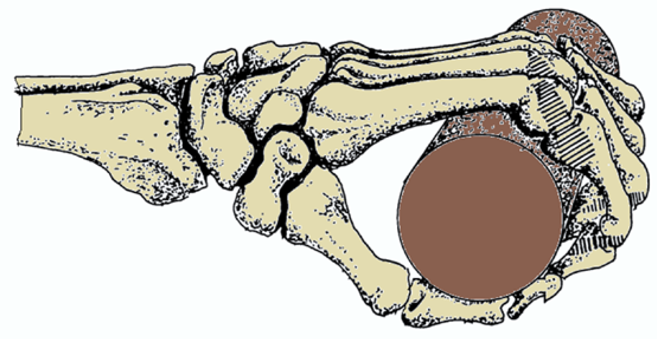
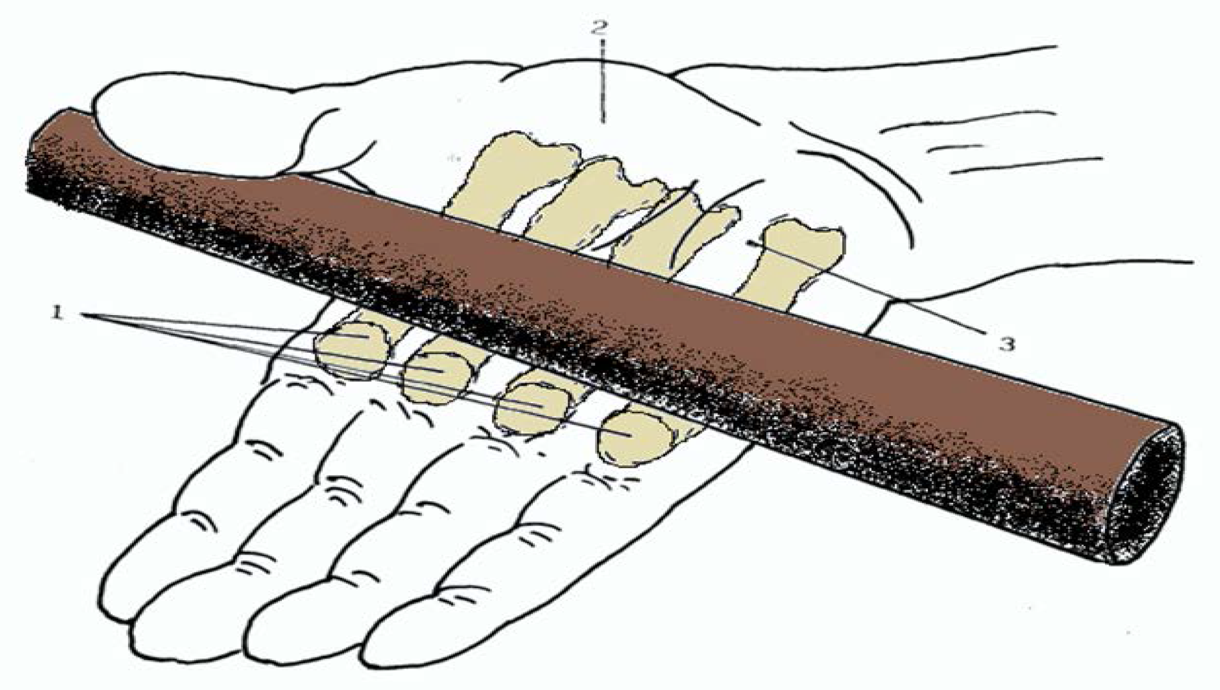
Palmar (or “full hand”) prehensility: It is a kind of prehensility that ensures the widest and most effective grip of the relatively heavy and voluminous objects. Also in this case their shape is usually cylindrical, spherical, or frustrum. In this action the hand is wrapped in flexion around the object and the axis of its palmar curvature corresponds to the axis of the cylindrical object that it holds. This kind of prehensility is normally used to hold the handle of working tools. In this grip the thumb plays a key role as it is wrapped around the object in opposition to the other fingers. The strength of the prehensility is strongly conditioned by the diameter of the held object: this one is optimal when the thumb touches the index.
Another kind of classification of motion by Long et al. [18] is “power grip”, which includes hook, squeeze, disc, and spherical grip; “precision grip”, which includes translation and rotation; and “pinch”. In all these motions both the extrinsic and the intrinsic muscles of the hand are involved. The kind of motion used in kayak paddling is the power grip. Long et al. demonstrated that the extrinsic muscles provide the major gripping force in the power grip and that they are all involved, while the major intrinsic muscles involved are the interossei and the metacarpophalangeal flexors.
The modality of prehensility involved in kayak paddling is the “palmar prehensility” (f). The latter is carried out also by the exogenous muscles in the hand [19,20]. The essential muscles include:
In the hand: The superficial and deep hand grip muscles such as; opponens digiti minimi, opponens pollicis, flexor digiti minimi brevis, flexor pollicis brevis, adductor pollicis, first dorsal interossei, and palmar interossei.
In the forearm: The superficial and deep flexor muscles (flexor pollicis longus, flexor digitorum superficialis, and flexor digitorum profundus) as well as extensor muscles (extensor digiti minimi, extensor digitorum, extensor indicis, extensor pollicis brevis, extensor pollicis longus) [21].
This type of prehensility results in maximum effectiveness, as the intrinsic contractility of the whole hand on the object to be taken is greatly enhanced by the flexor muscles of the forearm. This observation is taken into account during the design of the tool handles and it allows their proper construction.

3. The Prehensility of the Paddle

Palmar (or by “full hand”) prehensility is the one which realizes the only correct paddle grip able to ensure proper manoeuvrability during navigation [22,23]. Obviously, in the Olympic calm water paddling, the intensity of the impulsive force, and therefore the velocity of the kayak, are determined by and proportional to the volume of the water displaced with the paddle, among other factors including the weight of the kayak, the athlete, and environmental conditions, the latter increasing risk of injuries [5]. Another important factor is given by hand prehensility difficulties, which might be due to the complexity and anatomical variability of this unique organ. When a standard paddle is insufficiently gripped by an athlete, the strength and efficacy of the paddle grip can only be achieved by resorting to a more powerful and prolonged grasp, forcefully requiring an extremely elevated energy expense at the level of the flexor forearm muscles, resulting in prolonged isometric contraction of the superficialis and profundus flexor muscles of the fingers, with consequent increasing muscular hypoxia due to anaerobiosis. This inevitably provokes a painful contracture and hardening of these muscle masses and consequently a functional deficit of the hand grip [24,25]. Let us see how the prehensility of the paddle might be intended as correct and functionally sufficient [26,27,28,29,30]. Prehensility is an ability of modulated volumetric handgrip adaptation [31] to an object by the following essential articulations: radius-carpus, carpometacarpal, metacarpophalangeal, and interphalangeal. Of the above, especially the last two meet the specific conditions for holding an object [32,33].
A particular anatomical characteristic is evident at the observation of the articular heads of the joints: The distal epiphysis of each long bone of the hand has the semiovoid configuration, the base section of which is implanted on the metacarpal or phalangeal diaphysis.
Its sagittal section has, therefore, a semi-elliptical shape. This is common to all the metacarpal, metatarsal, and interphalangeal articular distal epiphysis, but it is a particularly important condition in the distal epiphysis of the metacarpal bones; this aspect is even more accentuated in the I° and II° metacarpal bone. Another relevant aspect is represented by the fact that the semiovoid joint presents a longitudinal axis that, in respect of that of the metacarpal diaphysis, appears slightly projected downwards: the two axes describe a very open angle, of about 160°–170°, with the hand in extension. This angle is open toward a palmar surface of the metacarpal bone. If the metacarpophalangeal joint is in extension, generally the longitudinal metacarpal axis (M) coincides with the longitudinal axis (A1) of the proximal phalanx (F1), although with a slight deviation at large obtuse angle open towards the palmar surface of the hand, due to the prevalence, under physiological resting conditions, of the flexor muscle tone of the fingers on the extensor one (Figure 2).
The semiovoid conformation of the metacarpal distal epiphysis head is, in our view, a very interesting observation. The significance of this unique anatomical structure is also emphasized by the insertions of the metacarpophalangeal and interphalangeal collateral ligaments (Figure 1). The latter are basically two: the collateral and the palmar one (Figure 3).
Of these two ligaments, the one which appears to be certainly the most interesting due to the particular dynamic metacarpal phalangeal joint (but also interphalangeal one), is undoubtedly the collateral ligament. As for the other important condyle diarthrosis (e.g., the knee), the metacarpophalangeal and interphalangeal collateral ligaments join the distal epiphysis of a bone segment with the proximal end of the next one. In these joints, the course of these ligaments, instead of being parallel to the major bone axis (the longitudinal one) relative to the two articular heads, presents an oblique course directed from proximal and dorsal travelling distally and ventral. These ligaments are inserted as follows: on the distal epiphysis of metacarpus they are inserted on the proximaldorsal border of the base of the articular capitellum, while on the proximal epiphysis of the following bone segment, they are inserted on the inferior-ventral border of the articular head. The course of these collateral ligaments is therefore dorsal-ventral. Because of these characteristics, and especially with regard to the metacarpophalangeal joint, the collateral ligaments in the hand are rather lax in extension, permitting sufficient lateral movements, whereas by flexing the joint—as happens in palmar prehension, these ligaments become taut and stabilize the joint. As already mentioned, this stabilisation is a result of the position and direction of the fibers of the collateral ligaments and of the semiovoidal conformation of the distal epiphysis head of the metacarpal bone. This anatomical condition of the joint allows a kind of click-like action and simultaneously permits a flexion, due to the fact that the semiovoidal conformation of the distal epyphisis heads of the metacarpal bones act as an eccentric. Of course, for actuating such a prehensility state, it is necessary that the longitudinal axis of the phalanx flexed on the gripped object (e.g., a paddle) is placed at right angle to the longitudinal axis of the bone with which it is articulated. If the angle between the longitudinal axes remains greater than 90°—whether due to the shortness or any other condition responsible of this discrepancy (joint contracture, osteoarthritisor arthritis, malunion, etc.) of the metacarpal bone and/or of the proximal phalanx, or to the largeness of the gripped object—the semiovoidal metacarpal distal epiphysis will be unable to fully function as an eccentric. In such state the correct flexion is not effectuated and consequently the collateral ligaments remain lax (Figure 4). This explains, therefore, why in case of anatomically articular insufficiency an athlete needs to compensate with the contraction supplied by the previously cited superficialis and profundus flexor muscles, giving rise so to the already specified negative implications. Thus, the metacarpal shafts should be of a proper length to ensure an adequately broad palmar surface of the hand, allowing both the right flexion angle between the phalanges and hence allowing the correct click-like flexion of the joint (Figure 5). This suggests also advantages for paddling, which are highlighted both in the traction phase in water and in the push phase.
(a)
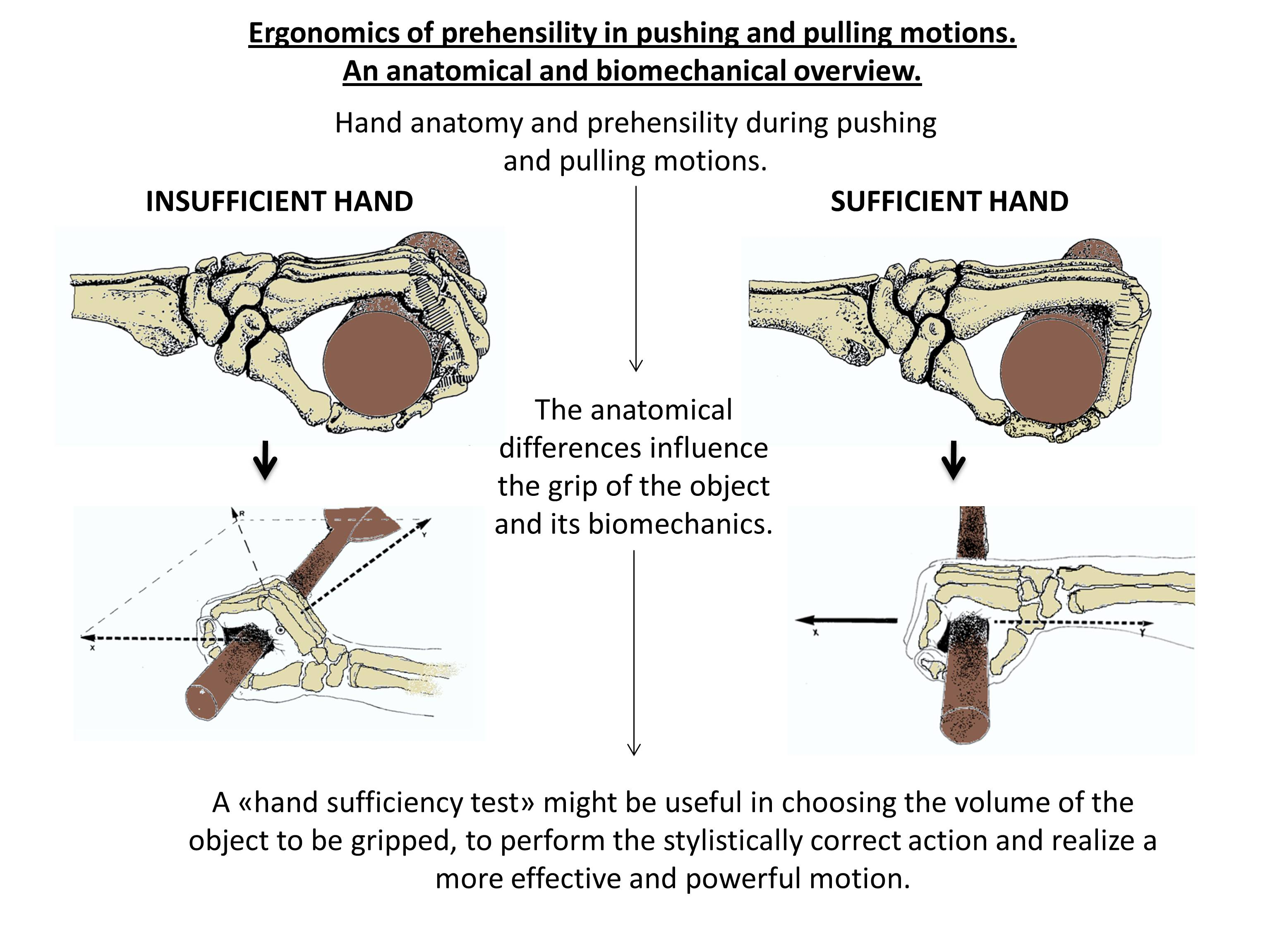
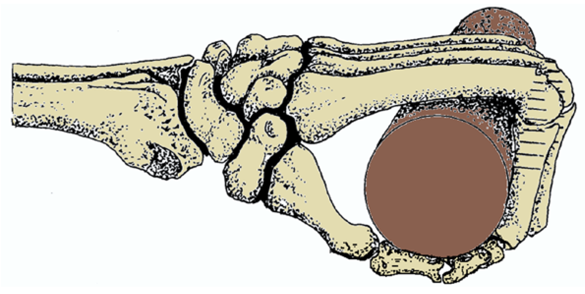
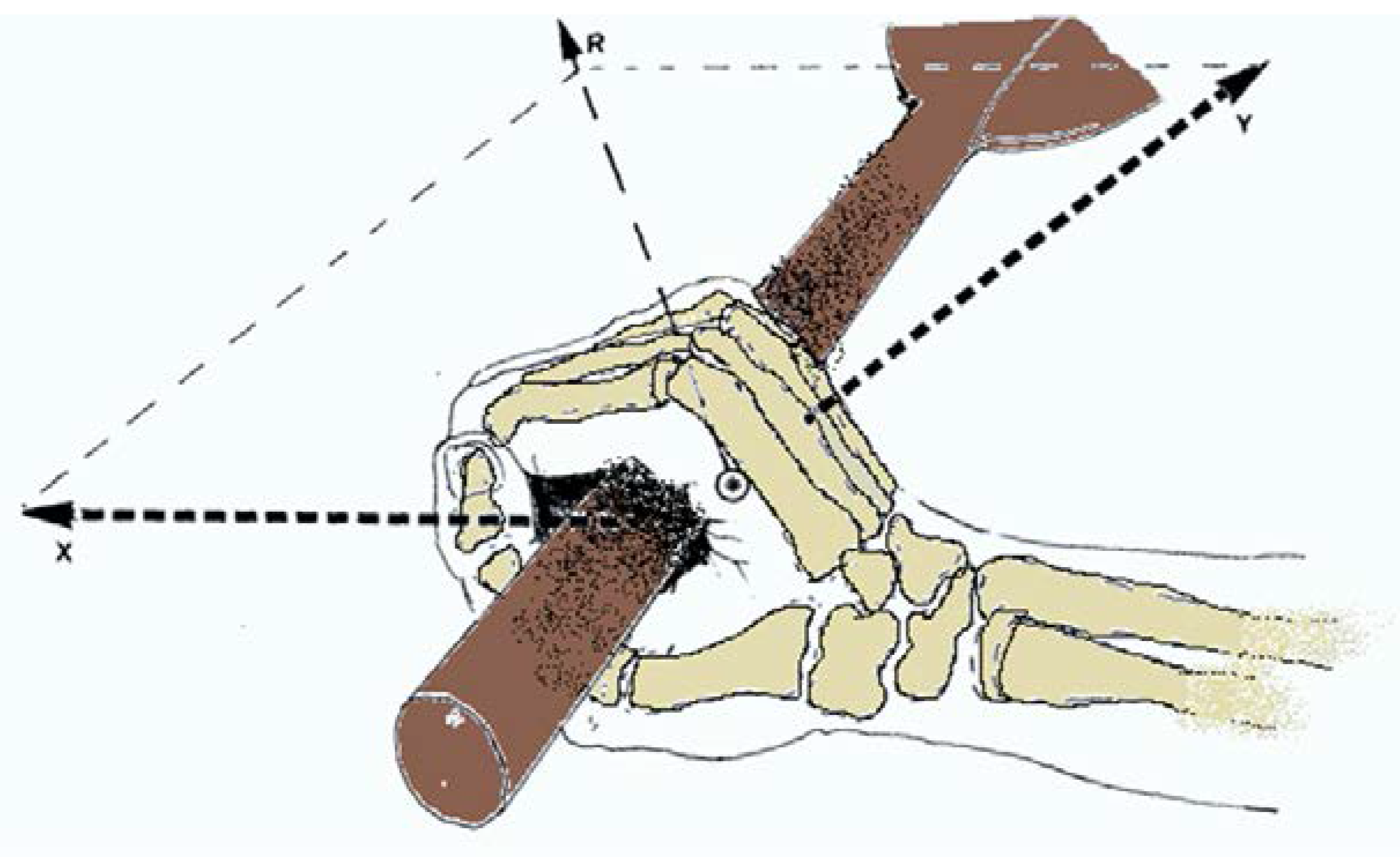
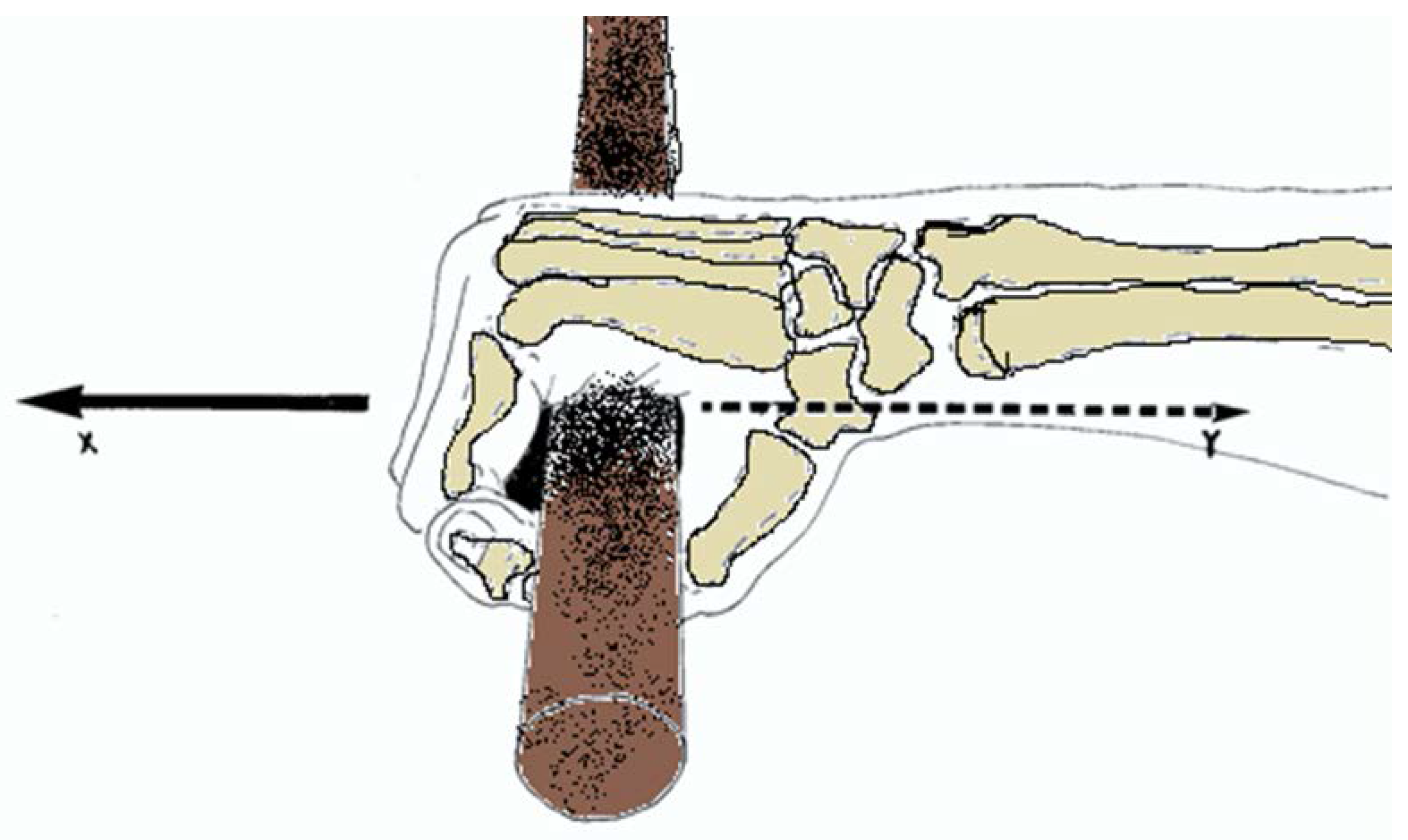
Traction phase: In the case of an insufficient grip (Figure 6), the resistance that the blade of the paddle meets along its course in the water moves on the phalanges that tighten the pole, which, not blocked in flexion, will tend to open up. The continuous use of the forearm flexor muscles allows one to maintain the flexion of the fingers on the pole, resulting in painful forearm hardening and progressive and inexorable loss of effectiveness of paddling. These negative effects will be unknown for the hand with long metacarpal shafts: this fact enables the “automatic” flexion of the metacarpophalangeal and interphalangeal joints on the pole of the paddle, with the benefit of detention of the forearm flexor muscles, which will thus be able to contribute to the upper limb function to realize the traction in the water. Moreover, another important factor for the traction phase might be given by the surface friction of the paddle, which seems to be very important for the breakaway strength of the hand grip [34].
(b)
Push phase: An inadequate length of the metacarpal shaft, leads to an aberrant prehensility pattern of the push phase of the paddling skill: the plane of the metacarpal portion of the hand will not be in register with the carpal and forearm axis (Figure 6). The paddle pole is then not recipient of the total push force dissipated by the resulting angulation developed by the deviation of the metacarpal-carpal vector forces. The consequence of this is that the push action will be produced almost exclusively by the metacarpal portion of the hand, with an obvious loss of this efficacy (Figure 6).
On the other hand, a sufficiently long metacarpal shaft (Figure 7), of which distal epiphysis (the “semiovoid” joint) overlaps the front of the pole of the paddle, by gripping it, may perform the flexion of the proximal phalanx, allowing in this way all the metacarpus to remain in axis with the forearm that pushes forward the paddle, and so fully using the pushing kinetic of the corresponding hemithorax and brachial triceps.

4. Conclusions

The presented topics at the beginning of this article, regarding the difficulties that kids face in the implementation of the grip on the cross bar, to let them hang down, or to rise above it, appear now evident. It would be enough if the cross bar was sufficiently thin to be appropriate to the gripping ability of the small hand of a child. This condition is not so far from the one in which a cadet or student paddler, or maybe a girl, encounters the volumetric grip of the paddle pole. A youngster is able to manage a standard kayak paddle, but probably, at an elevated cost in terms of exertion with a high risk of acquiring subsequently ineffective competition skills due to an early onset of fatigue, which might be difficult to eliminate later. Without mentioning the painful suffering of the excessively hardened forearm or risk of injury of overuse. For these reasons, we would propose a simple testing of the novice handgrip to verify its sufficiency (Figure 8).
The paddle is said to be successfully gripped if it is placed on the palmar surface of the extended hand and provided that its anterior margin is surpassed by the distal semiovoidal epiphyseal metacarpal head and its posterior margin is lying in front of the thenar and hypothenar eminences. The implementation of this simple test would most likely allow very considerable advantages of rowing not only to girls and boys, but certainly also to many high level athletes, juniors and seniors, whose somatic development, although already defined, remained in deficit with respect to the hand.
In conclusion, this review is an interesting way to unite science and, in particular, the notions of hand anatomy and biomechanics, with the world of sport, helping to increase the effectiveness of sport performance in kayak paddling.

Acknowledgments

This study was supported by a grant-in-aid from FIR 2016 (cod. 314509), University of Catania, Italy. The authors would like to thank Iain Halliday for commenting and making corrections to the paper. The decision to submit this paper for publication was not influenced by any of the funding bodies. Furthermore, the funders had no role in the design of the study, the collection and analysis of the data, the decision to publish, or the preparation of the manuscript.

Author Contributions

Each author wrote the highlight indicated, each author reviewed the proofs before publication.

Conflicts of Interest

The authors declare that there is no conflict of interest.

References

  1. Jones, L.A.; Lederman, S.J. Human Hand Function; Oxford University Press: New York, NY, USA, 2006; pp. 137–140. [Google Scholar]
  2. Ehrlich, P.F.; Young, J.G.; Ulin, S.; Woolley, C.; Armstrong, T.J.; Galecki, A.; Chen, S.; Ashton-Miller, J.A. Maximum hand-rung coupling forces in children: The effects of handhold diameter. Hum. Factors 2013, 55, 545–556. [Google Scholar] [CrossRef] [PubMed]
  3. Pawar, P.K.; Dadhich, A. Study of correlation between human height and hand length in residents of Mumbai. Int. J. Biol. Med. Res. 2012, 3, 2072–2075. [Google Scholar]
  4. Rumball, J.S.; Lebrun, C.M.; di Ciacca, S.R.; Orlando, K. Rowing injuries. Sports Med. 2005, 35, 537–555. [Google Scholar] [CrossRef] [PubMed]
  5. Newlands, C.; Reid, D.; Parmar, P. The prevalence, incidence and severity of low back pain among international-level rowers. Br. J. Sports Med. 2015, 49, 951–956. [Google Scholar] [CrossRef] [PubMed]
  6. Baugh, C.M.; Kerr, Z.Y. High school rowing injuries: National Athletic Treatment, Injury and Outcomes Network (NATION). J. Athl. Train. 2016, 51, 317–320. [Google Scholar] [CrossRef] [PubMed]
  7. Napier, J.R. The prehensile movements of the human hand. J. Bone Jt. Surg. Br. 1956, 38-B, 902–913. [Google Scholar]
  8. Landsmeer, J.M. Power grip and precision handling. Ann. Rheum. Dis. 1962, 21, 164–170. [Google Scholar] [CrossRef] [PubMed]
  9. Latash, M.L.; Zatsiorsky, V.M. Multi-finger prehension: Control of a redundant mechanical system. Adv. Exp. Med. Biol. 2009, 629, 597–618. [Google Scholar] [PubMed]
  10. Budgeon, M.K.; Latash, M.L.; Zatsiorsky, V.M. Digit force adjustments during finger addition/removal in multi-digit prehension. Exp. Brain. Res. 2008, 189, 345–359. [Google Scholar] [CrossRef] [PubMed]
  11. Bootsma, R.J.; Marteniuk, R.G.; MacKenzie, C.L.; Zaal, F.T. The speed-accuracy trade-off in manual prehension: Effects of movement amplitude, object size and object width on kinematic characteristics. Exp. Brain. Res. 1994, 98, 535–541. [Google Scholar] [CrossRef] [PubMed]
  12. Eastough, D.; Edwards, M.G. Movement kinematics in prehension are affected by grasping objects of different mass. Exp. Brain. Res. 2007, 176, 193–198. [Google Scholar] [CrossRef] [PubMed]
  13. Johansson, R.S.; Westling, G. Roles of glabrous skin receptors and sensorimotor memory in automatic control of precision grip when lifting rougher or more slippery objects. Exp. Brain. Res. 1984, 56, 550–564. [Google Scholar] [CrossRef] [PubMed]
  14. Loh, M.N.; Kirsch, L.; Rothwell, J.C.; Lemon, R.N.; Davare, M. Information about the weight of grasped objects from vision and internal models interacts within the primary motor cortex. J. Neurosci. 2010, 30, 6984–6990. [Google Scholar] [CrossRef] [PubMed]
  15. Qiu, D.; Fisher, H.C.; Kamper, D.G. Muscle activation patterns during force generation of the index finger. In Proceedings of the EMBC 2009 Annual International Conference of the IEEE, Minneapolis, MN, USA, 3–6 September 2009; pp. 3987–3990.
  16. Long, C.; Conrad, P.W.; Hall, E.A.; Furler, S.L. Intrinsic-extrinsic muscle control of the hand in power grip and precision handling. An electromyographic study. J. Bone Jt. Surg. Am. 1970, 52, 853–867. [Google Scholar] [CrossRef]
  17. Chong, H.I.; Kim, S.J.; Lee, E.K.; Yoo, G.E. Analysis of surface EMG activation in hand percussion playing depending on the grasping type and the tempo. J. Exerc. Rehabil. 2015, 11, 228–235. [Google Scholar] [CrossRef] [PubMed]
  18. Mogk, J.; Keir, P. The effects of posture on forearm muscle loading during gripping. Ergonomics 2003, 46, 956–975. [Google Scholar] [CrossRef] [PubMed]
  19. Hägg, G.M.; Milerad, E. Forearm extensor and flexor muscle exertion during simulated gripping work—An electromyographic study. Clin. Biomech. (Bristol, Avon) 1997, 12, 39–43. [Google Scholar] [CrossRef]
  20. Harih, G.; Dolsak, B. Comparison of subjective comfort ratings between anatomically shaped and cylindrical handles. Appl. Ergon. 2014, 45, 943–954. [Google Scholar] [CrossRef] [PubMed]
  21. Berguer, R.; Gerber, S.; Kilpatrick, G.; Remller, M.; BecLey, D. A comparison of forearm and thumb muscle electromyographic responses to the use of laparoscopic instruments with either a finger grasp or a palm grasp. Ergonomics 1999, 42, 1634–1645. [Google Scholar] [CrossRef] [PubMed]
  22. Abidin, M.R.; Thacker, J.G.; Becker, D.G.; Saunders, J.C.; Nayak, S.; Bacchetta, S.F.; Edlich, R.F. Hydrofitness devices for strengthening upper extremity muscles. J. Burn Care Rehabil. 1988, 9, 199–202. [Google Scholar] [PubMed]
  23. Duque, J.; Masset, D.; Malchaire, J. Evaluation of handgrip force from EMG measurements. J. Appl. Ergon. 1995, 26, 61–66. [Google Scholar] [CrossRef]
  24. Kong, Y.K.; Kim, D.M.; Lee, K.S.; Jung, M.C. Comparison of comfort, discomfort, and continuum ratings of force levels and hand regions during gripping exertions. Appl. Ergon. 2012, 43, 283–289. [Google Scholar] [CrossRef] [PubMed]
  25. Kong, Y.K.; Lowe, B. Optimal cylindrical handle diameter for grip force tasks. Int. J. Ind. Ergon. 2005, 35, 495–507. [Google Scholar] [CrossRef]
  26. Wu, J.Z.; Dong, R.G. Analysis of the contact interactions between fingertips and objects with different surface curvatures. Proc. Inst. Mech. Eng. H 2005, 219, 89–103. [Google Scholar] [CrossRef] [PubMed]
  27. Hoozemans, M.J.; van Dieën, J.H. Prediction of handgrip forces using surface EMG of forearm muscles. J. Electromyogr. Kinesiol. 2005, 15, 358–366. [Google Scholar] [CrossRef] [PubMed]
  28. Ong, K.B.; Ackand, T.R.; Hume, P.A.; Ridge, B.; Broad, E.; Kerr, D.A. Equipment set-up among Olympic sprint and slalom kayak paddlers. Sports Biomech. 2005, 4, 47–58. [Google Scholar] [CrossRef] [PubMed]
  29. Burstedt, M.K.; Flanagan, J.R.; Johansson, R.S. Control of grasp stability in humans under different frictional conditions during multidigit manipulation. J. Neurophysiol. 1999, 82, 2393–2405. [Google Scholar] [PubMed]
  30. Ansuini, C.; Giosa, L.; Turella, L.; Altoè, G.; Castiello, U. An object for an action, the same object for other actions: Effects on hand shaping. Exp. Brain Res. 2008, 185, 111–119. [Google Scholar] [CrossRef] [PubMed]
  31. Ansuini, C.; Santello, M.; Tubaldi, F.; Massaccesi, S.; Castiello, U. Control of handhaping in response to object shape perturbation. Exp. Brain Res. 2007, 180, 85–96. [Google Scholar] [CrossRef] [PubMed]
  32. Quéinnec, Y.; Daniellou, F. Designing for everyone. In Proceedings of the IIth Congress of the International Ergonomics Association, Paris-La Villet, France, 15–20 July 1991; Taylor & Francis: Paris, France, 1991; Volume 1, pp. 311–313. [Google Scholar]
  33. Young, J.G.; Woolley, C.; Armstrong, T.J.; Ashton-Miller, J.A. Hand-handhold coupling: Effect of handle shape, orientation, and friction on breakaway strength. Hum. Factors 2009, 51, 705–717. [Google Scholar] [CrossRef] [PubMed]
  34. Young, J.G.; Woolley, C.B.; Ashton-Miller, J.A.; Armstrong, T.J. The effect of handhold orientation, size, and wearing gloves on hand-handhold breakaway strength. Hum. Factors 2012, 54, 316–333. [Google Scholar] [CrossRef] [PubMed]
Figure 1. Schematic drawing showing the skeleton of a hand in extension. The collateral metacarpal (1) and interphalangeal (2) ligaments are shown (black lines). Their course is in the dorso-ventral direction, as to their attachments, the proximal ones are on the upper-dorsal epiphysis edge, while the distal ones end on the infero-ventral edge of the next epiphysis.
Jfmk 02 00006 g001
Figure 2. Graph of metacarpal phalangeal joint in extension (I) and in flexion (II). The articular distal head of metacarpal bone (M) has a semiovoide configuration: the radius 0–X, is clearly longer, in fact, than radius 0–X2. The result is a hemispherical area in excess (dotted area), that makes the metacarpal epiphysis assume the shape and the functions of an eccentric, which may become responsible for the flexion of the proximal phalanx F1 (II). 1. Joint cavity; 2. Joint capsule; 3. Articular cartilage. Longitudinal axis of the intermediate phalanx (A); Longitudinal axis of the proximal phalanx (A1); Proximal phalanx (F1).
Jfmk 02 00006 g002
Figure 3. Collateral and dorsal ligaments. 1, Flexor digitorum profundus and flexor digitorum superficialis; 2, Collateral ligament; 3, Extensor digitorum communis; 4, Palmar ligament or volar plate.
Jfmk 02 00006 g003
Figure 4. Drawing of the skeleton of a small hand, characterized by the short metacarpals, flexed into the grip of a paddle rod section of approximately 30 mm diameter (conventional paddle rod). The metacarpophalangeal and interphalangeal flexions are clearly incomplete. For this reason, the collateral ligaments remain lax and do not permit any lock in flexion: the grip of the paddle under such conditions will remain only by involving the flexor muscles of the superficial and deep fingers of the forearm.
Jfmk 02 00006 g004
Figure 5. Drawing of the skeleton of a “sufficient” hand, characterized by sufficiently long metacarpals, because their distal epiphysis surpasses the anterior edge of the paddle rod. The collateral ligaments in that case permit the lock of the metacarpophalangeal and interphalangeal joints, allowing the detension of the flexor muscles of the forearm fingers.
Jfmk 02 00006 g005
Figure 6. The grip of the paddle by an “insufficient” hand: Inevitably the push will be made by the metacarpus, which together with the carpus and with the longitudinal axis of the forearm will describe an obtuse angle. In such conditions, in respect of vector X corresponding to the push direction, an anti-vector Y will originate, and it will be deviant with respect to X. The resulting R will give a push, which will be directed upward, rather than forward; unless you operate a correction with the flexor carpi muscle in the forearm.
Jfmk 02 00006 g006
Figure 7. The grip of the paddle by a “sufficient” hand. The metacarpal bone overtakes the paddle. The lock in flexion is possible and the metacarpus remains in axis with the carpus and with the forearm longitudinal axis. Being anti-vector Y perfectly contrary to vector X, the push can be fully exploited in favour of kinetic progress.
Jfmk 02 00006 g007
Figure 8. “Test of sufficiency” of the hand against the rod of the paddle. 1, Articular distal heads of metacarpal bones; 2, Thenar eminence; 3, Hypothenar eminence.
Jfmk 02 00006 g008

