Article

Hyperglycemia and Hyperinsulinemia-Like Conditions Independently Induce Inflammatory Responses in Human Chondrocytes

by
Ana Teresa Rufino
1,2,
Madalena Ribeiro
1,2,
João Pinto Ferreira
1,
Fernando Judas
3,4 and
Alexandrina Ferreira Mendes
1,2,*
1
Centre for Neuroscience and Cell Biology, University of Coimbra, 3004-504 Coimbra, Portugal
2
Faculty of Pharmacy, University of Coimbra, 3000-548 Coimbra, Portugal
3
Orthopedics Department and Bone Bank, Hospital and University Centre of Coimbra, 3000-075 Coimbra, Portugal
4
Faculty of Medicine, University of Coimbra, 3000-548 Coimbra, Portugal
*
Author to whom correspondence should be addressed.
J. Funct. Morphol. Kinesiol. 2017, 2(2), 15; https://doi.org/10.3390/jfmk2020015
Submission received: 13 April 2017 / Revised: 8 May 2017 / Accepted: 11 May 2017 / Published: 16 May 2017
(This article belongs to the Special Issue Articular Cells and Tissues in Health and Osteoarthritis)

Abstract

:
To elucidate the mechanisms by which type 2 Diabetes Mellitus (DM2) constitutes a risk factor for the development and progression of osteoarthritis (OA), this work determined whether high glucose and/or high insulin, the hallmarks of DM2, are capable of activating the transcription factor, Nuclear Factor-κB (NF-κB), which plays a critical role in OA by inducing the expression of pro-inflammatory and catabolic genes. For this, we analyzed NF-κB activation by measuring the nuclear levels of p65 by western blot. As readouts of NF-κB activity, Interleukin-1β, Tumor Necrosis Factor-α, and inducible nitric oxide synthase (iNOS) expression were analyzed by real time RT-PCR and western blot. Culture of the human chondrocytic cell line, C28-I2, in high glucose (30 mM) increased nuclear NF-κB p65 levels in a time-dependent manner, relative to cells cultured in medium containing 10 mM glucose (regular culture medium). High glucose-induced NF-κB activation was inhibited by co-treatment with its specific inhibitor, Bay 11-7082, 5 µM. Culture of primary human chondrocytes under high glucose for 24 h increased IL-1β and TNF-α mRNA levels by 97% (p = 0.0066) and 85% (p = 0.0045), respectively, while iNOS mRNA and protein levels and NO production increased by 61% (p = 0.0017), 148% (p = 0.0089), and 70% (p = 0.049), respectively, relative to chondrocytes maintained in 10 mM glucose. Treatment of chondrocytic cells with 100 nM insulin was also sufficient to increase nuclear NF-κB p65 levels, independently of the glucose concentration in the culture medium. This study shows that hyperglycemia and hyperinsulinemia are independently sufficient to induce inflammatory responses in human chondrocytes, namely by activating NF-κB. This can be a relevant mechanism by which DM type 2 and other conditions associated with impaired glucose and insulin homeostasis, like obesity and the metabolic syndrome, contribute to the development and progression of OA.

Graphical Abstract

1. Introduction

Aging is the major risk factor for osteoarthritis (OA), but other factors have been recognized [1,2], including diabetes mellitus (DM), especially type 2 (DM2), and impaired glucose homeostasis. Recent epidemiologic studies demonstrated that DM is an independent risk factor for OA development and progression [3,4], supporting the notion of a specific diabetes-induced OA phenotype [1,2]. How DM and impaired glucose homeostasis contribute to cartilage and joint destruction is largely unknown.
Chronic low grade inflammation is a major OA driver, being associated with other mechanisms involved in OA pathogenesis, including cell senescence, oxidative stress, mitochondrial dysfunction, and impaired autophagy [5,6], which together contribute to cell depletion and imbalanced anabolic and catabolic responses, especially in chondrocytes [7].
All the above mechanisms have been associated with the effects of hyperglycemia and have been implicated in the pathogenesis of DM and its complications. Moreover, all these mechanisms have also been related to inflammation, which is currently accepted as an important effector mechanism by which DM2 causes cell and tissue damage [8].
Only a few studies have been conducted so far to identify direct effects of high glucose in articular chondrocytes, collectively indicating that hyperglycemia-like glucose concentrations favor catabolic processes and impair anabolic responses [9,10,11,12,13], especially in chondrocytes from OA/aged articular cartilage [11,12]. Moreover, high glucose has also been shown to directly induce the expression of interleukin (IL)-6 and cyclooxygenase 2 [13], two important inflammatory mediators, as well as to potentiate the same inflammatory responses induced by interleukin-1β (IL-1β) [14].
Given the central role of the transcription factor, Nuclear factor-κB (NF-κB), in inflammation and in OA [15], this study aimed at determining whether high, hyperglycemia-like, glucose concentrations are sufficient to activate this transcription factor and the expression of its pro-inflammatory target genes. Among these, IL-1β, Tumor Necrosis Factor (TNF)-α, and the inducible isoform of the Nitric Oxide Synthase (iNOS), the enzyme responsible for the production of large amounts of NO [16], play critical roles in OA pathophysiology [17]. For that purpose, we compared NF-κB activation in human chondrocytes cultured in regular (10 mM glucose) or high glucose media (30 mM) by measuring its nuclear accumulation. Since NF-κB activation is required for the expression of pro-inflammatory and catabolic genes in human chondrocytes, we also evaluated the expression of IL-1β, TNF-α, and iNOS in chondrocytes cultured in regular (10 mM) and high (30 mM) glucose containing media as readouts of NF-κB activity. As hyperinsulinemia often accompanies DM2 and it has been reported to have both protective and pathological roles in different cells, including inhibition of autophagy in OA models [5,6], we also evaluated its ability to modulate inflammatory responses in chondrocytes cultured in regular or in high glucose media.

2. Materials and Methods

2.1. Chondrocyte Isolation and Treatments

Human articular cartilage samples were collected and processed for cell isolation and culture within 24 h post-mortem from the distal femoral condyles of multi-organ donors (44–73 years old, mean = 59.4, n = 11) at the Orthopedic Department and Bone Bank of the Hospital and University Centre of Coimbra (CHUC). The cartilage samples were variably degraded, ranging from mild to severely damaged. Therefore, each sample was graded 0–4 according to the degree of macroscopic damage using the Outerbridge classification in which 0 corresponds to normal undamaged cartilage and 4 to severely damaged cartilage with full depth erosion. In this study, cartilage samples of grades 2 to 4 were used. All procedures were approved by the Health Ethics Committee of CHUC (authorization number: 0204/8654/DC, date: 12/16/2013).
Upon enzymatic isolation and to avoid chondrocyte dedifferentiation, confluent non-proliferating monolayer cultures were setup by plating 1 × 10⁶ chondrocytes/mL in the appropriate conditions as previously described [11]. The human chondrocytic cell line, C28/I2, was kindly provided by Prof. Mary Goldring (currently at the Hospital for Special Surgery, New York, NY, USA) and Harvard University. For each experiment, 0.5 × 10⁶ chondrocytes/mL were plated and allowed to recover for 24 h. Prior to any treatment, the cells were serum-starved for at least 8 h and then, cultured in serum-free conditions in regular Ham F-12 (Sigma-Aldrich, St. Louis, MO, USA), which contains 10 mM glucose, or in the same medium supplemented with glucose to a final concentration of 30 mM (high glucose) for the periods indicated in figure legends. Osmotic effects of high glucose were ruled out by adding 20 mM mannitol, a non-absorbable polysaccharide, to the regular culture medium. To confirm the mechanism involved in NF-κB activation, chondrocyte cultures in high glucose were simultaneously treated with 5 µM of the specific inhibitor, Bay-11-7082 (Sigma-Aldrich, St. Louis, MO, USA) [18]. To evaluate the effects of insulin, serum-deprived chondrocyte cultures in regular or high glucose medium were supplemented with insulin to achieve physiologic (10 nM) or supraphysiologic (100 nM) concentrations.

2.2. Nitric Oxide Production

NO production was evaluated as the concentration of nitrite accumulated in the supernatants of primary chondrocyte cultures maintained in 10 or 30 mM glucose for 72 h. Nitrite concentration was measured using the spectrophotometric method based on the Griess reaction [19].

2.3. Western Blot Analysis

Nuclear and total cell extracts were prepared and subjected to western blot as described previously [11]. The membranes were probed with monoclonal antibodies against human iNOS (R&D systems, Minneapolis, MN, USA) and NF-κB p65 (Cell Signalling, Boston, MA, USA) O/N at 4 °C and after extensive washings, with alkaline phosphatase-conjugated secondary antibodies (GE Healthcare, Buckinghamshire, UK) for 1 h at room temperature. Anti-human β-tubulin (Sigma-Aldrich) and anti-human Lamin B1 (Abcam, Cambridge, UK) were used to detect loading controls for total cell and nuclear extracts, respectively. Immune complexes were detected with the Enhanced ChemiFluorescence reagent (GE Healthcare) and the bands were analyzed using ImageQuant™ TL (version 7.0, GE Healthcare). The results were normalized by calculating the ratio between the intensities of the bands corresponding to the protein of interest and to the loading control.

2.4. Total RNA Extraction and Quantitative Real-Time RT-PCR (qRT-PCR)

Total RNA extraction, reverse transcription, and quantitative real-time PCR were performed as described previously [11]. Specific sets of primers for iNOS, IL-1β, TNF-α, and HPRT-1 (Gen Accession Numbers NM_000625.4, NM_000576.2, NM_000594.3, and NM_000194.2, respectively) were designed using Beacon Designer software (PREMIER Biosoft International, Palo Alto, CA, USA).
Gene expression changes were analyzed using the built-in iQ5 Optical system software v2 by the Pfaffl method, a variation of the ΔΔCT method corrected for gene-specific efficiencies [20].
The results for each gene of interest were normalized against HPRT-1, the housekeeping gene found to be the most stable under the experimental conditions used (data not shown).

2.5. Statistical Analysis

Results are presented as mean ± SEM representing the results of, at least, three independent experiments in which chondrocytes were isolated from, at least, three distinct donors. Cartilage from distinct donors was not pooled and each donor contributed only once for each experiment. Statistical analysis was performed using GraphPad Prism (version 5.00). The Kolmogorov-Smirnov test was used to assess the normality and homogeneity of variances to determine whether parametric tests could be used. As this test showed that the results were normally distributed in all experiments, the statistical analysis was performed using the two-tailed paired Student t-test between control and high glucose or high insulin conditions. Results were considered significant at p < 0.05.

3. Results

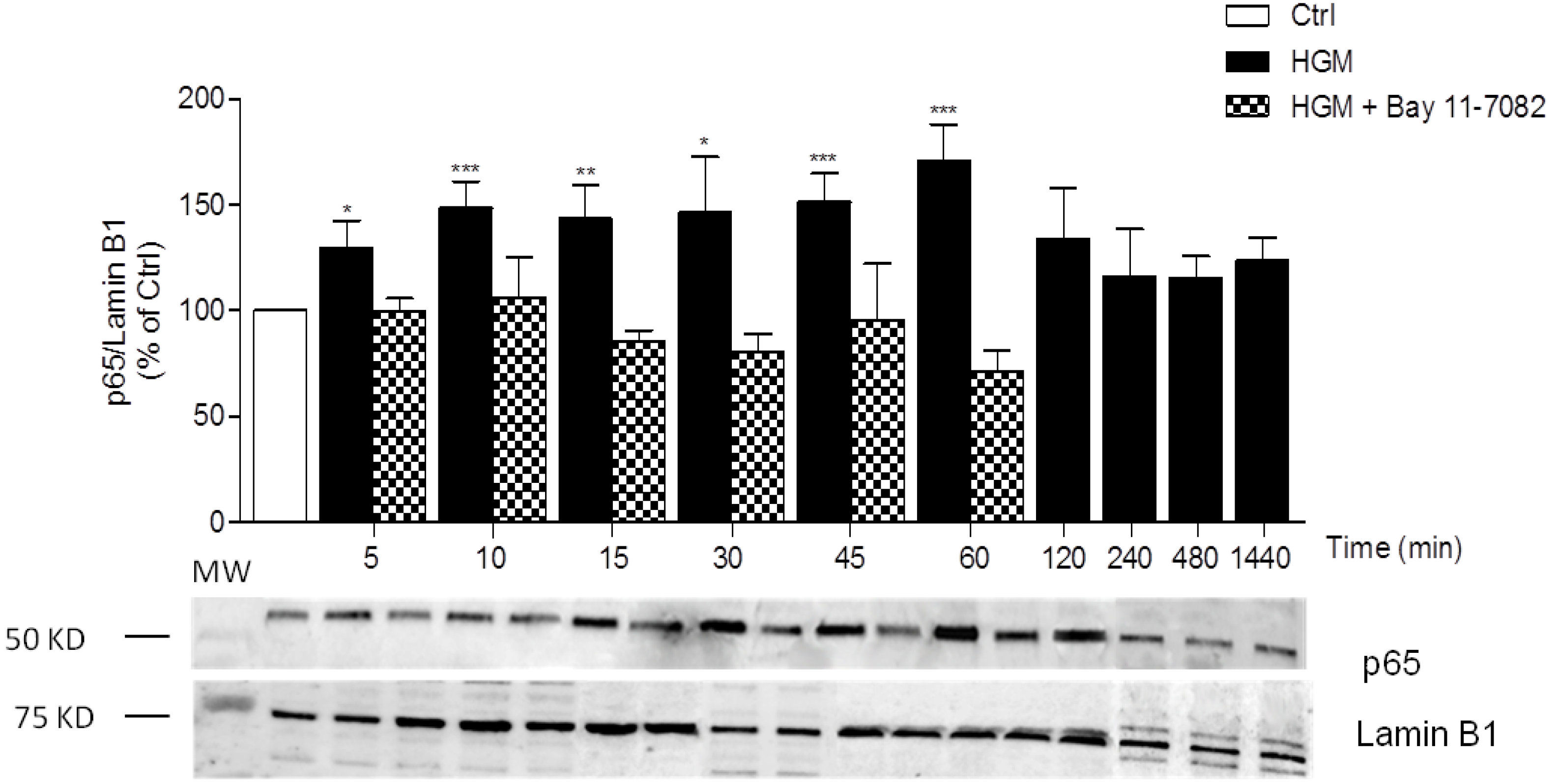
3.1. High-Glucose Induces NF-κB p65 Translocation to the Nucleus

As human cartilage samples are limited and a large number of cells was required, the human chondrocytic cell line, C-28/I2, was used to evaluate the ability of high glucose to activate the transcription factor, NF-κB. Activation of this transcription factor can occur through two distinct pathways of which the canonical pathway occurs in most cell types and requires the phosphorylation and subsequent ubiquitination and proteasomal degradation of inhibitory proteins, termed Inhibitor of NF-κB (IκB), which, in basal conditions, retain NF-κB dimers in the cytoplasm [18,21]. Once IκBs, in particular IκB-α, are degraded, NF-κB translocates to the nucleus promoting the transcription of its target genes. Therefore, activation of this transcription factor leads to its nuclear translocation that can be detected as the nuclear accumulation of p65 (Rel A), a major component of NF-κB dimers found in chondrocytes [22].
The results obtained show that nuclear p65 levels gradually increased during culture in high glucose, peaking at 1 h (Figure 1). After 2 h, nuclear levels of p65 were identical to those found in control conditions (10 mM glucose), and remained as such until up to, at least, 24 h, the longest period tested. No changes in nuclear p65 levels were observed in cells maintained in control conditions (10 mM glucose) for up to 1440 min (data not shown). The control in Figure 1 corresponds to cells maintained in these conditions for 120 min.
To further confirm the mechanism involved in high glucose-induced NF-κB activation, the cells were co-treated with the specific NF-κB inhibitor, Bay-11-7082. The results in Figure 1 show that, at the concentration of 5 µM which was shown to be sufficient to inhibit NF-κB activation in chondrocytes and other joint tissue cells [23,24], Bay 11-7082 completely reversed the effect induced by high glucose. This indicates that high glucose induces NF-κB activation through the canonic pathway, that is, phosphorylation, ubiquitination, and degradation of IκBs and consequent release and nuclear translocation of NF-κB dimers.
To rule out osmotic effects, the culture medium containing 10 mM glucose was supplemented with 20 mM mannitol, a non-absorbable polysaccharide, thus corresponding in terms of osmolarity, to the medium containing 30 mM glucose. No significant changes in the nuclear levels of p65 were observed in cells cultured in this medium for 1 h (mean =91.4 ± 11.9, p = 0.2574) relative to the control (Ctrl, 10 mM glucose), indicating that the effects detected with high glucose (HGM, 30 mM glucose) are not due to osmotic stimulation (Figure 2).

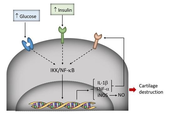
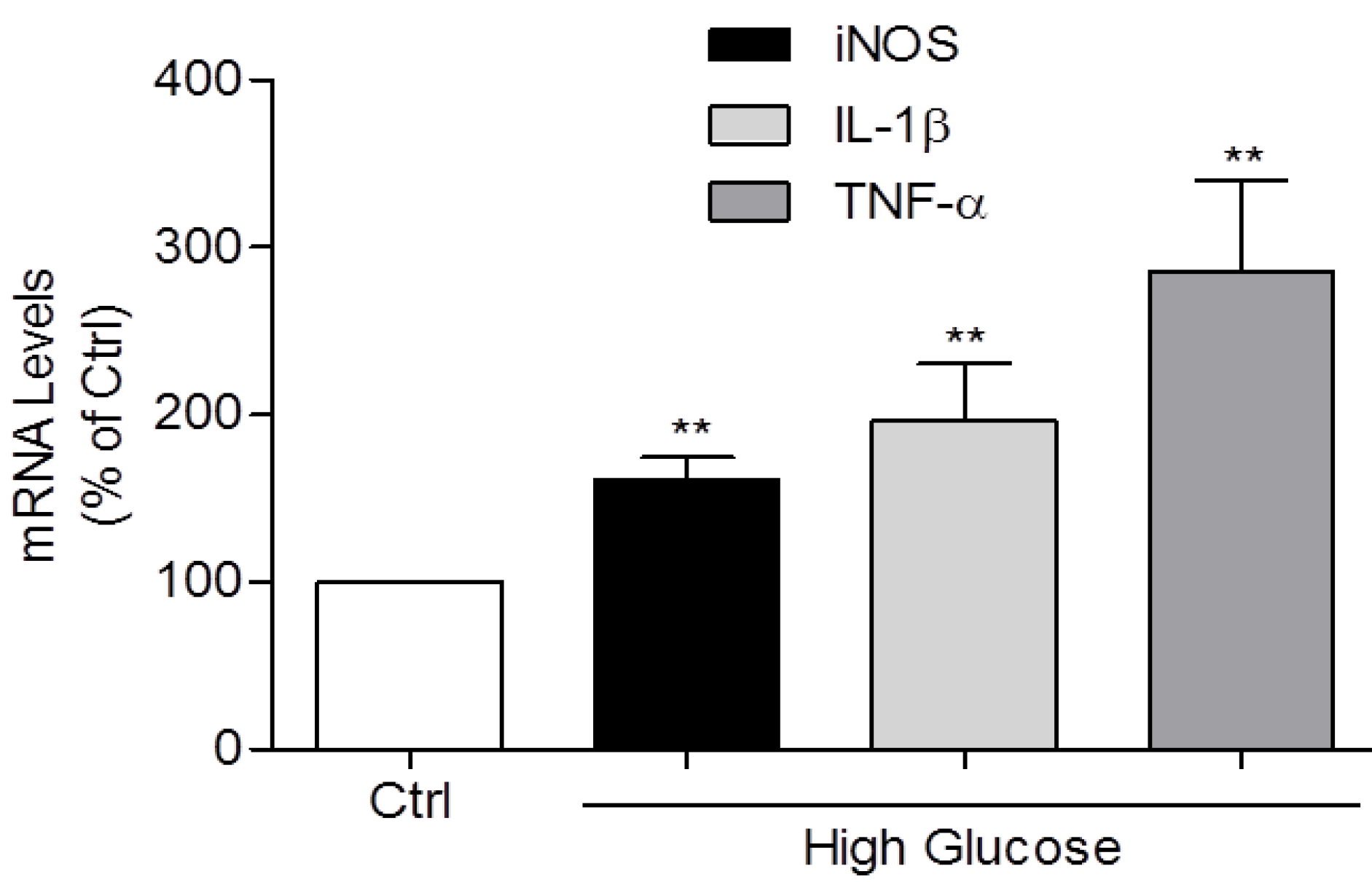
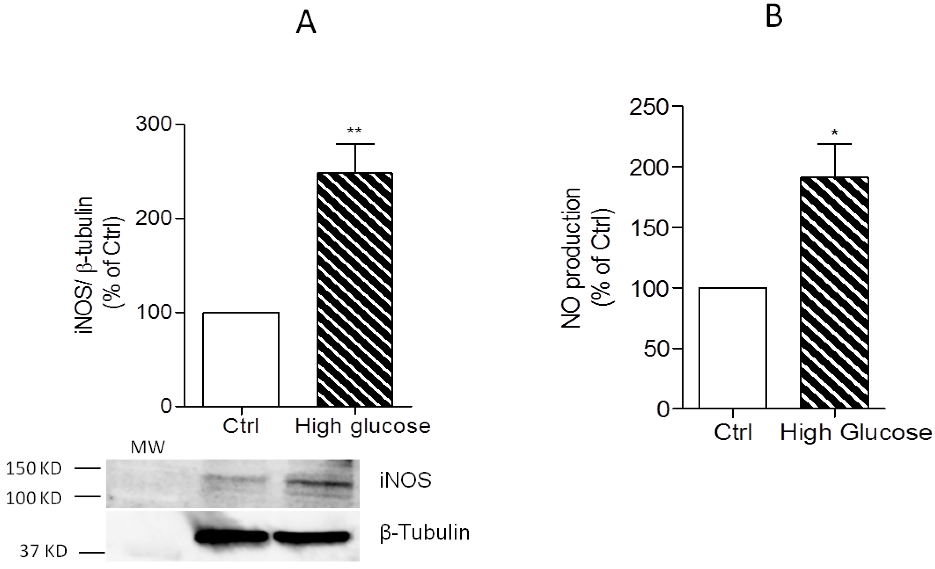
3.2. Induction of IL-1β, TNF-α, and iNOS Expression and NO Production by High Glucose

Figure 3 shows that culture of primary human chondrocytes in high glucose (30 mM)-containing medium significantly increased IL-1β, TNF-α, and iNOS mRNA levels relative to those found in chondrocytes maintained in regular culture medium (Ctrl, 10 mM glucose).
Moreover, the increase in iNOS mRNA induced by 30 mM glucose was accompanied by a similar increase of the protein level (Figure 4A) and enzyme activity, as shown by the augmented production of NO (Figure 4B).

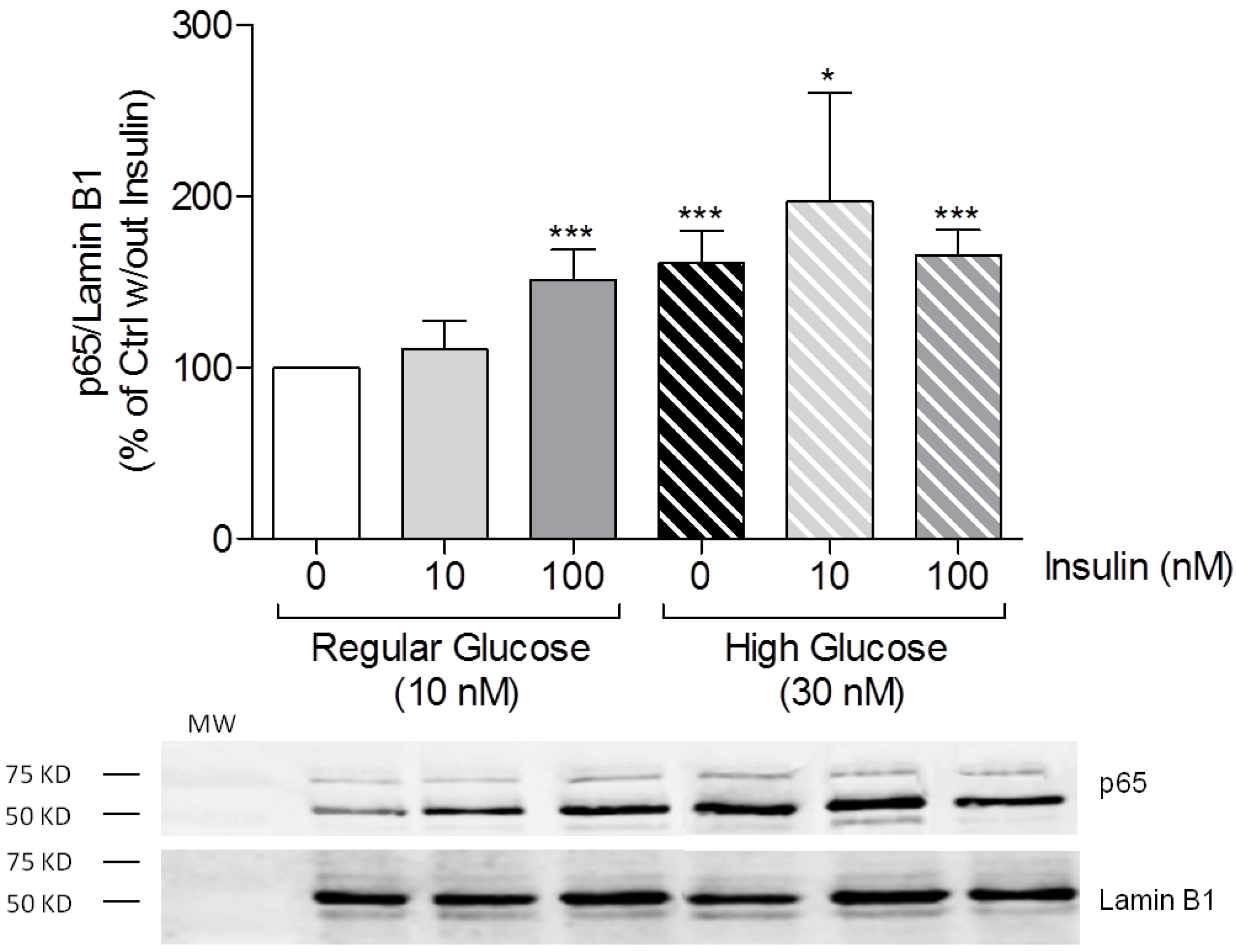
3.3. High Insulin Induced NF-κB p65 Translocation to the Nucleus

To determine whether insulin alone modulates the inflammatory state of human chondrocytes, we evaluated the effect of physiologic (10 nM) and supraphysiologic (100 nM) insulin concentrations on nuclear p65 levels in C28-I2 cells cultured in regular (Ctrl, 10 mM) glucose medium. Figure 5 shows that treatment of cells under regular glucose with 100 nM insulin is sufficient to increase nuclear p65 levels, and this increase is identical to that observed in cells cultured in high glucose (30 mM) alone. No changes in nuclear p65 levels were observed upon treatment with 10 nM insulin alone relative to control conditions (10 mM glucose).
To determine whether physiologic or supraphysiologic insulin concentrations modulate high glucose-induced NF-κB activation, chondrocyte cultures were treated simultaneously with 30 mM glucose and 10 or 100 nM insulin for 1 h. The results in Figure 5 show that although the mean value obtained in cells treated with 10 nM insulin in the presence of high glucose is higher than that obtained with high glucose alone, the difference did not reach statistical significance. Moreover, treatment of cells under high glucose with 100 nM insulin also did not change nuclear p65 levels relative to high glucose (30 mM) alone, thus indicating that insulin, either at physiologic or supraphysiologic concentrations, and high glucose neither synergize nor antagonize each other in inducing NF-κB activation.

4. Discussion

The results presented show that culture of human chondrocytes under high glucose is sufficient to induce NF-κB activation. The rapid nuclear translocation of p65 induced by high glucose suggests a direct effect on the NF-κB signaling pathway in chondrocytes. In a previous study, we reported that culture of human aged/OA chondrocytes under high glucose elicits the prolonged production of reactive oxygen species (ROS) [11], which were shown to mediate IL-1β-induced NF-κB activation and the expression of iNOS in chondrocytes [25]. Taking these studies together, it seems likely that high glucose-induced NF-κB activation can result, at least in part, from the increased production of ROS elicited by the intracellular glucose accumulation observed in aged/OA chondrocytes exposed to high glucose [11]. The observation that the selective inhibitor of the canonical NF-κB activation pathway, Bay 11-7082, completely abrogated high glucose-induced NF-κB activation shows that the response to high glucose occurs through this pathway and further supports a possible role for ROS, since these have been shown to be required for IL-1β-induced IκB-α degradation in chondrocytes [18,25]. Further studies will be directed at confirming this association and evaluating the efficacy of antioxidants in preventing high glucose-induced NF-κB activation and the expression of its dependent genes.
Moreover, the results presented also show that the expression of inflammatory mediators relevant in OA pathogenesis, namely IL-1β, TNF-α, and iNOS, was also upregulated by high glucose. Since the role of NF-κB in inducing the expression of these pro-inflammatory genes in chondrocytes is well established, these results indicate that NF-κB activation by high glucose is likely the underlying mechanism, although other processes may also be involved. Nonetheless, upregulated iNOS mRNA levels were detected upon culture in high glucose for 24 h, whereas measurable amounts of the protein and NO were detected after 72 h and required culture periods longer than, at least, 48 h. Moreover, NF-κB activation was maximal after exposure to high glucose for 1 h, while by 2 h the effect had returned to levels found in control cells cultured in regular medium (10 mM glucose) for the same period and remained as such for, at least, 24 h (1440 min). This kinetics of NF-κB activation is similar to that observed with other stimuli, like IL-1β, which allows iNOS protein to be detected much earlier [22] than observed here with high glucose. This suggests that other mechanisms required to induce iNOS expression and protein synthesis are delayed in response to high glucose when compared to IL-1β. One possibility is that high glucose-induced iNOS expression is preceded and mediated by the expression of IL-1β and TNF-α, which are potent inducers of iNOS expression in chondrocytes [16]. Other possibilities may be related to the modulation of other signaling pathways that contribute to increase NF-κB transcriptional activity after its release from IκB proteins. In this context, p65 phosphorylation and acetylation are known to increase its transcriptional activity [21], but how such modifications affect the expression of each NF-κB-dependent gene is largely unknown. Thus, it is possible that high glucose is not as efficient as IL-1β and TNF-α in inducing those modifications, thus requiring a longer period or even being unable to induce full NF-κB activity. Although out of the scope of this study, elucidating the molecular mechanisms involved in high glucose-induced NF-κB activation and how such mechanisms affect individual gene expression will be important to understand the mechanisms by which DM2 contributes to OA development and progression and eventually to design new preventive or therapeutic strategies.
Insulin is essential for maintenance of glucose homeostasis and for proper maintenance of the energetic balance, but also contributes to maintain anabolic processes in several tissues, including cartilage, as it was shown to induce anabolic and inhibit catabolic responses in adult chondrocytes and cartilage explant cultures from various species [12,26]. In DM2 and the metabolic syndrome, hyperinsulinemia occurs as an attempt at maintaining glucose homeostasis, at least while pancreatic β cells remain functional. Accumulating evidence, however, is unraveling a role for hyperinsulinemia in driving or, at least, contributing to the low-grade inflammation and tissue damage characteristic of those metabolic disorders and their complications [27]. For instance, supraphysiologic insulin concentrations, similar to or higher than those we used, were shown to induce NF-κB activation and synergize with TNF-α in cardiac myoblasts [28], while in chondrocytes they decreased autophagy and increased IL-1β and MMP-13 expression, contributing to cartilage degradation [5]. Thus, we hypothesized that physiologic and supraphysiologic insulin concentrations may have distinct effects in chondrocytes, either alone or in combination with high glucose. The results obtained confirmed this hypothesis, showing that supraphysiologic insulin concentrations are sufficient to activate NF-κB in chondrocytes while physiologic concentrations have no effect, either alone or in combination with high glucose. Nonetheless, we observed no additive or synergistic effects of the association of high insulin and high glucose in terms of NF-κB activation, while autophagy was found to be further impaired by high insulin in the presence of high glucose [5]. Taken together, the results presented here also suggest that by activating NF-κB, hyperinsulinemia can induce inflammatory responses in chondrocytes. Nonetheless, more studies are required to further elucidate the direct pro-inflammatory effects of hyperinsulinemia in primary human chondrocytes.

5. Conclusions

In summary, the results presented here, along with the pro-catabolic and anti-anabolic effects that we and others reported previously [11,12], further support the hypothesis that hyperglycemia per se can drive cartilage damage and OA, in agreement with in vivo studies that showed increased joint damage associated with a more intense inflammatory response in animal models of DM2 [13,14]. Moreover, the results obtained also indicate that hyperinsulinemia, which is characteristic of DM2 and metabolic syndrome, can also by itself contribute to activate an inflammatory state in human chondrocytes that can accelerate OA development and progression. Thus, these findings corroborate the hypothesis that hyperglycemia and hyperinsulinemia are relevant pro-inflammatory mediators in human chondrocytes, hence giving support to further studies both in vitro and in animal models of impaired glucose and insulin homeostasis in order to assess their relevance in OA development and progression. Such studies, although out of the scope of the current study, will elucidate the consequences to joint tissues homeostasis of the complex dynamic interactions between hyperglycemia and hyperinsulinemia. This knowledge will be essential for the development of new preventive and therapeutic strategies for DM-associated OA.

Acknowledgments

This work was supported by the Regional Development European Fund (FEDER), through the programs COMPETE 2020 and QREN of the European Community and the Portuguese Foundation for Science and Technology (FCT) under the project PTDC/EMS-TEC/3263/2014, the strategic projects, HealthyAging2020 CENTRO-01-0145-FEDER-000012 and UID/NEU/04539/2013, and the PhD studentships, SFRH/BD/47470/2008 and SFRH/BD/78188/2011.

Author Contributions

Alexandrina Ferreira Mendes and Ana Teresa Rufino conceived and designed the experiments; Ana Teresa Rufino, Madalena Ribeiro, and João Pinto Ferreira performed the experiments; Ana Teresa Rufino and Alexandrina Ferreira Mendes analyzed the data; Fernando Judas contributed cartilage samples and performed their macroscopic classification; Alexandrina Ferreira Mendes and Ana Teresa Rufino drafted the manuscript. All authors read and approved the final manuscript.

Conflicts of Interest

The authors declare no conflict of interest.

References

  1. Zhang, Y.; Jordan, J.M. Epidemiology of osteoarthritis. Clin. Geriatr. Med. 2010, 26, 355–369. [Google Scholar] [CrossRef] [PubMed]
  2. Berenbaum, F. Signaling transduction: Target in osteoarthritis. Curr. Opin. Rheumatol. 2004, 16, 616–622. [Google Scholar] [CrossRef] [PubMed]
  3. Schett, G.; Kleyer, A.; Perricone, C.; Sahinbegovic, E.; Iagnocco, A.; Zwerina, J.; Lorenzini, R.; Aschenbrenner, F.; Berenbaum, F.; D’Agostino, M.A.; et al. Diabetes Is an Independent Predictor for Severe Osteoarthritis: Results from a longitudinal cohort study. Diabetes Care 2012, 36, 403–409. [Google Scholar] [CrossRef] [PubMed]
  4. Nieves-Plaza, M.; Castro-Santana, L.E.; Font, Y.M.; Mayor, A.M.; Vila, L.M. Association of hand or knee osteoarthritis with diabetes mellitus in a population of Hispanics from Puerto Rico. J. Clin. Rheumatol. 2013, 19, 1–6. [Google Scholar] [CrossRef] [PubMed]
  5. Ribeiro, M.; Lopez de Figueroa, P.; Blanco, F.J.; Mendes, A.F.; Carames, B. Insulin decreases autophagy and leads to cartilage degradation. Osteoarthr. Cartil. 2015, 24, 731–739. [Google Scholar] [CrossRef] [PubMed]
  6. Ribeiro, M.; Lopez de Figueroa, P.; Nogueira-Recalde, U.; Centeno, A.; Mendes, A.F.; Blanco, F.J.; Carames, B. Diabetes-accelerated experimental osteoarthritis is prevented by autophagy activation. Osteoarthr. Cartil. 2016, 24, 2116–2125. [Google Scholar] [CrossRef] [PubMed]
  7. Lotz, M.K.; Carames, B. Autophagy and cartilage homeostasis mechanisms in joint health, aging and OA. Nat. Rev. Rheumatol. 2011, 7, 579–587. [Google Scholar] [CrossRef] [PubMed]
  8. Donath, M.Y.; Shoelson, S.E. Type 2 diabetes as an inflammatory disease. Nat. Rev. Immunol. 2011, 11, 98–107. [Google Scholar] [CrossRef] [PubMed]
  9. Kelley, K.M.; Johnson, T.R.; Ilan, J.; Moskowitz, R.W. Glucose regulation of the IGF response system in chondrocytes: Induction of an IGF-I-resistant state. Am. J. Physiol. 1999, 276, R1164–R1171. [Google Scholar] [PubMed]
  10. McNulty, A.L.; Stabler, T.V.; Vail, T.P.; McDaniel, G.E.; Kraus, V.B. Dehydroascorbate transport in human chondrocytes is regulated by hypoxia and is a physiologically relevant source of ascorbic acid in the joint. Arthritis Rheumatol. 2005, 52, 2676–2685. [Google Scholar] [CrossRef] [PubMed]
  11. Rosa, S.C.; Goncalves, J.; Judas, F.; Mobasheri, A.; Lopes, C.; Mendes, A.F. Impaired glucose transporter-1 degradation and increased glucose transport and oxidative stress in response to high glucose in chondrocytes from osteoarthritic versus normal human cartilage. Arthritis Res. Ther. 2009, 11, R80. [Google Scholar] [CrossRef] [PubMed]
  12. Rosa, S.C.; Rufino, A.T.; Judas, F.; Tenreiro, C.; Lopes, M.C.; Mendes, A.F. Expression and function of the insulin receptor in normal and osteoarthritic human chondrocytes: Modulation of anabolic gene expression, glucose transport and GLUT-1 content by insulin. Osteoarthr. Cartil. 2011, 19, 719–727. [Google Scholar] [CrossRef] [PubMed]
  13. Chen, Y.J.; Chan, D.C.; Lan, K.C.; Wang, C.C.; Chen, C.M.; Chao, S.C.; Tsai, K.S.; Yang, R.S.; Liu, S.H. PPARgamma is involved in the hyperglycemia-induced inflammatory responses and collagen degradation in human chondrocytes and diabetic mouse cartilages. J. Orthop. Res. 2015, 33, 373–381. [Google Scholar] [CrossRef] [PubMed]
  14. Laiguillon, M.C.; Courties, A.; Houard, X.; Auclair, M.; Sautet, A.; Capeau, J.; Feve, B.; Berenbaum, F; Sellam, J. Characterization of diabetic osteoarthritic cartilage and role of high glucose environment on chondrocyte activation: Toward pathophysiological delineation of diabetes mellitus-related osteoarthritis. Osteoarthr. Cartil. 2015, 23, 1513–1522. [Google Scholar] [CrossRef] [PubMed]
  15. Rosa, S.C.; Judas, F.; Lopes, M.C.; Mendes, A.F. Nitric oxide synthase isoforms and NF-κB activity in normal and osteoarthritic human chondrocytes: Regulation by inducible nitric oxide. Nitric Oxide 2008, 19, 276–283. [Google Scholar] [CrossRef] [PubMed]
  16. Stadler, J.; Stefanovic-Racic, M.; Billiar, T.R.; Curran, R.D.; McIntyre, L.A.; Georgescu, H.I.; Simmons, R.L.; Evans, C.H. Articular chondrocytes synthesize nitric oxide in response to cytokines and lipopolysaccharide. J. Immunol. 1991, 147, 3915–3920. [Google Scholar] [PubMed]
  17. Liu-Bryan, R.; Terkeltaub, R. Emerging regulators of the inflammatory process in osteoarthritis. Nat. Rev. Rheumatol. 2015, 11, 35–44. [Google Scholar] [CrossRef] [PubMed]
  18. Strickson, S.; Campbell, D.G.; Emmerich, C.H.; Knebel, A.; Plater, L.; Ritorto, M.S.; Shpiro, N.; Cohen, P. The anti-inflammatory drug BAY 11-7082 suppresses the MyD88-dependent signalling network by targeting the ubiquitin system. Biochem. J. 2013, 451, 427–437. [Google Scholar] [CrossRef] [PubMed]
  19. Green, L.C.; Wagner, D.A.; Glogowski, J.; Skipper, P.L.; Wishnok, J.S.; Tannenbaum, S.R. Analysis of nitrate, nitrite, and [15N] nitrate in biological fluids. Anal. Biochem. 1982, 126, 131–138. [Google Scholar] [CrossRef]
  20. Nolan, T.; Hands, R.E.; Bustin, S.A. Quantification of mRNA using real-time RT-PCR. Nat. Protoc. 2006, 1, 1559–1582. [Google Scholar] [CrossRef] [PubMed]
  21. Hayden, M.S.; Ghosh, S. Shared principles in NF-κB signaling. Cell 2008, 132, 344–362. [Google Scholar] [CrossRef] [PubMed]
  22. Mendes, A.F.; Carvalho, A.P.; Caramona, M.M.; Lopes, M.C. Role of nitric oxide in the activation of NF-κB, AP-1 and NOS II expression in articular chondrocytes. Inflamm. Res. 2002, 51, 369–375. [Google Scholar] [CrossRef] [PubMed]
  23. Frank, S.; Peters, M.A.; Wehmeyer, C.; Strietholt, S.; Koers-Wunrau, C.; Bertrand, J.; Heitzmann, M.; Hillmann, A.; Sherwood, J.; Seyfert, C.; et al. Regulation of matrixmetalloproteinase-3 and matrixmetalloproteinase-13 by SUMO-2/3 through the transcription factor NF-κB. Ann. Rheum. Dis. 2013, 72, 1874–1881. [Google Scholar] [CrossRef] [PubMed]
  24. Rufino, A.T.; Ribeiro, M.; Sousa, C.; Judas, F.; Salgueiro, L.; Cavaleiro, C.; Mendes, A.F. Evaluation of the anti-inflammatory, anti-catabolic and pro-anabolic effects of E-caryophyllene, myrcene and limonene in a cell model of osteoarthritis. Eur. J. Pharmacol. 2015, 750, 141–150. [Google Scholar] [CrossRef] [PubMed]
  25. Mendes, A.F.; Caramona, M.M.; Carvalho, A.P.; Lopes, M.C. Differential roles of hydrogen peroxide and superoxide in mediating IL-1-induced NF-κB activation and iNOS expression in bovine articular chondrocytes. J. Cell. Biochem. 2003, 88, 783–793. [Google Scholar] [CrossRef] [PubMed]
  26. Kellner, K.; Schulz, M.B.; Gopferich, A.; Blunk, T. Insulin in tissue engineering of cartilage: A potential model system for growth factor application. J. Drug Target. 2001, 9, 439–448. [Google Scholar] [CrossRef] [PubMed]
  27. Guo, S. Insulin signaling, resistance, and the metabolic syndrome: Insights from mouse models into disease mechanisms. J. Endocrinol. 2014, 220, T1–T23. [Google Scholar] [CrossRef] [PubMed]
  28. Madonna, R.; Geng, Y.J.; Bolli, R.; Rokosh, G.; Ferdinandy, P; Patterson, C.; De Caterina, R. Co-Activation of Nuclear Factor-κB and Myocardin/Serum Response Factor Conveys the Hypertrophy Signal of High Insulin Levels in Cardiac Myoblasts. J. Biol. Chem. 2014, 298, 19585–19598. [Google Scholar] [CrossRef] [PubMed]
Figure 1. Role of glucose concentrations on nuclear factor-κB (NF-κB) p65 levels. C28/I2 cells were cultured in control (Ctrl, 10 mM, 120 min) or high glucose (HGM; 30 mM) medium, with or without 5 µM Bay-11-7082, a specific NF-κB inhibitor, for the indicated time periods. Each column represents the mean ± SEM of, at least, four independent experiments. * p < 0.05, ** p < 0.01, and *** p < 0.001 relative to the Ctrl.
Jfmk 02 00015 g001
Figure 2. High glucose-induced nuclear p65 accumulation is not mediated by osmotic effects. C28/I2 cells were cultured in control (Ctrl, 10 mM), high glucose- (HGM; 30 mM), or mannitol (20 mM)-supplemented media for 1 h. Each column represents the mean ± SEM of, at least, five independent experiments. *** p < 0.001 relative to the Ctrl.
Jfmk 02 00015 g002
Figure 3. Role of high glucose in modulating pro-inflammatory cytokines and inducible nitric oxide synthase (iNOS) expression. mRNA levels of iNOS, interleukin (IL)-1β, and Tumor Necrosis Factor (TNF)-α were measured in primary human chondrocytes cultured for 24 h in control (Ctrl, 10 mM) or in high glucose (30 mM) medium. Each column represents the mean ± SEM of, at least, four independent experiments. ** p < 0.01 relative to Ctrl.
Jfmk 02 00015 g003
Figure 4. Role of high glucose in modulating iNOS protein levels and activity. Protein levels of iNOS (A) and NO production (B) were measured in primary human chondrocytes cultured for 72 h in control (Ctrl, 10 mM) or in high glucose (30 mM) media. Each column represents the mean ± SEM of, at least, four independent experiments. * p < 0.05 and ** p < 0.01 relative to Ctrl.
Jfmk 02 00015 g004
Figure 5. Role of Insulin concentrations on nuclear NF-κB p65 levels. Insulin, 10 or 100 nM, was added to C28-I2 cultures in regular (10 mM) or high glucose (30 mM) conditions for 1 h. Each column represents the mean ± SEM of, at least, four independent experiments. * p < 0.05 and *** p < 0.001 relative to the control (10 mM glucose).
Jfmk 02 00015 g005

