
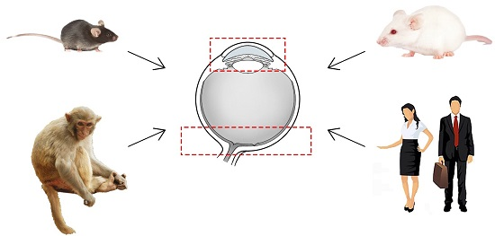
Comparative Anatomy of the Trabecular Meshwork, the Optic Nerve Head and the Inner Retina in Rodent and Primate Models Used for Glaucoma Research

Abstract
:
1. Introduction
2. Comparative Anatomy and Composition of the Trabecular Meshwork and Schlemm’s Canal
3. Comparative Anatomy and Composition of the Lamina Cribrosa
4. Comparative Anatomy of the Central Retinal Vessels
5. Comparative Anatomy of the Optic Nerve Head Blood Supply
6. Comparative Anatomy of the Ganglion Cell Layer
7. Summary of the Rodent and Primate Models Commonly Used for Glaucoma Research
8. Conclusions
Supplementary Materials
Acknowledgments
Author Contributions
Conflicts of Interest
References
- Quigley, H.A. Number of people with glaucoma worldwide. Br. J. Ophthalmol. 1996, 80, 389–393. [Google Scholar] [CrossRef] [PubMed]
- Quigley, H.A. The number of people with glaucoma worldwide in 2010 and 2020. Br. J. Ophthalmol. 2006, 90, 262–267. [Google Scholar] [CrossRef] [PubMed]
- Halpern, D.L.; Grosskreutz, C.L. Glaucomatous optic neuropathy: Mechanisms of disease. Ophthalmol. Clin. N. Am. 2002, 15, 61–68. [Google Scholar] [CrossRef]
- John, S.W.; Smith, R.S.; Savinova, O.V.; Hawes, N.L.; Chang, B.; Turnbull, D.; Davisson, M.; Roderick, T.H.; Heckenlively, J.R. Essential iris atrophy, pigment dispersion, and glaucoma in DBA/2J mice. Investig. Ophthalmol. Vis. Sci. 1998, 39, 951–962. [Google Scholar] [PubMed]
- Pang, I.H.; Clark, A.F. Rodent models for glaucoma retinopathy and optic neuropathy. J. Glaucoma 2007, 16, 483–505. [Google Scholar] [CrossRef] [PubMed]
- Johnson, T.V.; Tomarev, S.I. Rodent models of glaucoma. Brain Res. Bull. 2010, 81, 349–358. [Google Scholar] [CrossRef] [PubMed]
- Kolker, A.E.; Moses, R.A.; Constant, M.A.; Becker, B. The development of glaucoma in rabbits. Investig. Ophthalmol. Vis. Sci. 1963, 2, 316–321. [Google Scholar]
- Ruiz-Ederra, J.; Garcia, M.; Hernandez, M.; Urcola, H.; Hernandez-Barbachano, E.; Araiz, J.; Vecino, E. The pig eye as a novel model of glaucoma. Exp. Eye Res. 2005, 81, 561–569. [Google Scholar] [CrossRef] [PubMed]
- Dietrich, U. Feline glaucomas. Clin. Tech. Small Anim. Pract. 2005, 20, 108–116. [Google Scholar] [CrossRef] [PubMed]
- Brooks, D.E. Glaucoma in the dog and cat. Vet. Clin. N. Am. Small Anim. Pract. 1990, 20, 775–797. [Google Scholar] [CrossRef]
- Gelatt, K.N.; Peiffer, R.J.; Gwin, R.M.; Gum, G.G.; Williams, L.W. Clinical manifestations of inherited glaucoma in the beagle. Investig. Ophthalmol. Vis. Sci. 1977, 16, 1135–1142. [Google Scholar] [PubMed]
- Rasmussen, C.A.; Kaufman, P.L. Primate glaucoma models. J. Glaucoma 2005, 14, 311–314. [Google Scholar] [CrossRef] [PubMed]
- Burgoyne, C.F. The non-human primate experimental glaucoma model. Exp. Eye Res. 2015, 141, 57–73. [Google Scholar] [CrossRef] [PubMed]
- Bouhenni, R.A.; Dunmire, J.; Sewell, A.; Edward, D.P. Animal models of glaucoma. J. Biomed. Biotechnol. 2012, 2012, 1–11. [Google Scholar] [CrossRef] [PubMed]
- Gong, H.; Tripathi, R.C.; Tripathi, B.J. Morphology of the aqueous outflow pathway. Microsc. Res. Tech. 1996, 33, 336–367. [Google Scholar] [CrossRef]
- Goel, M.; Picciani, R.G.; Lee, R.K.; Bhattacharya, K.S. Aqueous humor dynamics: A review. Open Ophthalmol. J. 2010, 4, 52–59. [Google Scholar] [CrossRef] [PubMed]
- Alvarado, J.A.; Murphy, C.G. Outflow obstruction in pigmentary and primary open angle glaucoma. Arch. Ophthalmol. 1992, 110, 1769–1778. [Google Scholar] [CrossRef] [PubMed]
- Lutjen-Drecoll, E.; Futa, R.; Rohen, J.W. Ultrahistochemical studies on tangential sections of the trabecular meshwork in normal and glaucomatous eyes. Investig. Ophthalmol. Vis. Sci. 1981, 21, 563–573. [Google Scholar] [PubMed]
- Tan, J.C.; Peters, D.M.; Kaufman, P.L. Recent developments in understanding the pathophysiology of elevated intraocularpressure. Curr. Opin. Ophthalmol. 2006, 17, 168–174. [Google Scholar] [PubMed]
- Tripathi, R.C. Ultrastructure of the exit pathway of the aqueous in lower mammals: (A preliminary report on the “angular aqueous plexus”). Exp. Eye Res. 1971, 12, 311–314. [Google Scholar] [CrossRef]
- Van der Zypen, E. Experimental morphological study on structure and function of the filtration angel of the rat eye. Ophthalmologica 1977, 174, 285–298. [Google Scholar] [CrossRef] [PubMed]
- McMenamin, P.G.; Al-Shakarchi, M.J. The effect of various levels of intraocular pressure on the rat aqueous outflow system. J. Anat. 1989, 162, 67–82. [Google Scholar] [PubMed]
- Morrison, J.C.; Fraunfelder, F.W.; Milne, S.T.; Moore, C.G. Limbal microvasculature of the rat eye. Investig. Ophthalmol. Vis. Sci. 1995, 36, 751–756. [Google Scholar] [PubMed]
- Morrison, J.C.; Moore, C.G.; Deppmeier, L.M.; Gold, B.G.; Meshul, C.K.; Johnson, E.C. A rat model of chronic pressure-induced optic nerve damage. Exp. Eye Res. 1997, 64, 85–96. [Google Scholar] [CrossRef] [PubMed]
- Zhang, Z.; Dhaliwal, A.S.; Tseng, H.; Kim, J.D.; Schuman, J.S.; Weinreb, R.N.; Loewen, N.A. Outflow tract ablation using a conditionally cytotoxic feline immunodeficiency viral vector. Investig. Ophthalmol. Vis. Sci. 2014, 55, 935–940. [Google Scholar] [CrossRef] [PubMed]
- Johnson, A.W.; Ammar, D.A.; Kahook, M.Y. Two-Photon imaging of the mouse eye. Investig. Ophthalmol. Vis. Sci. 2011, 52, 4098–4105. [Google Scholar] [CrossRef] [PubMed]
- Ko, M.K.; Tan, J.C. Contractile markers distinguish structures of the mouse aqueous drainage tract. Mol. Vis. 2013, 19, 2561–2570. [Google Scholar] [PubMed]
- Overby, D.R.; Bertrand, J.; Schicht, M.; Paulsen, F.; Stamer, W.D.; Lutjen-Drecoll, E. The structure of the trabecular meshwork, its connections to the ciliary muscle, and the effect of pilocarpine on outflow facility in mice. Investig. Ophthalmol. Vis. Sci. 2014, 55, 3727–3736. [Google Scholar] [CrossRef] [PubMed]
- Smith, R.S.; Zabaleta, A.; Savinova, O.V.; John, S.W. The mouse anterior chamber angle and trabecular meshwork develop without cell death. BMC Dev. Biol. 2001, 1, 3. [Google Scholar] [CrossRef] [PubMed] [Green Version]
- Millar, J.C.; Phan, T.N.; Pang, I.H.; Clark, A.F. Strain and age effects on aqueous humor dynamics in the mouse. Investig. Ophthalmol. Vis. Sci. 2015, 56, 5764–5776. [Google Scholar] [CrossRef] [PubMed]
- Yan, Z.; Tian, Z.; Chen, H.; Deng, S.; Lin, J.; Liao, H.; Yang, X.; Ge, J.; Zhuo, Y. Analysis of a method for establishing a model with more stable chronic glaucoma in rhesus monkeys. Exp. Eye Res. 2015, 131, 56–62. [Google Scholar] [CrossRef] [PubMed]
- Holmberg, K.S. Schlemm’s canal and the trabecular meshwork. An electron microscopic study of the normal structure in man and monkey (Cercopithecus ethiops). Doc. Opthalmol. 1965, 19, 339–373. [Google Scholar] [CrossRef]
- Rohen, J. The histologic structure of the chamber angle in primates. Am. J. Opthalmol. 1961, 52, 529–539. [Google Scholar] [CrossRef]
- Epstein, D.L.; Rohen, J.W. Morphology of the trabecular meshwork and inner-wall endothelium after cationized ferritin perfusion in the monkey eye. Investig. Ophthalmol. Vis. Sci. 1991, 32, 160–171. [Google Scholar] [PubMed]
- Tian, B.; Geiger, B.; Epstein, D.L.; Kaufman, P.L. Cytoskeletal involvement in the regulation of aqueous humor outflow. Investig. Ophthalmol. Vis. Sci. 2000, 41, 619–623. [Google Scholar] [PubMed]
- Rohen, J.W.; Futa, R.; Lutjen-Drecoll, E. The fine structure of the cribriform meshwork in normal and glaucomatous eyes asseen in tangential sections. Investig. Ophthalmol. Vis. Sci. 1981, 21, 574–585. [Google Scholar] [PubMed]
- Hann, C.R.; Fautsch, M.P. The elastin fiber system between and adjacent to collector channels in the humanjuxtacanalicular tissue. Investig. Ophthalmol. Vis. Sci. 2011, 52, 45–50. [Google Scholar] [CrossRef] [PubMed]
- Chen, C.; Yeh, L.; Liu, C.; Kao, W.W.Y.; Samples, J.R.; Lin, S.; Hu, F.; Wang, I. Morphological differences between the trabecular meshworks of zebrafish and mammals. Curr. Eye Res. 2008, 33, 59–72. [Google Scholar] [CrossRef] [PubMed]
- Overby, D.R.; Bertrand, J.; Tektas, O.Y.; Boussommier-Calleja, A.; Schicht, M.; Ethier, C.R.; Woodward, D.F.; Stamer, W.D.; Lutjen-Drecoll, E. Ultrastructural changes associated with dexamethasone-induced ocular hypertension in mice. Investig. Ophthalmol. Vis. Sci. 2014, 55, 4922–4933. [Google Scholar] [CrossRef] [PubMed]
- De Kater, A.W.; Spurr-Michaud, S.J.; Gipson, I.K. Localization of smooth muscle myosin-containing cells in the aqueous outflow pathway. Investig. Ophthalmol. Vis. Sci. 1990, 31, 347–353. [Google Scholar] [PubMed]
- De Kater, A.W.; Shahsafaei, A.; Epstein, D.L. Localization of smooth muscle and nonmuscle actin isoforms in the human aqueous outflow pathway. Investig. Ophthalmol. Vis. Sci. 1992, 33, 424–429. [Google Scholar] [PubMed]
- Bunt-Milam, A.H.; Dennis, M.J.; Bensinger, R.E. Optic nerve head axonal transport in rabbits with hereditary glaucoma. Exp. Eye Res. 1987, 44, 537–551. [Google Scholar] [CrossRef]
- Brooks, D.E.; Arellano, E.; Kubilis, P.S.; Komaromy, A.M. Histomorphometry of the porcine scleral lamina cribrosa surface. Vet. Ophthalmol. 1998, 1, 129–135. [Google Scholar] [CrossRef] [PubMed]
- Radius, R.L.; Bade, B. The anatomy at the lamina cribrosa in the normal cat eye. Arch. Ophthalmol. 1982, 100, 1658–1660. [Google Scholar] [CrossRef] [PubMed]
- Brooks, D.E.; Samuelson, D.A.; Gelatt, K.N.; Smith, P.J. Morphologic changes in the lamina cribrosa of beagles with primary open-angle glaucoma. Am. J. Vet. Res. 1989, 50, 936–941. [Google Scholar] [PubMed]
- Morrison, J.C.; Johnson, E.; Cepurna, W.O. Rat models for glaucoma research. Prog. Brain Res. 2008, 173, 285–301. [Google Scholar] [PubMed]
- May, C.A.; Lutjen-Drecoll, E. Morphology of the murine optic nerve. Investig. Ophthalmol. Vis. Sci. 2002, 43, 2206–2212. [Google Scholar] [PubMed]
- Tansley, K. Comparison of the lamina cribrosa in mammalian species with good and with indifferent vision. Br. J. Ophthalmol. 1956, 40, 178–182. [Google Scholar] [CrossRef] [PubMed]
- Pazos, M.; Yang, H.; Gardiner, S.K.; Cepurna, W.O.; Johnson, E.C.; Morrison, J.C.; Burgoyne, C.F. Rat optic nerve head anatomy within 3D histomorphometric reconstructions of normal control eyes. Exp Eye Res. 2015, 139, 1–12. [Google Scholar] [CrossRef] [PubMed]
- Hildebrand, C.; Remahl, S.; Waxman, S.G. Axo-Glial relations in the retina-optic nerve junction of the adult rat: Electron-Microscopic observations. J. Neurocytol. 1985, 14, 597–617. [Google Scholar] [CrossRef] [PubMed]
- Johansson, J.O. The lamina cribrosa in the eyes of rats, hamsters, gerbils and guinea pigs. Cell Tissues Organs 1987, 128, 55–62. [Google Scholar] [CrossRef]
- May, C.A. The optic nerve head region of the aged rat: An immunohistochemical investigation. Curr. Eye Res. 2003, 26, 347–354. [Google Scholar] [CrossRef] [PubMed]
- Morrison, J.C.; Jerdan, J.A.; L’Hernault, N.L.; Quigley, H.A. The extracellular matrix composition of the monkey optic nerve head. Investig. Ophthalmol. Vis. Sci. 1988, 29, 1141–1150. [Google Scholar] [PubMed]
- Yang, H.; Downs, J.C.; Girkin, C.; Sakata, L.; Bellezza, A.; Thompson, H.; Burgoyne, C.F. 3-D Histomorphometry of the normal and early glaucomatous monkey optic nerve head: Lamina cribrosa and peripapillary scleral position and thickness. Investig. Ophthalmol. Vis. Sci. 2007, 48, 4597–4607. [Google Scholar] [CrossRef] [PubMed]
- Downs, J.C.; Yang, H.; Girkin, C.; Sakata, L.; Bellezza, A.; Thompson, H.; Burgoyne, C.F. Three-Dimensionalhistomorphometry of the normal and early glaucomatous monkey optic nerve head: Neural canal and subarachnoid space architecture. Investig. Ophthalmol. Vis. Sci. 2007, 48, 3195–3208. [Google Scholar] [CrossRef] [PubMed]
- Anderson, D.R. Ultrastructure of human and monkey lamina cribrosa and optic nerve head. Arch. Ophthalmol. 1969, 82, 800–814. [Google Scholar] [CrossRef] [PubMed]
- Jonas, J.B.; Mardin, C.Y.; Schlotzer-Schrehardt, U.; Naumann, G.O. Morphometry of the human lamina cribrosa surface. Investig. Ophthalmol. Vis. Sci. 1991, 32, 401–405. [Google Scholar] [PubMed]
- Johnson, E.C.; Morrison, J.C.; Farrell, S.; Deppmeier, L.; Moore, C.G.; McGinty, M.R. The effect of chronically elevated intraocular pressure on the rat optic nerve head extracellular matrix. Exp. Eye Res. 1996, 62, 663–674. [Google Scholar] [CrossRef] [PubMed]
- Morrison, J.C.; Dorman-Pease, M.E.; Dunkelberger, G.R.; Quigley, H.A. Optic nerve head extracellular matrix in primary optic atrophy and experimental glaucoma. Arch. Ophthalmol. 1990, 108, 1020–1024. [Google Scholar] [CrossRef] [PubMed]
- Quigley, H.A.; Dorman-Pease, M.E.; Brown, A.E. Quantitative study of collagen and elastin of the optic nerve head and sclera inhuman and experimental monkey glaucoma. Curr. Eye Res. 1991, 10, 877–888. [Google Scholar] [CrossRef] [PubMed]
- Fukuchi, T.; Sawaguchi, S.; Hara, H.; Shirakashi, M.; Iwata, K. Extracellular matrix changes of the optic nerve lamina cribrosa in monkey eyes with experimentally chronic glaucoma. Graefe’s Arch. Clin. Exp. Ophthalmol. 1992, 230, 421–427. [Google Scholar] [CrossRef] [PubMed]
- Hernandez, M.R.; Igoe, F.; Neufeld, A.H. Extracellular matrix of the human optic nerve head. Am. J. Opthalmol. 1986, 102, 139–148. [Google Scholar] [CrossRef]
- Hernandez, M.R.; Luo, X.X.; Igoe, F.; Neufeld, A.H. Extracellular matrix of the human lamina cribrosa. Am. J. Opthalmol. 1987, 104, 567–576. [Google Scholar] [CrossRef]
- Tengroth, B.; Rehnberg, M.; Amitzboll, T. A comparative analysis of the collagen type and distribution in the trabecular meshwork, sclera, lamina cribrosa and the optic nerve in the human eye. Acta Ophthalmol. 1985, 173, 91–93. [Google Scholar] [CrossRef]
- Albon, J.; Karwatowski, W.S.; Easty, D.L.; Sims, T.J.; Duance, V.C. Age related changes in the non-collagenous components of the extracellular matrix of the human lamina cribrosa. Br. J. Ophthalmol. 2000, 84, 311–317. [Google Scholar] [CrossRef] [PubMed]
- Fukuchi, T.; Sawaguchi, S.; Yue, B.Y.; Iwata, K.; Hara, H.; Kaiya, T. Sulfated proteoglycans in the lamina cribrosa of normal monkey eyes and monkey eyes with laser-induced glaucoma. Exp. Eye Res. 1994, 58, 231–243. [Google Scholar] [CrossRef] [PubMed]
- Sawaguchi, S.; Yue, B.Y.; Fukuchi, T.; Iwata, K.; Kaiya, T. Sulfated proteoglycans in the human lamina cribrosa. Investig. Ophthalmol. Vis. Sci. 1992, 33, 2388–2398. [Google Scholar] [PubMed]
- Sawaguchi, S.; Yue, B.Y.; Fukuchi, T.; Iwata, K.; Kaiya, T. Age-Related changes of sulfated proteoglycans in the human lamina cribrosa. Curr. Eye Res. 1993, 12, 685–692. [Google Scholar] [CrossRef] [PubMed]
- Caparas, V.L.; Cintron, C.; Hernandez-Neufeld, M.R. Immunohistochemistry of proteoglycans in human lamina cribrosa. Am. J. Opthalmol. 1991, 112, 489–495. [Google Scholar] [CrossRef]
- Jeffery, G.; Evans, A.; Albon, J.; Duance, V.; Neal, J.; Dawidek, G. The human optic nerve: Fascicular organisation and connective tissue types alongthe extra-fascicular matrix. Anat. Embryol. (Berl.) 1995, 191, 491–502. [Google Scholar] [CrossRef] [PubMed]
- Bisaria, K.K.; Sud, S.D. The central retinal artery in the albino rats (a histological study). Indian J. Opthalmol. 1977, 24, 23–26. [Google Scholar]
- Morrison, J.C.; Johnson, E.C.; Cepurna, W.O.; Funk, R.H.W. Microvasculature of the rat optic nerve head. Investig. Ophthalmol. Vis. Sci. 1999, 40, 1702–1709. [Google Scholar] [PubMed]
- Lieberman, M.F.; Maumenee, A.E.; Green, W.R. Histologic studies of the vasculature of the anterior optic nerve. Am. J. Opthalmol. 1976, 82, 405–423. [Google Scholar] [CrossRef]
- Pannarale, L.; Onori, P.; Ripani, M.; Gaudio, E. Precapillary patterns and perivascular cells in the retinal microvasculature. A scanning electron microscope study. J. Anat. 1996, 188, 693–703. [Google Scholar] [PubMed]
- Lassmann, H.; Pamperl, H.; Stockinger, G. Morphological-functional aspects of intima pads in the rat ophthalmic artery. Z. Mikrosk. Anat. Forsch. 1972, 85, 139–148. [Google Scholar] [PubMed]
- Sugiyama, K.; Gu, Z.B.; Kawase, C.; Yamamoto, T.; Kitazawa, Y. Optic nerve and peripapillarychoroidal microvasculature of the rat eye. Investig. Ophthalmol. Vis. Sci. 1999, 40, 3084–3090. [Google Scholar] [PubMed]
- Hayreh, S.S. The blood supply of the optic nerve head and the evaluation of it—Myth and reality. Prog. Retin. Eye Res. 2001, 20, 563–593. [Google Scholar] [CrossRef]
- Hayreh, S.S. Blood supply of the optic nerve head and its role in optic atrophy, glaucoma, and oedema of the optic disc. Br. J. Ophthalmol. 1969, 53, 721–748. [Google Scholar] [CrossRef] [PubMed]
- Goldberg, I.; Hollows, F.C.; Kass, M.A.; Becker, B. Systemic factors in patients with low-tension glaucoma. Br. J. Ophthalmol. 1981, 65, 56–62. [Google Scholar] [CrossRef] [PubMed]
- Hayreh, S.S. Anatomy and physiology of the optic nerve head. Trans. Am. Acad. Ophthalmol. Otolaryngol. 1974, 78, P240–P254. [Google Scholar]
- Hayreh, S.S. Microangioarchitecture of optic papilla. Jpn. J. Ophthalmol. 1989, 33, 519–525. [Google Scholar] [PubMed]
- Hayreh, S.S. Blood supply of the optic nerve head. Ophthalmologica 1996, 210, 285–295. [Google Scholar] [CrossRef] [PubMed]
- Jeon, C.; Strettoi, E.; Masland, R.H. The major cell populations of the mouse retina. J. Neurosci. 1998, 18, 8936. [Google Scholar] [PubMed]
- Carter-Dawson, L.D.; LaVail, M.M. Rods and cones in the mouse retina. I. Structural analysis using light and electron microscopy. J. Comp. Neurol. 1979, 188, 245–262. [Google Scholar] [CrossRef] [PubMed]
- Drager, U.C.; Olsen, J.F. Ganglion cell distribution in the retina of the mouse. Investig. Ophthalmol. Vis. Sci. 1981, 20, 285–293. [Google Scholar] [PubMed]
- Williams, R.W.; Strom, R.C.; Rice, D.S.; Goldowitz, D. Genetic and environmental control of variation in retinal ganglion cell number in mice. J. Neurosci. 1996, 16, 7193–7205. [Google Scholar] [PubMed]
- Hunter, A.; Bedi, K.S. A quantitative morphological study of interstrain variation in the developing rat optic nerve. J. Comp. Neurol. 1986, 245, 160–166. [Google Scholar] [CrossRef] [PubMed]
- Cavallotti, C.; Cavallotti, D.; Pescosolido, N.; Pacella, E. Age-Related changes in rat optic nerve: Morphological studies. Anat. Histol. Embryol. 2003, 32, 12–16. [Google Scholar] [CrossRef] [PubMed]
- Potts, R.A.; Dreher, B.; Bennett, M.R. The loss of ganglion cells in the developing retina of the rat. Brain Res. 1982, 255, 481–486. [Google Scholar] [CrossRef]
- Fukuda, Y.; Sugimoto, T.; Shirokawa, T. Strain differences in quantitative analysis of the rat optic nerve. Exp. Neurol. 1982, 75, 525–532. [Google Scholar] [CrossRef]
- Garcia-Ayuso, D.; Salinas-Navarro, M.; Agudo, M.; Cuenca, N.; Pinilla, I.; Vidal-Sanz, M.; Villegas-Perez, M.P. Retinal ganglion cell numbers and delayed retinal ganglion cell death in the P23H rat retina. Exp. Eye Res. 2010, 91, 800–810. [Google Scholar] [CrossRef] [PubMed]
- Perry, V.H. Evidence for an amacrine cell system in the ganglion cell layer of the rat retina. Neuroscience 1981, 6, 931–944. [Google Scholar] [CrossRef]
- Voigt, T. Cholinergic amacrine cells in the rat retina. J. Comp. Neurol. 1986, 248, 19–35. [Google Scholar] [CrossRef] [PubMed]
- Furuyoshi, N.; Furuyoshi, M.; May, C.A.; Hayreh, S.S.; Alm, A.; Lutjen-Drecoll, E. Vascular and glial changes in the retrolaminar optic nerve in glaucomatous monkey eyes. Ophthalmologica 2000, 214, 24–32. [Google Scholar] [CrossRef] [PubMed]
- Herbin, M.; Boire, D.; Ptito, M. Size and distribution of retinal ganglion cells in the St. Kitts green monkey (Cercopithecus aethiops sabeus). J. Comp. Neurol. 1997, 383, 459–472. [Google Scholar] [CrossRef]
- Kim, C.B.; Tom, B.W.; Spear, P.D. Effects of aging on the densities, numbers, and sizes of retinal ganglion cells in rhesus monkey. Neurobiol. Aging 1996, 17, 431–438. [Google Scholar] [CrossRef]
- Silveira, L.C.; Picanco-Diniz, C.W.; Sampaio, L.F.; Oswaldo-Cruz, E. Retinal ganglion cell distribution in the cebus monkey: A comparison with the cortical magnification factors. Vis. Res. 1989, 29, 1471–1483. [Google Scholar] [CrossRef]
- Curcio, C.A.; Allen, K.A. Topography of ganglion cells in human retina. J. Comp. Neurol. 1990, 300, 5–25. [Google Scholar] [CrossRef] [PubMed]
- Wassle, H.; Grunert, U.; Rohrenbeck, J.; Boycott, B.B. Retinal ganglion cell density and cortical magnification factor in the primate. Vis. Res. 1990, 30, 1897–1911. [Google Scholar] [CrossRef]
- Senatorov, V.; Malyukova, I.; Fariss, R.; Wawrousek, E.F.; Swaminathan, S.; Sharan, S.K.; Tomarev, S. Expression of mutated mouse myocilin induces open-angle glaucoma in transgenic mice. J. Neurosci. 2006, 26, 11903–11914. [Google Scholar] [CrossRef] [PubMed]
- Aihara, M.; Lindsey, J.D.; Weinreb, R.N. Ocular hypertension in mice with a targeted type I collagen mutation. Investig. Ophthalmol. Vis. Sci. 2003, 44, 1581–1585. [Google Scholar] [CrossRef] [PubMed]
- Mabuchi, F.; Lindsey, J.D.; Aihara, M.; Mackey, M.R.; Weinreb, R.N. Optic nerve damage in mice with a targeted type I collagen mutation. Investig. Ophthalmol. Vis. Sci. 2004, 45, 1841–1845. [Google Scholar] [CrossRef] [PubMed]
- Sawaguchi, K.; Nakamura, Y.; Nakamura, Y.; Sakai, H.; Sawaguchi, S. Myocilin gene expression in the trabecular meshwork of rats in a steroid-inducedocular hypertension model. Ophthalmic Res. 2005, 37, 235–242. [Google Scholar] [CrossRef] [PubMed]
- Toris, C.B.; Zhan, G.L.; Wang, Y.L.; Zhao, J.; McLaughlin, M.A.; Camras, C.B.; Yablonski, M.E. Aqueous humor dynamics in monkeys with laser-induced glaucoma. J. Ocul. Pharmacol. Ther. 2000, 16, 19–27. [Google Scholar] [CrossRef] [PubMed]
- Weber, A.J.; Zelenak, D. Experimental glaucoma in the primate induced by latex microspheres. J. Neurosci. Methods 2001, 111, 39–48. [Google Scholar] [CrossRef]
- Quigley, H.A.; Addicks, E.M. Chronic experimental glaucoma in primates. I. Production of elevated intraocularpressure by anterior chamber injection of autologous ghost red blood cells. Investig. Ophthalmol. Vis. Sci. 1980, 19, 126–136. [Google Scholar] [PubMed]
- Fujikawa, K.; Iwata, T.; Inoue, K.; Akahori, M.; Kadotani, H.; Fukaya, M.; Watanabe, M.; Chang, Q.; Barnett, E.M.; Swat, W. VAV2 and VAV3 as candidate disease genes for spontaneous glaucoma in mice and humans. PLoS ONE 2010, 5, e9050. [Google Scholar] [CrossRef] [PubMed]
- Gross, R.L.; Ji, J.; Chang, P.; Pennesi, M.E.; Yang, Z.; Zhang, J.; Wu, S.M. A mouse model of elevated intraocular pressure: Retina and optic nerve findings. Trans. Am. Ophthalmol. Soc. 2003, 101, 163–171. [Google Scholar] [PubMed]
- Ruiz-Ederra, J.; Verkman, A.S. Mouse model of sustained elevation in intraocular pressure produced by episcleral vein occlusion. Exp. Eye Res. 2006, 82, 879–884. [Google Scholar] [CrossRef] [PubMed]
- Shareef, S.R.; Garcia-Valenzuela, E.; Salierno, A.; Walsh, J.; Sharma, S.C. Chronic ocular hypertension following episcleral venous occlusion in rats. Exp. Eye Res. 1995, 61, 379–382. [Google Scholar] [CrossRef]
- Yu, S.; Tanabe, T.; Yoshimura, N. A rat model of glaucoma induced by episcleral vein ligation. Exp. Eye Res. 2006, 83, 758–770. [Google Scholar] [CrossRef] [PubMed]
- Levkovitch-Verbin, H.; Quigley, H.A.; Martin, K.R.; Valenta, D.; Baumrind, L.A.; Pease, M.E. Translimbal laser photocoagulation to the trabecular meshwork as a model of glaucoma in rats. Investig. Ophthalmol. Vis. Sci. 2002, 43, 402–410. [Google Scholar] [PubMed]
- Moreno, M.C.; Marcos, H.J.; Oscar, C.J.; Sande, P.H.; Campanelli, J.; Jaliffa, C.O.; Benozzi, J.; Rosenstein, R.E. A new experimental model of glaucoma in rats through intracameral injections of hyaluronic acid. Exp. Eye Res. 2005, 81, 71–80. [Google Scholar] [CrossRef] [PubMed]
- Bejjani, B.A.; Lewis, R.A.; Tomey, K.F.; Anderson, K.L.; Dueker, D.K.; Jabak, M.; Astle, W.F.; Otterud, B.; Leppert, M.; Lupski, J.R. Mutations in CYP1B1, the gene for cytochrome P4501B1, are the predominant cause of primary congenital glaucoma in Saudi Arabia. Am. J. Hum. Genet. 1998, 62, 325–333. [Google Scholar] [CrossRef] [PubMed]
- Libby, R.T.; Smith, R.S.; Savinova, O.V.; Zabaleta, A.; Martin, J.E.; Gonzalez, F.J.; John, S.W. Modification of ocular defects in mouse developmental glaucoma models by tyrosinase. Science 2003, 299, 1578–1581. [Google Scholar] [CrossRef] [PubMed]
- Young, C.; Festing, M.F.; Barnett, K.C. Buphthalmos (congenital glaucoma) in the rat. Lab. Anim. 1974, 8, 21–31. [Google Scholar] [CrossRef] [PubMed]
- Naskar, R.; Thanos, S. Retinal gene profiling in a hereditary rodent model of elevated intraocular pressure. Mol. Vis. 2006, 12, 1199–1210. [Google Scholar] [PubMed]
- Chang, B.; Smith, R.S.; Hawes, N.L.; Anderson, M.G.; Zabaleta, A.; Savinova, O.; Roderick, T.H.; Heckenlively, J.R.; Davisson, M.T.; John, S.W. Interacting loci cause severe iris atrophy and glaucoma in DBA/2J mice. Nat. Genet. 1999, 21, 405–409. [Google Scholar] [PubMed]
- Buckingham, B.P.; Inman, D.M.; Lambert, W.; Oglesby, E.; Calkins, D.J.; Steele, M.R.; Vetter, M.L.; Marsh-Armstrong, N.; Horner, P.J. Progressive ganglion cell degeneration precedes neuronal loss in a mouse model of glaucoma. J. Neurosci. 2008, 28, 2735–2744. [Google Scholar] [CrossRef] [PubMed]
- Filippopoulos, T.; Danias, J.; Chen, B.; Podos, S.M.; Mittag, T.W. Topographic and morphologic analyses of retinal ganglion cell loss in old DBA/2NNia mice. Investig. Ophthalmol. Vis. Sci. 2006, 47, 1968–1974. [Google Scholar] [CrossRef] [PubMed]
- Nagaraju, M.; Saleh, M.; Porciatti, V. IOP-Dependent retinal ganglion cell dysfunction in glaucomatous DBA/2J mice. Investig. Ophthalmol. Vis. Sci. 2007, 48, 4573–4579. [Google Scholar] [CrossRef] [PubMed]
- Fernandes, K.A.; Harder, J.M.; Williams, P.A.; Rausch, R.L.; Kiernan, A.E.; Nair, K.S.; Anderson, M.G.; John, S.W.; Howell, G.R.; Libby, R.T. Using genetic mouse models to gain insight into glaucoma: Past results and future possibilities. Exp. Eye Res. 2015, 141, 42–56. [Google Scholar] [CrossRef] [PubMed]
- Ishikawa, M.; Yoshitomi, T.; Zorumski, C.F.; Izumi, Y. Experimentally induced mammalian models of glaucoma. Biomed. Res. Int. 2015, 2015, 281214. [Google Scholar] [CrossRef] [PubMed]
- Boussommier-Calleja, A.; Bertrand, J.; Woodward, D.F.; Ethier, C.R.; Stamer, W.D.; Overby, D.R. Pharmacologic manipulation of conventional outflow facility in ex vivo mouse eyes. Investig. Ophthalmol. Vis. Sci. 2012, 53, 5838–5845. [Google Scholar] [CrossRef] [PubMed]
- Buie, L.K.; Karim, M.Z.; Smith, M.H.; Borras, T. Development of a model of elevated intraocular pressure in rats by gene transfer of bone morphogenetic protein 2. Investig. Ophthalmol. Vis. Sci. 2013, 54, 5441–5455. [Google Scholar] [CrossRef] [PubMed]
- McKee, C.T.; Wood, J.A.; Shah, N.M.; Fischer, M.E.; Reilly, C.M.; Murphy, C.J.; Russell, P. The effect of biophysical attributes of the ocular trabecular meshwork associated with glaucoma on the cell response to therapeutic agents. Biomaterials 2011, 32, 2417–2423. [Google Scholar] [CrossRef] [PubMed]
- Fujita, Y.; Imagawa, T.; Uehara, M. Comparative study of the lamina cribrosa and the pial septa in the vertebrate optic nerve and their relationship to the myelinated axons. Tissue Cell 2000, 32, 293–301. [Google Scholar] [CrossRef] [PubMed]
- Strouthidis, N.G.; Fortune, B.; Yang, H.; Sigal, I.A.; Burgoyne, C.F. Longitudinal change detected by spectral domain optical coherence tomography in the optic nerve head and peripapillary retina in experimental glaucoma. Investig. Ophthalmol. Vis. Sci. 2011, 52, 1206–1219. [Google Scholar] [CrossRef] [PubMed]
- Sigal, I.A.; Flanagan, J.G.; Lathrop, K.L.; Tertinegg, I.; Bilonick, R. Human lamina cribrosa insertion and age. Investig. Ophthalmol. Vis. Sci. 2012, 53, 6870–6879. [Google Scholar] [CrossRef] [PubMed]

| Species | Composition of TM | ||
|---|---|---|---|
| Elastic Fibers | Vimentin | α-Smooth Muscle Actin | |
| Mouse | + | N/A | + |
| Rat | N/A | + | + |
| Monkey | + | N/A | + |
| Human | + | + | + |
| Species | Presence of LC | Diameter of LC/Optic Nerve at the Level of the Sclera |
|---|---|---|
| Mouse | – | 193 ± 8 μm |
| Rat | (+) | 201 ± 1 μm |
| Monkey | + | 1717 ± 21 μm |
| Human [57] | + | 1580 ± 210 μm |
| Species | Central Retinal Artery | Choroid | Pial Vessels |
|---|---|---|---|
| Mouse | + | − | − |
| Rat | + | (+) | (+) |
| Monkey | + | + | + |
| Human | + | (+) | + |
| Species | Ganglion Cell Number | Percentage of Amacrine Cells of the GCL |
|---|---|---|
| Mouse | 32,000 to 87,000 | 59% |
| Rat | 70,000 to 120,000 | 50% |
| Monkey | 850,000 to 1,500,000 | Fovea 5%, nasal 30%, temporal 50% |
| Human | 700,000 to 1,500,000 | Fovea 3%, peripheral 80% |
| Glaucoma Type | Species | Method | Reference |
|---|---|---|---|
| POAG | Mouse | Transgenic mouse with myocilin mutation | [100] |
| Transgenic mouse with type I collagen mutation | [101,102] | ||
| Rat | Application of topical dexamethasone to rat eyes | [103] | |
| Monkey | Laser photocoagulation of entire TM | [104] | |
| Intracameral injection of sterilelatex microspheres | [105] | ||
| Intracameral injection of autologous fixed red blood cells | [106] | ||
| PACG | Mouse | Transgenic mouse with Vav2/Vav3 deficiency | [107] |
| Laser photocoagulation of the episcleral and limbal veins | [108] | ||
| Cauterization of episcleral veins | [109] | ||
| Rat | Cauterization of episcleral veins | [110] | |
| Ligation of episcleral veins | [111] | ||
| Laser photocoagulation of TM and episcleral veins | [112] | ||
| Episcleral veins injection of hypertonic saline | [24] | ||
| Intracameral injection of hyaluronic acid | [113] | ||
| PCG | Mouse | Transgenic mouse with CYP1B1 mutation | [114] |
| Transgenic mouse with CYP1B1 and Tyr mutation | [115] | ||
| Rat | Spontaneous inheritance of WAG strain | [116] | |
| Spontaneous inheritance of RCS-rdy-strain | [117] | ||
| Pigmentary | Mouse | DBA/2J strain | [118] |
© 2016 by the authors. Licensee MDPI, Basel, Switzerland. This article is an open access article distributed under the terms and conditions of the Creative Commons Attribution (CC-BY) license ( http://creativecommons.org/licenses/by/4.0/).
Share and Cite
Chen, L.; Zhao, Y.; Zhang, H. Comparative Anatomy of the Trabecular Meshwork, the Optic Nerve Head and the Inner Retina in Rodent and Primate Models Used for Glaucoma Research. Vision 2017, 1, 4. https://doi.org/10.3390/vision1010004
Chen L, Zhao Y, Zhang H. Comparative Anatomy of the Trabecular Meshwork, the Optic Nerve Head and the Inner Retina in Rodent and Primate Models Used for Glaucoma Research. Vision. 2017; 1(1):4. https://doi.org/10.3390/vision1010004
Chicago/Turabian StyleChen, Liwen, Yin Zhao, and Hong Zhang. 2017. "Comparative Anatomy of the Trabecular Meshwork, the Optic Nerve Head and the Inner Retina in Rodent and Primate Models Used for Glaucoma Research" Vision 1, no. 1: 4. https://doi.org/10.3390/vision1010004