Tick-, Flea-, and Louse-Borne Diseases of Public Health and Veterinary Significance in Nigeria

Jiann-Ping Hsu College of Public Health, Georgia Southern University, 501 Forest Drive, Statesboro, GA 30458-8015, USA
*
Author to whom correspondence should be addressed.
Trop. Med. Infect. Dis. 2018, 3(1), 3; https://doi.org/10.3390/tropicalmed3010003
Submission received: 12 November 2017
/
Revised: 15 December 2017
/
Accepted: 20 December 2017
/
Published: 3 January 2018
(This article belongs to the Special Issue The Past and Present Threat of Rickettsial Diseases)
Abstract
:Mosquito-borne diseases are common high-impact diseases in tropical and subtropical areas. However, other non-mosquito vector-borne pathogens (VBPs) may share their geographic distribution, seasonality, and clinical manifestations, thereby contributing their share to the morbidity and mortality caused by febrile illnesses in these regions. The purpose of this work was to collect and review existing information and identify knowledge gaps about tick, flea-, and louse-borne diseases of veterinary and public health significance in Nigeria. Full-length articles about VBPs were reviewed and relevant information about the vectors, their hosts, geographic distribution, seasonality, and association(s) with human or veterinary diseases was extracted. Specific laboratory tools used for detection and identification of VBPs in Nigeria were also identified. A total of 62 original publications were examined. Substantial information about the prevalence and impacts of ticks and fleas on pet and service dogs (18 articles), and livestock animals (23 articles) were available; however, information about their association with and potential for causing human illnesses was largely absent despite the zoonotic nature of many of these peri-domestic veterinary diseases. Recent publications that employed molecular methods of detection demonstrated the occurrence of several classic (Ehrlichia canis, Rickettsia africae, Bartonella sp.) and emerging human pathogens (R. aeschlimannii, Neoehrlichia mikurensis) in ticks and fleas. However, information about other pathogens often found in association with ticks (R. conorii) and fleas (R. typhi, R. felis) across the African continent was lacking. Records of louse-borne epidemic typhus in Nigeria date to 1947; however, its current status is not known. This review provides an essential baseline summary of the current knowledge in Nigeria of non-mosquito VBPs, and should stimulate improvements in the surveillance of the veterinary and human diseases they cause in Nigeria. Due to increasing recognition of these diseases in other African countries, veterinary and public health professionals in Nigeria should expand the list of possible diseases considered in patients presenting with fever of unknown etiology.
Keywords:
tick; flea; louse; vector-borne diseases; rickettsial diseases; public health; veterinary health; Nigeria1. Introduction
Vector-borne microorganisms, including numerous pathogens, persist in nature in various arthropods whose prevalence may be expanding due to global warming and human activities that promote favorable conditions for these arthropods. These activities include farming practices, community sports and amusements, globalization of travel and trade, and forest encroachment, which facilitate human exposure to agents propagated in these modified environments [1,2,3,4,5]. They may also ease the transmission of previously rare agents and increase the numbers of infected hosts and carriers, mostly through more frequent exposure of naïve hosts and domestic animals to these vectors.
Mosquito-borne diseases are common high-impact diseases in tropical and subtropical areas and receive most of the arthropod-directed attention from public health professionals. However, other vector-borne diseases and their pathogens (VBPs) may share their geographic distribution, seasonality, and clinical manifestations, thereby contributing their share to the morbidity and mortality caused by febrile illnesses in this region. Rickettsial diseases and their pathogens are one such largely silent contribution, as has been previously shown in the context of the overlapping epidemiology of malaria and infections caused by Rickettsia felis in sub-Saharan Africa [6]. Several other reviews and research articles demonstrated the broad spread and distribution of rickettsial pathogens in many regions on this continent [7,8,9]. However, in Nigeria, while there are numerous examples of veterinary research focused on tick-borne diseases in livestock from 1923 to 1966 and from 1966 to 1976 [10,11], recent updates from this area are largely missing.
The purpose of this work was to conduct an in-depth review of the historic and recent studies in Nigeria that focused on ticks, fleas, lice, and the pathogens they transmit. Full-length articles published from 1940 to the present were identified through PubMed, African Journals Online, and Google Scholar searches, and additional publications were retrieved by reviewing the references cited in every article identified (Figure 1). Articles without records of collection locations of ticks, fleas, lice, or the pathogens and diseases they transmit in Nigeria, were excluded. Also, articles without identifying the vector host of concern were excluded. Sixty-two peer-reviewed articles were identified. All articles were reviewed and relevant information about the vectors (ticks, fleas, and human lice), their hosts, geographic distribution, seasonality, and association(s) with human or veterinary diseases, was recorded. Laboratory tools used for detection and identification of VBPs were also examined.
Nigeria: Demographics, Geography, Climate, Ecology, and Practices
Nigeria is located on the west coast of Africa. It shares its boundary to the north with Niger, to the west with the Republic of Benin, and to the east with Cameroun and Chad (Figure 2).
Nigeria is only the 14th largest country in Africa (of 57), but it is the most populous with over 170 million people and more than 250 ethnic groups. The country spans about 923,768 sq. km. (356,668.67 sq. miles) in land area with a population density of 171.32 persons per sq. km (443.71 persons per sq. mile) [12]. Its geographical terrain ranges from high savannah-covered plateaus in the north, rainforests inland and swamps in the southern coast, and the oil-rich Niger Delta in the south. The climatic conditions differ significantly among major regions, with variable wet and dry seasons. There is a gradual decrease in the average yearly rainfall from the south to the north of the country ranging from 3000 mm a year in the southeast, 1800 mm a year in the southwest, to 500 mm a year in the far north [13]. The maximum mean temperature in the north is higher than in other regions, while the minimum mean temperature in the northern region is lower with high humidity for most part of the year [13]. In the southern coast and Niger Delta region, maximum temperatures seldom rise above 32 °C while the minimum temperature during the wet season is usually about 22 °C. In the northern region, especially northeastern and north–central region, the temperature can rise above 38 °C, while frost may appear during the night in the mountainous areas in the same season with temperatures dropping to about 12 °C in this part of the country [14,15].
There are various structures, establishments, and cultural practices like wildlife reserves, museums, and coastal beaches which attract tourists to the country with more than 2 million people visiting annually in the early 2000s [13]. The number of people visiting these historical structures and centers has significantly declined since 1999 due to recurrent periods of ethnic violence and the menace of the Nigerian Islamist Movement popularly known as ‘Boko Haram’, especially in the northern region of the country [16]. This political situation has led to the displacement of more than 2.8 million people, disruption of farming and animal rearing, hindered development in the region, and is of great concern as it could ultimately lead to uncontrolled propagation of ectoparasites on domestic stock and spread of their associated pathogens.
Below we summarize individual vector groups, the current knowledge base about ticks, fleas, human lice, and the pathogens they transmit in Nigeria, discuss their impact on human and veterinary health in the region, and outline future directions for the study and prevention of these diseases and approaches for control of the major vectors implicated in their transmission.
2. Ticks in Nigeria
There is a long history of studying ticks and tick-borne diseases in Nigeria mainly due to their significant impact on peri-domestic animals, including herds and service dogs. Classic studies were conducted to determine the distribution of ticks and tick-borne pathogens across the country; they relied on the morphological identification of ectoparasites and detection of pathogens through examination of hemolymph smears and other laboratory staining techniques [17,18,19,20,21,22,23]. The most recent studies included serological and molecular techniques [24,25,26,27,28,29].
2.1. Summary of Tick Species Known to Be Present in Nigeria
The Ixodid ticks of four hard tick genera—including Amblyomma, Rhipicephalus, Hyalomma, and Haemaphysalis—are commonly known and collected from animals in Nigeria (Table 1). Furthermore, the argasid soft tick, Ornithodoros moubata, is known to attack humans and transmits etiologic agents causing human relapsing fever and African swine fever [30]. As in other tropical countries, the brown dog tick, Rhipicephalus sanguineus, is the most reported tick in Nigeria [30]. This is mostly due to its high feeding preference for domestic dogs and its ability to survive on other vertebrate hosts [30]. It has been reported to occur in all regions in the country but most frequently in the southern regions of the country in contrast to the northern region where Rhipicephalus (Boophilus) decoloratus and Amblyomma variegatum occur more frequently.
Climatic factors have a significant impact on the natural development of ticks in Nigeria; they influence tick oviposition and larval hatching patterns for each species, duration of the various developmental periods, and the progression of their developmental stages [31,32,33,34,35,36,37,38,39,40]. Most of these studies focused on the oviposition and hatching pattern of varied species of ticks in natural conditions [33], in laboratory settings [31,32,35,37,38], or both [34]. In a study conducted by Dipeolu [33], the developmental periods of A. variegatum, Hyalomma rufipes, Hyalomma truncatum, Rhipicepahlus geigyi, and Rhipicephalus decoloratus collected from slaughter cattle in Ibadan (Oyo State, southwest coastal Nigeria) were recorded from egg to adult stages under natural conditions for a year. There was no variation in the attachment and engorgement periods of each tick species. Eggs of all the ticks except Rh. geigyi hatched faster in the dry season than the wet season. Eggs of A. variegatum hatched faster with a mean difference ranging from 2 to 18 days, while eggs of B. decoloratus had a mean hatch time difference ranging from 2 to 11 days [33]. The molting periods were longer during the wet season than the dry seasons for all the ticks [33]; A. variegatum molted for 2–13 days longer during the wet season than the dry season [33]. Overall, the ticks thrived better in the open areas under all observed natural conditions except for Rh. geigyi which preferred shaded areas [33]. Furthermore, at a certain time during the year the development of the tick eggs of some species halted or decelerated: A. variegatum did not hatch in December and January, Rh. Geigyi, did not hatch from December to February, while H. truncatum failed to hatch from August to October. The duration of the life cycle of A. variegatum increased from 41 to 65 days during the dry season to 44–77 days at the beginning of the rainy season, and to 77–116 days at the peak of the rainy season [33]. Similar changes were recorded in other tick species studied except Rh. geigyi where a slight reduction in the duration of the life cycle was observed. Therefore, it is believed that alterations in the duration of the life cycle affect the prevalence of these ticks and their geographic and seasonal variations in Nigeria [33]. The presence of Rh. geigyi has been documented in the savannah and forest zones but not in the hot and dry Sahel zones of the northern region, while H. rufipes is established in the wet and humid southern region [33]. Similar observations were obtained in studies where the natural conditions were reproduced in the laboratory to observe varied tick species [31,35]. The ticks were observed to lay more eggs when fully engorged; oviposition and eclosion periods were shorter at high temperatures (≥24 °C) [31]. At 15 °C, all the eggs, except those of H. rufipes, did not hatch, suggesting that H. rufipes is the most tolerant of these species [31]. Lastly, the duration of oviposition and the size and number of eggs laid were highest among same tick species collected from cattle, followed by those from sheep and then horses [35].
Some of the traditional control measures employed locally by livestock farmers in Nigeria include: maintaining a hygienic breeding area, manual removal of ticks, use of herbs and incantations, bush burning, and movement of animals to other breeding sites [27,39,65,66]. The contemporary control measures and surveillance system adopted in Nigeria are based on the Integrated Disease Surveillance and Response (IDSR) program—a strategy developed by the WHO in 2000 to improve historically poor surveillance systems in the African sub-region [67]. This surveillance system is still limited and weak, mostly due to the paucity of the qualified public health workforce needed to operate the system [68,69]. To fill this gap, a public health competency-based training program was initiated, in conjunction with the US CDC, in 2007 [70]. This program was established to foster and implement a feasible, effective, and adequately staffed multi-disease public health surveillance system—including zoonotic diseases—in all states and local government areas in the country by 2020 [70]. There was little or no support from the government through legislation or funding to control these ticks in the past [65], although these recent efforts were undertaken to improve the situation [70]. Nevertheless, ticks remain a critical focus of concern in regard to public health safety in Nigeria.
2.2. Impacts of Ticks and Tick-Borne Diseases on Dogs and Cats
People own dogs and cats in Nigeria for various reasons based on their social status, culture, and occupation [25,58,71,72]. Dogs raised and retained in residential areas are primarily used for herding cattle, especially in northern regions, and for security purposes. Most of these dogs are not kept in strict confinement but are often allowed to roam and stray. Due to these practices, the dogs are exposed to a variety of potential health hazards such as infections from parasites and their vectors, while simultaneously serving as reservoirs for potential transmission of infectious diseases to humans. In addition to these purposes for owning dogs, they are also used as a food source among some ethnic groups in Nigeria [25,71]. There are abundant but poorly documented populations of owned and stray dogs and cats with little effort towards controlling their numbers. In an early pilot study, it was determined that the dog to human ratio was 1:21 for urban areas and 1:45 for rural areas suggesting a greater population of dogs exists in urban areas of Nigeria [72]. These estimates are low compared to what is recorded in other regions like the USA, Mexico, and some European countries [73,74]; however, no recent studies or census numbers have been found for assessing the current population of pets in Nigeria.
The brown dog tick, Rh. sanguineus, is the most prevalent tick found on dogs in Nigeria [21,40,47,55,58]. Its prevalence varies both geographically and seasonally in Nigeria. In a recent study, the prevalence of Rh. sanguineus infestation in 200 dogs was reported to be 80% in Lokoja, Kogi State (south-central Nigeria) compared to only about 10.8% in Borno, in the northeastern part of Nigeria where the Rhipicephalus (Boophilus) cattle ticks are more prevalent [21,45]. Most of the dogs examined in Kogi state were used as pets, house-guards, or for hunting, while all the dogs examined in Borno state were stray dogs. Assessing the cumulative pattern, it was observed that ticks were more prevalent during the rainy season in Borno State [21], which contrasts with the findings from an older study conducted by Dipeolu [33] in Oyo State.
Similar to other parts of the world [75,76], Nigerian Rh. sanguineus preferentially feeds on dogs but will also bite humans and other animals; it is also capable of house infestations. In a report about a heavy house infestation in Lagos [40], Rh. sanguineus was found on three dogs, the outside walls of the house, and in the garage where the dogs were kept at night. These observations indicate a potential for establishing an endemic focus, which may lead to an outbreak of diseases in a scenario with little awareness about associated pathogens transmitted by these ticks as has been previously described in other countries [75].
Several recent investigations focused on detection of various pathogens found in association with dogs and ticks collected on dogs [20,21,22,24,25,28,61,62,77]. Most of the studies except one were conducted in the central eastern region especially in the Plateau State [20,24,25,28,61]. Three species of ticks were predominantly collected, including Rh. sanguineus with an infestation rate ranging from 0.3 to 80%, A. variegatum (0.3–70.2%) and H. leachi (4.4–33.2%) in different studies [21,24,25,28,45,47,48,54,57]. DNA of Babesia spp., Anaplasma spp., and Rickettsia spp. were detected most frequently [20,21,24,25,28,61].
Kamani et al. [25] tested 181 blood samples and 258 ticks (Rh. sanguineus, Rh. turanicus, and Haemaphysalis leachi) collected from 42 dogs in 4 states (Plateau: east-central; Kaduna: north-central; Kwara: west-central; and Rivers: mid-east-coastal). DNA of Hepatozoon canis was the most prevalent (41.4%) in the blood samples, while DNA of Ehrlichia canis (23.7%) was the most prevalent among the 76 tick pools tested. DNA of Anaplasma platys, another canine pathogen was detected for the first time in Nigeria in 6.6% of canine bloods and one tick pool of Rh. sanguineus. DNA of Rickettsia spp. was detected using gltA PCR in 6.6% of canine bloods and 10.5% of tick pools; DNA of Rickettsia conorii israelensis was detected in one dog and one Rh. sanguineus pool DNA while DNA of Candidatus Neoehrlichia mikurensis was detected in four (5.3%) of the tick pools tested, including two Rh. sanguineus, and one each of Rh. turanicus and H. adleri. This was the first detection of Candidatus N. mikurensis in Nigeria. This agent is an emerging pathogen affecting humans and animals in Europe and Asia where it has been detected in association with Ixodes spp. ticks, rats, and dogs [78,79,80]. Whether the PCR positive ticks had acquired Candidatus N. mikurensis from infected animals is unknown since only engorged ticks were tested. Physicians should consider this pathogen to rule out complicated unexplained fever of unknown etiology in Nigerian patients. Furthermore, a role of co-infections is not excluded, since nine (11.8%) of the tick pools tested positive for two or more pathogens [25].
Application of PCR and reverse line blot (RLB) assays to screen for DNA of tick-borne pathogens in canine blood samples from Jos, Plateau State, corroborated previous findings and expanded the list of veterinary pathogens that have been confirmed to be present in the region using molecular techniques [28]. The dogs were predominantly infested by Rh. sanguineus (73% of all ticks collected) and H. leachi (18%); while six other tick species were collected on those dogs, no testing was reported. Based on the RLB testing, 72% of the canine blood samples (N = 100) tested positive for one or more pathogen [28]. DNA for seven pathogens was detected including Babesia rossi (53%), Theileria spp. (13%), E. canis (7%), Anaplasma spp. (7%), T. equi (4%), B. vogeli (1%), and Ehrlichia ruminantium (1%); evidence of co-infections was obtained from 23 dogs. In comparison, Ogo et al. [24] applied PCR for detection of rickettsial pathogens and RLB to test for Babesia spp. and Theileria sp. in partially engorged A. variegatum and Rh. decoloratus from cattle and Rh. sanguineus from dogs from three areas in Plateau and Nasarawa states (mid-east central region). In total, 15.1% of ticks tested positive by either method. Babesia spp. was only detected in 11% of 153 A. variegatum including DNA of B. bigemina (1.3%) and B. divergens (0.6%), while DNA of Theileria spp. was not found. DNA of R. africae was detected in all tick species collected in this study including 7.8% of A. variegatum, 4.4% Rh. decoloratus and 5% Rh. sanguineus; Ehrlichia spp. including E. ruminantium were not detected at all; nine Rh. decoloratus tested PCR positive for DNA of A. marginale. These contrasting results may be attributed to the differences in the source of ticks collected and/or variable sensitivity of the detection methods used.
Exposure to R. africae is usually associated with cattle [24,81], or is reported by tourists after walking through grasslands and being exposed to questing ticks [82]. However, finding R. africae in A. variegatum from a dog in Plateau State [28] suggests that R. africae may be disseminated to human domiciles on dogs even if they are not susceptible to infection. PCR detection of R. africae in A. variegatum collected from a dog was previously reported from Senegal, a West African country [83]. Furthermore, previous work conducted in the southern district of the Kruger National Park in South Africa reported wild dog infestations with numerous ticks including A. hebraeum, and detection of antibodies to R. conorii and/or R. africae in 27 of 29 dogs tested by indirect fluorescent antibody technique, suggestive of their possible exposure to this or related spotted fever group rickettsiae [84].
Only one study was found that mentions tick-borne diseases of cats in Nigeria [11]. Hepatozoon felis was detected in the polymorphonuclear leucocytes of a cat that was part of a summary survey of tick-borne diseases in domestic animals in northern Nigeria during 1966–1976 [11]. It is likely that this pathogen is prevalent in Nigerian felids as has been reported from other African countries, and introduction of molecular methods will substantially improve its detection and identification in this part of the continent [85,86,87].
2.3. Ticks and Tick-Borne Diseases in Large Livestock Animals
In Nigeria, ticks and tick-transmitted pathogens represent a major constraint to livestock health and productivity [88]. The most reliable census on livestock populations in Nigeria was conducted in 1990 through a World Bank assisted government project that estimated that there were 13.9 million cattle and 90% of them were in the northern two-thirds of the country [42,89]. Livestock production is a significant contributor to the Nigerian economy. Based on the 2016 estimation by the Ministry of Agriculture and Rural Development, livestock production contributes 20–25% of agricultural gross domestic product (GDP) and 6–8% of the total GDP [90]. About 70% of the Nigerian population is engaged in agricultural practices either for domestic or commercial purpose [90,91]. To evaluate the economic impact of Babesia bigemina, an etiological agent of bovine babesiosis (also called tick fever or cattle fever), on cattle productivity, six Zebu cattle were left to graze on a Rhipicephalus (Boophilus sp.)-infested pasture at the Ministry of Agriculture demonstration farms in Ibadan where ticks infected with B. bigemina were present [17]. Following the infection, an average lean mass weight loss of 9 kg in these cattle was observed and a significant estimated average annual loss of 360 million Naira was predicted. The error in this small study cannot be estimated but may even be underestimated since cattle are consistently involved in extensive grazing in forest and pasture, and exposed to ticks, especially in the wet season occurring from April to October, but less in the dry season, from November to March [27]. While there are no indigenous camels in Nigeria, one-humped camels (Camelus dromedarius) are often imported from Chad, Mali, Libya, and Niger, mainly for transportation and meat purposes. The estimated population of camels in northern Nigeria was 90,000 in 1990 and 74,360 in 1992 [89,92] but no census on the current population of these animals in Nigeria could be found.
Several articles evaluated the extent of tick infestation of livestock animals in Nigeria [23,42,44,46,48,56,60,93]. Hyalomma sp., Rhipicephalus (Boophilus sp.), A. variegatum, and Rh. sanguineus were the most common ticks identified in these studies. Other tick species—including H. leachii, Ornithodoros sp., and Amblyomma lepidum—were found to infect livestock animals less frequently [23,42,48]. The earliest study of tick infestation of cattle in Nigeria was conducted by Unsworth [60] to determine the distribution of the species of ticks that infest cattle in Nigeria. Ticks were collected monthly from cattle in their rearing areas in the north for a year. There were 17 species of ticks collected over this period but A. variegatum and Rh. decoloratus were the most common and widely-distributed species collected [60]. In Borno State (Northeast most) and adjacent Yobe State, 2200 cattle, and 1600 camels were examined for ectoparasites [48]. Among 3620 ticks collected from the camels, Hyalomma rufipes (34.86%), H. dromedarii (30.38%), Rh. decoloratus (24.20%), and H. truncatum (10.55%) were identified. 81.8% of the cattle were infested; Rh. decoloratus (21.78%), H. truncatum (17.9%), Rh. evertsi (17.84%), H. rufipes (11.46%), A. variegatum (11.37%), Rh. sanguineus (8.9%), H. leachii (8.77%), and A. lepidum (1.97%) were identified among the 5391 ticks collected. The cattle were infested by the same ticks collected from camels except H. dromedarii exhibited host specificity. Similar observations were recorded when tick infestations of cattle and camels were investigated in Oyo and Nasarawa states [50,51].
We have identified only one study where questing and feeding ticks collected from cattle from the same collection site were tested simultaneously [81]. Accordingly, 700 questing ticks were collected by cloth dragging and direct hand-picking from vegetation at 7 locations and 136 feeding ticks from 11 herds of cattle from 4 locations in Oyo State in southwestern Nigeria [81]. Questing ticks from the vegetation were all Rh. evertsi while Rh. annulatus (37.5%), A. variegatum (33.8%), H. impeltatum (14.7%), and Rh. Evertsi, (14%) were collected from the cattle. Feeding ticks tested PCR positive for DNA of Anaplasma spp. (11%), Rickettsia spp. (12.5%), Theileria mutans (2.9%), and Coxiella burnetii (14%). DNA of Anaplasmataceae was detected in all four feeding tick species tested; Anaplasma marginale and A. centrale were the most prevalent (53.3%; 8/15); DNA of both E. chaffeensis and E. ewingii was detected only in a single tick, and unidentified Ehrlichia spp. were detected in five ticks. DNA of R. africae-like species was commonly detected in all four species of feeding ticks (82.4%; 14/17) with R. aeschlimannii being the second most predominant Rickettsia species (17.6%; 3/17). Based on these findings, it was estimated that 45.5% (5/11) of herds were infected with A. marginale/centrale, 45.5% (5/11) with Ehrlichia sp., 9.1% (1/11) with E. chaffeensis, and 9.1% (1/11) with E. ewingii. The estimated infection rate of cattle in Rickettsia-positive herds ranged from 15.4 to 50% for R. africae and 7.7 to 100% with R. aeschlimannii, although these numbers must be interpreted with caution since only a single tick or/and animal tested positive in these herds. PCR positivity of questing ticks was lower and only DNAs from Rickettsia spp. (3.1%), C. burnetii (0.1%), and Borrelia spp. (0.4%) were detected. Sequencing of a 339-nt fragment of the 17-kDa protein gene of rickettsiae permitted identification of R. massiliae in R. evertsi questing ticks from all seven locations of tick collections [81]. It was also reported that DNA of a Rickettsia sp. belonging to the R. rickettsii group was detected in one of the ticks; however, it is likely a different Rickettsia sp., since the 17 kDa protein gene does not allow accurate species identification due to its low nucleotide diversity [94]. DNA from C. burnetii was detected both in ticks collected from cattle and the vegetation; at least one tick of each species was PCR positive [81]. In total, 63.6% (N = 11) of herds were infested with ticks positive for Coxiella and in most cases (57.1%, N = 7), more than one animal per herd was involved.
Amblyomma variegatum is prevalent in the North central part of Nigeria, which includes Plateau, Kaduna, Kwara, Niger, Nasarawa, and Benue States, and the Federal Capital Territory [27]. These areas are the major cattle-rearing centers. In Plateau State, this tick was common on cattle [24,27,41]. Ticks were also collected from Zebu cattle imported from Uganda [41]. DNA of SFG Rickettsia was detected by PCR in 62% of A. variegatum (141 ticks) collected from cattle in Plateau State. Amblyomma variegatum was also collected in some studies conducted in other states (Oyo: east-central; Nasarawa: central, and Borno and Yobe in the northeast) [48,49,50,51]. The highest tick infestation rate of 43.8% was recorded in Borno and Yobe state by Opara et al. [49] when 3150 randomly selected cattle were examined for ticks by manual handpicking.
Because of their veterinary and economic impacts on cattle, assessments of the circulation of two Anaplasmataceae, E. ruminantium, and A. marginale/centrale, in ticks are very important for Nigeria, as in many other African countries. Ehrlichia ruminantium causes heartwater (cowdriosis). The presence of this pathogen has been reported in Nigeria [63,95,96,97,98,99,100]. It was first briefly discussed in two consecutive systematic reviews summarizing veterinary research conducted in 1923–1966 and 1966–1976 [10,11]. Heartwater is endemic in northern Nigeria [96,100]; since it affects cattle, goats and sheep, strains of E. ruminantium with different tropisms and genetic make-up may exist in Nigeria as in other African countries [101,102].
Lorusso et al. [63] determined the occurrence of tick-borne pathogens in cattle in Plateau State using PCR-based reverse line blotting and sequencing. 82.6% of the 774 cattle blood samples had DNA from at least one pathogen [63]. However, DNA of E. ruminantium was detected in only eight (1.1%) of the blood samples using the 460–520-bp long fragment from the V1 hypervariable region of the 16S rRNA gene for Ehrlichia spp. [63]. The authors attributed the low prevalence of E. ruminantium to the biology of its infection (low reproduction of microorganism in the endothelial cells of the capillaries and infrequent release into a bloodstream [63]. On the other hand, Anaplasma sp. (Omatjenne) was another frequently detected microorganism in this study at a prevalence of 34.7%. This bacterium was originally isolated from H. truncatum in Namibia; after passage through three generations in A. hebraeum, it became infectious when experimentally inoculated in sheep and caused a disease indistinguishable from heartwater [103]; the Omatjenne genotype has also been detected in ticks collected from Nigerian dogs in the same area as the study by Lorusso et al. [28,63]. A. marginale (39.1%), A. centrale (6.3%), A. platys (3.9%), R. massiliae (3.5%), and B. bovis (2.0%) were also detected; however, Theileria spp. (39.5–66.3%) were the most prevalent microorganisms detected. The most critical finding of the study was that 69.6% of cattle were positive for two or more microorganisms simultaneously; 77 of various combinations of microorganisms were diagnosed [63].
Anaplasma marginale is the most prevalent tick-borne livestock pathogen in Nigeria. It affects economic production of cattle because it causes significant loss of milk production and weight, and it often leads to death of the infected cattle [104]. Control measures are important to prevent infection because once infected, cattle are usually carriers for life [104]. Anaplasma marginale has been detected historically in livestock animals in Nigeria through examination of blood smears [10,105,106]. Subsequently, serological methods were adopted to detect this pathogen [24,64]. In the study conducted by Ajayi & Dipeolu [64] in 10 northern states, serum samples were collected randomly from 500 cattle to assess the presence of A. marginale, Babesia bigemina, and B. bovis using multiple serological tests. 79.4% of the serum samples were positive for A. marginale by indirect fluorescent antibody test, 40% by card agglutination test, and 25% by capillary tube-agglutination test [59].
Camels may be essential vertebrate hosts for maintaining ticks and associated tick-borne pathogens as suggested by finding DNA of R. aeshlimannii in 43.3% (42/97) pools of 197 Hyalomma sp. ticks collected off 51 C. dromedarius in Kano, northern Nigeria [29]. Furthermore, the gltA fragment of Rickettsia spp. was also detected by PCR in 18.8% (N = 170) of camel blood samples tested. Unfortunately, their sequence identification was not completed, but it is possible camels are the reservoirs for R. aeschlimannii, a known agent of spotted fever rickettsiosis in humans [7].
2.4. Ticks and Tick-Borne Diseases in Other Livestock Animals
There were 34.5 million goats, 22.1 million sheep, and 3.5 million pigs in Nigeria in 2009 [89]. Few studies in Nigeria have focused on tick infestation of these livestock animals [23,44,48,59]. James-Rugu and Jidayi [48] reported that 26.5% of 400 goats and 43% of 500 sheep were infested by ticks, while no ticks were identified on pigs in four selected local government areas in Borno and Yobe States in Northeast Nigeria. A. variegatum and Rh. decoloratus were collected from the sheep; however, information about tick species collected from the goats was not reported [48]. In a 2016 study conducted in Nasarawa state [23], 72 and 32 blood samples were collected from goats and sheep, respectively, at an abattoir and examined for the presence of haemoparasites through microscopic examination of blood smears. Anaplasma (13.5%), Babesia (4.8%) and Trypanosoma (1.9%) were detected in blood from 21 animals including 23.61% of the goats and 12.5% of the sheep; however, species identification of these three pathogens was not reported.
2.5. Ticks and Tick-Borne Diseases in Humans
Despite the increasing incidence and prevalence of tick-borne diseases in humans in many developing countries, similar information from Nigeria has not been reported [107]. An outbreak of dermatitis was reported in 1973 among school children and soldiers in Nsukka, Enugu State [108]. Larvae of A. variegatum that were found in great numbers on grass blades within the school compound were implicated as the source of the outbreak. Although it was not recognized at the time, it cannot be excluded that the cases were due to infection with R. africae. Rickettsia africae causes African tick-bite fever, which presents with ‘flu-like symptoms, a characteristic rash and one or more eschars arising from infected larval tick bites [7].
Several cases of Rh. sanguineus infestation of humans and animals in 2001 were reported from Owerri, Imo State (southeast coastal region) [57] (11 human, 2 canine, and 3 sheep infestations). Multi-host parasitism was believed to be due to extensive bush clearing in the area resulting in non-availability of alternative animal hosts. The presence of the pathogens causing African tick-bite fever, babesiosis, and tick typhus have been well documented in Nigeria [24,25,41,77,81,93]; however, the diagnosis of these diseases in humans is rarely reported mostly because their primary symptoms are similar to other highly endemic diseases in Nigeria such as malaria and typhoid fever and confirmatory laboratory testing is rare.
3. Fleas in Nigeria
In comparison to studies on ticks in Nigeria, more studies and reports on flea infestation of humans as well as their role in the transmission of pathogens were found [109,110,111,112,113]. This could be because flea infestation causes more profound and unpleasant acute symptoms than tick infestation including intense skin itching and pain, generalized rash, and severe allergic reactions. This makes them important to clinicians and laboratory researchers. Several species of fleas have been identified on humans and domesticated animals in Nigeria including the human flea (Pulex irritans), dog flea (Ctenocephalides canis), jigger flea (Tunga penetrans), Oriental rat flea (Xenopsylla cheopis) and cat flea (Ctenocephalides felis) (Table 2).
Tunga penetrans (chigoe flea or jigger) is usually found in human dwellings but it commonly also infests dogs, cows and pigs, as well as humans, in Nigeria [111]. Echidnophaga gallinacea, commonly known as the hen flea, stickfast flea, or sticktight flea, was collected from dogs in a study conducted in Anambra State (east central coastal region) [114]. This flea is known to infest other animals and humans but does not transmit any known disease [115]. One study reported its presence on dogs in Nigeria [114]. In this study, 338 dogs, including pets and hunting dogs, were brought to veterinary clinics in Enugu State (south central region); E. gallinacea was collected from seven of the dogs [114].
3.1. Fleas and Flea-Borne Pathogens in Rats
Knowledge regarding the population sizes and distribution of rat species in Nigeria is limited, but these rodents are known to live in close proximity to human dwellings especially in overcrowded communities and unhygienic environments [26,116]. The Oriental rat flea, Xenopsylla cheopis, has only been detected on Rattus rattus and R. norvegicus in Nigeria [26,43,117,118].
Rattus rattus, R. norvegicus, and Cricetomys gambianus were suggested to be a part of the natural cycle of several Bartonella spp. [26]. Accordingly, 32 ectoparasites were collected from 177 rodents in Plateau State, Nigeria. Blood samples from the rodents and the ectoparasite samples (Ctenophthalmus spp., X. cheopsis) were analyzed using PCR for Bartonella spp. DNA of Bartonella was detected 26% of blood samples from rodents and 28% of ectoparasite samples. Among the Bartonella sequences derived from R. norvegicus, 26 were 98–100% similar to B. elizabethae, 9 were 97–98% similar to B. tribicorum, and 1 was 98% similar to B. grahamii [26]. The majority of Bartonella DNA detected in blood of R. rattus and all the sequences from C. gambianus exhibited 98–100% similarity to the homologous sequences of B. elizabethae [26]. These pathogens are associated with clinical diseases of humans [119]; however, further work is needed to understand the details of their ecology and the epidemiology of human exposure in the country.
Murine typhus is a zoonotic disease caused by the bacterium, Rickettsia typhi, which is most commonly transmitted by X. cheopsis. The seroprevalence of R. typhi detected among African countries, like Ivory Coast and Mali, varied from 1 to 24% [120], and 34% in Madagascar [121]. Although no reports regarding the occurrence of murine typhus in Nigeria were found, the potential for the transfer of this disease from neighboring countries, especially by shipping, should not be neglected due to the cosmopolitan nature of its vector and rat host. Although C. felis is common in Nigeria, it is not known if it also transmits R. typhi as it has been implicated in other regions [122,123].
3.2. Fleas and Flea-Borne Pathogens in Dogs and Cats
Dogs and cats are usually infested by common species of fleas which transmit flea-borne diseases to humans. In Nigeria, C. canis, P. irritans, E. gallinacea, and T. penetrans are most commonly found on dogs and cats [44,53,114]. The cat flea, Ctenocephalides felis, is closely related to C. canis and it is believed to be more common on dogs than C. canis globally [124,125]. However, different observations were obtained in Kwara State (West Central), Nigeria [53]. Dog fleas, C. canis (32.1%) were the most prevalent, flea detected on 155 of 396 dogs followed by P. irritans (6.6%) and T. penetrans (0.5%). Similarly, C. canis was also the most prevalent flea detected on dogs in the southeastern region (Edo, Delta, Anambra) of Nigeria [52,114]. Echidnophaga gallinacea was detected on 2.1% of the 338 dogs examined by Chukwu [124]. No Nigerian studies were found which focused on flea-borne pathogens in canine and feline blood. The impact of these pathogens is probably significant given the levels of flea infestation that occur, but zoonotic peridomestic transmission of flea-borne pathogens is largely overlooked in Nigeria.
3.3. Fleas and Flea-Borne Pathogens in Livestock
Fleas parasitizing dogs and cats can also infest livestock animals and be a major nuisance in livestock operations. Several flea species—including C. felis, C. canis, P. irritans, T. penetrans, and X. cheopsis—can breed in poultry houses and other livestock farming operations, especially within bedding, straw, sawdust, and sand where these animals are housed [48,107,129]. Consequently, the animals, workers, and owners of these farms can also get infested. Most of the studies conducted on fleas affecting livestock in Nigeria were focused on determining the prevalence of infestation (Table 2).
Heavy infestation of C. canis in goats and sheep in Nigeria has been reported [44,107,126,130]. One case report documented infestation of 7 out of 25 Fresian calves on a dairy farm in Nigeria [128]. Ctenocephalides felis was identified as the cause of the infestation based on examination of ectoparasites and skin scrapings examined at the parasitology laboratory in Plateau State [128]. Although blood samples were collected to screen for haemoparasites, no infections were reported. In a study by Tongjura et al. [50], 1200 cattle, 1200 sheep, and 1200 goats were examined for common ectoparasites of livestock in Nasarawa State (central region); the prevalence of flea infestation was highest among cattle (50.7%) followed by sheep (41.8%), and goats (32.6%); unfortunately, the species of fleas collected were not reported.
3.4. Fleas and Flea-Borne Pathogens in Humans
Only P. irritans is typically referred to as a human flea; however, other fleas may come in contact with humans through interaction with dogs, cats, and livestock animals and transmit zoonotic pathogens. Similarly, P. irritans is often seen on dogs, cats, rats, goats and other livestock animals, so it actually has a cosmopolitan distribution [124].
Fagbemi et al. [109] described a massive infestation of humans by C. canis in a household surrounded by lawns and shrubs which occurred after a large scale elimination of stray dogs in the area. In contrast, reports of human exposure to C. felis in Nigeria were not found, despite the cosmopolitan nature of this flea. In other countries across Africa there are numerous reports of exposure to cat flea-borne R. felis and increased recognition of cases of cat flea rickettsiosis in patients suffering from fever of unexplained origin [131,132,133,134,135]. For example, the prevalences of cat flea rickettsiosis among patients in Senegal and Kenya were 4.4% and 3.7%, respectively [136,137]. Furthermore, cat flea rickettsiosis and malaria share some similarities in their geographic distribution, seasonality, and clinical symptoms [6]. Studies were conducted among patients from Senegal (2075), Mali (100), Gabon (50), Tunisia (183), Algeria (266), Morocco (48), and France (400) from June 2010 to March 2012 [6]. Medical assessments were done, and blood samples were collected to evaluate and compare the epidemiology of R. felis and Plasmodium spp. by PCR (R. felis and Plasmodium spp.) and Giemsa staining (Plasmodium spp.). The attack rate of R. felis infection was highest among patients from Senegal (15%) compared to patients from the other countries (1–10%) [6]. Also, the attack rate was higher during the rainy season than the dry season. When compared to malaria, the attack rates of R. felis infection were also high in countries with high attack rates of malaria (Senegal, Gabon, and Mali) and vice versa (Algeria, Tunisia, Morocco, and France) [6]. Most of the countries with higher attack rates were in West Africa. In Nigeria, the rate of malaria infection is among the highest globally [138,139,140] but there is no information on the presence of R. felis infections. Based on the results from this study and the endemicity of malaria in Nigeria, it is very likely that R. felis infections also occur in Nigeria. The lack of recognition of this disease correlation in Nigeria indicates the need for its further investigation.
Tungiasis is an inflammatory skin disease caused by the flea Tunga penetrans. It occurs when the female parasite burrows into the skin. Initial burrowing by the gravid female is usually painless; symptoms, including itching and irritation, usually start to develop as the females become fully-developed into the engorged state [141]. Inflammation and ulceration may become severe, and multiple lesions in the feet can lead to difficulty in walking. Several published reports on tungiasis indicate a high prevalence of 30–50% of this condition in Nigeria [110,111,112,113]. In an early study on primary and post-primary school pupils in Rivers State (East Coastal region), 30.4% of 480 pupils suffered from tungiasis, more male pupils (33.8%) than female pupils (27.1%) were infested and a progressive decline in prevalence of tungiasis with age was observed [111]. A cross-sectional study was conducted among 615 students for the prevalence of tungasiasis in two government-run primary schools in Lagos state [127]. Tungiasis skin lesions were detected in 468 (76.1%) of the students [127]. School children infested with this disease often miss school due to the severe discomfort, from itching and pain. This flea needs further attention because T. penetrans was positive for R. felis DNA in a study in the Democratic Republic of Congo [142]; T. penetrans may thus serve as an unrecognized vector for the transmission of R. felis to humans in Nigeria.
4. Lice and Louse-Borne Diseases in Nigeria
Contemporary information about the presence and prevalence of human pediculosis in Nigeria is mostly limited to public health reports focused on head louse pediculosis and its prevalence in pediatric populations (Table 3) [143,144,145,146,147,148,149].
Historically, the only well-described outbreak of clinical typhus occurred between 4 June and 15 September of 1945 in Jos, native town on the Bauchi Plateau in northern Nigeria, was the first typhus epidemic in West Africa [150,151]. Jos situated at a height of 4134 ft (~1260 m) and has a climate which is considerably cooler, especially at night, compared to elsewhere in Nigeria. This epidemic resulted in 126 cases, of which 32 (25%) died [150]. It was discovered that 20,000 of the population of Jos native town lived in overcrowded buildings, about half of the population did not possess a complete change of clothing, garment laundering and personal ablutions were limited, and almost everybody was infested with body lice, Pediculus humanis corporis [150]. Head lice, crab lice, and bed bugs were also found. Clinical symptoms of the patients were compatible with classical description of epidemic typhus, although the investigators emphasized rapid development of encephalitis, which dominated the clinical picture [152]. In a follow up laboratory study, isolates of Rickettsia sp. from body lice collected from the sick people or those living in the same compound during this outbreak were made by intraperitoneal inoculation of guinea pigs [151]. Ten Rickettsia sp. isolates were established from the blood of nine patients drawn on days 3–8 of acute illness (median = 5.5, mode = 5). All strains, whether isolated from patient’s blood or lice, produced a similar clinical picture in guinea pigs, including an incubation period between 5 and 10 days and temperature 40–40.6 °C (104–105 °F), death on of the day 2–3 of fever, and occasional development of scrotal reaction in male guinea pigs [151]. To complete investigation, serological testing was completed using the Weil-Felix test, rickettsial agglutination and complement fixation tests [151]. Accordingly, 82 out of 92 sera tested exhibited a high titer with OX19 (suggestive of a typhus group reaction) and 10/92 tested positive with OX2. Of 31 sera tested in the agglutination assay, 29 (93.5%) exhibited a higher titer with R. prowazekii antigen. The results of the complement fixation test corroborated these findings, thus implicating R. prowazekii as the etiological agent responsible for this deadly outbreak.
Later observations were made on patients with a diagnosis of a pyrexia of unknown origin in Enugu State (south central Nigeria); an infection with either or both scrub fever typhus and epidemic typhus was based on positive Weil-Felix reactions to Proteus mirabilis OXK and OX19 antigens on specimen sera from 211 patients examined [153]. Out of 211 samples examined, 154 sera tested positive with both Proteus OXK antigen and Proteus OX19 antigen, 57 sera tested positive with OXK antigen only. An outbreak of epidemic typhus is unlikely in the absence of body louse infestations; however, based on new discoveries of Orientia in Africa, a scrub typhus-like infection cannot be excluded [154,155].
The only published report of the occurrence of another louse-borne illness, relapsing fever due to Borrelia recurrentis, is known from Lagos State (western coastal region) [156]. Seven cases were identified and attributed to the return of Senegalese troops from the European war after the first outbreak was recorded in a British territory (Accra, Ghana) in 1922. Improved transport capabilities led to the introduction of the disease into Nigeria, along the Niger, through Delta State (central coastal region) to Lagos State. The current occurrence of B. recurrentis in Nigeria is not known but it should be considered as another health burden if body lice are present.
Head louse infestation is a common occurrence especially among children living in communities with poor hygienic practices. Multiple studies have reported the occurrence of head louse infestations in Nigeria [143,144,145,146,147,148,149] with most of them reporting a prevalence ranging from <1% to 45.6%. Also, there were significant differences in prevalence rates of infestation in rural (0–3% in Oyo State; one case detected in a school in Kwara State) and urban (4.2–6.9% in Oyo State; 0.1–3.1% in Kwara State) areas [143,145]. Some controversies on the role of the head louse as a vector for the transmission of diseases in humans persist. Acinetobacter baumannii has been detected by PCR in head lice from studies conducted in Thailand [157], Ethiopia [158], and Senegal [159] with prevalence of 1.45, 47, and 4% in the head lice respectively. No report on the detection of A. baumannii in head lice from Nigeria has been identified. However, one study detected multi-drug resistant (especially to imipenem) A. baumannii in clinical isolates in the University College Hospital, Oyo State [160]. Three out of five isolates analyzed from March to May 2015 were positive for molecular markers of resistance to imipenem [160]. Two other studies from Nigeria reported multidrug-resistant strains of A. baumannii from body fluids in patients admitted in tertiary hospitals [161,162]. Isolates with similar antibiotic-resistance profile and carrying blaTEM, blaCTX-M, and blaOXA genes have been identified from other global studies of A. baumannii [158,161,163,164,165]. Presently, there is no evidence of A. baumannii in head lice in Nigeria, but the role of lice as a vector of A. baumannii in Nigeria should be evaluated.
In a study conducted by Sangaré et al. [166], the presence of B. quintana was evaluated in nine African countries including Senegal and Mali, which are near neighbors of Nigeria. In total, 616 head lice and 424 body lice were tested for B. quintana DNA using PCR, and 2% of the head lice and 54% of the body lice tested positive. DNA of B. quintana was detected in two (0.52%) head lice tested in Senegal, indicating that it could be a possible vector for this pathogen. The occurrence of B. quintana in Nigeria is not proven yet; however, its presence is likely due to increased trade and migration activities among these countries. No other Nigerian studies were found on lice and louse-borne diseases.
5. Conclusions and Future Directions
The introductory section of this manuscript discussed the issues associated with the increasing impacts of non-mosquito vector-borne diseases on human populations, their known clinical and geographic overlap with mosquito-borne diseases, and the difficulties presented by other less-frequently recognized acute febrile illnesses caused by rickettsial pathogens in Africa in both humans and domestic animals. Our review and summary of retrieved literature has led us to the conclusion that Nigeria, as in many other countries in tropical and subtropical regions, is likely to have a broad spectrum of pathogens transmitted by various ectoparasites. However, the diseases they cause are generally overlooked due to the lack of public health and veterinary resources, the instability of the political environment, and the resulting widespread health crisis in the region [167]. We summarized here the scientific evidence available for the presence of ticks, fleas, and lice in Nigeria, the sine qua non for the vector-borne diseases they can transmit. However, precise information about the incidence and prevalence of specific disease transmitted by each of these vectors among sylvatic and domestic animals, and to people is very limited, and this is necessary to drive the flow of additional resources. Early investigations were focused on the veterinary economic impacts of these ectoparasites, while some recent publications using molecular methods give more accurate and specific data on the magnitude and complexity of the circulation of multiple non-mosquito vector-borne pathogens in Nigeria.
Because resources are limited, it is desirable to try to prioritize studies that may lead to the greatest impact. With respect to public health, the absence of reports related to human exposure to R. typhi, R. africae, and R. felis is puzzling; this appears to be similar to the situations in many other parts of the continent [6,131,137] and may be due to the erroneous perception that they are self-limiting and unlikely to be fatal. However, R. africae can have an impact on tourism as it is one of the most frequently-reported travel-associated infections; it causes less severe infections than R. conorii, which is also probably greatly underreported. Reports of louse-borne typhus are not known in Nigeria since the 1940s, suggesting existing adequate public health measures are in place coupled with the near extinction of circulating body lice as in many other parts of the globe. However, louse-borne typhus remains on the WHO epidemic threat list for Africa and the continuing political instability and health crisis in many African countries may be a classic background for new outbreaks in conflict areas, as previously happened in Burundi in 1995–1997 [168] and Rwanda in 2012 [169]. One other rickettsial disease, scrub typhus, has recently received more attention in the context of the diagnosis of scrub-typhus-like cases outside of its classic endemic area [170,171], evidence for human exposure during passive surveillance [154,172], and molecular detection of Orientia DNA in these areas [173,174], so it may have been overlooked in Nigeria as well. Furthermore, circulation of emerging pathogens such as Candidatus Neoehrlichia mikurenis [25], potential human pathogenicity of E. ruminantium [175], and other yet unidentified pathogens is likely to contribute to the list of febrile illnesses. Addressing these diseases contributes to several potential public health outcomes. Previous observations made in Sri Lanka have clearly demonstrated that including rickettsial diseases in the list of differential diagnoses improved patients’ outcomes, due to correct treatment leading to faster recovery and release from the hospital [176], and a significant reduction in hospitalisation costs for readily-treated diseases.
Morbidity and mortality of rickettsial diseases may also be masked by tick-borne haemorrhagic viral infections especially Crimean–Congo haemorrhagic fever (CCHF). CCHF virus is commonly transmitted by H. rufipes and H. truncatum, although other tick species have been implicated [177]. Continued exposure of the human population to CCHFV is well established in Nigeria [178]. Other zoonotic tick-borne viruses—including Jos virus, Dugbe virus, Bhanja virus, and Thogoto virus—may cause severe illness in humans but they are diagnosed infrequently [179]. The impact of these viruses on livestock animals is not fully understood despite frequent isolation of these viruses from ticks collected from livestock hosts [180].
Given the economic importance of cattle to Nigeria, it is even easier to make the argument that enhanced diagnosis and treatment of important domestic stock will pay for itself relatively quickly by reducing or eliminating the economic burden of stock diseases, especially tick-borne diseases. Nigeria is no stranger to the global trend toward urbanization, so improved treatment of companion animals will also reduce exposure of their owners to these endemic tropical diseases. Therefore, it is indeed important to conduct comprehensive epidemiological and ecological studies on human and veterinary vector-borne rickettsial diseases in Nigeria using the increasingly refined molecular tools now available. These studies must be linked to improvement in the training of public health, medical, and veterinary personnel to increase their awareness about these illnesses so they can provide adequate measures and approaches to their diagnosis, surveillance, control, and prevention. Furthermore, public education of physicians and other medical care providers should emphasize critical aspects of recognizing these illnesses which can be confused with malaria, chickungunya, and hemorrhagic fevers but are so much more treatable.
Acknowledgments
We thank Gregory A. Dasch of the US Center for Disease Control and Prevention for his review and comments on the manuscript.
Author Contributions
All authors contributed to the literature search, review of the articles, and drafting the manuscript. M.E.E. developed the review concept, supervised development of the manuscript, and completed final editing.
Conflicts of Interest
The authors declare no conflict of interest.
References
- Ogden, N.H.; Lindsay, L.R. Effects of climate and climate change on vectors and vector-borne diseases: Ticks are different. Trends Parasitol. 2016, 32, 646–656. [Google Scholar] [CrossRef] [PubMed]
- Wu, X.; Lu, Y.; Zhou, S.; Chen, L.; Xu, B. Impact of climate change on human infectious diseases: Empirical evidence and human adaptation. Environ. Int. 2016, 86, 14–23. [Google Scholar] [CrossRef] [PubMed]
- Campbell-Lendrum, D.; Manga, L.; Bagayoko, M.; Sommerfeld, J. Climate change and vector-borne diseases: What are the implications for public health research and policy? Philos. Trans. R. Soc. B 2015, 370, 20130552. [Google Scholar] [CrossRef] [PubMed]
- Parham, P.E.; Waldock, J.; Christophides, G.K.; Hemming, D.; Agusto, F.; Evans, K.J.; Fefferman, N.; Gaff, H.; Gumel, A.; LaDeau, S. Climate, environmental and socio-economic change: Weighing up the balance in vector-borne disease transmission. Philos. Trans. R. Soc. B 2015, 370, 20130551. [Google Scholar] [CrossRef] [PubMed]
- Atehmengo, N.L.; Nnagbo, C.S. Emerging animal parasitic diseases: A global overview and appropriate strategies for their monitoring and surveillance in Nigeria. Open Microbiol. J. 2014, 8, 87–94. [Google Scholar] [CrossRef] [PubMed]
- Mediannikov, O.; Socolovschi, C.; Edouard, S.; Fenollar, F.; Mouffok, N.; Bassene, H.; Diatta, G.; Tall, A.; Niangaly, H.; Doumbo, O.; et al. Common epidemiology of Rickettsia felis infection and malaria, Africa. Emerg. Infect. Dis. 2013, 19, 1775–1783. [Google Scholar] [CrossRef] [PubMed]
- Parola, P.; Paddock, C.D.; Socolovschi, C.; Labruna, M.B.; Mediannikov, O.; Kernif, T.; Abdad, M.Y.; Stenos, J.; Bitam, I.; Fournier, P.-E.; et al. Update on tick-borne rickettsioses around the world: A geographic approach. Clin. Microbiol. Rev. 2013, 26, 657–702. [Google Scholar] [CrossRef] [PubMed]
- Angelakis, E.; Mediannikov, O.; Parola, P.; Raoult, D. Rickettsia felis: The complex journey of an emergent human pathogen. Trends Parasitol. 2016, 32, 554–564. [Google Scholar] [CrossRef] [PubMed]
- Aung, A.K.; Spelman, D.W.; Murray, R.J.; Graves, S. Rickettsial infections in southeast Asia: Implications for local populace and febrile returned travelers. Am. J. Trop. Med. Hyg. 2014, 91, 451–460. [Google Scholar] [CrossRef] [PubMed]
- Leeflang, P. Tick-borne diseases of domestic animals in northern Nigeria I. Historical review, 1923–1966. Trop. Anim. Health Prod. 1977, 9, 147–152. [Google Scholar] [CrossRef] [PubMed]
- Leeflang, P.; Ilemobade, A.A. Tick-borne diseases of domestic animals in northern Nigeria. II. Research summary, 1966 to 1976. Trop. Anim. Health Prod. 1977, 9, 211–218. [Google Scholar] [CrossRef] [PubMed]
- National Geographic Atlas of the World-Nigeria Facts. Available online: http://travel.nationalgeographic.com/travel/countries/nigeria-facts/ (accessed on 11 July 2017).
- Kirk-Greene, A.H.; Udo, R.K. Nigeria. Available online: https://www.britannica.com/place/Nigeria (accessed on 20 July 2017).
- Average Monthly Rainfall, Sunshine, Temperatures, Humidity, Wind Speed. Available online: https://weather-and-climate.com/average-monthly-Rainfall-Temperature-Sunshine-in-Nigeria (accessed on 13 July 2017).
- Falola, T.O.; Ajayi, J.F.A.; Kirk-Greene, A.H.M.; Udo, R.K. Nigeria. Available online: https://www.britannica.com/place/Nigeria/Climate (accessed on 28 June 2017).
- Blanchard, L.P. Nigeria’s Boko Haram: Frequently asked questions. Curr. Politics Econ. Afr. 2014, 7, 109–140. [Google Scholar] [CrossRef]
- Akinboade, O.A.; Akinboade, C.Y. The effect of Babesia bigemina infections caused by cattle ticks on Nigerian economy. Rev. Elev. Med. Vet. Pays Trop. 1985, 38, 250–252. [Google Scholar] [CrossRef]
- Akinboade, O.; Dipeolu, O. Comparison of blood smear and indirect fluorescent antibody techniques in detection of haemoparasite infections in trade cattle in Nigeria. Vet. Parasitol. 1984, 14, 95–104. [Google Scholar] [CrossRef]
- Dipeolu, O.; Amoo, A. The presence of kinetes of a Babesia species in the haemolymph smears of engorged Hyalomma ticks in Nigeria. Vet. Parasitol. 1984, 17, 41–46. [Google Scholar] [CrossRef]
- Jegede, O.; Obeta, S.; Faisal, B. Infection of dogs with Babesia canis in Gwagwalada metropolis of Federal Capital Territory, Abuja, Nigeria. Sokoto J. Vet. Sci. 2014, 12, 37–41. [Google Scholar] [CrossRef]
- Konto, M.; Biu, A.; Ahmed, M.; Charles, S. Prevalence and seasonal abundance of ticks on dogs and the role of Rhipicephalus sanguineus in transmitting Babesia species in Maidugiri, Northeastern Nigeria. Vet. World 2014, 7, 119–124. [Google Scholar] [CrossRef]
- Paul, B.; Bello, A.; Ngari, O.; Mana, H.; Gadzama, M.; Abba, A.; Malgwi, K.; Balami, S.; Dauda, J.; Abdullahi, A. Risk factors of haemoparasites and some haematological parameters of slaughtered trade cattle in Maiduguri, Nigeria. J. Vet. Med. Anim. Health 2016, 8, 83–88. [Google Scholar] [CrossRef]
- Opara, M.; Santali, A.; Mohammed, B.; Jegede, O. Prevalence of haemoparasites of small ruminants in Lafia Nassarawa state: A guinea savannah zone of Nigeria. J. Vet. Adv. 2016, 6, 1251–1257. [Google Scholar] [CrossRef]
- Ogo, N.I.; de Mera, I.G.F.; Galindo, R.C.; Okubanjo, O.O.; Inuwa, H.M.; Agbede, R.I.; Torina, A.; Alongi, A.; Vicente, J.; Gortázar, C. Molecular identification of tick-borne pathogens in Nigerian ticks. Vet. Parasitol. 2012, 187, 572–577. [Google Scholar] [CrossRef] [PubMed]
- Kamani, J.; Baneth, G.; Mumcuoglu, K.Y.; Waziri, N.E.; Eyal, O.; Guthmann, Y.; Harrus, S. Molecular detection and characterization of tick-borne pathogens in dogs and ticks from Nigeria. PLoS Negl. Trop. Dis. 2013, 7, e2108. [Google Scholar] [CrossRef] [PubMed]
- Kamani, J.; Morick, D.; Mumcuoglu, K.Y.; Harrus, S. Prevalence and diversity of Bartonella species in commensal rodents and ectoparasites from Nigeria, West Africa. PLoS Negl. Trop. Dis. 2013, 7, e2246. [Google Scholar] [CrossRef] [PubMed]
- Lorusso, V.; Picozzi, K.; de Bronsvoort, B.M.; Majekodunmi, A.; Dongkum, C.; Balak, G.; Igweh, A.; Welburn, S.C. Ixodid ticks of traditionally managed cattle in central Nigeria: Where Rhipicephalus (Boophilus) microplus does not dare (yet?). Parasites Vectors 2013, 6, 171–180. [Google Scholar] [CrossRef] [PubMed] [Green Version]
- Adamu, M.; Troskie, M.; Oshadu, D.O.; Malatji, D.P.; Penzhorn, B.L.; Matjila, P.T. Occurrence of tick-transmitted pathogens in dogs in Jos, Plateau state, Nigeria. Parasites Vectors 2014, 7, 119–126. [Google Scholar] [CrossRef] [PubMed]
- Kamani, J.; Baneth, G.; Apanaskevich, D.; Mumcuoglu, K.; Harrus, S. Molecular detection of Rickettsia aeschlimannii in Hyalomma spp. ticks from camels (Camelus dromedarius) in Nigeria, West Africa. Med. Vet. Entomol. 2015, 29, 205–209. [Google Scholar] [CrossRef] [PubMed]
- Walker, A.R. Ticks of Domestic Animals in Africa: A Guide to Identification of Species; Bioscience Reports: Edinburgh, UK, 2003. [Google Scholar]
- Dipeolu, O. Studies on ticks of veterinary importance in Nigeria VI. Comparisons of oviposition and the hatching of eggs of Hyalomma species. Vet. Parasitol. 1983, 13, 251–265. [Google Scholar] [CrossRef]
- Dipeolu, O. Studies on ticks of veterinary importance in Nigeria. XVI. The oviposition pattern of engorged Boophilus and Hyalomma species when subjected in the laboratory to artificially created factors. Acarologia 1984, 25, 232–240. [Google Scholar] [PubMed]
- Dipeolu, O. Development of ixodid ticks under natural conditions in Nigeria. Trop. Anim. Health Prod. 1984, 16, 13–20. [Google Scholar] [CrossRef] [PubMed]
- Dipeolu, O. Studies on ticks of veterinary importance in Nigeria XII. Oviposition and eclosion in five species of ixodid ticks in contrasting habitats. Exp. Appl. Acarol. 1985, 1, 45–62. [Google Scholar] [CrossRef] [PubMed]
- Dipeolu, O.; Adeyafa, C. Studies on ticks of veterinary importance in Nigeria. VIII. Differences observed in the biology of ticks which fed on different domestic animal hosts. Folia Parasitol. 1984, 31, 53–61. [Google Scholar] [PubMed]
- Dipeolu, O.; Amoo, A.; Akinboade, O. Studies on ticks of veterinary importance in Nigeria: Intrinsic factors influencing oviposition and egg-hatch of Amblyomma variegatum under natural conditions. Folia Parasitol. 1991, 38, 63–74. [Google Scholar] [CrossRef] [PubMed]
- Adejinmi, J.O. Effect of water flooding on the oviposition capacity of engorged adult females and hatchability of eggs of dog ticks: Rhipicephalus sanguineus and Haemaphysalis leachi leachi. J. Parasitol. Res. 2011, 2011, 824162. [Google Scholar] [CrossRef] [PubMed]
- Adejinmi, J.O.; Akinboade, O.A. Sizes and developmental viability of sequentially oviposited eggs of dog ticks: Rhipicephalus sanguineus and Haemaphysalis leachi leachi. Afr. J. Med. Med. Sci. 2012, 41, 55–60. [Google Scholar] [PubMed]
- Bayer, W.; Maina, J.A. Seasonal pattern of tick load in Bunaji cattle in the subhumid zone of Nigeria. Vet. Parasitol. 1984, 15, 301–307. [Google Scholar] [CrossRef]
- Dipeolu, O.O.; Akinboade, O.A.; Ogunji, F.O. Observations on the epidemiology of house infesting Rhipicephalus sanguineus in a household in Lagos, Nigeria. Bull. Anim. Health Prod. Afr. 1982, 30, 29–30. [Google Scholar] [PubMed]
- Lorusso, V.; Gruszka, K.A.; Majekodunmi, A.; Igweh, A.; Welburn, S.C.; Picozzi, K. Rickettsia africae in Amblyomma variegatum ticks, Uganda and Nigeria. Emerg. Infect. Dis. 2013, 19, 1705–1707. [Google Scholar] [CrossRef] [PubMed]
- Musa, H.I.; Jajere, S.M.; Adamu, N.B.; Atsanda, N.N.; Lawal, J.R.; Adamu, S.G.; Lawal, E.K. Prevalence of tick infestation in different breeds of cattle in Maiduguri, Northeastern Nigeria. Bangladesh J. Vet. Med. 2014, 12, 161–166. [Google Scholar] [CrossRef]
- Ugbomoiko, U.S.; Obiamiwe, B.A. Distribution and incidence of ectoparasites on small mammals in a rainforest belt of Southern Nigeria. Angew. Parasitol. 1991, 32, 143–148. [Google Scholar] [PubMed]
- Omudu, E.; Amuta, E. Parasitology and urban livestock farming in Nigeria: Prevalence of ova in faecal and soil samples and animal ectoparasites in Makurdi. J. S. Afr. Vet. Assoc. 2007, 78, 40–45. [Google Scholar] [CrossRef] [PubMed]
- Abah, O.O.I.; Audu, P.A. Prevalence of brown dog tick (Rhipicephalus sanguineus) infestation of dogs in Lokoja metropolis, Kogi State, North-Central Nigeria. Niger. J. Parasitol. 2013, 34, 91–98. [Google Scholar]
- Obadiah, H.; Shekaro, A. Survey of tick infestation in cattle in Zaria abattoir, Nigeria. J. Vet. Adv. 2012, 2, 81–87. [Google Scholar]
- Agbolade, O.; Soetan, E.; Awesu, A.; Ojo, J.; Somoye, O.; Raufu, S. Ectoparasites of domestic dogs in some Ijebu communities, Southwest Nigeria. World Appl. Sci. J. 2008, 3, 916–920. [Google Scholar]
- James-Rugu, N.N.; Jidayi, S. A survey on the ectoparasites of some livestock from some areas of Borno and Yobe States. Niger. Vet. J. 2004, 25, 48–55. [Google Scholar] [CrossRef]
- Opara, M.N.O.; Ezeh, N.O. Ixodid ticks of cattle in Borno and Yobe states of Northeastern Nigeria: Breed and coat colour preference. Anim. Res. Int. 2016, 8, 1359–1365. [Google Scholar]
- Tongjura, J.; Amuga, G.; Ombugadu, R.; Azamu, Y.; Mafuiya, H. Ectoparasites infesting livestock in three local government areas (LGAs) of Nasarawa State, Nigeria. Sci. World J. 2012, 7, 15–17. [Google Scholar]
- Ameen, S.; Odetokun, I.; Ghali-Muhammed, L.; Azeez, O.; Raji, L.; Kolapo, T.; Adedokun, R. Status of ticks infestation in ruminant animals in Ogbomoso area of Oyo State, Nigeria. J. Environ. Issues Agric. Dev. Ctries. 2014, 6, 48–53. [Google Scholar]
- Ugochukwu, E.I.; Nnadozie, C.C. Ectoparasitic infestation of dogs in Bendel state, Nigeria. Int. J. Zoonoses 1985, 12, 308–312. [Google Scholar] [PubMed]
- Ugbomoiko, U.S.; Ariza, L.; Heukelbach, J. Parasites of importance for human health in Nigerian dogs: High prevalence and limited knowledge of pet owners. BMC Vet. Res. 2008, 4, 49–57. [Google Scholar] [CrossRef] [PubMed]
- Amuta, E.U.; Houmsou, R.S.; Ogabiela, M. Tick infestation of dogs in Makurdi metropolis, Benue state, Nigeria. Int. J. Vet. Med. 2010, 7, 3. [Google Scholar]
- Isaac, C.; Igbinosa, I.; Nmorsi, O. Parasites and pathogens of ticks (Rhipicephalus species Acari: Ixodidae) among dogs in Edo state, Nigeria. Niger. J. Parasitol. 2016, 37, 129–134. [Google Scholar] [CrossRef]
- Ikpeze, O.; Eneanya, C.; Chinweoke, O.; Aribodor, D.; Anyasodor, A. Species diversity, distribution and predilection sites of ticks (Acarina: Ixodidae) on trade cattle at Enugu and Anambra States, South-Eastern Nigeria. Zoologist 2011, 9, 1–8. [Google Scholar]
- Okoli, I.C.; Okoli, C.G.; Opara, M. Environmental and multi-host infestation of the brown dog tick, Rhipicephalus sanguineus in Owerri, South-East Nigeria—A case report. Vet. Arch. 2006, 76, 93–100. [Google Scholar]
- Arong, G.A.; Adetunji, B.A.; Mowang, D.A.; Odu, A.E. Comparative distribution of ticks on dogs in the Calabar Metropolis, South-South Nigeria. Eur. J. Zool. Res. 2013, 2, 14–18. [Google Scholar]
- Ugochukwu, E.; Apeh, A. Prevalence of ectoparasites of small ruminants in Nsukka, Nigeria. Int. J. Zoonoses 1985, 12, 313–317. [Google Scholar] [PubMed]
- Unsworth, K. The ixodid parasites of cattle in Nigeria, with particular reference to the northern territories. Ann. Trop. Med. Parasitol. 1952, 46, 331–336. [Google Scholar] [CrossRef] [PubMed]
- Obeta, S.; Idris, H.; Azare, B.; Simon, M.; Jegede, C. Prevalence of haemoparasites of dogs in Federal Capital Territory, Abuja, Nigeria. Niger. Vet. J. 2009, 30, 73–76. [Google Scholar]
- Kamani, J.; Lee, C.-C.; Haruna, A.M.; Chung, P.-J.; Weka, P.R.; Chung, Y.-T. First detection and molecular characterization of Ehrlichia canis from dogs in Nigeria. Res. Vet. Sci. 2013, 94, 27–32. [Google Scholar] [CrossRef] [PubMed]
- Lorusso, V.; Wijnveld, M.; Majekodunmi, A.O.; Dongkum, C.; Fajinmi, A.; Dogo, A.G.; Thrusfield, M.; Mugenyi, A.; Vaumourin, E.; Igweh, A.C. Tick-borne pathogens of zoonotic and veterinary importance in Nigerian cattle. Parasites Vectors 2016, 9, 217–229. [Google Scholar] [CrossRef] [PubMed]
- Ajayi, S.; Dipeolu, O. Prevalence of Anaplasma marginale, Babesia bigemina and B. bovis in Nigerian cattle using serological methods. Vet. Parasitol. 1986, 22, 147–149. [Google Scholar] [CrossRef]
- Iwuala, M.; Ejezie, G. Control of arthropod vectors of disease in Nigeria—A review. Bull. Anim. Health Prod. Afr. 1980, 28, 197–213. [Google Scholar] [PubMed]
- Adekunle, O.; Oladele, O.; Olukaiyeja, T. Indigenous control methods for pests and diseases of cattle in Northern Nigeria. Livest. Res. Rural Dev. 2002, 14, 66–75. [Google Scholar]
- Isere, E.E.; Fatiregun, A.A.; Ajayi, I.O. An overview of disease surveillance and notification system in Nigeria and the roles of clinicians in disease outbreak prevention and control. Niger. Med. J. 2015, 56, 161–168. [Google Scholar] [CrossRef] [PubMed]
- Abubakar, A.; Idris, S.; Sabitu, K.; Shehu, A.; Sambo, M. Emergency preparedness and the capability to identify outbreaks: A case study of Sabon gari local government area, Kaduna state. Ann. Niger. Med. 2010, 4, 21–27. [Google Scholar] [CrossRef]
- Abubakar, A.A.; Sambo, M.N.; Idris, S.H.; Sabitu, K.; Nguku, P. Assessment of integrated disease surveillance and response strategy implementation in selected local government areas of Kaduna state. Ann. Niger. Med. 2013, 7, 14–19. [Google Scholar] [CrossRef]
- Nguku, P.; Oyemakinde, A.; Sabitu, K.; Olayinka, A.; Ajayi, I.; Fawole, O.; Babirye, R.; Gitta, S.; Mukanga, D.; Waziri, N.; et al. Training and service in public health, Nigeria. Field epidemiology and laboratory training, 2008–2014. Pan Afr. Med. J. 2014, 18, 2. [Google Scholar] [CrossRef] [PubMed]
- Murray, S. Dog’s Dinners Prove Popular in Nigeria. Available online: http://news.bbc.co.uk/1/hi/world/africa/6419041.stm (accessed on 20 October 2017).
- Oboegbulem, S.I.; Nwakonobi, I.E. Population density and ecology of dogs in Nigeria: A pilot study. Rev. Sci. Tech. 1989, 8, 733–745. [Google Scholar] [CrossRef]
- Rangel, M.; Cardenas Lara, J.; De Aluja, A. Canine population of Mexico City: An estimative study. Anim. Regul. Stud. 1981, 3, 281–290. [Google Scholar]
- Bögel, K.; Meslin, F. Economics of human and canine rabies elimination: Guidelines for programme orientation. Bull. World Health Organ. 1990, 68, 281–291. [Google Scholar] [PubMed]
- Demma, L.J.; Traeger, M.S.; Nicholson, W.L.; Paddock, C.D.; Blau, D.M.; Eremeeva, M.E.; Dasch, G.A.; Levin, M.L.; Singleton, J., Jr.; Zaki, S.R. Rocky mountain spotted fever from an unexpected tick vector in Arizona. N. Engl. J. Med. 2005, 353, 587–594. [Google Scholar] [CrossRef] [PubMed]
- Fox, M.; Sykes, T. Establishment of the tropical dog tick, Rhipicephalus sanguineus, in a house in London. Vet. Rec. 1985, 116, 661–662. [Google Scholar] [CrossRef] [PubMed]
- Sasaki, M.; Omobowale, O.; Tozuka, M.; Ohta, K.; Matsuu, A.; Nottidge, H.O.; Hirata, H.; Ikadai, H.; Oyamada, T. Molecular survey of Babesia canis in dogs in Nigeria. J. Vet. Med. Sci. 2007, 69, 1191–1193. [Google Scholar] [CrossRef] [PubMed]
- Li, H.; Jiang, J.-F.; Liu, W.; Zheng, Y.-C.; Huo, Q.-B.; Tang, K.; Zuo, S.-Y.; Liu, K.; Jiang, B.-G.; Yang, H. Human infection with Candidatus Neoehrlichia mikurensis, China. Emerg. Infect. Dis. 2012, 18, 1636–1639. [Google Scholar] [CrossRef] [PubMed]
- Naitou, H.; Kawaguchi, D.; Nishimura, Y.; Inayoshi, M.; Kawamori, F.; Masuzawa, T.; Hiroi, M.; Kurashige, H.; Kawabata, H.; Fujita, H. Molecular identification of Ehrlichia species and ‘Candidatus Neoehrlichia mikurensis’ from ticks and wild rodents in Shizuoka and Nagano prefectures, Japan. Microbiol. Immunol. 2006, 50, 45–51. [Google Scholar] [CrossRef] [PubMed]
- Derdáková, M.; Václav, R.; Pangrácova-Blaňárová, L.; Selyemová, D.; Koči, J.; Walder, G.; Špitalská, E. Candidatus Neoehrlichia mikurensis and its co-circulation with Anaplasma phagocytophilum in Ixodes ricinus ticks across ecologically different habitats of central Europe. Parasites Vectors 2014, 7, 160–163. [Google Scholar] [CrossRef] [PubMed]
- Reye, A.L.; Arinola, O.G.; Hübschen, J.M.; Muller, C.P. Pathogen prevalence in ticks collected from the vegetation and livestock in Nigeria. Appl. Environ. Microbiol. 2012, 78, 2562–2568. [Google Scholar] [CrossRef] [PubMed]
- Jensenius, M.; Fournier, P.-E.; Raoult, D. Tick-borne rickettsioses in international travellers. Int. J. Infect. Dis. 2004, 8, 139–146. [Google Scholar] [CrossRef] [PubMed]
- Mediannikov, O.; Trape, J.-F.; Diatta, G.; Parola, P.; Fournier, P.-E.; Raoult, D. Rickettsia africae, Western Africa. Emerg. Infect. Dis. 2010, 16, 571–573. [Google Scholar] [CrossRef] [PubMed]
- Van Heerden, M. An investigation into the health status and diseases of wild dogs (Lycaon pictus) in the Kruger National Park. J. S. Afr. Vet. Assoc. 1995, 66, 18–27. [Google Scholar] [PubMed]
- Kelly, P.; Marabini, L.; Dutlow, K.; Zhang, J.; Loftis, A.; Wang, C. Molecular detection of tick-borne pathogens in captive wild felids, Zimbabwe. Parasites Vectors 2014, 7, 514–519. [Google Scholar] [CrossRef] [PubMed]
- Williams, B.M.; Berentsen, A.; Shock, B.C.; Teixiera, M.; Dunbar, M.R.; Becker, M.S.; Yabsley, M.J. Prevalence and diversity of Babesia, Hepatozoon, Ehrlichia, and Bartonella in wild and domestic carnivores from Zambia, Africa. Parasitol. Res. 2014, 113, 911–918. [Google Scholar] [CrossRef] [PubMed]
- Noden, B.H.; Soni, M. Vector-borne diseases of small companion animals in Namibia: Literature review, knowledge gaps and opportunity for a One Health approach. J. S. Afr. Vet. Assoc. 2015, 86, 1–7. [Google Scholar] [CrossRef] [PubMed]
- Rabana, J.L.; Kumshe, H.A.; Kamani, J.; Hafsat, G.; Turaki, U.A.; Dilli, H.K. Effects of Parasitic Infections on Erythrocyte Indices of Camels in Nigeria. Vet. Res. Forum 2011, 2, 59–63. [Google Scholar]
- Bourn, D.; Wint, W.; Blench, R.; Woolley, E. Nigerian livestock resources survey. World Anim. Rev. 1994, 78, 49–58. [Google Scholar]
- Onusi, A. Retreat on Livestock and Dairy Development in Nigeria—Keynote Address Delivered by the Hon. Minister of Agriculture and Rural Development, Chief Audu Ogbeh. Available online: http://fmard.gov.ng/retreat-on-livestock-and-dairy-development-in-nigeria-keynote-address-delivered-by-the-hon-minister-of-agriculture-and-rural-development-chief-audu-ogbeh/ (accessed on 7 June 2017).
- Adesina, A. Nigeria’s Agriculture Minister on the Country’s Historical Failures, and a Reform Agenda to Push Nigeria into the Big League of Agriculture Producers. Available online: http://www.thisisafricaonline.com/Analysis/Interview-Akinwumi-Adesina-Minister-of-Agriculture-Nigeria (accessed on 28 September 2017).
- Mohammed, I.; Hoffmann, I. Management of draught camels (Camelus dromedarius) in crop-livestock production systems in Northwest Nigeria. Livest. Res. Rural Dev. 2006, 18. Available online: http://www.lrrd.cipav.org.co/lrrd18/1/moha18016.htm (accessed on 18 December 2017).
- Egbe-Nwiyi, T. Haematological and pathological studies of camel babesiosis in Nigeria. Bull. Anim. Health Prod. Afr. 1994, 42, 287–290. [Google Scholar]
- Anderson, B.E.; Tzianabos, T. Comparative sequence analysis of a genus-common rickettsial antigen gene. J. Bacteriol. 1989, 171, 5199–5201. [Google Scholar] [CrossRef] [PubMed]
- Isoun, T.; Akpokodje, J.; Ikede, B.; Fayemi, O. Heartwater in imported brown Swiss breed of cattle in western Nigeria. Bull. Epizoot. Dis. Afr. 1974, 22, 331–334. [Google Scholar] [PubMed]
- Ilemobade, A. Heartwater in Nigeria. I. The susceptibility of different local breeds and species of domestic ruminants to heartwater. Trop. Anim. Health Prod. 1977, 9, 177–180. [Google Scholar] [CrossRef] [PubMed]
- Ilemobade, A.; Blotkamp, C. Heartwater in Nigeria. II. The isolation of Cowdria ruminantium from live and dead animals and the importance of routes of inoculation. Trop. Anim. Health Prod. 1978, 10, 39–44. [Google Scholar] [CrossRef] [PubMed]
- Okoh, A.; Oyetunde, I.; Ibu, J. Heartwater infection (cowdriosis) in a sitatunga (Tragelaphus spekei) in Nigeria. J. Wildl. Dis. 1987, 23, 211–214. [Google Scholar] [CrossRef] [PubMed]
- Mahan, S.; Peter, T.; Semu, S.; Simbi, B.; Norval, R.; Barbet, A. Laboratory reared Amblyomma hebraeum and Amblyomma variegatum ticks differ in their susceptibility to infection with Cowdria ruminantium. Epidemiol. Infect. 1995, 115, 345–353. [Google Scholar] [CrossRef] [PubMed]
- Ilemobade, A.; Leeflang, P. Epidemiology of heartwater in Nigeria. Revue D’élevage et de Médecine Vétérinaire des Pays Tropicaux 1977, 30, 149–155. [Google Scholar] [CrossRef] [PubMed]
- Nakao, R.; Morrison, L.J.; Zhou, L.; Magona, J.W.; Jongejan, F.; Sugimoto, C. Development of multiple-locus variable-number tandem-repeat analysis for rapid genotyping of Ehrlichia ruminantium and its application to infected Amblyomma variegatum collected in heartwater endemic areas in Uganda. Parasitology 2012, 139, 69–82. [Google Scholar] [CrossRef] [PubMed]
- Adakal, H.; Stachurski, F.; Konkobo, M.; Zoungrana, S.; Meyer, D.F.; Pinarello, V.; Aprelon, R.; Marcelino, I.; Alves, P.M.; Martinez, D. Efficiency of inactivated vaccines against heartwater in Burkina Faso: Impact of Ehrlichia ruminantium genetic diversity. Vaccine 2010, 28, 4573–4580. [Google Scholar] [CrossRef] [PubMed]
- Du Plessis, J.L. Increased pathogenicity of an Ehrlichia-like agent after passage through Amblyomma hebraeum: A preliminary report. Onderstepoort J. Vet. Res. 1990, 57, 233–237. [Google Scholar] [PubMed]
- Aubry, P.; Geale, D. A review of bovine anaplasmosis. Transbound. Emerg. Dis. 2011, 58, 1–30. [Google Scholar] [CrossRef] [PubMed]
- Folkers, C.; Kuil, H. Blood-parasites in cattle, sheep and goats in Northern Nigeria. Bull. Epizoot. Dis. Afr. 1967, 15, 121–123. [Google Scholar] [PubMed]
- Dipeolu, O.O. Survey of blood parasites in domestic animals in Nigeria. Bull. Anim. Health Prod. Afr. 1975, 23, 155–164. [Google Scholar]
- Vesco, U.; Knap, N.; Labruna, M.B.; Avšič-Županc, T.; Estrada-Peña, A.; Guglielmone, A.A.; Bechara, G.H.; Gueye, A.; Lakos, A.; Grindatto, A. An integrated database on ticks and tick-borne zoonoses in the tropics and subtropics with special reference to developing and emerging countries. Exp. Appl. Acarol. 2011, 54, 65–83. [Google Scholar] [CrossRef] [PubMed]
- Nduaka, O.; Ikeme, M.M. Human skin lesions in east central state, Nigeria due to the larvae of Amblyomma variegatum (Fabricius, 1794). Niger. Med. J. 1973, 3, 140–143. [Google Scholar] [PubMed]
- Fagbemi, B.O.; Ogunji, F.; Dipeolu, O.O. Anthropophilic deflection of Ctenocephalides canis (dog flea). Int. J. Zoonoses 1981, 8, 97–99. [Google Scholar] [PubMed]
- Ejezie, G.C. The parasitic diseases of school children in Lagos state, Nigeria. Acta Trop. 1981, 38, 79–84. [Google Scholar] [PubMed]
- Arene, F.O.I. The prevalence of sand flea (Tunga penetrans) among primary and post-primary school pupils in Choba area of the Niger Delta. Public Health 1984, 98, 282–283. [Google Scholar] [CrossRef]
- Ugbomoiko, U.S.; Ariza, L.; Ofoezie, I.E.; Heukelbach, J. Risk factors for tungiasis in Nigeria: Identification of targets for effective intervention. PLoS Negl. Trop. Dis. 2007, 1, e87. [Google Scholar] [CrossRef] [PubMed]
- Ugbomoiko, U.S.; Ofoezie, I.E.; Heukelbach, J. Tungiasis: High prevalence, parasite load, and morbidity in a rural community in Lagos State, Nigeria. Int. J. Dermatol. 2007, 46, 475–481. [Google Scholar] [CrossRef] [PubMed]
- Chukwu, C. Prevalence of fleas on dogs in Anambra state of Nigeria. Int. J. Zoonoses 1985, 12, 192–195. [Google Scholar] [PubMed]
- Koehler, P.G.; Pereira, R.; Kaufman, P. Sticktight Flea, Echidnophaga Gallinacea; University of Florida Cooperative Extension Service, Institute of Food and Agriculture Sciences, EDIS: Gainsville, FL, USA, 1991. [Google Scholar]
- Mafiana, C.; Osho, M.; Sam-Wobo, S. Gastrointestinal helminth parasites of the black rat (Rattus rattus) in Abeokuta, southwest Nigeria. J. Helminthol. 1997, 71, 217–220. [Google Scholar] [CrossRef] [PubMed]
- Ogunniyi, T.; Balogun, H.; Shasanya, B. Ectoparasites and endoparasites of peridomestic house-rats in Ile-Ife, Nigeria and implication on human health. Iran J. Parasitol. 2014, 9, 134–140. [Google Scholar] [PubMed]
- Pearse, A. Ecology of the ectoparasites of Nigerian rodents and insectivores. J. Mammal. 1929, 10, 229–239. [Google Scholar] [CrossRef]
- Breitschwerdt, E.B. Bartonellosis, One Health and all creatures great and small. Vet. Dermatol. 2017, 28, 96-e21. [Google Scholar] [CrossRef] [PubMed]
- Dupont, H.T.; Brouqui, P.; Faugere, B.; Raoult, D. Prevalence of antibodies to Coxiella burnetii, Rickettsia conorii, and Rickettsia typhi in seven African countries. Clin. Infect. Dis. 1995, 21, 1126–1133. [Google Scholar] [CrossRef] [PubMed]
- Rakotonanahary, R.J.L.; Harrison, A.; Maina, A.N.; Jiang, J.; Richards, A.L.; Rajerison, M.; Telfer, S. Molecular and serological evidence of flea-associated typhus group and spotted fever group rickettsial infections in Madagascar. Parasites Vectors 2017, 10, 125. [Google Scholar] [CrossRef] [PubMed]
- Noden, B.H.; Davidson, S.; Smith, J.L.; Williams, F. First detection of Rickettsia typhi and Rickettsia felis in fleas collected from client-owned companion animals in the southern Great Plains. J. Med. Entomol. 2017, 54, 1093–1097. [Google Scholar] [CrossRef] [PubMed]
- Maina, A.N.; Fogarty, C.; Krueger, L.; Macaluso, K.R.; Odhiambo, A.; Nguyen, K.; Farris, C.M.; Luce-Fedrow, A.; Bennett, S.; Jiang, J.; et al. Rickettsial infections among Ctenocephalides felis and host animals during a flea-borne rickettsioses outbreak in Orange County, California. PLoS ONE 2016, 11, e0160604. [Google Scholar] [CrossRef] [PubMed]
- Bitam, I.; Dittmar, K.; Parola, P.; Whiting, M.F.; Raoult, D. Fleas and flea-borne diseases. Int. J. Infect. Dis. 2010, 14, e667–e676. [Google Scholar] [CrossRef] [PubMed]
- Dobler, G.; Pfeffer, M. Fleas as parasites of the family Canidae. Parasites Vectors 2011, 4, 139–150. [Google Scholar] [CrossRef] [PubMed]
- Opasina, B. Ctenocephalides canis infestation of goats. Trop. Anim. Health Prod. 1983, 15, 106. [Google Scholar] [CrossRef] [PubMed]
- Ugbomoiko, U.S.; Ariza, L.; Babamale, A.O.; Heukelbach, J. Prevalence and clinical aspects of tungiasis in South-West Nigerian schoolchildren. Trop. Dr. 2017, 47, 34–38. [Google Scholar] [CrossRef] [PubMed]
- Shaibu, S.; Oyetunde, I.; Jwander, L.; Tanko, J.; Ikpa, L.; Adamu, J. Flea bite dermatitis in a herd of dairy calves in Vom Nigeria. Niger. Vet. J. 2011, 32. [Google Scholar] [CrossRef]
- Krasnov, B.R. Functional and Evolutionary Ecology of Fleas. A Model for Ecological Parazitology; Cambridge University Press: New York, NY, USA, 2008. [Google Scholar]
- Dipeolu, O.O.; Ayoade, G.O. The epizootiology of infestation of sheep with Ctenocephalides canis in a livestock farm in Nigeria. Bull. Anim. Health Prod. Afr. 1982, 30, 31–34. [Google Scholar] [PubMed]
- Raoult, D.; La Scola, B.; Enea, M.; Fournier, P.E.; Roux, V.; Fenollar, F.; Galvao, M.A.; de Lamballerie, X. A flea-associated Rickettsia pathogenic for humans. Emerg. Infect. Dis. 2001, 7, 73–81. [Google Scholar] [CrossRef] [PubMed]
- Rolain, J.-M.; Bourry, O.; Davoust, B.; Raoult, D. Bartonella quintana and Rickettsia felis in Gabon. Emerg. Infect. Dis. 2005, 11, 1742–1744. [Google Scholar] [CrossRef] [PubMed]
- Pérez-Osorio, C.E.; Zavala-Velázquez, J.E.; León, J.J.A.; Zavala-Castro, J.E. Rickettsia felis as emergent global threat for humans. Emerg. Infect. Dis. 2008, 14, 1019–1023. [Google Scholar] [CrossRef] [PubMed]
- Berrelha, J.; Briolant, S.; Muller, F.; Rolain, J.M.; Marie, J.L.; Pagés, F.; Raoult, D.; Parola, P. Rickettsia felis and Rickettsia massiliae in Ivory Coast, Africa. Clin. Microbiol. Infect. 2009, 15, 251–252. [Google Scholar] [CrossRef] [PubMed]
- Reif, K.E.; Macaluso, K.R. Ecology of Rickettsia felis: A review. J. Med. Entomol. 2009, 46, 723–736. [Google Scholar] [CrossRef] [PubMed]
- Socolovschi, C.; Mediannikov, O.; Sokhna, C.; Tall, A.; Diatta, G.; Bassene, H.; Trape, J.-F.; Raoult, D. Rickettsia felis-associated uneruptive fever, Senegal. Emerg. Infect. Dis. 2010, 16, 1140–1142. [Google Scholar] [CrossRef] [PubMed]
- Richards, A.L.; Jiang, J.; Omulo, S.; Dare, R.; Abdirahman, K.; Ali, A.; Sharif, S.K.; Feikin, D.R.; Breiman, R.F.; Njenga, M.K. Human infection with Rickettsia felis, Kenya. Emerg. Infect. Dis. 2010, 16, 1081–1086. [Google Scholar] [CrossRef] [PubMed]
- Udoudo, M.G.M.; Umoh, G.S.; Akpaeti, A.J. Malaria and agricultural production in Nigeria. Asian Dev. Policy Rev. 2016, 4, 91–99. [Google Scholar] [CrossRef]
- World Health Organization. World Malaria Report 2015; World Health Organization: Geneva, Switzerland, 2016. [Google Scholar]
- Yemi, K. Annual Abstract of Statistics, 2012; National Bureau of Statistics: Beijing, China, 2012; pp. 176–177.
- Tungiasis. Available online: https://www.cdc.gov/dpdx/tungiasis/index.html (accessed on 13 July 2017).
- Sackal, C.; Laudisoit, A.; Kosoy, M.; Massung, R.; Eremeeva, M.E.; Karpathy, S.E.; Van Wyk, K.; Gabitzsch, E.; Zeidner, N.S. Bartonella spp. and Rickettsia felis in fleas, Democratic Republic of Congo. Emerg. Infect. Dis. 2008, 14, 1972–1974. [Google Scholar] [CrossRef] [PubMed]
- Ogunrinade, A.F.; Oyejide, C.O. Pediculosis capitis among rural and urban schoolchildren in Nigeria. Trans. R. Soc. Trop. Med. Hyg. 1984, 78, 590–592. [Google Scholar] [CrossRef]
- Jinadu, M.K. Pediculosis humanus capitis among primary school children in Ile-ife, Nigeria. J. R. Soc. Health 1985, 105, 25–27. [Google Scholar] [CrossRef] [PubMed]
- Ebomoyi, E. Pediculosis capitis among primary schoolchildren in urban and rural areas of Kwara state, Nigeria. J. Sch. Health 1988, 58, 101–103. [Google Scholar] [CrossRef] [PubMed]
- Ebomoyi, E.W. Pediculosis capitis among urban school children in Ilorin, Nigeria. J. Natl. Med. Assoc. 1994, 86, 861–864. [Google Scholar] [PubMed]
- Arene, F.O.; Ukaulor, A.L. Prevalence of head louse (Pediculus capitis) infestation among inhabitants of the Niger Delta. Trop. Med. Parasitol. 1985, 36, 140–142. [Google Scholar] [PubMed]
- Gboeloh, L.B.; Elele, K. Incidence of head lice (Pediculus humanus capitis) among primary school children in five rural schools in Khana local government area, Rivers state, Nigeria. Res. Zool. 2013, 3, 75–79. [Google Scholar] [CrossRef]
- Okwa, O.O.; Omoniyi, O.A.O. The prevalence of head lice (Pediculus humanus capitus) and bed bugs (Cimex hemipterus) in selected human settlement areas in southwest, Lagos state, Nigeria. J. Parasitol. Vector Biol. 2010, 2, 8–13. [Google Scholar]
- Montgomery, T.H.L.; Budden, F.H. Typhus in Northern Nigeria. I. Epidemiological studies. Trans. R. Soc. Trop. Med. Hyg. 1947, 41, 327–337. [Google Scholar] [CrossRef]
- Findlay, G.M.; Elmes, B.G.T. Typhus in Northern Nigeria. II. Laboratory investigations. Trans. R. Soc. Trop. Med. Hyg. 1947, 41, 339–352. [Google Scholar] [CrossRef]
- Montgomery, T.H.L.; Budden, F.H. Typhus in Northern Nigeria. III. Clinical studies. Trans. R. Soc. Trop. Med. Hyg. 1947, 41, 353–362. [Google Scholar] [CrossRef]
- Emejuaiwe, S.O.; Njoku-Obi, A.N. Serological evidence for the presence of rickettsial infections in parts of Nigeria. Niger. Med. J. 1978, 8, 514–517. [Google Scholar] [PubMed]
- Thiga, J.W.; Mutai, B.K.; Eyako, W.K.; Ng’ang’a, Z.; Jiang, J.; Richards, A.L.; Waitumbi, J.N. High seroprevalence of antibodies against spotted fever and scrub typhus bacteria in patients with febrile illness, Kenya. Emerg. Infect. Dis. 2015, 21, 688–691. [Google Scholar] [CrossRef] [PubMed]
- Galan, M.; Razzauti, M.; Bard, E.; Bernard, M.; Brouat, C.; Charbonnel, N.; Dehne-Garcia, A.; Loiseau, A.; Tatard, C.; Tamisier, L.; et al. 16S rRNA amplicon sequencing for epidemiological surveys of bacteria in wildlife. mSystems 2016, 1, e00032-16. [Google Scholar] [CrossRef] [PubMed]
- Sharp, N.A.D. Epidemic disease in West Africa: The menace of the future. Trans. R. Soc. Trop. Med. Hyg. 1925, 19, 256–264. [Google Scholar] [CrossRef]
- Sunantaraporn, S.; Sanprasert, V.; Pengsakul, T.; Phumee, A.; Boonserm, R.; Tawatsin, A.; Thavara, U.; Siriyasatien, P. Molecular survey of the head louse Pediculus humanus capitis in Thailand and its potential role for transmitting Acinetobacter spp. Parasites Vectors 2015, 8, 127–133. [Google Scholar] [CrossRef] [PubMed]
- Kempf, M.; Abdissa, A.; Diatta, G.; Trape, J.-F.; Angelakis, E.; Mediannikov, O.; La Scola, B.; Raoult, D. Detection of Acinetobacter baumannii in human head and body lice from Ethiopia and identification of new genotypes. Int. J. Infect. Dis. 2012, 16, e680–e683. [Google Scholar] [CrossRef] [PubMed] [Green Version]
- Kempf, M.; Rolain, J.-M.; Diatta, G.; Azza, S.; Samb, B.; Mediannikov, O.; Gassama Sow, A.; Diene, S.M.; Fenollar, F.; Raoult, D. Carbapenem resistance and Acinetobacter baumannii in Senegal: The paradigm of a common phenomenon in natural reservoirs. PLoS Negl. Trop. Dis. 2012, 7, e39495. [Google Scholar] [CrossRef] [PubMed] [Green Version]
- Olaitan, A.O.; Berrazeg, M.; Fagade, O.E.; Adelowo, O.O.; Alli, J.A.; Rolain, J.M. Emergence of multidrug-resistant Acinetobacter baumannii producing OXA-23 carbapenemase, Nigeria. Int. J. Infect. Dis. 2013, 17, e469–e470. [Google Scholar] [CrossRef] [PubMed]
- Nwadike, V.U.; Ojide, C.K.; Kalu, E.I. Multidrug resistant acinetobacter infection and their antimicrobial susceptibility pattern in a nigerian tertiary hospital ICU. Afr. J. Infect. Dis. 2014, 8, 14–18. [Google Scholar] [CrossRef] [PubMed]
- Odewale, G.; Adefioye, O.J.; Ojo, J.; Adewumi, F.A.; Olowe, O.A. Multidrug resistance of Acinetobacter baumannii in Ladoke Akintola University Teaching Hospital, Osogbo, Nigeria. Eur. J. Microbiol. Immunol. 2016, 6, 238–243. [Google Scholar] [CrossRef] [PubMed]
- Mugnier, P.D.; Poirel, L.; Naas, T.; Nordmann, P. Worldwide dissemination of the bla(OXA-23) carbapenemase gene of Acinetobacter baumannii. Emerg. Infect. Dis. 2010, 16, 35–40. [Google Scholar] [CrossRef] [PubMed]
- Poirel, L.; Naas, T.; Nordmann, P. Diversity, epidemiology, and genetics of class D β-lactamases. Antimicrob. Agents Chemother. 2010, 54, 24–38. [Google Scholar] [CrossRef] [PubMed]
- Poirel, L.; Nordmann, P. Carbapenem resistance in Acinetobacter baumannii: Mechanisms and epidemiology. Clin. Microbiol. Infect. 2006, 12, 826–836. [Google Scholar] [CrossRef] [PubMed]
- Sangaré, A.K.; Boutellis, A.; Drali, R.; Socolovschi, C.; Barker, S.C.; Diatta, G.; Rogier, C.; Olive, M.-M.; Doumbo, O.K.; Raoult, D. Detection of Bartonella quintana in African body and head lice. Am. J. Trop. Med. Hyg. 2014, 91, 294–301. [Google Scholar] [CrossRef] [PubMed]
- World Health Organization. Nigeria Crisis. Available online: http://www.who.int/emergencies/nigeria/en/ (accessed on 14 September 2017).
- Raoult, D.; Ndihokubwayo, J.B.; Tissot-Dupont, H.; Roux, V.; Faugere, B.; Abegbinni, R.; Birtles, R.J. Outbreak of epidemic typhus associated with trench fever in Burundi. Lancet 1998, 352, 353–358. [Google Scholar] [CrossRef]
- Umulisa, I.; Omolo, J.; Muldoon, K.A.; Condo, J.; Habiyaremye, F.; Uwimana, J.M.; Muhimpundu, M.A.; Galgalo, T.; Rwunganira, S.; Dahourou, A.G.; et al. A mixed outbreak of epidemic typhus fever and trench fever in a youth rehabilitation center: Risk factors for illness from a case-control study, Rwanda, 2012. Am. J. Trop. Med. Hyg. 2016, 95, 452–456. [Google Scholar] [CrossRef] [PubMed]
- Weitzel, T.; Dittrich, S.; Lopez, J.; Phuklia, W.; Martinez-Valdebenito, C.; Velasquez, K.; Blacksell, S.D.; Paris, D.H.; Abarca, K. Endemic scrub typhus in South America. N. Engl. J. Med. 2016, 375, 954–961. [Google Scholar] [CrossRef] [PubMed]
- Izzard, L.; Fuller, A.; Blacksell, S.D.; Paris, D.H.; Richards, A.L.; Aukkanit, N.; Nguyen, C.; Jiang, J.; Fenwick, S.; Day, N.P.; et al. Isolation of a novel Orientia species (O. chuto sp. nov.) from a patient infected in Dubai. J. Clin. Microbiol. 2010, 48, 4404–4409. [Google Scholar] [CrossRef] [PubMed]
- Horton, K.C.; Jiang, J.; Maina, A.; Dueger, E.; Zayed, A.; Ahmed, A.A.; Pimentel, G.; Richards, A.L. Evidence of Rickettsia and Orientia infections among abattoir workers in Djibouti. Am. J. Trop. Med. Hyg. 2016, 95, 462–465. [Google Scholar] [CrossRef] [PubMed]
- Cosson, J.F.; Galan, M.; Bard, E.; Razzauti, M.; Bernard, M.; Morand, S.; Brouat, C.; Dalecky, A.; Ba, K.; Charbonnel, N.; et al. Detection of Orientia sp. DNA in rodents from Asia, West Africa and Europe. Parasites Vectors 2015, 8, 172. [Google Scholar] [CrossRef] [PubMed] [Green Version]
- Kolo, A.O.; Sibeko-Matjila, K.P.; Maina, A.N.; Richards, A.L.; Knobel, D.L.; Matjila, P.T. Molecular detection of zoonotic Rickettsiae and Anaplasma spp. in domestic dogs and their ectoparasites in Bushbuckridge, South Africa. Vector-Borne Zoonotic Dis. 2016, 16, 245–252. [Google Scholar] [CrossRef] [PubMed]
- Esemu, S.N.; Ndip, L.M.; Ndip, R.N. Ehrlichia species, probable emerging human pathogens in sub-Saharan Africa: Environmental exacerbation. Rev. Environ. Health 2011, 26, 269–279. [Google Scholar] [CrossRef] [PubMed]
- Premaratna, R.; Halambarachchige, L.P.; Nanayakkara, D.M.; Chandrasena, T.G.; Rajapakse, R.P.; Bandara, N.K.; de Silva, H.J. Evidence of acute rickettsioses among patients presumed to have chikungunya fever during the chikungunya outbreak in Sri Lanka. Int. J. Infect. Dis. 2011, 15, e871–e873. [Google Scholar] [CrossRef] [PubMed]
- Hoogstraal, H. Review Article 1: The epidemiology of tick-borne Crimean-Congo hemorrhagic fever in Asia, Europe, and Africa. J. Med. Entomol. 1979, 15, 307–417. [Google Scholar] [CrossRef] [PubMed]
- Bukbuk, D.N.; Dowall, S.D.; Lewandowski, K.; Bosworth, A.; Baba, S.S.; Varghese, A.; Watson, R.J.; Bell, A.; Atkinson, B.; Hewson, R. Serological and virological evidence of Crimean-Congo haemorrhagic fever virus circulation in the human population of Borno State, northeastern Nigeria. PLoS Negl. Trop. Dis. 2016, 10, e0005126. [Google Scholar] [CrossRef] [PubMed]
- Moore, D.L.; Causey, O.R.; Carey, D.E.; Reddy, S.; Cooke, A.R.; Akinkugbe, F.M.; David-West, T.S.; Kemp, G.E. Arthropod-borne viral infections of man in Nigeria, 1964–1970. Ann. Trop. Med. Parasitol. 1975, 69, 49–64. [Google Scholar] [CrossRef] [PubMed]
- Lutomiah, J.; Musila, L.; Makio, A.; Ochieng, C.; Koka, H.; Chepkorir, E.; Mutisya, J.; Mulwa, F.; Khamadi, S.; Miller, B.R. Ticks and tick-borne viruses from livestock hosts in arid and semiarid regions of the eastern and northeastern parts of Kenya. J. Med. Entomol. 2014, 51, 269–277. [Google Scholar] [CrossRef] [PubMed]
Figure 1.
Flow diagram for the selection of articles for review.

Figure 2.
Geographic location of Nigeria and identification of major areas where vector-borne diseases were studied. Numbers indicate references reporting vector-borne diseases in the corresponding state.
Figure 2.
Geographic location of Nigeria and identification of major areas where vector-borne diseases were studied. Numbers indicate references reporting vector-borne diseases in the corresponding state.
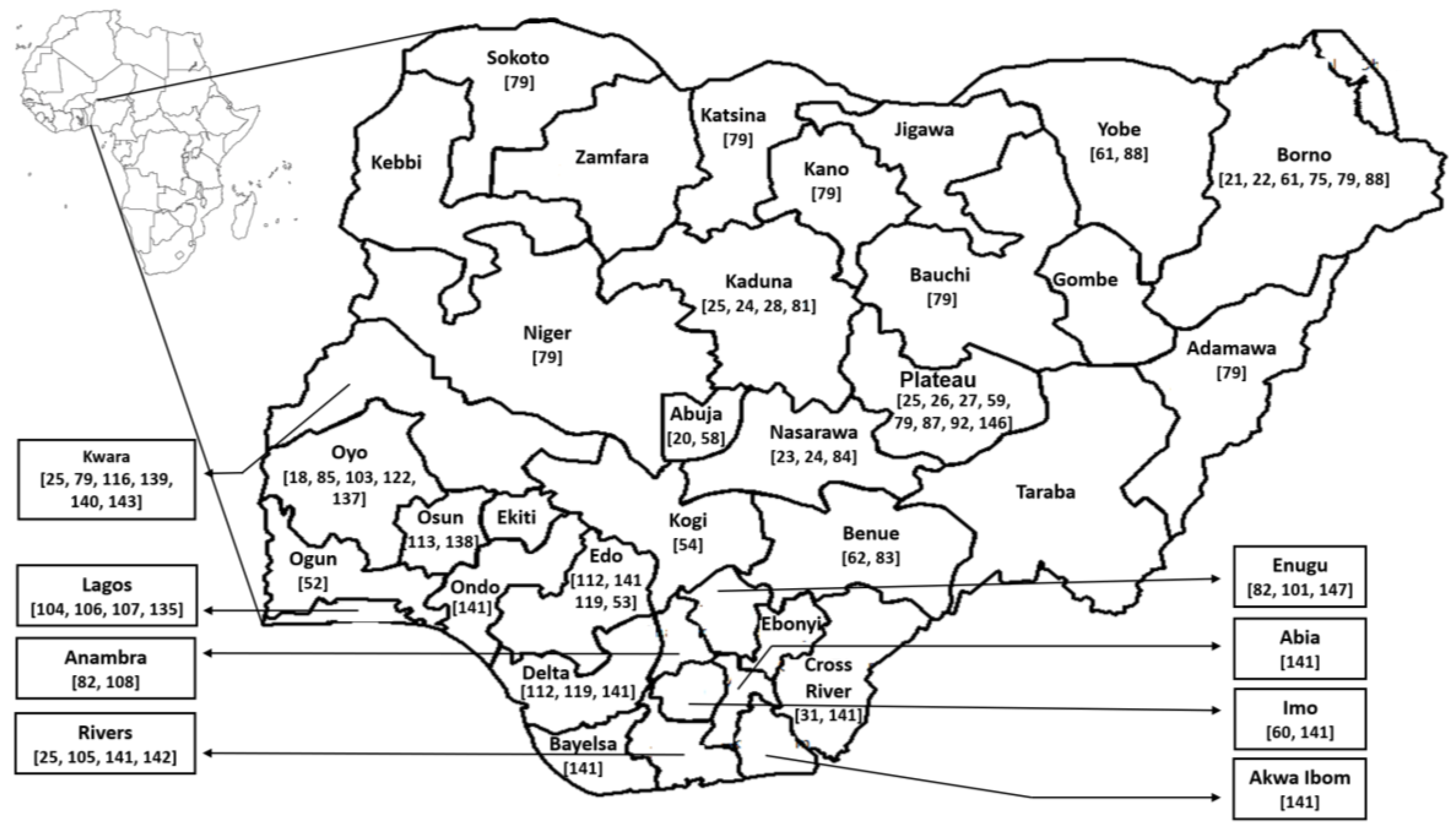

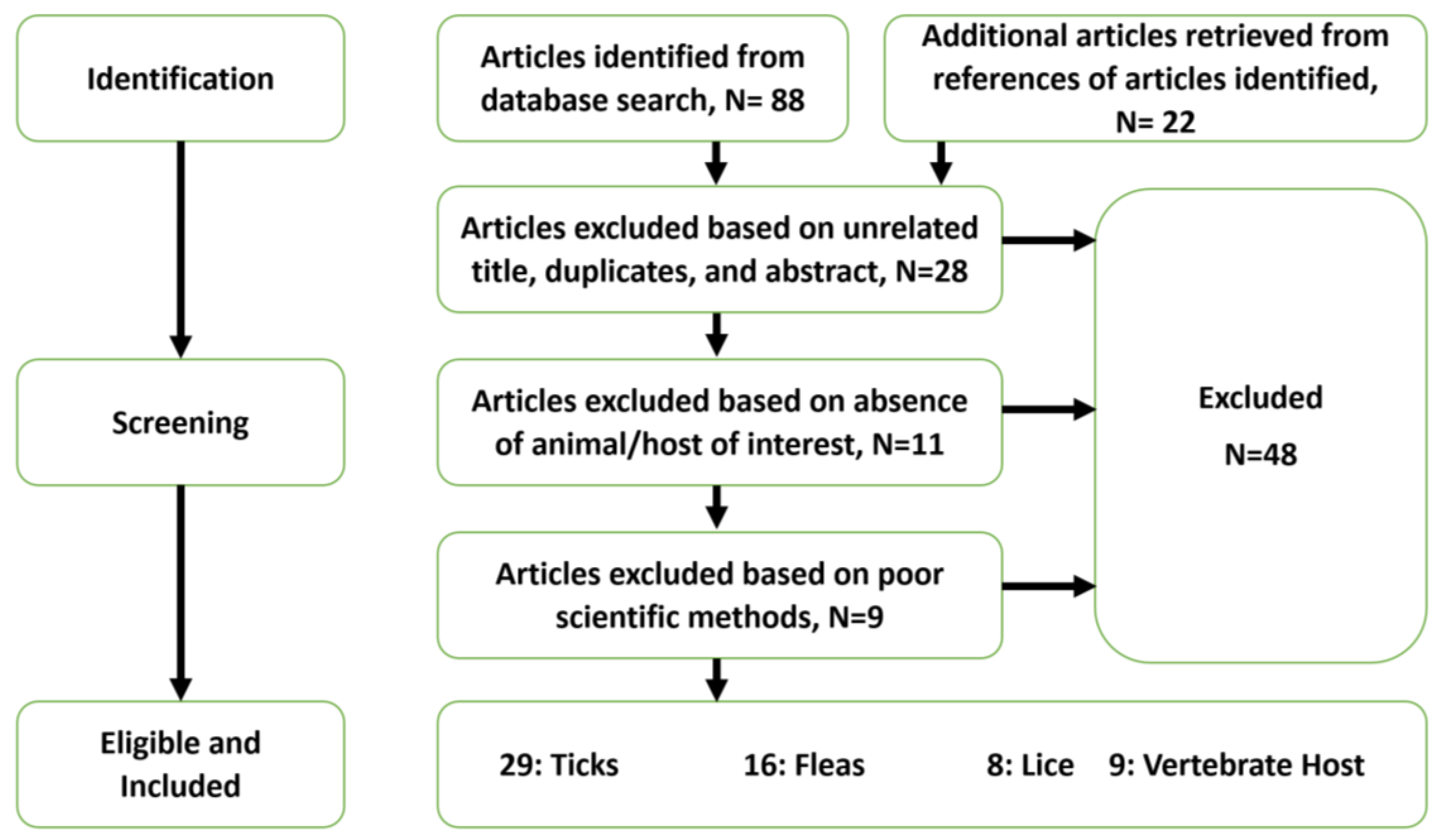
Table 1.
Summary of ticks and tick-borne pathogens in Nigeria.
| Tick Species Collected | Vertebrate Host (Number) | Location | Infestation a (%) | Pathogen (%) b | Detection Method | Study [Reference] | |
|---|---|---|---|---|---|---|---|
| Host Blood | Tick | ||||||
| Hyalomma impeltatum | Camels (170) | Kano | 59.9 | Rickettsia | Rickettsia | PCR | Kamani et al. 2015 [29] |
| - | - | - | - | aeschlimannii (18.8) | aeschlimannii (4.2–50.0) c | - | - |
| Hyalomma rufipes | - | - | 20.3 | - | Rickettsia | PCR | - |
| - | - | - | - | - | aeschlimannii (56.3–94.1) c | - | - |
| Hyalomma dromedarii | - | - | 18.8 | - | Rickettsia | PCR | - |
| - | - | - | - | - | aeschlimannii (13.3) c | - | - |
| Hyalomma impressum | - | - | 1 | - | ND | PCR | - |
| Rhipicephalus sanguineus | Dogs (181) | Plateau | NR | Hepatozoon canis (41.4) | Ehrlichia canis (23.7) | PCR | Kamani et al. 2013 [25] |
| Haemaphysalis leachi | - | Rivers | NR | Ehrlichia canis (12.7) | Hepatozoon canis (21.1) | PCR | - |
| Rhipicephalus turanicus | - | Kaduna | NR | Rickettsia spp. (8.8) | Rickettsia spp. (10.5) | PCR | - |
| - | - | Kwara | - | Babesia rossi (6.6) | Candidatus Neoehrlichia | - | - |
| - | - | - | - | - | mikurensis (5.3) | - | - |
| - | - | - | - | Anaplasma platys (6.6) | Anaplasma platys (1.9) | - | - |
| - | - | - | - | Rickettsia conorii | Rickettsia conorii | - | - |
| - | - | - | - | israelensis (NR) | israelensis (NR) d | - | - |
| Rhipicephalus sanguineus | Dogs (100) | Plateau | 73 | Babesia rossi (53) e | NT | PCR & RLB | Adamu et al. 2014 [28] |
| Haemaphysalis leachi | - | - | 18 | Theileria spp. (12.5) e | NT | - | - |
| Rhipicephalus turanicus | - | - | 2 | Ehrlichia canis (6.9) e | NT | - | - |
| - | - | - | - | Anaplasma spp. (6.9) e | - | - | - |
| - | - | - | - | Theileria equi (4.2) e | - | - | - |
| Amblyomma variegatum | Dogs (NR) | Plateau | 70.2 | NT | Babesia spp.(3.9) f | PCR | Ogo et al. 2012 [24] |
| Rhipicephalus | Cattle (NR) | Nassarawa | 20.6 | NT | Babesia bigemina (1.3) f | PCR | - |
| (Boophilus) decoloratus | - | - | - | - | - | - | - |
| Rhipicephalus sanguineus | - | - | 9.2 | - | Babesia divergens (0.6) f | PCR | - |
| - | - | - | - | - | Anaplasma marginale (20.0) g | - | - |
| - | - | - | - | - | Rickettsia africae (4.4–7.8) h | - | - |
| Rhipicephalus sanguineus | Rattus rattus (48) | Plateau | 46.5 | Bartonella spp. (18.8) | Bartonella spp. (10.0) i | PCR & Blood culture j | Kamani et al. 2013 [26] |
| Haemaphysalis leachi | R. norvegicus (121) | - | 3.5 | Bartonella spp. (29.8) | ND | PCR & Blood culture j | - |
| - | Mus musculus (6) | - | - | - | ND | PCR & Blood culture j | - |
| - | Cricetomys gambianus (2) | - | - | - | Bartonella spp. (50.0) | PCR & Blood culture j | - |
| Hyalomma rufipes | Cattle (NR) | Nigeria | 34 k | NT | Babesia kinetes (88) | Haemolymph | Dipeolu et al. 1984 [19] |
| - | - | - | - | - | - | smear stain | - |
| Hyalomma truncatum | - | - | 32 k | NT | Babesia kinetes (56) | Haemolymph | - |
| - | - | - | - | - | - | smear stain | - |
| Hyalomma impressum | - | - | 19 k | NT | Babesia kinetes (36) | Haemolymph | - |
| - | - | - | - | - | - | smear stain | - |
| Hyalomma marginatum | - | - | 18 k | NT | Babesia kinetes (72) | Haemolymph | - |
| - | - | - | - | - | - | smear stain | - |
| Hyalomma impeltatum | - | - | 7 k | NT | Babesia kinetes (43) | Haemolymph | - |
| - | - | - | - | - | - | smear stain | - |
| Amblyomma variegatum | Cattle (NR) | Plateau | 141 k | NT | Rickettsia africae (62) | PCR | Lorusso et al. 2013 [41] |
| Rhipicephalus | Cattle (120) | Nigeria | NR | Babesia bigemina (80) | NT | Blood smear stain | Akinboade et al. 1985 [17] |
| (Boophilus) spp. | - | - | - | - | - | - | - |
| Rhipicephalus | Dogs (400) | Borno | 88 | Babesia canis (12.0) | NT | Blood smear stain | Konto et al. 2014 [21] |
| (Boophilus) spp. | - | - | - | - | - | - | - |
| Rhipicephalus sanguineus | - | - | 10.8 | - | NT | - | - |
| Hyalomma spp. | - | - | 0.9 | - | NT | - | - |
| Amblyomma variegatum | - | - | 0.3 | - | NT | - | - |
| Rhipicephalus | Cattle (205) | Borno | 63.4 l | NT | NT | N/A | Musa et al. 2014 [42] |
| (Boophilus) microplus | - | - | - | - | - | - | - |
| Amblyomma variegatum | - | - | - | - | NT | - | - |
| Hyalomma spp. | - | - | - | - | NT | - | - |
| Rhipicephalus sanguineus | - | - | - | - | NT | - | - |
| Ornithodorus spp. | - | - | - | - | NT | - | - |
| Amblyomma variegatum | Arvincanthis | Delta | 7.4 | NT | NT | N/A | Ugbomoiko et al. 1991 [43] |
| - | niloticus (NR) | - | - | - | - | - | - |
| Ixodes sp. | Lophuromys | Edo | 5.1 | NT | NT | - | - |
| - | sikapusi (NR) | - | - | - | - | - | - |
| - | Mastomys | - | - | NT | - | - | - |
| - | natalensis (NR) m | - | - | - | - | - | - |
| - | Mus minutoides (NR) | - | - | NT | - | - | - |
| - | Leminiscomys | - | - | NT | - | - | - |
| - | striatus (NR) m | - | - | - | - | - | - |
| Rhipicephalus | Cattle (228) | Plateau | 41.4 | NT | NT | N/A | Lorusso et al. 2013 [27] |
| (Boophilus) decoloratus | - | - | - | - | - | - | - |
| Rhipicephalus | - | - | 15.4 | - | NT | - | - |
| (Boophilus) annulatus | - | - | - | - | - | - | - |
| Rhipicephalus guilhoni | - | - | 12 | - | NT | - | |
| Rhipicephalus | - | - | 7.6 | - | NT | - | - |
| (Boophilus) geigyi | - | - | - | - | - | - | - |
| Hyalomma truncatum | - | - | 7.4 | - | NT | - | - |
| Amblyomma variegatum | - | 6.3 | - | NT | - | - | |
| Rhipicephalus | - | - | 4.1 | - | NT | - | |
| (Boophilus) spp. | - | - | - | - | - | - | - |
| Rhipicephalus | - | - | 4 | - | NT | - | - |
| simus group | - | - | - | - | - | - | - |
| Rhipicephalus turanicus | - | - | 1.2 | NT | - | - | |
| Rhipicephalus sanguineus | - | - | 0.3 | - | NT | - | - |
| Hyalomma rufipes | - | - | 0.2 | - | NT | - | - |
| Rhipicephalus lunulatus | - | - | <0.1 | - | NT | - | - |
| Rhipicephalus spp. | Dogs (44) | Benue | 34.1 | NT | NT | N/A | Omudu et al. 2007 [44] |
| Rhipicephalus | - | - | 29.5 | NT | NT | - | - |
| (Boophilus) spp. | - | - | - | - | - | - | - |
| Amblyomma spp. | - | - | 22.7 | NT | NT | - | - |
| Amblyomma spp. | Goats (45) | Benue | 20.1 | NT | NT | N/A | Omudu et al. 2007 [44] |
| Rhipicephalus spp. | - | - | 13.3 | NT | NT | - | - |
| Rhipicephalus | - | - | 11.1 | NT | NT | - | - |
| (Boophilus) spp. | - | - | - | - | - | - | - |
| Hyalomma spp. | - | - | 8.9 | NT | NT | - | - |
| Amblyomma spp. | Cattle (43) | Benue | 25.6 | NT | NT | N/A | Omudu et al. 2007 [44] |
| Hyalomma spp. | - | - | 18.6 | NT | NT | - | - |
| Rhipicephalus | - | - | 16.3 | NT | NT | - | - |
| (Boophilus) spp. | - | - | - | - | - | - | - |
| Rhipicephalus spp. | - | - | 9.3 | NT | NT | - | - |
| Amblyomma spp. | Pigs (44) | Benue | 20.5 | NT | NT | N/A | Omudu et al. 2007 [44] |
| Hyalomma spp. | - | - | 20.5 | NT | NT | - | - |
| Rhipicephalus | - | - | 15.9 | NT | NT | - | - |
| (Boophilus) spp. | - | - | - | - | - | - | - |
| Amblyomma spp. | Sheep (45) | Benue | 13.3 | NT | NT | N/A | Omudu et al. 2007 [44] |
| Rhipicephalus | - | - | 4.4 | NT | NT | - | - |
| (Boophilus) spp. | - | - | - | - | - | - | - |
| Hyalomma spp. | - | - | 8.9 | NT | NT | - | - |
| Rhipicephalus sanguineus | Dogs (200) | Kogi | 80 | NT | NT | N/A | Abah et al. 2013 [45] |
| Amblyomma variegatum | Cattle (120) | Kaduna | 22.5 | NT | NT | N/A | Obadiah et al. 2012 [46] |
| Rhipicephalus | - | - | 17.5 | - | NT | - | - |
| (Boophilus) decoloratus | - | - | - | - | - | - | - |
| Hyalomma sp. | - | - | 6.7 | - | NT | - | - |
| Rhipicephalus sanguineus | - | - | 3.3 | - | NT | - | - |
| Rhipicephalus sanguineus | Dogs (202) | Ogun | 89.6 | NT | NT | N/A | Agbolade et al. 2008 [47] |
| Haemaphysalis leachi | - | 78.7 | - | NT | - | - | |
| Hyalomma rufipes | Camels (1600) | Borno | 34.9 | NT | NT | N/A | James-Rugu et al. 2004 [48] |
| Hyalomma dromedarii | - | Yobe | 30.4 | - | NT | - | - |
| Hyalomma truncatum | - | - | 10.6 | - | NT | - | - |
| Rhipicephalus | - | - | 24.2 | - | NT | - | - |
| (Boophilus) decoloratus | - | - | - | - | - | - | - |
| Amblyomma variegatum | Dogs (230) | Borno | 23.5 | NT | NT | N/A | James-Rugu et al. 2004 [48] |
| Rhipicephalus spp. | - | Yobe | 21.2 | - | NT | - | - |
| Rhipicephalus (Boophilus) | - | - | 40.8 | - | NT | - | - |
| decoloratus | - | - | - | - | - | - | - |
| Amblyomma lepidum | - | - | 7.1 | - | NT | - | - |
| Haemaphysalis leachi | - | - | 6.9 | - | NT | - | - |
| Amblyomma variegatum | Cattle (2200) | Borno | 11.4 | NT | NT | N/A | James-Rugu et al. 2004 [48] |
| Amblyomma lepidum | - | Yobe | 1.97 | - | NT | - | - |
| Hyalomma truncatum | - | - | 17.9 | - | NT | - | - |
| Hyalomma rufipes | - | - | 11.5 | - | NT | - | - |
| Haemaphysalis leachi | - | - | 8.8 | - | NT | - | - |
| Rhipicephalus evertsi | - | - | 17.8 | - | NT | - | - |
| Rhipicephalus (Boophilus) | - | - | 21.8 | - | NT | - | - |
| decoloratus | - | - | - | - | - | - | - |
| Rhipicephalus sanguineus | - | - | 8.9 | - | NT | - | - |
| Amblyomma variegatum | Sheep (500) | Borno | 50.0 | NT | NT | N/A | James-Rugu et al. 2004 [48] |
| Rhipicephalus (Boophilus) | - | Yobe | 50.0 | - | NT | - | - |
| decoloratus | - | - | - | - | - | - | - |
| Amblyomma variegatum | Cattle (3150) | Borno | 43.8 | NT | NT | N/A | Opara et al. 2016 [49] |
| Hyalomma sp. | - | Yobe | 24.7 | - | NT | - | - |
| Rhipicephalus (Boophilus) | - | - | 21.9 | - | NT | - | - |
| microplus | - | - | - | - | - | - | - |
| Dermacentor variabilis | - | - | 9.6 | - | NT | - | - |
| Amblyomma variegatum | Cattle (1200) | Nasarawa | 20.8 | NT | NT | N/A | Tongjura et al. 2012 [50] |
| Amblyomma lepidum | - | - | 14.3 | - | NT | - | - |
| Rhipicephalus (Boophilus) | - | - | 14.3 | - | NT | - | - |
| decoloratus | - | - | - | - | - | - | - |
| Rhipicephalus (Boophilus) | - | - | 12.8 | - | NT | - | - |
| annulatus | - | - | - | - | - | - | - |
| Hyalomma truncatum | - | - | 6.8 | - | NT | - | - |
| Amblyomma variegatum | Sheep (1200) | Nasarawa | 18.2 | NT | NT | N/A | Tongjura et al. 2012 [50] |
| Amblyomma lepidum | - | - | 8.6 | - | NT | - | - |
| Hyalomma truncatum | - | - | 5.6 | - | NT | - | - |
| Rhipicephalus (Boophilus) | - | - | 10.0 | - | NT | - | - |
| decoloratus | - | - | - | - | - | - | - |
| Rhipicephalus (Boophilus) | - | - | 7.6 | - | NT | - | - |
| annulatus | - | - | - | - | - | - | - |
| Amblyomma variegatum | Goats (1200) | Nasarawa | 15.5 | NT | NT | N/A | Tongjura et al. 2012 [50] |
| Rhipicephalus (Boophilus) | - | - | 7.1 | - | NT | - | - |
| decoloratus | - | - | - | - | - | - | - |
| Amblyomma lepidum | - | - | 6.6 | - | NT | - | - |
| Rhipicephalus (Boophilus) | - | - | 6.4 | - | NT | - | - |
| annulatus | - | - | - | - | - | - | - |
| Hyalomma truncatum | - | - | 3.7 | - | NT | - | - |
| Rhipicephalus evertsi | Cattle (317) | Oyo | 37.6 | NT | NT | N/A | Ameen et al. 2014 [51] |
| evertsi | - | - | - | - | - | - | - |
| Rhipicephalus (Boophilus) | - | - | 22.1 | - | NT | - | - |
| decoloratus | - | - | - | - | - | - | - |
| Rhipicephalus (Boophilus) | - | - | 17.8 | - | NT | - | - |
| annulatus | - | - | - | - | - | - | - |
| Amblyomma variegatum | - | - | 15.8 | - | NT | - | - |
| Rhipicephalus | - | - | 10.7 | - | NT | - | - |
| appendiculatus | - | - | - | - | - | - | - |
| Haemaphysalis leachi | - | - | 5.7 | - | NT | - | - |
| Rhipicephalus evertsi | Goats (210) | Oyo | 33.3 | NT | NT | N/A | Ameen et al. 2014 [51] |
| evertsi | - | - | - | - | - | - | - |
| Rhipicephalus (Boophilus) | - | - | 24.8 | - | NT | - | - |
| decoloratus | - | - | - | - | - | - | - |
| Rhipicephalus (Boophilus) | - | - | 18.1 | - | NT | - | - |
| annulatus | - | - | - | - | - | - | - |
| Amblyomma variegatum | - | - | 11.9 | - | NT | - | - |
| Rhipicephalus | - | - | 6.2 | - | NT | - | - |
| appendiculatus | - | - | - | - | - | - | - |
| Haemaphysalis leachi | - | - | 2.9 | - | NT | - | - |
| Rhipicephalus evertsi | Sheep (104) | Oyo | 28.8 | NT | NT | N/A | Ameen et al. 2014 [51] |
| evertsi | - | - | - | - | - | - | - |
| Rhipicephalus (Boophilus) | - | - | 18.3 | - | NT | - | - |
| decoloratus | - | - | - | - | - | - | - |
| Rhipicephalus (Boophilus) | - | - | 11.5 | - | NT | - | - |
| annulatus | - | - | - | - | - | - | - |
| Amblyomma variegatum | - | - | 9.6 | - | NT | - | - |
| Rhipicephalus | - | 8.7 | - | NT | - | - | |
| appendiculatus | - | - | - | - | - | - | - |
| Haemaphysalis leachi | - | - | 4.8 | - | NT | - | - |
| Rhipicephalus sanguineus | Dogs (820) | Edo | 19.5 | NT | NT | N/A | Ugochukwu et al. 1985 [52] |
| - | - | Delta | - | - | - | - | - |
| Rhipicephalus sanguineus | Dogs (396) | Kwara | 19.2 | NT | NT | N/A | Ugbomoiko et al. 2008 [53] |
| Ixodes sp. | - | - | 4.5 | - | NT | - | - |
| Rhipicephalus sanguineus | Dogs (130) | Benue | 80.5 | NT | NT | N/A | Amuta et al. 2010 [54] |
| Rhipicephalus (Boophilus) | - | - | 14.6 | - | NT | - | - |
| annulatus | - | - | - | - | - | - | - |
| Hyalomma truncatum | - | - | 4.9 | - | NT | - | - |
| Rhipicephalus sanguineus | Dogs (157) | Edo | 53.6 | NT | NT | N/A | Isaac et al. 2016 [55] |
| Rhipicephalus pulchellus | - | - | 42.3 | - | NT | - | - |
| Rhipicephalus | - | - | 7.4 | - | NT | - | - |
| (Boophilus) decoloratus | - | - | - | - | - | - | - |
| Hyalomma truncatum | Cattle (450) | Anambra | 34.8 | NT | NT | N/A | Ikpeze et al. 2011 [56] |
| Rhipicephalus | - | Enugu | 23.1 | - | NT | - | - |
| appendiculatus | - | - | - | - | - | - | - |
| Rhipicephalus | - | - | 22.1 | - | NT | - | - |
| (Boophilus) annulatus | - | - | - | - | - | - | - |
| Amblyomma variegatum | - | - | 20.1 | - | NT | - | - |
| Rhipicephalus sanguineus | Humans (11) | Imo | NR | NT | NT | N/A | Okoli et al. 2006 [57] |
| - | - | Dogs (2) | - | - | NT | - | - |
| - | - | Sheep (3) | - | - | NT | - | - |
| Rhipicephalus sanguineus | Dogs (150) | Cross River | NR | NT | NT | N/A | Arong et al. 2013 [58] |
| Haemaphysalis leachi | - | - | - | - | NT | - | - |
| Rhipicephalus | - | - | - | - | NT | - | - |
| (Boophilus) decoloratus | - | - | - | - | - | - | - |
| Rhipicephalus | Goats (NR) | Enugu | NR | NT | NT | N/A | Ugochukwu et al. 1985 [59] |
| (Boophilus) decoloratus | - | - | - | - | - | - | - |
| Amblyomma variegatum | - | - | NR | - | NT | - | - |
| Amblyomma variegatum | Cattle (NR) | Kano | NR | NT | NT | N/A | Unsworth K. 1952 [60] |
| Amblyomma splendidum | - | Katsina | NR | - | NT | - | - |
| Rhipicephalus | - | Plateau | NR | - | NT | - | - |
| (Boophilus) decoloratus | - | - | - | - | - | - | - |
| Hyalomma spp. | - | Niger | NR | - | NT | - | - |
| Rhipicephalus spp. | - | Sokoto | NR | - | NT | - | - |
| - | - | Adamawa | - | - | - | - | - |
| - | - | Bauchi | - | - | - | - | - |
| - | - | Borno | - | - | - | - | - |
| - | - | Kwara | - | - | - | - | - |
| NR | Cattle (120) | Borno | N/A | Anaplasma spp. (5.8) | N/A | Blood smear stain | Paul et al. 2016 [22] |
| - | - | - | - | Babesia spp. (4.2) | - | - | - |
| NR | Goats (72) | Nassarawa | N/A | Anaplasma spp. (15.3) | N/A | Blood smear stain | Opara et al. 2016 [23] |
| - | - | - | - | Babesia spp. (5.6) | - | - | - |
| NR | Sheep (32) | Nassarawa | N/A | Anaplasma spp. (9.4) | N/A | Blood smear stain | Opara et al. 2016 [23] |
| - | - | - | - | Babesia spp. (3.1) | - | - | - |
| NR | Dogs (101) | Abuja | N/A | Babesia canis (8.9) | N/A | Blood smear stain | Jegede et al. 2014 [20] |
| NR | Dogs (120) | Abuja | N/A | Babesia spp. | N/A | Blood smear stain | Obeta et al. 2009 [61] |
| NR | Dogs (100) | Plateau | N/A | Ehrlichia canis (11) | N/A | PCR | Kamani et al. 2013 [62] |
| NR | Cattle (704) | Plateau | N/A | Theileria mutans (61.8) | N/A | PCR, RLB | Lorusso et al. 2016 [63] |
| - | - | - | - | Theileria velifera (49.4) | - | - | - |
| - | - | - | - | Anaplasma marginale (38.1) | - | - | - |
| - | - | - | - | Theileria taurotragi (36.9) | - | - | - |
| - | - | - | - | Anaplasma sp. (Omatjenne) (33.9) | - | - | - |
| - | - | - | - | Anaplasma centrale (8.1) | - | - | - |
| - | - | - | - | Babesia bigemina (8.1) | - | - | - |
| - | - | - | - | Rickettsia spp. (2.7) | - | - | - |
| - | - | - | - | Babesia bovis (2.3) | - | - | - |
| - | - | - | - | Erhlichia ruminantium (1.1) | - | - | - |
| - | - | - | - | Anaplasma platys (3.8) | - | - | - |
| NR | Cattle (100) | Oyo | N/A | Babesia bigemina (9, 93.0) n | N/A | Blood smear stain | Akinboade et al. 1984 [18] |
| - | - | - | - | - | - | IFA | - |
| - | - | - | - | Anaplasma marginale (8.9, 68.0) n | - | Blood smear stain | - |
| - | - | - | - | - | - | IFA | - |
| - | - | - | - | Babesia bovis (3.3, 54.5) n | - | Blood smear stain | - |
| - | - | - | - | - | - | IFA | - |
| - | - | - | - | Anaplasma centrale (0.75) | - | Blood smear stain | - |
| - | - | - | - | Eperythrozoon (0.75) | - | Blood smear stain | - |
| - | - | - | - | Theleria spp. (0.75) | - | Blood smear stain | - |
| NR | Cattle (500) | Nigeria | N/A | Anaplasma marginale | N/A | IFA, CT, CA | Ajayi et al. 1986 [64] |
| - | - | - | - | (79.4, 40.0, 25.0) | - | - | - |
| - | - | - | - | Babesia bigemina (29.4) | - | IFA | - |
| - | - | - | - | Babesia bovis (14.1) | - | IFA | - |
NT: Not tested, NR: Not recorded, N/A: Not applicable, ND: Not detected, IFA: Indirect fluorescent antibody test, CT: Card agglutination, CA: Capillary tube-agglutination, PCR: Polymerase chain reaction, RLB: Reverse line blotting. a Level of infestation (%) is compared to the total pool of ticks or vertebrate host examined in the study otherwise where stated. b Level of infection (%) is compared to the total ticks or hosts tested in the study otherwise where stated. c Prevalence of SFG Rickettsia detected in pools of ticks using different set of tick primers and PCR methods. d Pathogen was detected only in one pool of Rh. sanguineus collected. e Prevalence is based on the number of dogs positive for at least one pathogen. f Pathogens were detected only in pools of A. variegatum. g Pathogens were detected only in pools of Rh. decoloratus. h R. africae was detected in all pools of ticks with varying prevalence. i Prevalence is based on pools of Rh. sanguineus tested. j Bartonella sp. was cultured from only 30 rodent blood samples. k Number of each species of tick detached from cattle. l Prevalence of infestation was for all tick pools collected in the study. m Ixodes sp. was not collected from the hosts. n Prevalence of B. bigemina, A. marginale, and B. bovis from blood smear stain and IFA respectively. o Prevalence of A. marginale from IFA, CT, and CA respectively.
Table 2.
Summary of fleas and their associated pathogens in Nigeria.
| Species Collected | Vertebrate Host (Number) | Location | Infestation (%) | Pathogen (%) | Detection Method | Study [Reference] | |
|---|---|---|---|---|---|---|---|
| Host Blood | Fleas | ||||||
| Xenopsylla cheopsis | Rattus rattus (48) | Plateau | NR | Bartonella spp. (18.8) | Bartonella spp. (50) a | PCR, Blood culture b | Kamani et al. 2013 [26] |
| Ctenophthalmus sp. | R. norvegicus (121) | - | NR | Bartonella spp. (29.8) | Bartonella spp. (66.7) c | PCR, Blood culture b | - |
| - | Mus musculus (6) | - | - | ND | - | PCR, Blood culture b | - |
| - | Cricetomys gambianus (2) | - | - | Bartonella spp. (50.0) | - | PCR, Blood culture b | - |
| Ctenocephalides canis | Goats (2) | Oyo | NR | Babesia motasi | NT | NR | Opasina. 1983 [126] |
| Ctenocephalides canis | Dogs (396) | Kwara | 32.1 | NT | NT | N/A | Ugbomoiko et al. 2008 [53] |
| Pulex irritans | - | - | 6.6 | NT | - | - | - |
| Tunga penetrans | - | - | 0.5 | NT | - | - | - |
| Ctenocephalides sp. | Dogs (44) | Benue | 38.6 | NT | NT | N/A | Omudu et al. 2007 [44] |
| Pulex sp. | - | - | 6.8 | NT | NT | - | - |
| Tunga sp. | Pigs (44) | Benue | 77.3 | NT | NT | N/A | Omudu et al. 2007 [44] |
| Tunga sp. | Goats (45) | Benue | 22.2 | NT | NT | N/A | Omudu et al. 2007 [44] |
| Tunga sp. | Sheep (45) | Benue | 26.7 | NT | NT | N/A | Omudu et al. 2007 [44] |
| Tunga penetrans | Humans (5595) | Lagos | 49.5 | NT | NT | N/A | Ejezie G. 1981 [110] |
| Tunga penetrans | Humans (480) | Rivers | 30.4 | NT | NT | N/A | Arene, F. O. I. 1984 [111] |
| Xenopsylla cheopsis | Arvincanthis | Delta | 22.4 | NT | NT | N/A | Ugbomoiko et al. 1991 [43] |
| - | niloticus (NR) | - | - | - | - | - | - |
| Xenopsylla braziliensis | Lophuromys | Edo | 5.1 | NT | NT | - | - |
| - | sikapusi (NR) d | - | - | - | - | - | |
| - | Mastomys | - | - | NT | - | - | - |
| - | natalensis (NR) | - | - | - | - | - | - |
| - | Mus minutoides (NR) d | - | - | NT | - | - | - |
| - | Leminiscomys | - | - | NT | - | - | - |
| - | striatus (NR) d | - | - | - | - | - | - |
| - | Rattus rattus (NR) | - | - | NT | - | - | - |
| Tunga penetrans | Humans (547) | Lagos | 45.2 | NT | NT | N/A | Ugbomoiko et al. 2007 [113] |
| Tunga penetrans | Humans (545) | Lagos | 22.4 | NT | NT | N/A | Ugbomoiko et al. 2017 [127] |
| Ctenocephalides canis | Dogs (202) | Ogun | 13.4 | NT | NT | N/A | Agbolade et al. 2008 [47] |
| Xenopsylla cheopsis | Rattus rattus (50) | Osun | 36 | NT | NT | N/A | Ogunniyi et al. 2014 [117] |
| Ctenocephalides canis | Dogs (338) | Anambra | 26.3 | NT | NT | N/A | Chukwu et al. 1985 [114] |
| Echidnophaga gallinacean | - | - | 2.1 | - | NT | - | - |
| Ctenocephalides canis | Dogs (820) | Edo | NR | NT | NT | N/A | Ugochukwu et al. 1985 [52] |
| - | - | Delta | - | - | - | - | - |
| Ctenocephalides canis | Humans (NR) | Oyo | NR | NT | NT | N/A | Fagbemi et al. 1981 [109] |
| Ctenocephalides felis | Cattle (NR) | NR | NR | NT | NT | N/A | Shaibu et al. 2011 [128] |
NT: Not tested; NR: Not recorded; N/A: Not applicable; PCR: Polymerase chain reaction. a Prevalence of Bartonella spp. in pools of X. cheopsis. b Bartonella sp. was cultured from only 30 rodent blood samples. c Prevalence of Bartonella spp. in pools of Ctenophthalmus sp. d Only X. cheopsis was collected on the vertebrate host.
Table 3.
Summary of lice and their associated pathogens in Nigeria.
| Species Collected | Vertebrate Host (Number) | Location | Infestation (%) | Pathogen (%) | Detection Method | Study [Reference] | |
|---|---|---|---|---|---|---|---|
| Host Blood | Lice | ||||||
| Pediculus humanus corporis | Humans (126) | Plateau | NR | Rickettsia prowazekii (100) a | NT | Weil-Felix | Montgomery et al. 1947 [152] |
| Pediculus humanus capitis | Humans (2333) | Oyo | 4.8 b | NT | NT | N/A | Ogunrinade et al. 1984 [143] |
| Pediculus humanus capitis | Humans (2704) | Osun | 12.7 b | NT | NT | N/A | Jinadu, M. K. 1985 [144] |
| Pediculus humanus capitis | Humans (7360) | Abia, Imo | 5.7 b | NT | NT | N/A | Arene et al. 1985 [147] |
| - | - | Akwa Ibom | - | - | - | - | - |
| - | - | Bayelsa, Delta | - | - | - | - | - |
| - | - | Cross River | - | - | - | - | - |
| - | - | Edo, Ondo | - | - | - | - | - |
| - | - | Rivers | - | - | - | - | - |
| Pediculus humanus capitis | Humans (6882) | Kwara | 3.7 b | NT | NT | N/A | Ebomoyi, E. 1994 [146] |
| Pediculus humanus capitis | Humans (2898) | Kwara | 2.0 b | NT | NT | N/A | Ebomoyi, E. 1988 [145] |
| Pediculus humanus capitis | Humans (1000) | Kwara | 8.8 b | NT | NT | N/A | Okwa et al. 2010 [149] |
| Pediculus humanus capitis | Humans (726) | Rivers | 45.6 b | NT | NT | N/A | Gboeloh et al. 2013 [148] |
| NR | Humans (NR) | Enugu | N/A | Rickettsia prowazekii | N/A | Weil-Felix | Emejuaiwe et al. 1978 [153] |
NT: Not tested, NR: Not recorded, N/A: Not applicable. a Testing was performed on hospitalized patients. b Overall prevalence of infestation in the study.
© 2018 by the authors. Licensee MDPI, Basel, Switzerland. This article is an open access article distributed under the terms and conditions of the Creative Commons Attribution (CC BY) license (http://creativecommons.org/licenses/by/4.0/).
Share and Cite
MDPI and ACS Style
Oguntomole, O.; Nwaeze, U.; Eremeeva, M.E. Tick-, Flea-, and Louse-Borne Diseases of Public Health and Veterinary Significance in Nigeria. Trop. Med. Infect. Dis. 2018, 3, 3. https://doi.org/10.3390/tropicalmed3010003
AMA Style
Oguntomole O, Nwaeze U, Eremeeva ME. Tick-, Flea-, and Louse-Borne Diseases of Public Health and Veterinary Significance in Nigeria. Tropical Medicine and Infectious Disease. 2018; 3(1):3. https://doi.org/10.3390/tropicalmed3010003
Chicago/Turabian StyleOguntomole, Oluwaseun, Ugochukwu Nwaeze, and Marina E. Eremeeva. 2018. "Tick-, Flea-, and Louse-Borne Diseases of Public Health and Veterinary Significance in Nigeria" Tropical Medicine and Infectious Disease 3, no. 1: 3. https://doi.org/10.3390/tropicalmed3010003