Review

New Immuno-Epidemiological Biomarker of Human Exposure to Aedes Vector Bites: From Concept to Applications †

by
André B. Sagna
1,2,‡,
Mabo C. Yobo
2,3,‡,
Emmanuel Elanga Ndille
1,4 and
Franck Remoue
1,*
1
MIVEGEC, IRD, CNRS, Univ. Montpellier, BP 64501, 34394 Montpellier, France
2
Institut Pierre Richet (IPR), Institut Nationale de la Santé Publique (INSP), BP 1500 Bouaké, Côte d’Ivoire
3
UFR Sciences de la Nature, Université Nangui Abrogoua, Abidjan, Côte d’Ivoire, BP 801 Abidjan, Côte d’Ivoire
4
Department of Medical Entomology, Centre for Research in Infectious Diseases (CRID), P.O. Box 13591, Yaoundé, Cameroun
*
Author to whom correspondence should be addressed.
In memory of Mabo C. Yobo, died on Friday, 11 May 2018.
These authors contributed equally to this work.
Trop. Med. Infect. Dis. 2018, 3(3), 80; https://doi.org/10.3390/tropicalmed3030080
Submission received: 2 July 2018 / Revised: 23 July 2018 / Accepted: 30 July 2018 / Published: 1 August 2018
(This article belongs to the Special Issue Epidemiology of Dengue: Past, Present and Future)

Abstract

:
Arthropod-borne viruses (arboviruses) such as dengue virus (DENV), chikungunya virus (CHIKV), Zika virus (ZIKV), and yellow fever virus (YFV) are the most important ‘emerging pathogens’ because of their geographic spread and their increasing impact on vulnerable human populations. To fight against these arboviruses, vector control strategies (VCS) remain one of the most valuable means. However, their implementation and monitoring are labour intensive and difficult to sustain on large scales, especially when transmission and Aedes mosquito densities are low. To increase the efficacy of VCS, current entomological methods should be improved by new complementary tools which measure the risk of arthropod-borne diseases’ transmission. The study of human–Aedes immunological relationships can provide new promising serological tools, namely antibody-based biomarkers, allowing to accurately estimate the human–Aedes contact and consequently, the risk of transmission of arboviruses and the effectiveness of VCS. This review focuses on studies highlighting the concept, techniques, and methods used to develop and validate specific candidate biomarkers of human exposure to Aedes bites. Potential applications of such antibody-based biomarkers of exposure to Aedes vector bites in the field of operational research are also discussed.

1. Introduction

Mosquitoes are vectors of numerous human pathogens that kill or debilitate millions of humans each year. Those belonging to the genus Aedes are vectors of the major viral infections including yellow fever, dengue, chikungunya, and Zika fever [1]. It is estimated that 200,000 new cases of yellow fever occur each year with 30,000 deaths [2], despite the existence of a preventive vaccine. There are an estimated 3.9 billion people at risk of infection with dengue virus (DENV) worldwide [3] and 390 million DENV infections a year, including 500,000 severe forms and over 20,000 deaths [4]. Recent large outbreaks of chikungunya and Zika fever have affected the immunologically naive populations of the Indian and Pacific oceans and of the Americas [5,6,7]. As an example, 693,000 and 500,000 cases of chikungunya and Zika, respectively, were suspected in the Americas in 2016 [8]. All these viruses are transmitted between humans by the bite of infected Aedes mosquitoes, mainly Aedes aegypti and Aedes albopictus [9,10].
The lack and limitations of prophylactic treatment in the vast majority of these diseases forces the use of vector control methods to interrupt transmission. This involves extensive surveillance of these mosquito populations to estimate risk of transmission, to plan vector control strategies, and to assess their impact on the diseases. This surveillance is currently based on entomological methods to identify positive breeding sites, to sample adult mosquitoes by traps, intra-housing spraying, and human landing catches [11]. However, these methods have many limitations in efficiency and accuracy that make them difficult to apply on large scales. Indeed, estimation of immature stage densities is time consuming and expensive with problems of inaccessibility to all potential breeding sites. Furthermore, larval and pupal indicators are focused on immature stages that do not perfectly correlate to the density of adult mosquitoes. The estimation of adult female densities using human landing catches could be more significant for sampling host-seeking mosquitoes and then determining human exposure levels [11,12]. However, due to the infection risk of exposed volunteers to mosquito vector bites, this method evokes obvious ethical concerns that limit its application. Furthermore, entomological methods are mainly applicable at the community level and cannot be used to evaluate the heterogeneity of exposure to mosquito bites between individuals.
Given all these shortcomings, there is an urgent need for more efficient and sensitive complementary tools to improve the impact of vector control strategies (VCS) and predict the risk of transmission of mosquito-borne diseases, and if possible at both the community and individual levels. The development of such tools needs to explore the close interactions between the human host and Aedes mosquitoes. The physical contact between these two protagonists involves physiological interactions through salivary proteins of Aedes mosquitoes. Indeed, upon feeding, mosquitoes inject a mixture of bioactive molecules into the host that assist with blood meal acquisition. These molecules include factors that are anticlotting, vasomodulatory, and antiplatelet. Interestingly, some of arthropod salivary proteins are antigenic and initiate an anti-saliva antibody response in the human host [13,14,15,16]. One approach is to quantify the human antibody response to mosquito salivary proteins to define a biomarker of exposure to mosquito bites [17,18,19,20,21,22,23,24]. Indeed, the human antibody response against mosquito salivary gland proteins varies temporally and spatially, and correlates with mosquito density or host biting activity, suggesting the human antibody response can be used to study mosquito–human contact [25]. In addition, biomarkers of mosquito bite exposure can be used to define the ranges of medically important mosquitoes, identify areas of risk for vector-borne diseases, and evaluate vector control interventions.
Even though this approach may appear to be valid, the use of whole saliva or salivary gland extracts (SGE) of Aedes vectors as a biomarker of exposure could be inadequate because some salivary factors are shared between mosquitoes or arthropods from different genera that induce immune cross-reaction. In the objective to optimise this promising indicator and to use it on a large scale with high reproducibility, the identification of specific and antigenic salivary proteins or peptides, or both, of Aedes has been done. This led to the identification of the putative 34 kDa family secreted salivary protein that appeared to be antigenic and specific to the Aedes genus [26]. Interestingly, one peptide (the Nterm-34 kDa) from Ae. aegypti saliva was recently identified and validated as a pertinent and specific candidate biomarker of human exposure to bites of major Aedes species (Ae. aegypti and Ae. albopictus) in several countries: Benin, Côte d’Ivoire, Lao PDR, and La Reunion Island [27,28,29,30].
This review focuses on studies highlighting the epidemiological approaches, techniques, and methods used to develop and validate specific candidate biomarkers of human exposure to Aedes bites. Potential applications of such antibody-based biomarkers of exposure to Aedes vector bites in the field of operational research for arboviral diseases control are highlighted.

2. Development of Antibody-Based Biomarker of Exposure to Aedes Bites
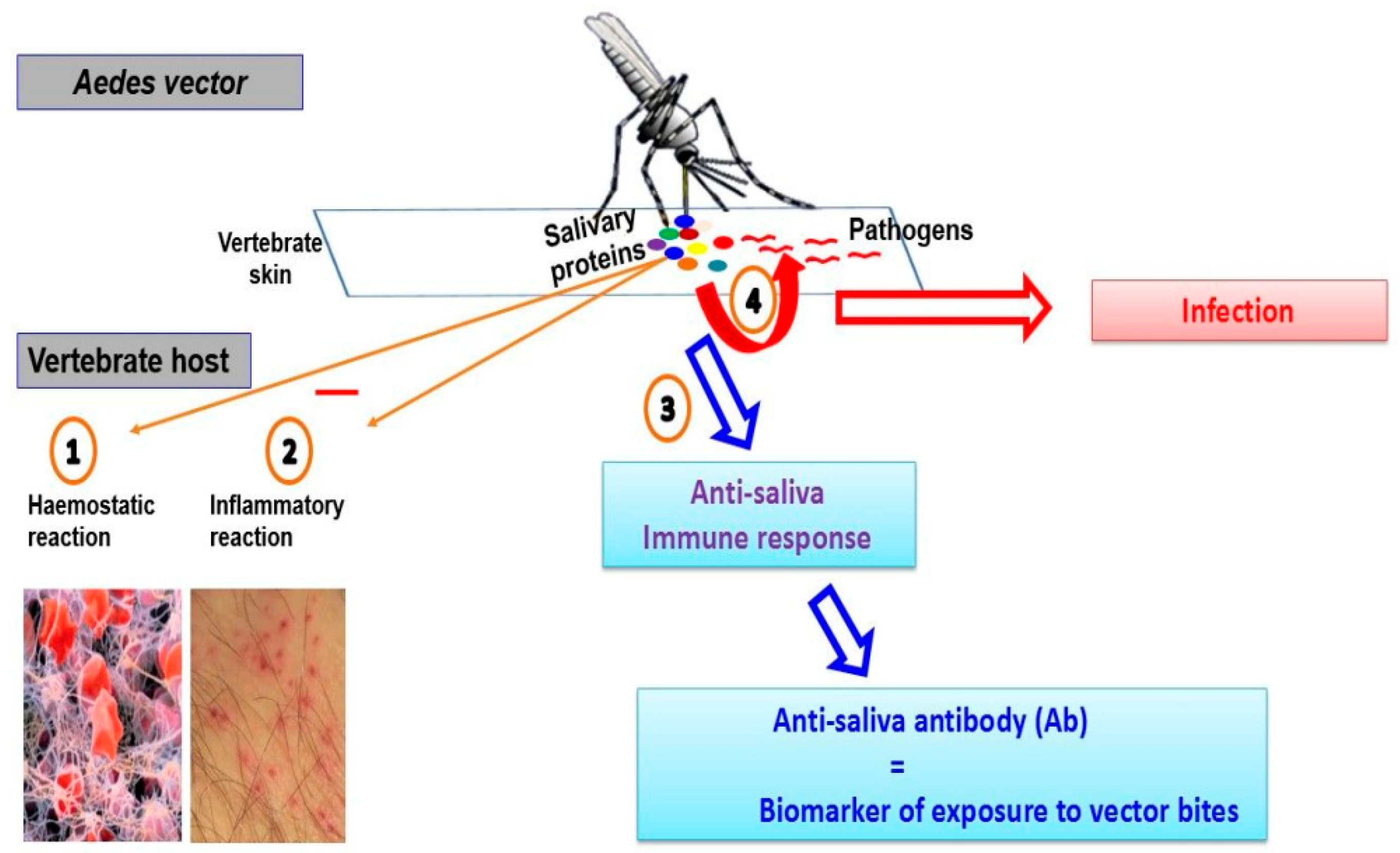
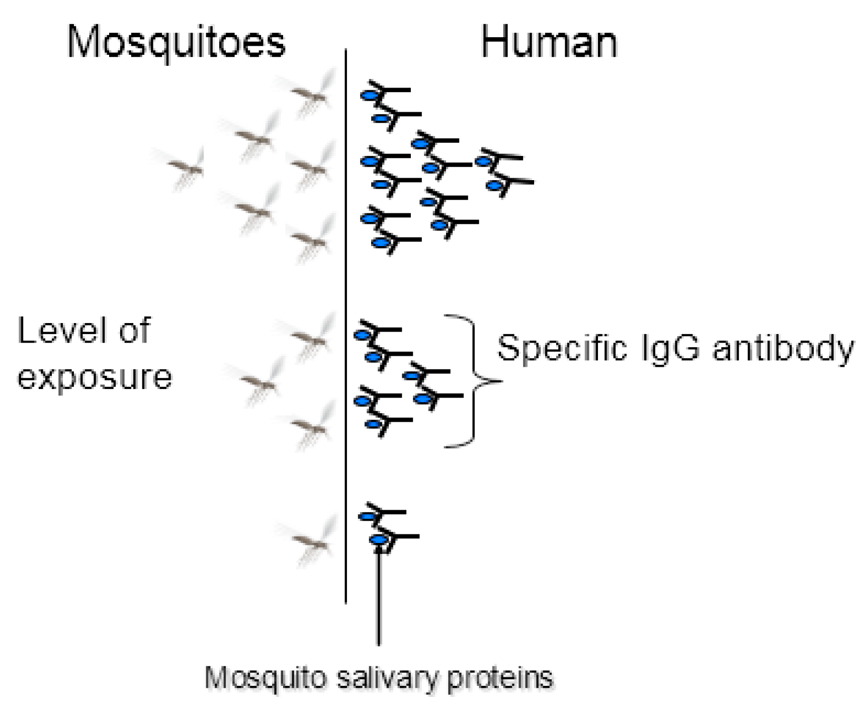
2.1. The Concept

The penetration of haematophagous arthropod mouthparts into the host skin elicits haemostatic, inflammatory, and immune responses by the vertebrate host (animal, human). These reactions aiming to prevent blood loss, promote tissue repair, and prevent colonisation of the damaged tissues by opportunistic pathogens, can largely impair haematophagous arthropod blood intake. Blood-feeding behaviour then requires solutions to counteract vertebrate host defence responses to successfully obtain a blood meal. Thus upon feeding, the female Aedes, like all haematophagous arthropods, injects into the mammal host a mixture of bioactive molecules that assist with blood intake by inhibiting physiological responses of the host (Figure 1) [13,31]. Interestingly, some of them (salivary proteins) are antigenic and initiate an antibody response in the host [13,14].
The first studies on the effects of mosquito salivary proteins on the immune system of the host concerned allergic reactions due to Aedes and Culex mosquito bites. These mosquitoes express in their saliva a panel of allergens responsible for a production of specific immunoglobulin (Ig) E antibodies [18,32]. Some of these salivary allergens are now well characterised. For example, four recombinant Aedes aegypti salivary allergens corresponding to a 68 kDa salivary apyrase (rAed a1), a 37 kDa protein belonging to the D7 family (rAed a2), a 30 kDa salivary gland allergen (rAed a3), and a 67 kDa α-glucosidase (rAed a4) elicit predominantly specific IgE responses in mosquito-allergic individuals [33,34,35,36]. The authors concluded that these recombinant proteins can be used as tools for diagnosis and treatment of allergic reactions due to mosquito bites.
A panel of serological studies have demonstrated a relation between the exposure of various hematophagous arthropod density and the anti-saliva Ab response levels [21,37]. Regarding Aedes mosquitoes, several studies in Finnish Lapland [25], Canada [38], Senegal [17], Bolivia and Reunion Island [23,39], have reported an increasing anti-saliva IgG Ab during the peak of human exposure or in persons living in sites with higher Aedes density. Anti-saliva IgG responses have shown to potentially wane just 15 days after a decrease in Aedes albopictus densities [40]. Other studies performed in domestic animals showed that IgM antibodies against the saliva of triatomine bugs can be detected after one day of exposure and waned rapidly (18 days) [41,42].
Since the acquisition of an Ab response against mosquitoes’ (and hematophagous arthropods’) saliva depended on the level of exposure of an individual to mosquito bites, it seemed logical to develop serological tools for monitoring human–vector contacts. The concept ‘anti-saliva Ab response = biomarker of vector bite exposure’ had just emerged. The idea was that the level of specific Ab of an individual is proportional, as a proxy, to the intensity of mosquito bites received by the individual (Figure 2). For example, it has been shown that an individual slightly exposed to Aedes bites thus produces a low-level Ab response (≤0.70 ΔOD) to Aedes salivary antigens, while a highly-exposed individual to Aedes bites produces a high-level Ab response (≥1.2 ΔOD) to Aedes salivary antigens [40]. The advantage of such a tool is that it is quantitative (values are expressed in optical densities (OD)) and individual (one OD value represents the level of exposure to bites of one individual).
A low-exposed individual to Aedes bites would develop a low-level Ab response to salivary antigens while a highly exposed one would develop a high Ab response to Aedes salivary antigens.

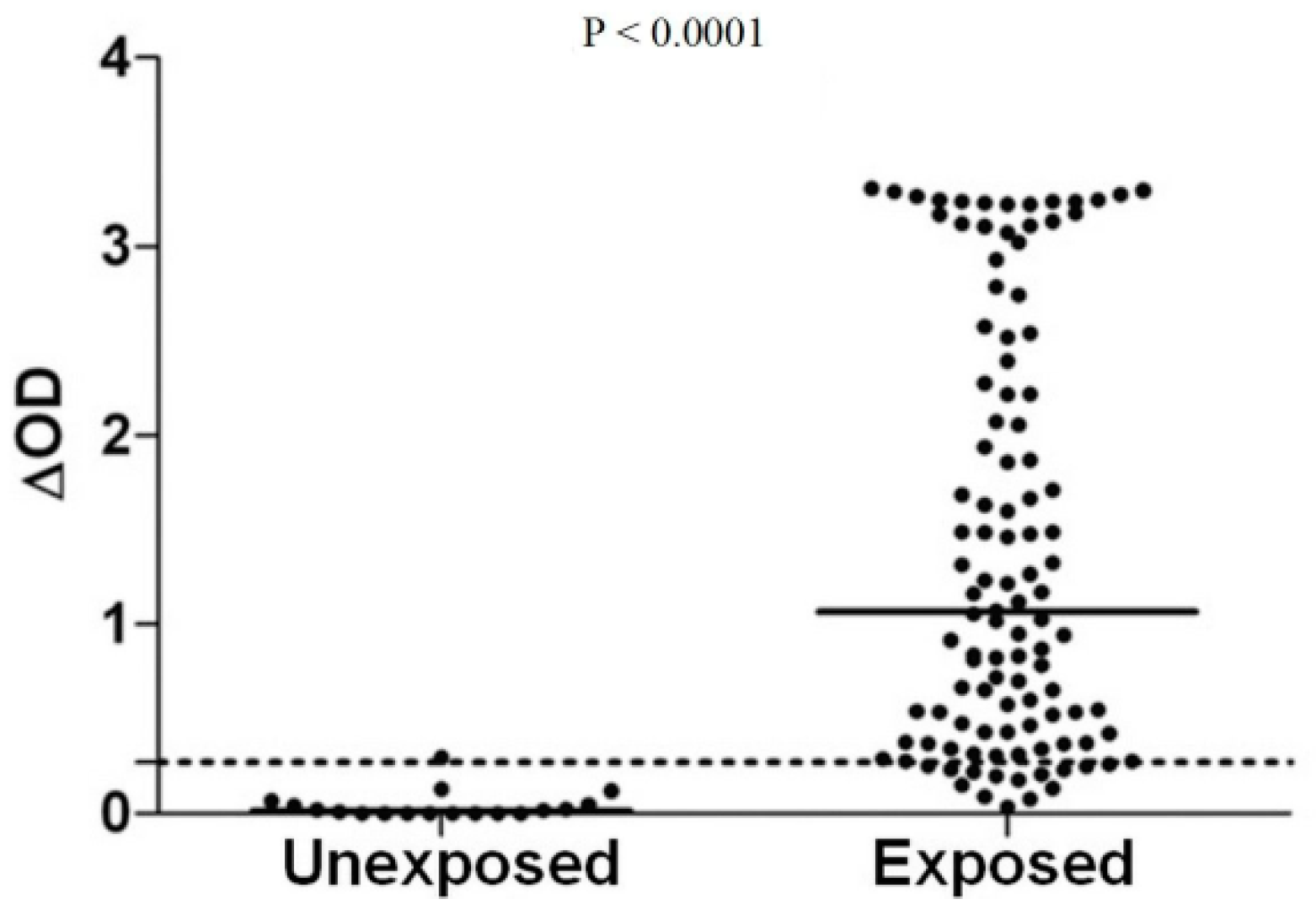
2.2. Validation of the Concept

To validate the proof of concept ‘anti-saliva Ab response = biomarker of vector bite exposure’, it was necessary to find creative methods to reach this goal. But curiously, all studies undertaken for this purpose were directly carried out in the field. Although this approach could seem surprising, it represents a great benefit for testing the validity of the Aedes salivary biomarker as it allows the taking into consideration of specific conditions closely related to the epidemiology of mosquito-borne diseases. To be valid, the Aedes salivary biomarker should fulfil four fundamental criteria: (i) to discriminate exposed individuals to Aedes vector bites from those who are not exposed, (ii) to evaluate the density and fluctuations of vector populations that can bite human populations, (iii) to classify individuals according to their level of exposure to Aedes vectors, and (iv) to be usable at a population level. To demonstrate that the Aedes salivary biomarker met these criteria, Ae. aegypti SGE were then collected and run on ELISA in serum samples from young Senegalese children living in an area of exposure to the Ae. aegypti mosquitoes [20]. Specific IgE and IgG4 responses increased during the rainy season of high exposure to Aedes bites, compared to the dry season. In addition, the evolution of anti-saliva isotype levels during the rainy season presented spatial heterogeneity between studied villages. These preliminary results supported the potential approach of using anti-saliva Ab responses for evaluating exposure to Aedes vectors and the risk of emerging arboviral infections.
From these results, several studies were undertaken to evaluate the relevance of using salivary biomarkers to determine the risk of exposure to Aedes bites, thus to the viruses that they transmit. For example, several Colombian and French travellers were reported to develop specific Ab responses against Ae. aegypti saliva after a stay of 2 to 5 months in areas with Ae. aegypti mosquitoes and their specific Ab responses strongly decreased several weeks after the end of their trip [20,24]. In three sites in south-eastern France, a positive association was observed between the average levels of IgG responses against Ae. caspius SGE and spatial Ae. caspius densities. Additionally, the average level of IgG responses increased significantly during the peak exposure to Ae. caspius at each site and returned to baseline four months later [22]. A rapid decrease of anti-Ae. albopictus SGE IgG levels was also reported just two weeks after the implementation of control measures (elimination of all breeding sites and insecticide space spraying with deltamethrin) in an urban city in Reunion Island [40]. Other studies using Ae. aegypti and Ae. albopictus SGE also demonstrated that exposed individuals to these Aedes mosquito species developed a high anti-SGE IgG Abs compared to unexposed ones (Figure 3). In addition, anti-SGE IgG Abs were shown to vary according to the level of exposure (measured by entomological methods) and age of individuals [23,39].
The relationship between anti-SGE Ab responses and arbovirus infections was also investigated in some studies. Londono-Renteria and colleagues found significantly higher anti-SGE IgG Abs in DENV positive patients with some difference in exposure to Ae. aegypti bites among DENV serotypes (Figure 4) [24]. Similarly, the same authors reported higher Ab responses to Ae. aegypti SGE in people exposed to DENV, with a positive correlation between anti-SGE IgM and IgG to DENV antigens. In addition, a significant higher level of anti-SGE IgG in subjects living in houses with Ae. aegypti aquatic stages was found [43].
Taken together, these results demonstrate that the estimation of human Ab responses to Aedes SGEs could be a valuable indicator or a biomarker for evaluating the level of human exposure to Aedes bites, the risk of arbovirus infection or transmission, and the effectiveness of vector control strategies. However, a biomarker of exposure based on the use of Aedes SGE or whole saliva may not be very reliable for epidemiological purposes. Indeed, saliva is a cocktail of various molecular components with different nature and biological functions, and some of which are shared between species, genus, families, orders, or classes of bloodsucking Diptera or arthropods [13]. Therefore, the evaluation of vector control effectiveness or the risk of arbovirus infection based on the immunogenicity of SGE could be under or overestimated due to possible cross-reactivity between common epitopes. In addition, the collection of saliva or SGEs is tedious and time consuming and saliva batches appeared to be not reproducible in terms of protein quantities and antigenicity. Therefore, it will be difficult or even impossible to have an adequate production of mosquito saliva needed for large scale epidemiological studies. Also, the standardisation of immunological assay using whole saliva appeared difficult and time consuming. An alternative for optimising the specificity of this immunological test would be to identify Aedes genus-specific proteins or peptides [44].

2.3. From the Whole Saliva to Synthetic Salivary Peptide of Aedes

Recent progress in sialotranscriptomic studies have allowed the identification of more specific antigens that have enhanced the specificity of Aedes salivary biomarkers [45,46,47]. Two main approaches have been used for that purpose: (i) the recombinant protein strategy based on the identification of genus-specific salivary proteins and (ii) bioinformatics analysis of the sialotranscriptomic data used to design specific and antigenic peptides.

2.3.1. The Recombinant Protein Approach

Proteomic studies on arthropods’ saliva aim to identify which salivary proteins are antigenic and recognised by the vertebrate host during blood intake. These studies revealed the antigenicity of a broad range of arthropod vector salivary proteins [26,48]. This proteomic approach confirmed the Ab cross-reactivity observed in the field with Aedes SGE. Western blot analyses on Aedes mosquitoes’ saliva revealed common antigens between Aedes species. Individuals exposed to Aedes bites recognised the 36 and 22 kDa salivary proteins which belong to several Aedes species, notably Ae. communis and Ae. aegypti [18]. Similarly, the screening of Ae. albopictus salivary protein using two-dimensional electrophoresis technique combined with immunoblotting and mass spectrometry analysis revealed extensive immune cross-reactivity with Ae. aegypti salivary proteins [49]. However, some salivary proteins that may represent candidate biomarkers of exposure at a genus or species level were identified by proteomic tools and produced in their recombinant form so as to be tested under different scenarios. This process led to the development of recombinant proteins such as rTisP14.6 derived from Triatoma infestans [50], rLJM17 and rLJM11 proteins produced from Lutzomyia longipalpis [51,52], gSG6 from An. gambiae [53,54], and fSG6 derived from An. funestus [55]. For Aedes, one protein, the ‘family secreted salivary putative 34 kDa protein’, was identified and described as specific to the Aedes genus [46]. Immunological assays with the recombinant form of this protein in serum samples of rabbits exposed to Ae. aegypti, An. gambiae, and Cx. quinquefasciatus confirmed its antigenicity and its specificity to Aedes mosquitoes [56]. Obviously, this protein seemed to constitute a relevant candidate biomarker of exposure to Aedes bites. However, the production of recombinant proteins is not an easy task and it is also challenging to produce protein with a high degree of purity and with total reproducibility between production batches to ensure a correct assessment of the anti-saliva Ab response. In addition, recombinant proteins may carry more than one epitope which could increase the risk of immune cross-reactivity, impairing the specificity of candidate biomarkers. To address these limitations linked to the production of recombinant proteins, a peptide approach was developed.

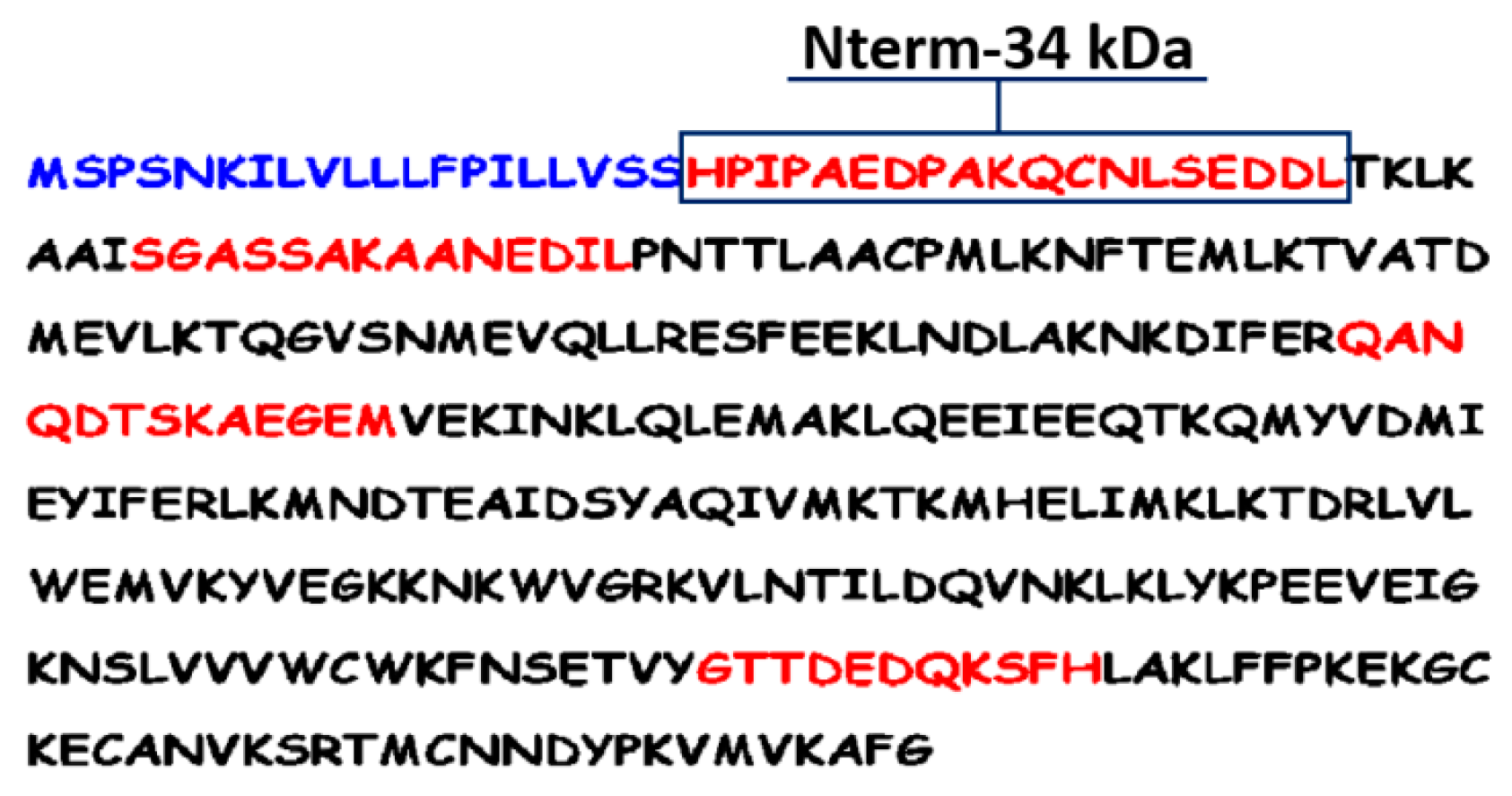
2.3.2. The Peptide Approach

The peptide approach is based on the exploitation of sialotranscriptome databases and an in silico analysis to identify potential epitopes based on physicochemical properties of amino acids. This peptide design approach was previously used for the identification of the famous and well validated gSG6-P1 (gambiae salivary gland protein-peptide 1) salivary peptide, biomarker of human exposure to major malaria vectors [57,58,59,60]. Basically, several algorithms were employed for prediction of potential immunogenic sites of the putative 34 kDa protein by using bioinformatics. The prediction of immunogenicity was based on the determination of physicochemical properties of the amino-acid (AA) sequences with BcePred and FIMM databases and on the identification of major histocompatibility complex class 2 binding regions using the ProPred-2 online service. This led to defining four peptides (highlighted in red) of 11 to 19 AA residues in length (Figure 5).
Similarities and specificity were also searched using the Blast family programs, including both the genome and EST libraries of other vector arthropods available in Vectorbase and of pathogens and organisms in non-redundant GenBank CDS databases. All these sequence alignments predicted that only the N-terminal peptide (Nterm-34 kDa) would be very specific to Ae. aegypti species and potentially antigenic (Figure 5) [27]. This peptide was then selected and chemically synthetised to be validated as a potential candidate biomarker of human exposure to Ae. aegypti mosquito bites.

2.3.3. Validation of the Synthetic Aedes Nterm-34 kDa Salivary Peptide

The evolution of specific IgG levels to the Nterm-34 kDa salivary peptide was evaluated during dry and rainy seasons in a cohort of 420 under five-year-old children and compared to the accumulated monthly rainfall recorded in the same studied area [27]. A positive association was found between specific IgG Ab responses against the Nterm-34 kDa and the rainfall. Indeed, IgG Ab responses to the Nterm-34 kDa salivary peptide increased significantly from the dry (low exposure to Aedes) to the rainy season (high exposure to Aedes) (Figure 6). These findings indicate that IgG Ab to Nterm-34 kDa salivary peptide may represent a reliable biomarker to detect the difference, heterogeneity, and evolution in human exposure to Ae. aegypti bites, and thus to evaluate the risk of arbovirus transmission.
This peptide appeared then to satisfy several requirements that an exposure biomarker should fulfil. First, so far, it appears to be specific to Aedes genus and therefore, no relevant cross-reactivity phenomena with epitopes from other proteins of arthropods or pathogens would be expected. Second, its synthetic nature guarantees high reproducibility of the immunological assay. Third, it elicits a specific Ab response which correlates well with the level of exposure to Ae. aegypti bites.
Now having a specific biomarker of exposure to Ae. aegypti bites, the question is what could be the potential applications of such a tool in epidemiological contexts?

3. Potential Applications of Aedes Salivary Biomarker in the Field

In endemic areas for mosquito-transmitted diseases like arboviruses, the estimation of the level of human–mosquito contact is an important indicator to measure the risk of transmission of such diseases. For this purpose, discovering that mosquito salivary proteins and peptides can elicit into the human host a specific Ab response that is correlated to its level of exposure to mosquito bites has stimulated researchers to identify specific biomarkers for mosquito bite exposure [17,23,39,57]. Recently, the human IgG Ab response to Ae. aegypti Nterm-34 kDa salivary peptide has been validated as biomarker of Ae. aegypti and Ae. albopictus bite exposure, the main vectors of arboviruses [27]. The availability of such a tool could help to accurately identify individuals exposed to Aedes vector bites, consequently at risk of arbovirus transmission, and to evaluate the effectiveness of VCS implemented by sanitary authorities on human–Aedes contact.

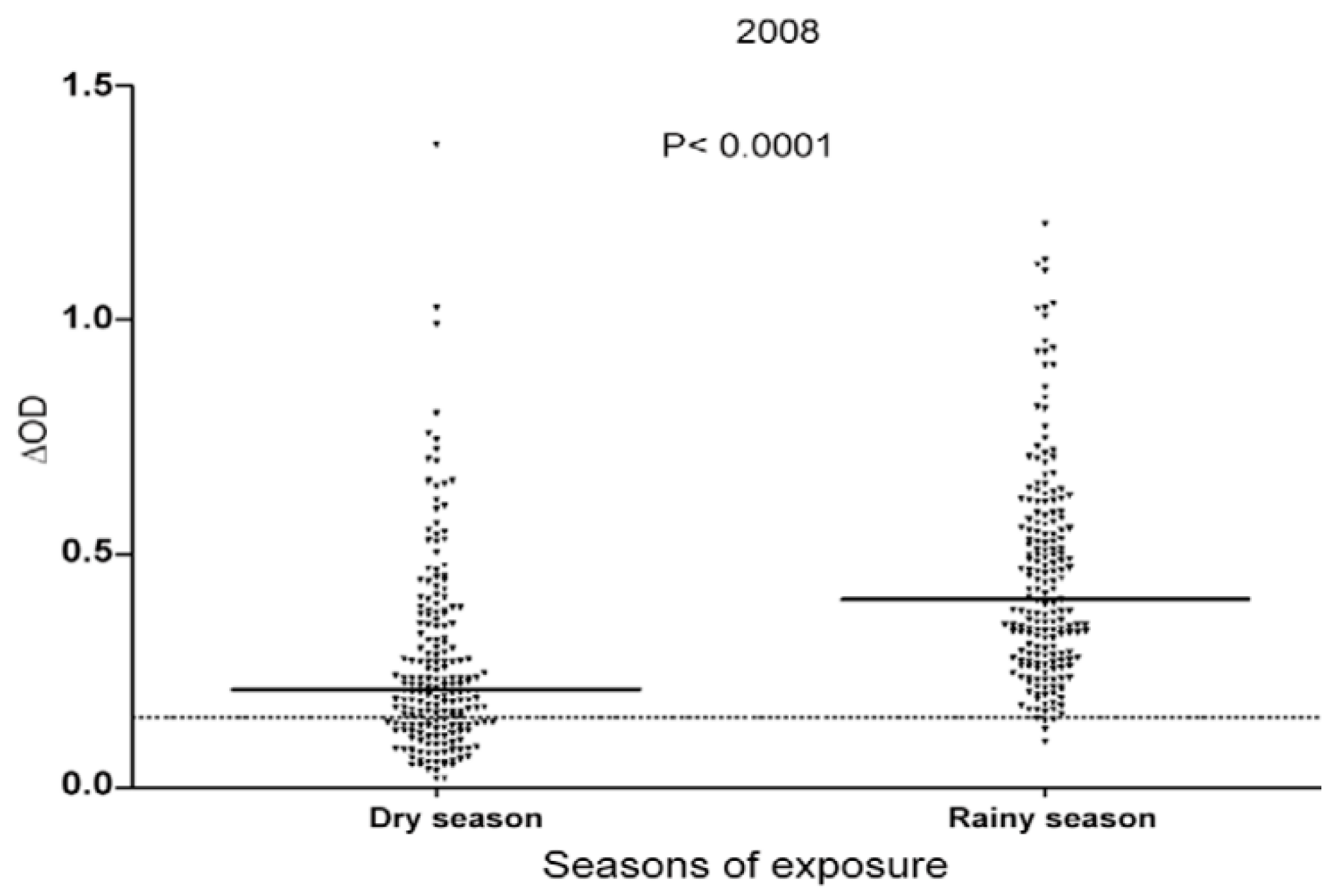
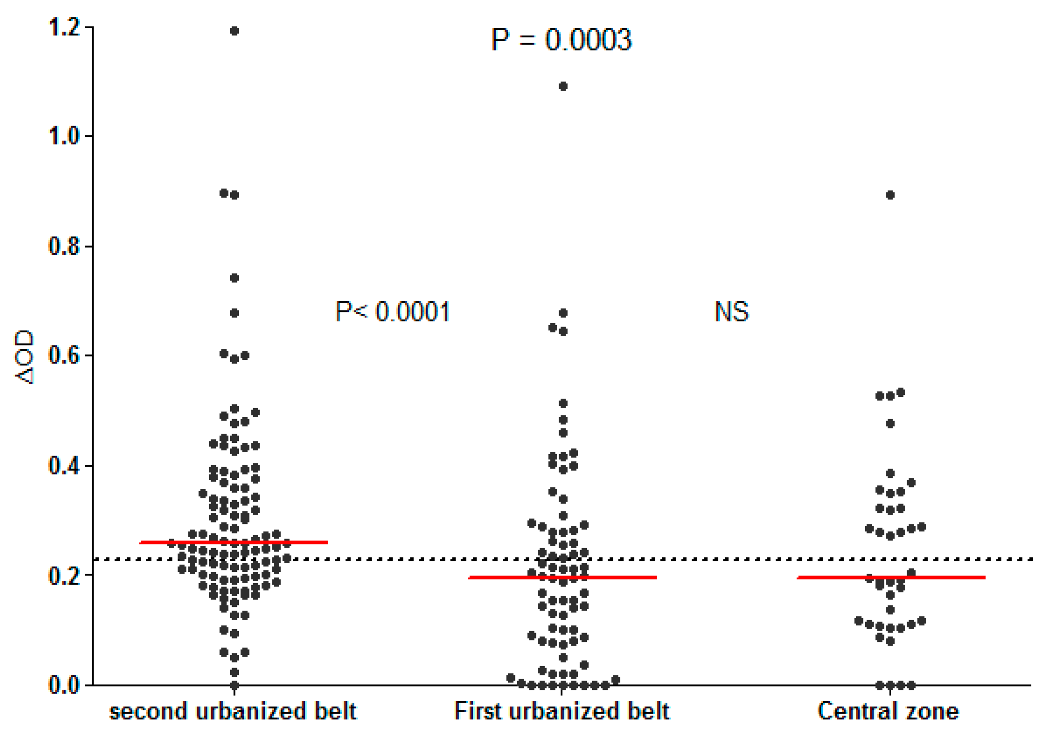
3.1. Evaluation of Human Exposure to Aedes Bites

There is an obvious need of new indicators and methods to evaluate the heterogeneity of human exposure to Aedes mosquito bites so as to focus and prioritise interventions in areas, seasons, or groups of individuals at high risk of arbovirus transmission. The relevance of using human IgG Ab responses to the Nterm-34Da peptide as a biomarker of Aedes bite exposure was firstly tested in rural areas of Benin, West Africa. Specific IgG Abs to the Nterm-34 Da salivary peptide were evaluated in the same children, during dry and rainy seasons, which generally represented periods of low and high exposure to Aedes bites, respectively, and compared with cumulated monthly rainfall registered at the same periods. Specific IgG responses showed significant seasonal variations in the study site. Indeed, specific IgG responses were detected in 29% of children in the dry season, whereas this percentage reached 99% in the rainy season. In addition, the median level of specific IgG Ab responses significantly increased between the dry and the rainy season (Figure 6) [27]. This result perfectly correlated with the intensity of rainfall at the same periods and highlighted the sensitivity and specificity of the Nterm-34 kDa epitope(s). This points out the relevance of using the biomarker of Aedes bite exposure for monitoring the risk of arbovirus transmission in areas colonised by Aedes mosquito vectors. Recently, another study in urban area of Senegal (Saint-Louis city) revealed considerable individual variations in anti-Nterm-34 kDa IgG levels between and within districts [61]. Similarly, heat maps of IgG responses to the Nterm-34 kDa indicated variations in the spatial distribution of the intensity of Ab responses inside urban districts. Of the four studied urban districts, hot spots of arbovirus transmission risk were located in some patches in the north of one district and were dispersed throughout the other urban district. The two remaining districts showed no hot spots of arbovirus transmission [61]. These results emphasised the significance of using such a serological tool to identify, target, and prioritise vector control strategies in areas with high risk of arbovirus transmission.
So far, studies aiming to validate anti-Nterm-34 kDa IgG Ab as a biomarker of Aedes bite exposure were only carried out in areas where Ae. aegypti was widely implanted. The study conducted in La Reunion Island revealed a very high immune cross-reactivity between the Nterm-34 kDa peptide of Ae. aegypti and that of Ae. albopictus [29], the second principal vector of arboviruses worldwide. Indeed, IgG Ab response against the Nterm-34 kDa peptide identified in Ae. aegypti saliva was measured in individuals only exposed to Ae. albopictus bites. Results indicated that individuals exposed to Ae. albopictus recognised the Nterm-34 kDa peptide of Ae. aegypti, suggesting that this peptide could be used to evaluate human exposure to these two major arbovirus vectors.

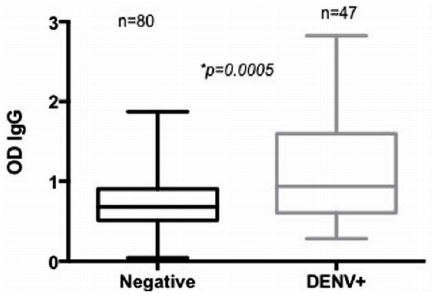
3.2. Evaluation of Arboviral Transmission Risk

Previous works provided data on human exposure to Aedes saliva and its relationship with dengue infection in the Americas [24,43]. Using the Nterm-34 kDa salivary peptide, one study aimed to evaluate the risk of dengue transmission in urban settings of Vientiane city, Lao PDR [28]. The authors reported no significant difference in anti-Nterm-34 kDa IgG levels between DENV-positive and DENV-negative individuals (evaluated by anti-DENV IgM assays to detect current dengue infection). However, the level of IgG Ab response to Nterm-34 kDa peptide deferred significantly according to the neighbourhoods’ degree of urbanisation (Figure 7). Specific IgG response among individuals living in the second urbanised belt (the periphery with low urbanisation) was higher than those from the first urbanised belt or from the central highly urbanised area. Interestingly, the prevalence of current DENV infection was higher in individuals living in the periphery (4.5%) than those living the central zone (2.2%). This study indicates the potential usefulness of human IgG Ab response to Nterm-34 kDa salivary peptide for predicting areas with a higher risk of dengue virus transmission in urban settings of developing countries. Nonetheless, further studies in different epidemiologic settings are required before validating this biomarker as a proxy of dengue transmission risk.

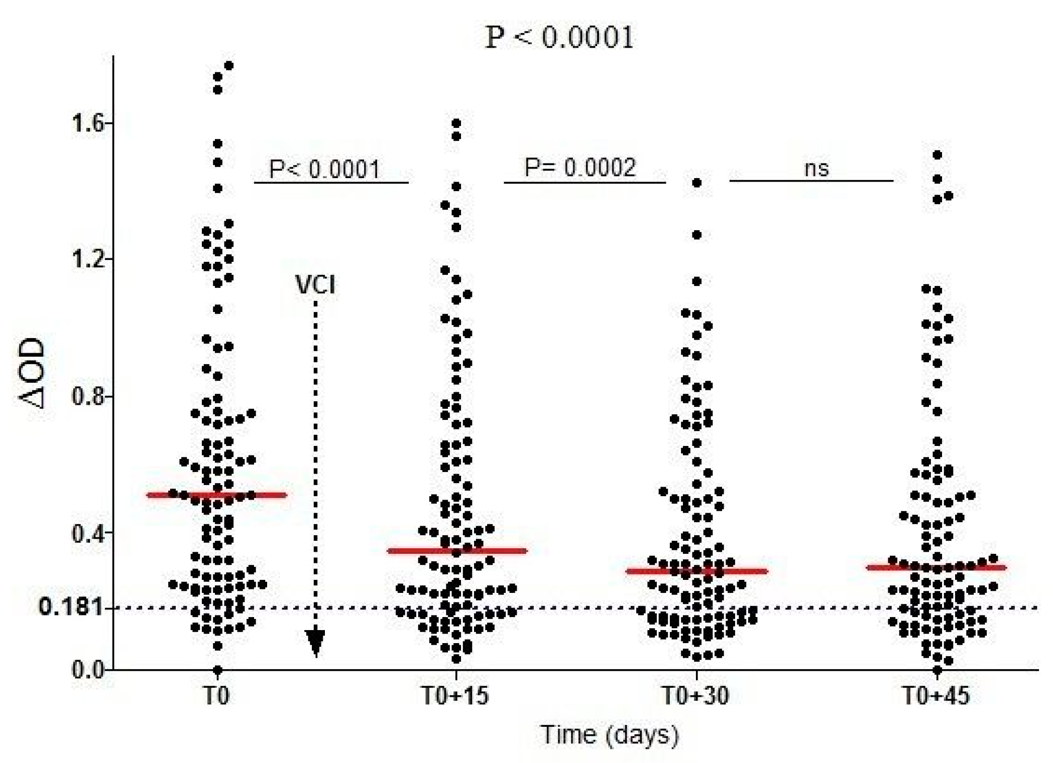
3.3. Evaluation of Vector Control Strategies’ Effectiveness

A recent study has reported that human IgG response to the Ae. aegypti Nterm-34 kDa peptide could be used for assessing the effectiveness of VCS against Aedes [29]. During this study, specific IgG response was assessed from 102 individuals living in urban area in La Reunion Island before and after the implementation of VCS against Ae. albopictus mosquito species (elimination of all breeding sites and insecticide space spraying with deltamethrin). A rapid and significant decrease of specific IgG response was reported only 15 days after the VCS implementation. This specific IgG response continued to decrease until 30 days after VCS implementation and then remained unchanged at 45 days (Figure 8). A similar trend was observed with the percentage of immune responders to the peptide. The proportion of immune responders decreased from 88% (before VCS implementation) to 68%, 30 days after VCS implementation. However, a slight increase was observed 45 days after VCS implementation. This decrease in specific IgG response was associated with the decline of Aedes mosquito densities, as estimated by entomological parameters and closely correlated to the efficacy of vector control implementation. The impact of implemented VCS were also assessed according the initial level of IgG Ab response in individuals.
All these results showed that this candidate biomarker can detect the short-time variations of human exposure to Aedes mosquito bite after vector control implementation. This immuno-epidemiological tool appears to be relevant to assess the effectiveness of VCS against the vectors of arboviruses.

4. Conclusions and Perspectives

This review emphasizes the potential of using biomarkers of human exposure to Aedes vectors, based on antibody responses to Aedes salivary proteins to evaluate: (i) the level of human population exposure to Aedes bites and its heterogeneity, (ii) the risk of arbovirus transmission or infection, and (iii) the effectiveness of vector control means implemented to prevent arbovirus transmission. To improve the specificity of salivary biomarkers, a peptide approach was developed to identify species and genera-specific salivary antigens to Aedes. One peptide, the Nterm-34 kDa, was identified as specific to Aedes species. IgG Ab to this peptide was validated as biomarker of human exposure to both Ae. aegypti and Ae. albopictus species. The availability of such a tool allows the assessment of the risk of arbovirus transmission in low-level exposure and transmission areas and to accurately evaluate the effectiveness of current or experimental VCS. For a direct impact in the fight against arboviruses directly in the field and at a large scale, the biomarker is currently in development into a rapid diagnosis test (under its auto-reactive dipstick form) for direct assessment of the level of exposure and the effectiveness of vector control means by sanitary authorities.

Author Contributions

F.R. conceived the intellectual content of the manuscript. A.B.S. and M.C.Y. collected results presented here and drafted the manuscript. E.E.N. and F.R. participated in the formation of the final version of the manuscript.

Funding

The post-doctoral fellowship of A.B.S. was supported by the Foundation Infectiopole Sud, Marseille.

Conflicts of Interest

The authors declare they have no conflict of interests.

References

  1. Gubler, D.J. The global emergence/resurgence of arboviral diseases as public health problems. Arch. Med. Res. 2002, 33, 330–342. [Google Scholar] [CrossRef]
  2. World Health Organization (WHO). A Global Brief on Vector-Borne Diseases; World Health Organization: Geneva, Switzerland, 2014; p. 54. [Google Scholar]
  3. Brady, O.J.; Gething, P.W.; Bhatt, S.; Messina, J.P.; Brownstein, J.S.; Hoen, A.G.; Moyes, C.L.; Farlow, A.W.; Scott, T.W.; Hay, S.I. Refining the global spatial limits of dengue virus transmission by evidence-based consensus. PLoS Negl. Trop. Dis. 2012, 6, e1760. [Google Scholar] [CrossRef] [PubMed] [Green Version]
  4. Bhatt, S.; Gething, P.W.; Brady, O.J.; Messina, J.P.; Farlow, A.W.; Moyes, C.L.; Drake, J.M.; Brownstein, J.S.; Hoen, A.G.; Sankoh, O.; et al. The global distribution and burden of dengue. Nature 2013, 496, 504–507. [Google Scholar] [CrossRef] [PubMed] [Green Version]
  5. Weaver, S.C.; Lecuit, M. Chikungunya virus and the global spread of a mosquito-borne disease. N. Engl. J. Med. 2015, 372, 1231–1239. [Google Scholar] [CrossRef] [PubMed]
  6. Musso, D. Zika Virus transmission from French Polynesia to Brazil. Emerg. Infect. Dis. 2015, 21, 1887. [Google Scholar] [CrossRef] [PubMed]
  7. Zhang, J.; Jin, X.; Zhu, Z.; Huang, L.; Liang, S.; Xu, Y.; Liao, R.; Zhou, L.; Zhang, Y.; Wilder-Smith, A. Early detection of Zika virus infection among travellers from areas of ongoing transmission in China. J. Travel Med. 2016, 23, taw047. [Google Scholar] [CrossRef] [PubMed]
  8. World Health Organization (WHO). Global Vector Control Response 2017–2030; World Health Organization: Geneva, Switzerland, 2017; p. 53. [Google Scholar]
  9. Reiter, P.; Fontenille, D.; Paupy, C. Aedes albopictus as an epidemic vector of chikungunya virus: Another emerging problem? Lancet Infect. Dis. 2006, 6, 463–464. [Google Scholar] [CrossRef]
  10. Tsetsarkin, K.A.; Vanlandingham, D.L.; McGee, C.E.; Higgs, S. A single mutation in chikungunya virus affects vector specificity and epidemic potential. PLoS Pathog. 2007, 3, e201. [Google Scholar] [CrossRef] [PubMed]
  11. Focks, D.A. A Review of Entomological Sampling Methods and Indicators for Dengue Vectors; World Health Organization: Geneva, Switzerland, 2004. [Google Scholar]
  12. Dibo, M.R.; Chierotti, A.P.; Ferrari, M.S.; Mendonca, A.L.; Chiaravalloti Neto, F. Study of the relationship between Aedes (Stegomyia) aegypti egg and adult densities, dengue fever and climate in Mirassol, state of Sao Paulo, Brazil. Mem. Inst. Oswaldo Cruz 2008, 103, 554–560. [Google Scholar] [CrossRef] [PubMed]
  13. Ribeiro, J.M.; Francischetti, I.M. Role of arthropod saliva in blood feeding: Sialome and post-sialome perspectives. Annu. Rev. Entomol. 2003, 48, 73–88. [Google Scholar] [CrossRef] [PubMed]
  14. Billingsley, P.F.; Baird, J.; Mitchell, J.A.; Drakeley, C. Immune interactions between mosquitoes and their hosts. Parasite Immunol. 2006, 28, 143–153. [Google Scholar] [CrossRef] [PubMed]
  15. Remoue, F.; Cisse, B.; Ba, F.; Sokhna, C.; Herve, J.P.; Boulanger, D.; Simondon, F. Evaluation of the antibody response to Anopheles salivary antigens as a potential marker of risk of malaria. Trans. R. Soc. Trop. Med. Hyg. 2006, 100, 363–370. [Google Scholar] [CrossRef] [PubMed]
  16. Fontaine, A.; Diouf, I.; Bakkali, N.; Misse, D.; Pages, F.; Fusai, T.; Rogier, C.; Almeras, L. Implication of haematophagous arthropod salivary proteins in host-vector interactions. Parasites Vectors 2011, 4, 187. [Google Scholar] [CrossRef] [PubMed]
  17. Remoue, F.; Alix, E.; Cornelie, S.; Sokhna, C.; Cisse, B.; Doucoure, S.; Mouchet, F.; Boulanger, D.; Simondon, F. IgE and IgG4 antibody responses to Aedes saliva in African children. Acta Trop. 2007, 104, 108–115. [Google Scholar] [CrossRef] [PubMed]
  18. Reunala, T.; Brummer-Korvenkontio, H.; Palosuo, K.; Miyanij, M.; Ruiz-Maldonado, R.; Love, A.; Francois, G.; Palosuo, T. Frequent occurrence of IgE and IgG4 antibodies against saliva of Aedes communis and Aedes aegypti mosquitoes in children. Int. Arch. Allergy Immunol. 1994, 104, 366–371. [Google Scholar] [CrossRef] [PubMed]
  19. Brummer-Korvenkontio, H.; Palosuo, K.; Palosuo, T.; Brummer-Korvenkontio, M.; Leinikki, P.; Reunala, T. Detection of mosquito saliva-specific IgE antibodies by capture ELISA. Allergy 1997, 52, 342–345. [Google Scholar] [CrossRef] [PubMed]
  20. Orlandi-Pradines, E.; Almeras, L.; Denis de Senneville, L.; Barbe, S.; Remoue, F.; Villard, C.; Cornelie, S.; Penhoat, K.; Pascual, A.; Bourgouin, C.; et al. Antibody response against saliva antigens of Anopheles gambiae and Aedes aegypti in travellers in tropical Africa. Microbes Infect. 2007, 9, 1454–1462. [Google Scholar] [CrossRef] [PubMed]
  21. Drame, P.M.; Poinsignon, A.; Besnard, P.; Le Mire, J.; Dos-Santos, M.A.; Sow, C.S.; Cornelie, S.; Foumane, V.; Toto, J.C.; Sembene, M.; et al. Human antibody response to Anopheles gambiae saliva: An immuno-epidemiological biomarker to evaluate the efficacy of insecticide-treated nets in malaria vector control. Am. J. Trop. Med. Hyg. 2010, 83, 115–121. [Google Scholar] [CrossRef] [PubMed]
  22. Fontaine, A.; Pascual, A.; Orlandi-Pradines, E.; Diouf, I.; Remoue, F.; Pages, F.; Fusai, T.; Rogier, C.; Almeras, L. Relationship between exposure to vector bites and antibody responses to mosquito salivary gland extracts. PLoS ONE 2011, 6, e29107. [Google Scholar] [CrossRef] [PubMed]
  23. Doucoure, S.; Mouchet, F.; Cornelie, S.; DeHecq, J.S.; Rutee, A.H.; Roca, Y.; Walter, A.; Herve, J.P.; Misse, D.; Favier, F.; et al. Evaluation of the human IgG antibody response to Aedes albopictus saliva as a new specific biomarker of exposure to vector bites. PLoS Negl. Trop. Dis. 2012, 6, e1487. [Google Scholar] [CrossRef] [PubMed]
  24. Londono-Renteria, B.; Cardenas, J.C.; Cardenas, L.D.; Christofferson, R.C.; Chisenhall, D.M.; Wesson, D.M.; McCracken, M.K.; Carvajal, D.; Mores, C.N. Use of anti-Aedes aegypti salivary extract antibody concentration to correlate risk of vector exposure and dengue transmission risk in Colombia. PLoS ONE 2013, 8, e81211. [Google Scholar] [CrossRef] [PubMed]
  25. Palosuo, K.; Brummer-Korvenkontio, H.; Mikkola, J.; Sahi, T.; Reunala, T. Seasonal increase in human IgE and IgG4 antisaliva antibodies to Aedes mosquito bites. Int. Arch. Allergy Immunol. 1997, 114, 367–372. [Google Scholar] [CrossRef] [PubMed]
  26. Waitayakul, A.; Somsri, S.; Sattabongkot, J.; Looareesuwan, S.; Cui, L.; Udomsangpetch, R. Natural human humoral response to salivary gland proteins of Anopheles mosquitoes in Thailand. Acta Trop. 2006, 98, 66–73. [Google Scholar] [CrossRef] [PubMed]
  27. Elanga Ndille, E.; Doucoure, S.; Damien, G.; Mouchet, F.; Drame, P.M.; Cornelie, S.; Noukpo, H.; Yamadjako, S.; Djenontin, A.; Moiroux, N.; et al. First attempt to validate human IgG antibody response to Nterm-34 kDa salivary peptide as biomarker for evaluating exposure to Aedes aegypti bites. PLoS Negl. Trop. Dis. 2012, 6, e1905. [Google Scholar] [CrossRef] [PubMed]
  28. Ndille, E.E.; Dubot-Peres, A.; Doucoure, S.; Mouchet, F.; Cornelie, S.; Sidavong, B.; Fournet, F.; Remoue, F. Human IgG antibody response to Aedes aegypti Nterm-34 kDa salivary peptide as an indicator to identify areas at high risk for dengue transmission: A retrospective study in urban settings of Vientiane city, Lao PDR. Trop. Med. Int. Health 2014, 19, 576–580. [Google Scholar] [CrossRef] [PubMed]
  29. Elanga Ndille, E.; Doucoure, S.; Poinsignon, A.; Mouchet, F.; Cornelie, S.; D’Ortenzio, E.; DeHecq, J.S.; Remoue, F. Human IgG antibody response to Aedes Nterm-34 kDa salivary peptide, an epidemiological tool to assess vector control in chikungunya and dengue transmission area. PLoS Negl. Trop. Dis. 2016, 10, e0005109. [Google Scholar] [CrossRef] [PubMed]
  30. Yobo, M.C.; Sadia-Kacou, A.M.C.; Adja, A.M.; Elanga-Ndille, E.; Sagna, A.B.; Guindo-Coulibaly, N.; Poinsignon, A.; Remoue, F.; Koudou, B.G. Evaluation of human exposure to Aedes bites in rubber and palm cultivations using an immuno-epidemiological biomarker. BioMed Res. Int. 2018. accepted. [Google Scholar]
  31. Ribeiro, J.M. Role of saliva in blood-feeding by arthropods. Annu. Rev. Entomol. 1987, 32, 463–478. [Google Scholar] [CrossRef] [PubMed]
  32. Brummer-Korvenkontio, H.; Lappalainen, P.; Reunala, T.; Palosuo, T. Detection of mosquito saliva-specific IgE and IgG4 antibodies by immunoblotting. J. Allergy Clin. Immunol. 1994, 93, 551–555. [Google Scholar] [CrossRef]
  33. Peng, Z.; Xu, W.; James, A.A.; Lam, H.; Sun, D.; Cheng, L.; Simons, F.E. Expression, purification, characterization and clinical relevance of rAed a 1—A 68-kDa recombinant mosquito Aedes aegypti salivary allergen. Int. Immunol. 2001, 13, 1445–1452. [Google Scholar] [CrossRef] [PubMed]
  34. Peng, Z.; Xu, W.; Lam, H.; Cheng, L.; James, A.A.; Simons, F.E. A new recombinant mosquito salivary allergen, rAed a 2: Allergenicity, clinical relevance, and cross-reactivity. Allergy 2006, 61, 485–490. [Google Scholar] [CrossRef] [PubMed]
  35. Peng, Z.; Xu, W.W.; Sham, Y.; Lam, H.; Sun, D.; Cheng, L.; Rasic, N.F.; Guan, Q.; James, A.A.; Simons, F.E. Mosquito salivary allergen Aed a 3: Cloning, comprehensive molecular analysis, and clinical evaluation. Allergy 2016, 71, 621–628. [Google Scholar] [CrossRef] [PubMed]
  36. Peng, Z.; Caihe, L.; Beckett, A.N.; Guan, Q.; James, A.A.; Simons, F.E. rAed a 4: A new 67-kDa Aedes aegypti mosquito salivary allergen for the diagnosis of mosquito allergy. Int. Arch. Allergy Immunol. 2016, 170, 206–210. [Google Scholar] [CrossRef] [PubMed]
  37. Schwartz, B.S.; Ribeiro, J.M.; Goldstein, M.D. Anti-tick antibodies: An epidemiologic tool in Lyme disease research. Am. J. Epidemiol. 1990, 132, 58–66. [Google Scholar] [CrossRef] [PubMed]
  38. Peng, Z.; Rasic, N.; Liu, Y.; Simons, F.E. Mosquito saliva-specific IgE and IgG antibodies in 1059 blood donors. J. Allergy Clin. Immunol. 2002, 110, 816–817. [Google Scholar] [CrossRef] [PubMed]
  39. Doucoure, S.; Mouchet, F.; Cournil, A.; Le Goff, G.; Cornelie, S.; Roca, Y.; Giraldez, M.G.; Simon, Z.B.; Loayza, R.; Misse, D.; et al. Human antibody response to Aedes aegypti saliva in an urban population in Bolivia: A new biomarker of exposure to dengue vector bites. Am. J. Trop. Med. Hyg. 2012, 87, 504–510. [Google Scholar] [CrossRef] [PubMed]
  40. Doucoure, S.; Mouchet, F.; Cornelie, S.; Drame, P.M.; D’Ortenzio, E.; DeHecq, J.S.; Remoue, F. Human antibody response to Aedes albopictus salivary proteins: A potential biomarker to evaluate the efficacy of vector control in an area of chikungunya and dengue virus transmission. BioMed Res. Int. 2014, 2014, 746509. [Google Scholar] [CrossRef] [PubMed]
  41. Schwarz, A.; Sternberg, J.M.; Johnston, V.; Medrano-Mercado, N.; Anderson, J.M.; Hume, J.C.; Valenzuela, J.G.; Schaub, G.A.; Billingsley, P.F. Antibody responses of domestic animals to salivary antigens of Triatoma infestans as biomarkers for low-level infestation of triatomines. Int. J. Parasitol. 2009, 39, 1021–1029. [Google Scholar] [CrossRef] [PubMed]
  42. Schwarz, A.; Medrano-Mercado, N.; Billingsley, P.F.; Schaub, G.A.; Sternberg, J.M. IgM-antibody responses of chickens to salivary antigens of Triatoma infestans as early biomarkers for low-level infestation of triatomines. Int. J. Parasitol. 2010, 40, 1295–1302. [Google Scholar] [CrossRef] [PubMed]
  43. Londono-Renteria, B.; Cardenas, J.C.; Giovanni, J.E.; Cardenas, L.; Villamizar, P.; Rolon, J.; Chisenhall, D.M.; Christofferson, R.C.; Carvajal, D.J.; Perez, O.G.; et al. Aedes aegypti anti-salivary gland antibody concentration and dengue virus exposure history in healthy individuals living in an endemic area in Colombia. Biomedica 2015, 35, 572–581. [Google Scholar] [CrossRef] [PubMed]
  44. Doucoure, S.; Drame, P.M. Salivary biomarkers in the control of mosquito-borne diseases. Insects 2015, 6, 961–976. [Google Scholar] [CrossRef] [PubMed]
  45. Arca, B.; Lombardo, F.; Francischetti, I.M.; Pham, V.M.; Mestres-Simon, M.; Andersen, J.F.; Ribeiro, J.M. An insight into the sialome of the adult female mosquito Aedes albopictus. Insect Biochem. Mol. Biol. 2007, 37, 107–127. [Google Scholar] [CrossRef] [PubMed]
  46. Ribeiro, J.M.; Arca, B.; Lombardo, F.; Calvo, E.; Phan, V.M.; Chandra, P.K.; Wikel, S.K. An annotated catalogue of salivary gland transcripts in the adult female mosquito, Aedes aegypti. BMC Genom. 2007, 8, 6. [Google Scholar] [CrossRef] [PubMed]
  47. Ribeiro, J.M.; Martin-Martin, I.; Arca, B.; Calvo, E. A deep insight into the sialome of male and female Aedes aegypti mosquitoes. PLoS ONE 2016, 11, e0151400. [Google Scholar] [CrossRef] [PubMed]
  48. Cornelie, S.; Remoue, F.; Doucoure, S.; Ndiaye, T.; Sauvage, F.X.; Boulanger, D.; Simondon, F. An insight into immunogenic salivary proteins of Anopheles gambiae in African children. Malar. J. 2007, 6, 75. [Google Scholar] [CrossRef] [PubMed]
  49. Doucoure, S.; Cornelie, S.; Patramool, S.; Mouchet, F.; Demettre, E.; Seveno, M.; Dehecq, J.S.; Rutee, H.; Herve, J.P.; Favier, F.; et al. First screening of Aedes albopictus immunogenic salivary proteins. Insect Mol. Biol. 2013, 22, 411–423. [Google Scholar] [CrossRef] [PubMed]
  50. Schwarz, A.; Helling, S.; Collin, N.; Teixeira, C.R.; Medrano-Mercado, N.; Hume, J.C.; Assumpcao, T.C.; Marcus, K.; Stephan, C.; Meyer, H.E.; et al. Immunogenic salivary proteins of Triatoma infestans: Development of a recombinant antigen for the detection of low-level infestation of triatomines. PLoS Negl. Trop. Dis. 2009, 3, e532. [Google Scholar] [CrossRef] [PubMed] [Green Version]
  51. Souza, A.P.; Andrade, B.B.; Aquino, D.; Entringer, P.; Miranda, J.C.; Alcantara, R.; Ruiz, D.; Soto, M.; Teixeira, C.R.; Valenzuela, J.G.; et al. Using recombinant proteins from Lutzomyia longipalpis saliva to estimate human vector exposure in visceral leishmaniasis endemic areas. PLoS Negl. Trop. Dis. 2010, 4, e649. [Google Scholar] [CrossRef] [PubMed]
  52. Teixeira, C.; Gomes, R.; Collin, N.; Reynoso, D.; Jochim, R.; Oliveira, F.; Seitz, A.; Elnaiem, D.E.; Caldas, A.; de Souza, A.P.; et al. Discovery of markers of exposure specific to bites of Lutzomyia longipalpis, the vector of Leishmania infantum chagasi in Latin America. PLoS Negl. Trop. Dis. 2010, 4, e638. [Google Scholar] [CrossRef] [PubMed]
  53. Rizzo, C.; Ronca, R.; Fiorentino, G.; Verra, F.; Mangano, V.; Poinsignon, A.; Sirima, S.B.; Nebie, I.; Lombardo, F.; Remoue, F.; et al. Humoral response to the Anopheles gambiae salivary protein gSG6: A serological indicator of exposure to Afrotropical malaria vectors. PLoS ONE 2011, 6, e17980. [Google Scholar] [CrossRef] [PubMed]
  54. Rizzo, C.; Ronca, R.; Fiorentino, G.; Mangano, V.D.; Sirima, S.B.; Nebie, I.; Petrarca, V.; Modiano, D.; Arca, B. Wide cross-reactivity between Anopheles gambiae and Anopheles funestus SG6 salivary proteins supports exploitation of gSG6 as a marker of human exposure to major malaria vectors in tropical Africa. Malar. J. 2011, 10, 206. [Google Scholar] [CrossRef] [PubMed]
  55. Ali, Z.M.; Bakli, M.; Fontaine, A.; Bakkali, N.; Vu Hai, V.; Audebert, S.; Boublik, Y.; Pages, F.; Remoue, F.; Rogier, C.; et al. Assessment of Anopheles salivary antigens as individual exposure biomarkers to species-specific malaria vector bites. Malar. J. 2012, 11, 439. [Google Scholar] [CrossRef] [PubMed]
  56. Mouchet, F.; (MIVEGEC-IRD, Montpellier, France). IgG antibody response against Aedes aegypti saliva in a Bolivian urban population: Towards a new biomarker of exposure to dengue vector bites. Personal communication, 2009. [Google Scholar]
  57. Poinsignon, A.; Cornelie, S.; Mestres-Simon, M.; Lanfrancotti, A.; Rossignol, M.; Boulanger, D.; Cisse, B.; Sokhna, C.; Arca, B.; Simondon, F.; et al. Novel peptide marker corresponding to salivary protein gSG6 potentially identifies exposure to Anopheles bites. PLoS ONE 2008, 3, e2472. [Google Scholar] [CrossRef] [PubMed]
  58. Poinsignon, A.; Samb, B.; Doucoure, S.; Drame, P.M.; Sarr, J.B.; Sow, C.; Cornelie, S.; Maiga, S.; Thiam, C.; Rogerie, F.; et al. First attempt to validate the gSG6-P1 salivary peptide as an immuno-epidemiological tool for evaluating human exposure to Anopheles funestus bites. Trop. Med. Int. Health 2010, 15, 1198–1203. [Google Scholar] [CrossRef] [PubMed]
  59. Londono-Renteria, B.; Drame, P.M.; Weitzel, T.; Rosas, R.; Gripping, C.; Cardenas, J.C.; Alvares, M.; Wesson, D.M.; Poinsignon, A.; Remoue, F.; et al. An. gambiae gSG6-P1 evaluation as a proxy for human-vector contact in the Americas: A pilot study. Parasites Vectors 2015, 8, 533. [Google Scholar] [CrossRef] [PubMed]
  60. Ya-Umphan, P.; Cerqueira, D.; Parker, D.M.; Cottrell, G.; Poinsignon, A.; Remoue, F.; Brengues, C.; Chareonviriyaphap, T.; Nosten, F.; Corbel, V. Use of an Anopheles salivary biomarker to assess malaria transmission risk along the Thailand-Myanmar Border. J. Infect. Dis. 2017, 215, 396–404. [Google Scholar] [CrossRef] [PubMed]
  61. Sagna, A.B.; Kassie, D.; Couvray, A.; Hermann, E.; Riveau, G.; Salem, G.; Fournet, F.; Remoue, F. Spatial risk of human exposure to Anopheles and Aedes mosquito bites in African urban settings using antibody-based biomarkers. Unpublished data.
Figure 1. Human-vector relationships during arthropod-borne diseases. During the bite, the vector (Aedes) injects its saliva in the human skin. Once in the skin, salivary proteins take the control of (1) the human haemostatic system by inhibiting the platelet activation and clotting mechanism, and (2) the inflammatory system. (3) The salivary proteins modulate the human immune response and promote the production of anti-salivary antibodies. (4) If ever the (Aedes) vector carries a pathogen, the salivary proteins contribute to its transmission into the human.
Tropicalmed 03 00080 g001
Figure 2. The concept of salivary biomarkers to vector bites.
Tropicalmed 03 00080 g002
Figure 3. IgG Ab response to Ae. albopictus salivary gland extracts (SGEs) between exposed and unexposed individuals to Ae. albopictus bites in La Reunion Island [23]. Black points indicate individual IgG response (ΔOD) and bars represent the median value in each group. Dotted line represents the cut-off of specific Ab response (ΔOD > 0.181) and p value was calculated using the Mann–Whitney U test.
Tropicalmed 03 00080 g003
Figure 4. Levels of anti-Ae. aegypti SGE antibodies according to the acute dengue infection status in Los Patios in subjects with DENV (dengue virus) (+) (DENV+) and subjects without infection (DENV−) [24]. Statistical significance of Mann–Whitney test * p < 0.05.
Tropicalmed 03 00080 g004
Figure 5. Amino acids’ sequence of the 34 kDa salivary protein of female Ae. aegypti mosquito (gi|94468336 [46]). The blue sequence corresponds to the signal peptide.
Tropicalmed 03 00080 g005
Figure 6. Evolution of individual IgG response to Nterm-34 kDa peptide between the dry and the rainy season [27]. Results are presented for the peak of the dry (February) and the rainy (July) season in 2008. Black points indicate individual IgG response (ΔOD) and bars indicate the median value for each group. Dotted line represents the threshold (TR) of specific Ab response (ΔOD = 0.151) and the statistical significant difference between medians is indicated (non-parametric Wilcoxon test).
Tropicalmed 03 00080 g006
Figure 7. IgG Ab response to Nterm-34 kDa salivary peptide in the studied population according the level of urbanisation of their residential areas in Vientiane city [28]. Black dots indicate individual IgG response (ΔOD), and bars represent the median value in each group. Dotted line represents the cut-off of a specific Ab response (ΔOD > 0.250). Statistically significant differences between the three groups (P = 0.0003, nonparametric Kruskal–Wallis test) and between the first and second urbanised belts (P < 0.0001, Mann–Whitney U test) are indicated.
Tropicalmed 03 00080 g007
Figure 8. IgG Ab response to Nterm-34 kDa salivary peptide from individuals exposed to Ae. albopictus bites before and after vector control implementation [29]. Individual IgG Ab response (ΔOD) is presented just before (T0) and then 15, 30, and 45 days after vector control implementation. Bars indicated the median value in the population at each time point and the dotted line represents the cut-off of immune response. P-values indicating differences in IgG response level at the overall time points (Kruskal–Wallis test) or between two different time points (Wilcoxon matched pair test) are presented. Vertical solid grey line indicates timing of Vector Control Implementation (VCI).
Tropicalmed 03 00080 g008

