Toxicity of Nanoparticles in the Lung: Environmental and Medical Aspects (Closed)

A topical collection in Nanomaterials (ISSN 2079-4991). This collection belongs to the section "Biology and Medicines".

Viewed by 14570

Editor


E-Mail Website
Collection Editor

Topical Collection Information

Dear Colleagues,

With the broad use of nanoparticles in many products, accumulation in the environment and increased exposure of humans may occur. Adverse effects of non-intentionally inhaled nanoparticles in humans may be caused by environmental exposure, at the workplace, and by exposure to medical products. The biocompatibility of nanoparticles has been intensely studied in vitro and in vivo over the last decade, and the role of the respiratory system as the most permeable and vulnerable portal of entry was confirmed. Nevertheless, action on the immune system, effects on repeated exposure, organ-specificity, etc., are not well known. Translation of experimental data to estimation of human risk is complicated by a lack of representative models and exposure conditions, inter-individual differences in exposure levels and in predisposing biological factors.

In addition to unintended toxicity, nanoparticles can be used to increase the efficacy of cytostatic drugs, mainly for the treatment of lung cancer. Better encapsulation of poorly-soluble drugs, delivery of small molecules, and enhanced permeability and retention effect increase anticancer action.

Similar nanoparticle properties, such as cellular accumulation, decreased clearance, and modulation of immune effects, are linked to undesired effects in the lung; however, the same effects that cause the undesired effects of environmental nanoparticles in the lung are exploited to increase efficacy of nanoparticles in medical treatments.

This Topical Collection will be dedicated to reactions of nanoparticles in the respiratory system. Toxic reactions to air-borne particles and medical particles as well as differences in reactions of the respiratory system compared to other organs are of interest. We welcome the submission of comprehensive/mini reviews, original research articles, and communications.

Prof. Dr. Eleonore Fröhlich
Guest Editor

Manuscript Submission Information

Manuscripts should be submitted online at www.mdpi.com by registering and logging in to this website. Once you are registered, click here to go to the submission form. Manuscripts can be submitted until the deadline. All submissions that pass pre-check are peer-reviewed. Accepted papers will be published continuously in the journal (as soon as accepted) and will be listed together on the collection website. Research articles, review articles as well as short communications are invited. For planned papers, a title and short abstract (about 100 words) can be sent to the Editorial Office for announcement on this website.

Submitted manuscripts should not have been published previously, nor be under consideration for publication elsewhere (except conference proceedings papers). All manuscripts are thoroughly refereed through a single-blind peer-review process. A guide for authors and other relevant information for submission of manuscripts is available on the Instructions for Authors page. Nanomaterials is an international peer-reviewed open access semimonthly journal published by MDPI.

Please visit the Instructions for Authors page before submitting a manuscript. The Article Processing Charge (APC) for publication in this open access journal is 2900 CHF (Swiss Francs). Submitted papers should be well formatted and use good English. Authors may use MDPI's English editing service prior to publication or during author revisions.

Keywords

  • Environmental nanoparticles
  • Engineered nanoparticles
  • In-vitro models
  • Chronic effects
  • In silico modeling
  • Nano-based formulations
  • Mechanisms of nanotoxicity
  • Lung cancer treatment
  • Inhalation treatment

Published Papers (3 papers)

2021

Jump to: 2019, 2018

18 pages, 47995 KiB  
Article
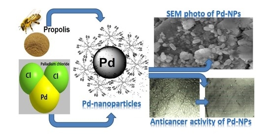
Characterization, Antimicrobial and Anticancer Properties of Palladium Nanoparticles Biosynthesized Optimally Using Saudi Propolis
by Maged S. Al-Fakeh, Samir Osman Mohammed Osman, Malek Gassoumi, Mokded Rabhi and Mohamed Omer
Nanomaterials 2021, 11(10), 2666; https://doi.org/10.3390/nano11102666 - 11 Oct 2021
Cited by 17 | Viewed by 3058
Abstract
Due to their unique physicochemical characteristics, palladium nanoparticles (Pd-NPs) have shown tremendous promise in biological applications. The biosynthesis of Pd-NPs employing Saudi propolis has been designed to be environmental, fast, controlled, and cost-effective. The formation and stability of biosynthesized Pd-NPs by Saudi propolis [...] Read more.
Due to their unique physicochemical characteristics, palladium nanoparticles (Pd-NPs) have shown tremendous promise in biological applications. The biosynthesis of Pd-NPs employing Saudi propolis has been designed to be environmental, fast, controlled, and cost-effective. The formation and stability of biosynthesized Pd-NPs by Saudi propolis extract were proved by ultraviolet–visible spectrophotometry (UV-Vis), Fourier-transform infrared spectroscopy (FT-IR), and Zeta potential analysis. Transmission electron microscopy (TEM), scanning electron microscopy (SEM), and X-ray diffraction (XRD) findings show that the average particle size of Pd-NPs is between 3.14 and 4.62 nm, which is in quantum scale. The Saudi propolis enhanced the antimicrobial activity against B. subtilis, S. aureus, E. coli, K. pneumoniae, and C. albicans. Pd-NPs show effective anticancer activity against ductal carcinoma (MCF-7) with IC50 of 104.79 µg/mL. Full article
Show Figures

Graphical abstract

2019

Jump to: 2021, 2018

15 pages, 1469 KiB  
Article
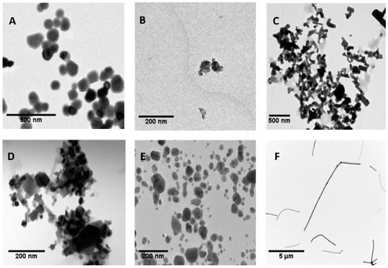
Micronuclei Detection by Flow Cytometry as a High-Throughput Approach for the Genotoxicity Testing of Nanomaterials
by Alba García-Rodríguez, Liliya Kazantseva, Laura Vila, Laura Rubio, Antonia Velázquez, María José Ramírez, Ricard Marcos and Alba Hernández
Nanomaterials 2019, 9(12), 1677; https://doi.org/10.3390/nano9121677 - 24 Nov 2019
Cited by 17 | Viewed by 3000
Abstract
Thousands of nanomaterials (NMs)-containing products are currently under development or incorporated in the consumer market, despite our very limited understanding of their genotoxic potential. Taking into account that the toxicity and genotoxicity of NMs strongly depend on their physicochemical characteristics, many variables must [...] Read more.
Thousands of nanomaterials (NMs)-containing products are currently under development or incorporated in the consumer market, despite our very limited understanding of their genotoxic potential. Taking into account that the toxicity and genotoxicity of NMs strongly depend on their physicochemical characteristics, many variables must be considered in the safety evaluation of each given NM. In this scenario, the challenge is to establish high-throughput methodologies able to generate rapid and robust genotoxicity data that can be used to critically assess and/or predict the biological effects associated with those NMs being under development or already present in the market. In this study, we have evaluated the advantages of using a flow cytometry-based approach testing micronucleus (MNs) induction (FCMN assay). In the frame of the EU NANoREG project, we have tested six different NMs—namely NM100 and NM101 (TiO2NPs), NM110 (ZnONPs), NM212 (CeO2NPs), NM300K (AgNPs) and NM401 (multi-walled carbon nanotubes (MWCNTs)). The obtained results confirm the ability of AgNPs and MWCNTs to induce MN in the human bronchial epithelial BEAS-2B cell line, whereas the other tested NMs retrieved non-significant increases in the MN frequency. Based on the alignment of the results with the data reported in the literature and the performance of the FCMN assay, we strongly recommend this assay as a reference method to systematically evaluate the potential genotoxicity of NMs. Full article
Show Figures

Figure 1

2018

Jump to: 2021, 2019

11 pages, 3224 KiB  
Communication
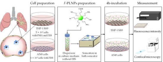
Surface Charge-Dependent Cellular Uptake of Polystyrene Nanoparticles
by Soyeon Jeon, Jessica Clavadetscher, Dong-Keun Lee, Sunay V. Chankeshwara, Mark Bradley and Wan-Seob Cho
Nanomaterials 2018, 8(12), 1028; https://doi.org/10.3390/nano8121028 - 10 Dec 2018
Cited by 120 | Viewed by 6895
Abstract
The evaluation of the role of physicochemical properties in the toxicity of nanoparticles is important for the understanding of toxicity mechanisms and for controlling the behavior of nanoparticles. The surface charge of nanoparticles is suggested as one of the key parameters which decide [...] Read more.
The evaluation of the role of physicochemical properties in the toxicity of nanoparticles is important for the understanding of toxicity mechanisms and for controlling the behavior of nanoparticles. The surface charge of nanoparticles is suggested as one of the key parameters which decide their biological impact. In this study, we synthesized fluorophore-conjugated polystyrene nanoparticles (F-PLNPs), with seven different types of surface functional groups that were all based on an identical core, to evaluate the role of surface charge in the cellular uptake of nanoparticles. Phagocytic differentiated THP-1 cells or non-phagocytic A549 cells were incubated with F-PLNP for 4 h, and their cellular uptake was quantified by fluorescence intensity and confocal microscopy. The amount of internalized F-PLNPs showed a good positive correlation with the zeta potential of F-PLNPs in both cell lines (Pearson’s r = 0.7021 and 0.7852 for zeta potential vs. cellular uptake in THP-1 cells and nonphagocytic A549 cells, respectively). This result implies that surface charge is the major parameter determining cellular uptake efficiency, although other factors such as aggregation/agglomeration, protein corona formation, and compositional elements can also influence the cellular uptake partly or indirectly. Full article
Show Figures

Graphical abstract

Back to TopTop