Mechanisms of Mycobacterium tuberculosis Pathogenesis

A special issue of Pathogens (ISSN 2076-0817).

Deadline for manuscript submissions: closed (31 December 2017) | Viewed by 38085

Special Issue Editor


E-Mail Website1 Website2
Guest Editor
1. Department of Microbiology, School of Medicine, The University of Alabama at Birmingham, Birmingham, AL 35294, USA
2. Africa Health Research Institute, University of KwaZulu-Natal, Durban 4001, South Africa
Interests: Mtb bioenergetics; energy metabolism; immunometabolism; human TB pathogenesis; host gasotransmitters

Special Issue Information

Dear Colleagues,

Tuberculosis (TB) is a major global public health problem and, despite substantial progress, TB is one of the most important infectious causes of morbidity and death in the world. The unique ability of M. tuberculosis (Mtb) to persist in millions of people in a dormant, drug-resistant state, sometimes reactivating to cause TB decades after the primary infection, has puzzled scientists for decades. Alarmingly, during the past decade a substantial increase in the number of patients with multidrug resistant TB and extensively drug-resistant TB has been noted.

The microenvironment within the Mtb-infected lung is dynamic and contains a wide range of different carbon sources, proteins and metabolites. This represents a biological predicament for investigation as the metabolic state of bacilli within the spectrum of lesions will differ from one another. Not surprisingly, ample data suggest that Mtb adjusts its metabolism in response to the availability of nutrients during different stages of infection.

The global TB burden anti-TB drug crisis has made understanding the molecular mechanisms of pathogenesis, and responses to actively growing and dormant Mtb, essential for improving existing therapies and identifying innovative therapeutic approaches. This Special Issue on “Mechanisms of Mycobacterium tuberculosis Pathogenesis” will focus on our current understanding of how Mtb causes disease, and opportunities for therapeutic intervention. We invite you to submit research and review manuscripts that cover any aspect of Mtb genetics, metabolism, and drug resistance, host pathogen interaction, immunology, human TB, and TB pathogenesis.

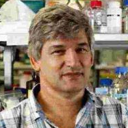
Prof. Adrie J.C. Steyn
Guest Editor

Manuscript Submission Information

Manuscripts should be submitted online at www.mdpi.com by registering and logging in to this website. Once you are registered, click here to go to the submission form. Manuscripts can be submitted until the deadline. All submissions that pass pre-check are peer-reviewed. Accepted papers will be published continuously in the journal (as soon as accepted) and will be listed together on the special issue website. Research articles, review articles as well as short communications are invited. For planned papers, a title and short abstract (about 100 words) can be sent to the Editorial Office for announcement on this website.

Submitted manuscripts should not have been published previously, nor be under consideration for publication elsewhere (except conference proceedings papers). All manuscripts are thoroughly refereed through a single-blind peer-review process. A guide for authors and other relevant information for submission of manuscripts is available on the Instructions for Authors page. Pathogens is an international peer-reviewed open access monthly journal published by MDPI.

Please visit the Instructions for Authors page before submitting a manuscript. The Article Processing Charge (APC) for publication in this open access journal is 2700 CHF (Swiss Francs). Submitted papers should be well formatted and use good English. Authors may use MDPI's English editing service prior to publication or during author revisions.

Keywords

  • dormancy
  • nonreplicating persistence
  • drug resistance
  • genetics redox homeostasis
  • bioenergetics
  • pathology
  • immunology
  • cell biology

Published Papers (4 papers)

Order results
Result details
Select all
Export citation of selected articles as:

Research

Jump to: Review

12 pages, 1136 KiB  
Article
Soluble CD14 as a Diagnostic Biomarker for Smear-Negative HIV-Associated Tuberculosis
by Yanyan Liu, Okechukwu C. Ndumnego, Tingting Chen, Ryung S. Kim, Elizabeth R. Jenny-Avital, Thumbi Ndung’u, Douglas Wilson and Jacqueline M. Achkar
Pathogens 2018, 7(1), 26; https://doi.org/10.3390/pathogens7010026 - 27 Feb 2018
Cited by 10 | Viewed by 4735
Abstract
Sputum smear-negative HIV-associated active tuberculosis (TB) is challenging to diagnose. CD14 is a pattern recognition receptor that is known to mediate monocyte activation. Prior studies have shown increased levels of soluble CD14 (sCD14) as a potential biomarker for TB, but little is known [...] Read more.
Sputum smear-negative HIV-associated active tuberculosis (TB) is challenging to diagnose. CD14 is a pattern recognition receptor that is known to mediate monocyte activation. Prior studies have shown increased levels of soluble CD14 (sCD14) as a potential biomarker for TB, but little is known about its value in detecting smear-negative HIV-associated TB. We optimized a sandwich ELISA for the detection of sCD14, and tested sera from 56 smear-negative South African (39 culture-positive and 17 culture-negative) HIV-infected pulmonary TB patients and 24 South African and 43 US (21 positive and 22 negative for tuberculin skin test, respectively) HIV-infected controls. SCD14 concentrations were significantly elevated in smear-negative HIV-associated TB compared with the HIV-infected controls (p < 0.0001), who had similar concentrations, irrespective of the country of origin or the presence or absence of latent M. tuberculosis infection (p = 0.19). The culture-confirmed TB group had a median sCD14 level of 2199 ng/mL (interquartile range 1927–2719 ng/mL), versus 1148 ng/mL (interquartile range 1053–1412 ng/mL) for the South African controls. At a specificity of 96%, sCD14 had a sensitivity of 95% for culture-confirmed smear-negative TB. These data indicate that sCD14 could be a highly accurate biomarker for the detection of HIV-associated TB. Full article
(This article belongs to the Special Issue Mechanisms of Mycobacterium tuberculosis Pathogenesis)
Show Figures

Figure 1

Review

Jump to: Research

30 pages, 15400 KiB  
Review
Bioenergetics of Mycobacterium: An Emerging Landscape for Drug Discovery
by Iram Khan Iqbal, Sapna Bajeli, Ajit Kumar Akela and Ashwani Kumar
Pathogens 2018, 7(1), 24; https://doi.org/10.3390/pathogens7010024 - 23 Feb 2018
Cited by 44 | Viewed by 11282
Abstract
Mycobacterium tuberculosis (Mtb) exhibits remarkable metabolic flexibility that enables it to survive a plethora of host environments during its life cycle. With the advent of bedaquiline for treatment of multidrug-resistant tuberculosis, oxidative phosphorylation has been validated as an important target and a vulnerable [...] Read more.
Mycobacterium tuberculosis (Mtb) exhibits remarkable metabolic flexibility that enables it to survive a plethora of host environments during its life cycle. With the advent of bedaquiline for treatment of multidrug-resistant tuberculosis, oxidative phosphorylation has been validated as an important target and a vulnerable component of mycobacterial metabolism. Exploiting the dependence of Mtb on oxidative phosphorylation for energy production, several components of this pathway have been targeted for the development of new antimycobacterial agents. This includes targeting NADH dehydrogenase by phenothiazine derivatives, menaquinone biosynthesis by DG70 and other compounds, terminal oxidase by imidazopyridine amides and ATP synthase by diarylquinolines. Importantly, oxidative phosphorylation also plays a critical role in the survival of persisters. Thus, inhibitors of oxidative phosphorylation can synergize with frontline TB drugs to shorten the course of treatment. In this review, we discuss the oxidative phosphorylation pathway and development of its inhibitors in detail. Full article
(This article belongs to the Special Issue Mechanisms of Mycobacterium tuberculosis Pathogenesis)
Show Figures

Figure 1

11 pages, 5359 KiB  
Review
Pathogenesis and Animal Models of Post-Primary (Bronchogenic) Tuberculosis, A Review
by Robert L. Hunter, Jefrey K. Actor, Shen-An Hwang, Arshad Khan, Michael E. Urbanowski, Deepak Kaushal and Chinnaswamy Jagannath
Pathogens 2018, 7(1), 19; https://doi.org/10.3390/pathogens7010019 - 06 Feb 2018
Cited by 27 | Viewed by 7780
Abstract
Primary and post-primary tuberculosis (TB) are different diseases caused by the same organism. Primary TB produces systemic immunity. Post-primary TB produces cavities to support massive proliferation of organisms for transmission of infection to new hosts from a person with sufficient immunity to prevent [...] Read more.
Primary and post-primary tuberculosis (TB) are different diseases caused by the same organism. Primary TB produces systemic immunity. Post-primary TB produces cavities to support massive proliferation of organisms for transmission of infection to new hosts from a person with sufficient immunity to prevent systemic infection. Post-primary, also known as bronchogenic, TB begins in humans as asymptomatic bronchial spread of obstructive lobular pneumonia, not as expanding granulomas. Most lesions regress spontaneously. However, some undergo caseation necrosis that is coughed out through the necrotic bronchi to form cavities. Caseous pneumonia that is not expelled through the bronchi is retained to become the focus of fibrocaseous disease. No animal reproduces this entire process. However, it appears that many mammals utilize similar mechanisms, but fail to coordinate them as do humans. Understanding this makes it possible to use human tuberculous lung sections to guide manipulation of animals to produce models of particular human lesions. For example, slowly progressive and reactivation TB in mice resemble developing human bronchogenic TB. Similarly, bronchogenic TB and cavities resembling those in humans can be induced by bronchial infection of sensitized rabbits. Granulomas in guinea pigs have characteristics of both primary and post primary TB. Mice can be induced to produce a spectrum of human like caseating granulomas. There is evidence that primates can develop bronchogenic TB. We are optimistic that such models developed by coordinated study of human and animal tissues can be used with modern technologies to finally address long-standing questions about host/parasite relationships in TB, and support development of targeted therapeutics and vaccines. Full article
(This article belongs to the Special Issue Mechanisms of Mycobacterium tuberculosis Pathogenesis)
Show Figures

Figure 1

16 pages, 787 KiB  
Review
Mycobacterium tuberculosis Molecular Determinants of Infection, Survival Strategies, and Vulnerable Targets
by Davide M. Ferraris, Riccardo Miggiano, Franca Rossi and Menico Rizzi
Pathogens 2018, 7(1), 17; https://doi.org/10.3390/pathogens7010017 - 01 Feb 2018
Cited by 31 | Viewed by 13322
Abstract
Mycobacterium tuberculosis is the causative agent of tuberculosis, an ancient disease which, still today, represents a major threat for the world population. Despite the advances in medicine and the development of effective antitubercular drugs, the cure of tuberculosis involves prolonged therapies which complicate [...] Read more.
Mycobacterium tuberculosis is the causative agent of tuberculosis, an ancient disease which, still today, represents a major threat for the world population. Despite the advances in medicine and the development of effective antitubercular drugs, the cure of tuberculosis involves prolonged therapies which complicate the compliance and monitoring of drug administration and treatment. Moreover, the only available antitubercular vaccine fails to provide an effective shield against adult lung tuberculosis, which is the most prevalent form. Hence, there is a pressing need for effective antitubercular drugs and vaccines. This review highlights recent advances in the study of selected M. tuberculosis key molecular determinants of infection and vulnerable targets whose structures could be exploited for the development of new antitubercular agents. Full article
(This article belongs to the Special Issue Mechanisms of Mycobacterium tuberculosis Pathogenesis)
Show Figures

Figure 1

Back to TopTop