


Toxins 2023, 15(2), 93; https://doi.org/10.3390/toxins15020093 - 18 Jan 2023
Cited by 11 | Viewed by 3777
Abstract
►
Show Figures
To combat the ineffectiveness of currently available pharmaceutical medications, caused by the emergence of increasingly resistant bacterial and fungal strains, novel antibacterial and antifungal medications are urgently needed. Novel natural compounds with antimicrobial activities can be obtained by exploring underexplored habitats such as
[...] Read more.
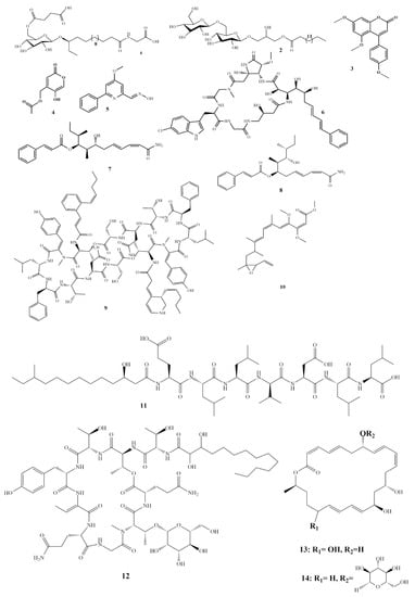
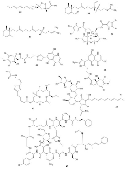
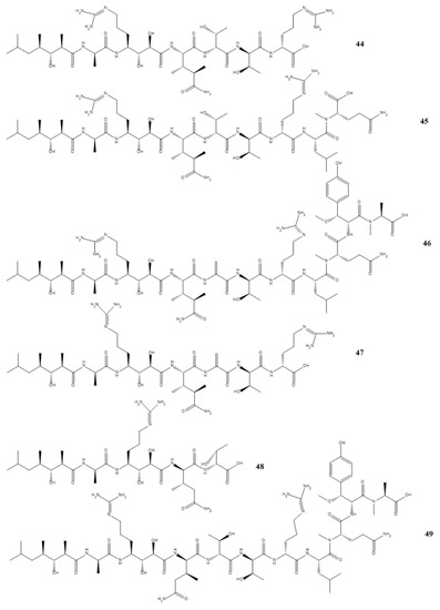
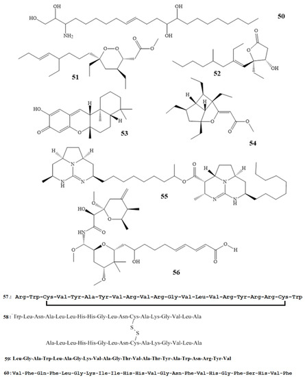
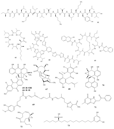
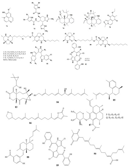
To combat the ineffectiveness of currently available pharmaceutical medications, caused by the emergence of increasingly resistant bacterial and fungal strains, novel antibacterial and antifungal medications are urgently needed. Novel natural compounds with antimicrobial activities can be obtained by exploring underexplored habitats such as the world’s oceans. The oceans represent the largest ecosystem on earth, with a high diversity of organisms. Oceans have received some attention in the past few years, and promising compounds with antimicrobial activities were isolated from marine organisms such as bacteria, fungi, algae, sea cucumbers, sea sponges, etc. This review covers 56 antifungal and 40 antibacterial compounds from marine organisms. These compounds are categorized according to their chemical structure groups, including polyketides, alkaloids, ribosomal peptides, and terpenes, and their organismal origin. The review provides the minimum inhibitory concentration MIC values and the bacterial/fungal strains against which these chemical compounds show activity. This study shows strong potential for witnessing the development of new novel antimicrobial drugs from these natural compounds isolated and evaluated for their antimicrobial activities.
Full article
Scheme 1


