Rapid Detection of Bacterial Toxins
A topical collection in Toxins (ISSN 2072-6651). This collection belongs to the section "Bacterial Toxins".
Viewed by 88796
Share This Topical Collection
Editor
 Dr. Xiaohua He
Dr. Xiaohua He
E-Mail
Website
Collection Editor
Research Molecular Biologist, USDA, ARS, WRRC, 800 Buchanan Street, Albany, CA 94710, USA
Interests: molecular tools and technologies for rapid; accurate; and sensitive detection and quantification of zoonotic pathogens and toxins in food; mechanisms of interactions between bacterial toxins and host cells; binding between antigen and antibody or receptors
Special Issues, Collections and Topics in MDPI journals
Topical Collection Information
Dear Colleagues,
Bacterial toxins, including endotoxins and exotoxins, are major virulence factors that promote infection and disease caused by certain bacterial pathogens. Some of them, such as botulinum neurotoxins, are the most potent natural-occurring toxins known. Ingestion of even a small amount of bacterial toxins present in contaminated food could result in severe food poisoning. Conventional methods for detecting bacterial toxins, such as in vivo animal bioassays or in vitro cell-based toxicity assays, are usually labor and time intensive. With the advancement of biotechnology, the diagnostic procedures used in toxin detection are continually evolving. This Special Issue highlights the recent advancements in rapid methods for detecting bacterial toxins and explores their applications for indirect assessment of the presence of the associated bacteria in different matrices.
Dr. Xiaohua He
Collection Editor
Submission
Manuscripts for the topical collection can be submitted online at www.mdpi.com by registering and logging in to this website. Once you are registered, click here to go to the submission form. All papers will be peer-reviewed. Accepted papers will be published continuously in the journal (as soon as accepted) and will be listed together on this website. The topical collection considers regular research articles, short communications and review articles. A guide for authors and other relevant information for submission of manuscripts is available on the Instructions for Authors page.
Submitted manuscripts should not have been published previously, nor be under consideration for publication elsewhere (except conference proceedings papers). All manuscripts are refereed through a peer-review process. A guide for authors and other relevant information for submission of manuscripts is available on the Instructions for Authors page. Toxins is an international peer-reviewed Open Access monthly journal published by MDPI.
Please visit the Instructions for Authors page before submitting a manuscript. The Article Processing Charge (APC) for publication in this open access journal is 1200 CHF (Swiss Francs).
Keywords
- Bacterial toxin
- Endotoxin
- Exotoxin
- Toxin activity
- Immunoassay
- Mass spectrometry
- Polymerase chain reaction
- Biosensor
- Receptor binding assay
Related Special Issue
Published Papers (15 papers)
Open AccessArticle
Single-Domain Antibody Multimers for Detection of Botulinum Neurotoxin Serotypes C, D, and Their Mosaics in Endopep-MS
by
Michiel M. Harmsen, Jan C. Cornelissen, Fimme J. van der Wal, Jan H. W. Bergervoet and Miriam Koene
Viewed by 1127
Abstract
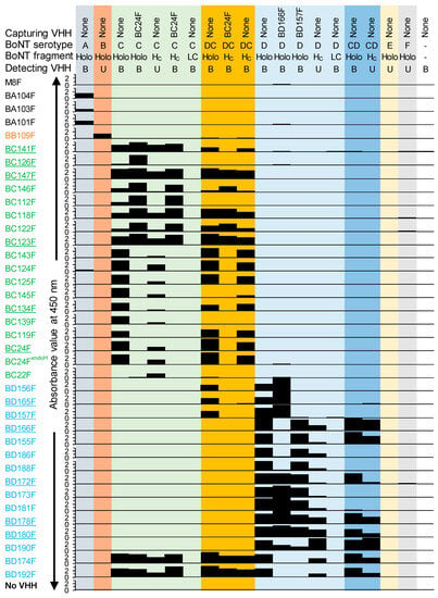
Botulinum neurotoxins (BoNTs) are highly toxic proteins that require high-affinity immunocapture reagents for use in endopeptidase-based assays. Here, 30 novel and 2 earlier published llama single-domain antibodies (VHHs) against the veterinary-relevant BoNT serotypes C and D were yeast-produced. These VHHs recognized 10 independent
[...] Read more.
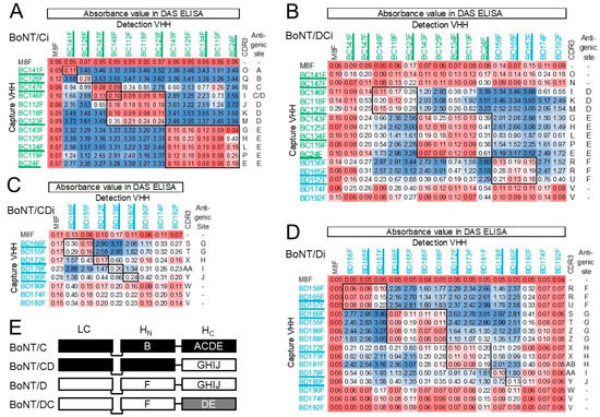
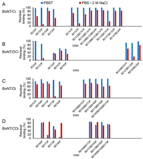
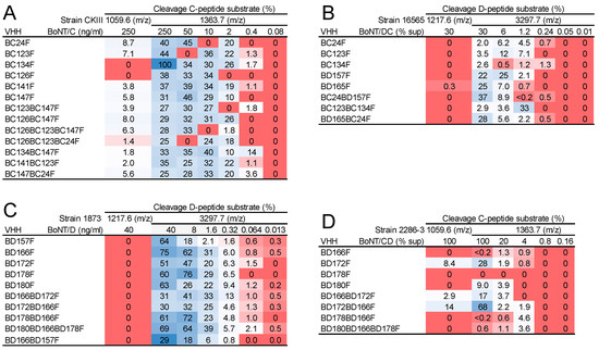
Botulinum neurotoxins (BoNTs) are highly toxic proteins that require high-affinity immunocapture reagents for use in endopeptidase-based assays. Here, 30 novel and 2 earlier published llama single-domain antibodies (VHHs) against the veterinary-relevant BoNT serotypes C and D were yeast-produced. These VHHs recognized 10 independent antigenic sites, and many cross-reacted with the BoNT/DC and CD mosaic variants. As VHHs are highly suitable for genetically linking to increase antigen-binding affinity, 52 VHH multimers were produced and their affinity for BoNT/C, D, DC, and CD was determined. A selection of 15 multimers with high affinity (
KD < 0.1 nM) was further shown to be resilient to a high salt wash that is used for samples from complex matrices and bound native BoNTs from culture supernatants as shown by Endopep-MS. High-affinity multimers suitable for further development of a highly sensitive Endopep-MS assay include four multimers that bind both BoNT/D and CD with
KD of 14–99 pM, one multimer for BoNT/DC (65 pM) that also binds BoNT/C (75 pM), and seven multimers for BoNT/C (<1–19 pM), six of which also bind BoNT/DC with lower affinity (93–508 pM). In addition to application in diagnostic tests, these VHHs could be used for the development of novel therapeutics for animals or humans.
Full article
►▼
Show Figures
Open AccessFeature PaperArticle
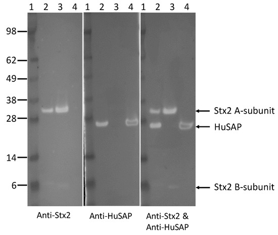
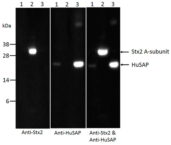
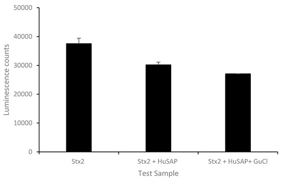
An Improved Method for the Sensitive Detection of Shiga Toxin 2 in Human Serum
by
Xiaohua He, Gianluigi Ardissino, Stephanie Patfield, Luisa W. Cheng, Christopher J. Silva and Maurizio Brigotti
Cited by 14 | Viewed by 4044
Abstract
Shiga toxins (Stx) released by Stx-producing
E. coli (STEC) are virulence factors that are most closely associated with hemolytic uremic syndrome (HUS), a life-threatening complication of intestinal infections by STEC. Stx have to enter into the circulatory system before they are delivered to
[...] Read more.
Shiga toxins (Stx) released by Stx-producing
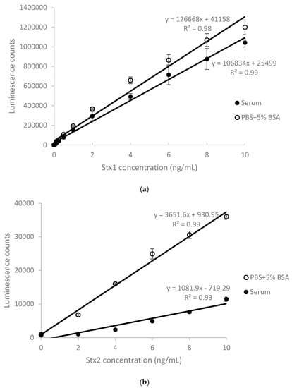
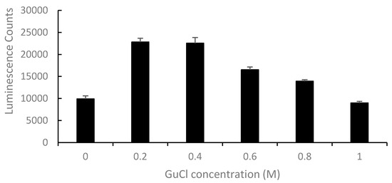
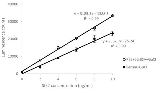
E. coli (STEC) are virulence factors that are most closely associated with hemolytic uremic syndrome (HUS), a life-threatening complication of intestinal infections by STEC. Stx have to enter into the circulatory system before they are delivered to target organs and cause damage. The presence of Stx in sera could be a risk indicator for HUS development. However, the detection of Stx, particularly Stx2, has been difficult due to the presence of Stx2-binding components in human serum. Here, we report new ELISA-based methods for the detection of Stx1 and Stx2 in human serum and the effect of guanidinium chloride on enhancing the sensitivity for the detection of Stx2. The recovery rate for Stx2 was 62% when Stx2-spiked serum samples were treated with guanidinium chloride at a concentration of 200 mM, in contrast to 17% without guanidinium chloride treatment. The effectiveness of guanidinium chloride treatment for the detection of Stx2 in human serum was validated using sera from STEC-infected patients. Coimmunoprecipitation results indicated a specific physical interaction between Stx2 and the human serum amyloid P component (HuSAP) in human serum samples. Our in vitro study demonstrated that the inhibition from HuSAP alone for the detection of Stx2 was only 20%, much less than 69.6% from human serum at Stx2 level 10 ng/mL, suggesting that there may be other factors that bind Stx2 in human serum. This study indicates that treatment of serum samples with guanidinium chloride may be useful for the early and sensitive detection of Stx2 in sera of STEC-infected patients, so preventive measures can be adopted in a timely manner.
Full article
►▼
Show Figures
Open AccessArticle
Implementing the Bruker MALDI Biotyper in the Public Health Laboratory for C. botulinum Neurotoxin Detection
by
Michael J. Perry, Dominick A. Centurioni, Stephen W. Davis, George E. Hannett, Kimberlee A. Musser and Christina T. Egan
Cited by 13 | Viewed by 6592
Abstract
Currently, the gold standard method for active botulinum neurotoxin (BoNT) detection is the mouse bioassay (MBA). A Centers for Disease Control and Prevention-developed mass spectrometry (MS)-based assay that detects active BoNT was successfully validated and implemented in a public health laboratory in clinical
[...] Read more.
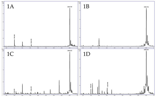
Currently, the gold standard method for active botulinum neurotoxin (BoNT) detection is the mouse bioassay (MBA). A Centers for Disease Control and Prevention-developed mass spectrometry (MS)-based assay that detects active BoNT was successfully validated and implemented in a public health laboratory in clinical matrices using the Bruker MALDI-TOF MS (Matrix-assisted laser desorption ionization–time of flight mass spectrometry) Biotyper. For the first time, a direct comparison with the MBA was performed to determine MS-based assay sensitivity using the Bruker MALDI Biotyper. Mice were injected with BoNT/A, /B, /E, and /F at concentrations surrounding the established MS assay limit of detection (LOD) and analyzed simultaneously. For BoNT/B, /E, and /F, MS assay sensitivity was equivalent or better than the MBA at 25, 0.3, and 8.8 mLD
50, respectively. BoNT/A was detected by the MBA between 1.8 and 18 mLD
50, somewhat more sensitive than the MS method of 18 mLD
50. Studies were performed to compare assay performance in clinical specimens. For all tested specimens, the MS method rapidly detected BoNT activity and serotype in agreement with, or in the absence of, results from the MBA. We demonstrate that the MS assay can generate reliable, rapid results while eliminating the need for animal testing.
Full article
►▼
Show Figures
Open AccessArticle
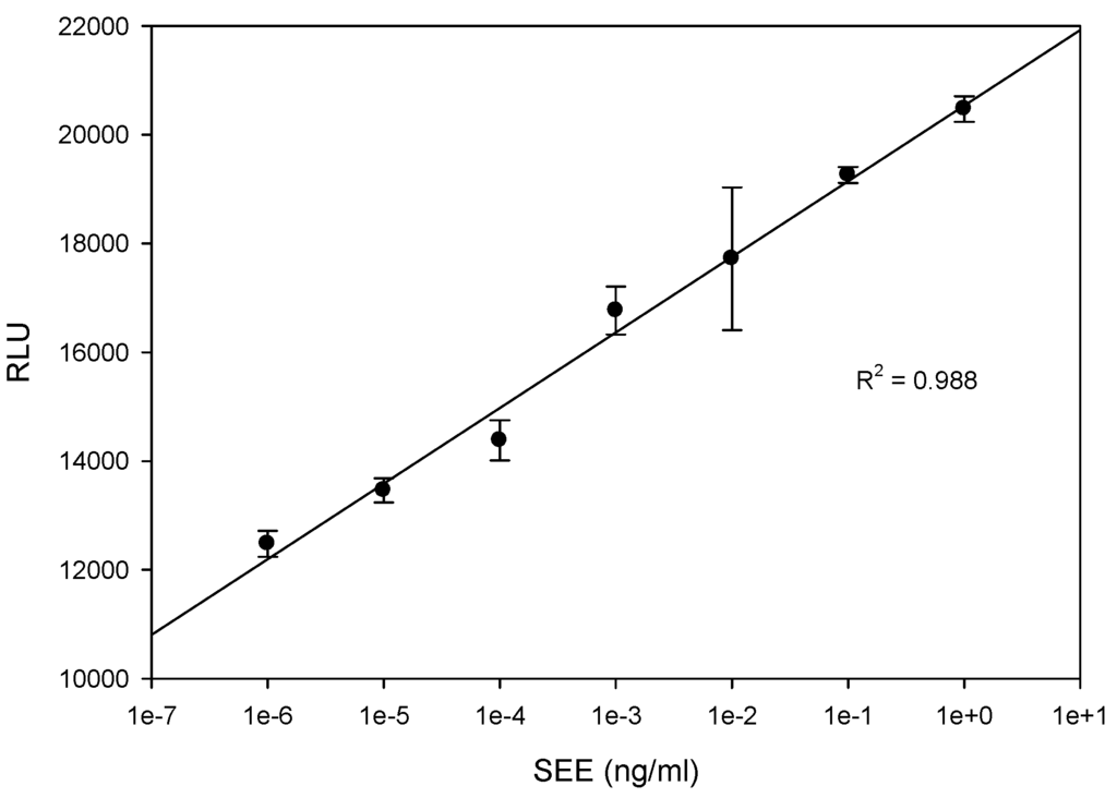
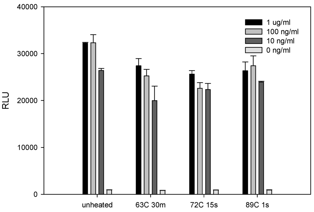
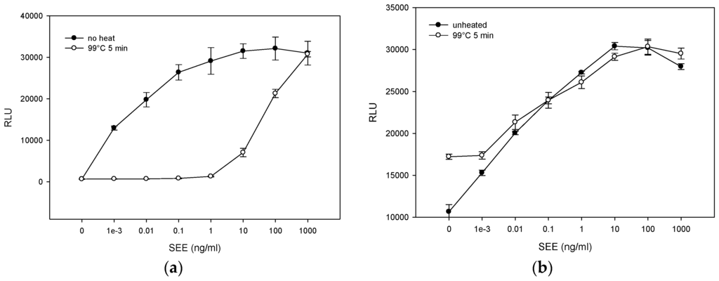
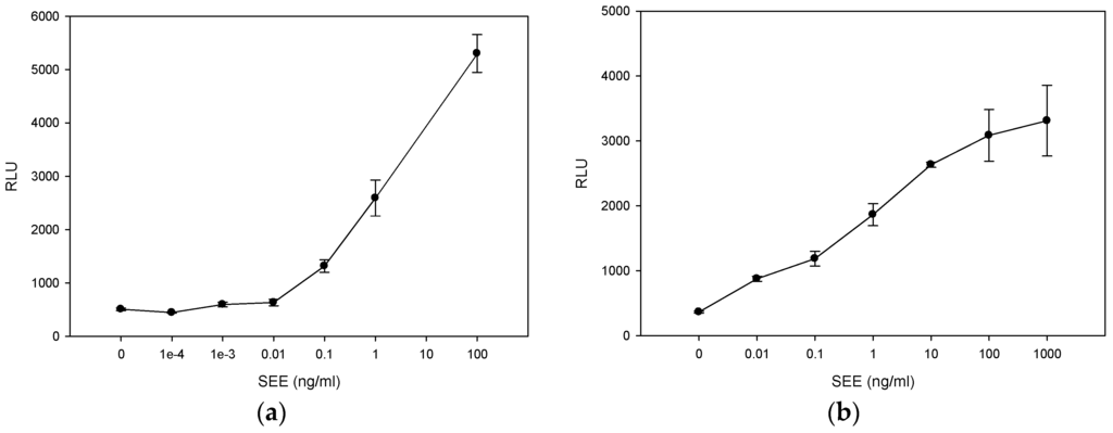
Sensitive, Rapid, Quantitative and in Vitro Method for the Detection of Biologically Active Staphylococcal Enterotoxin Type E
by
Reuven Rasooly, Paula Do and Bradley Hernlem
Cited by 7 | Viewed by 5080
Abstract
Staphylococcus aureus is a major bacterial cause of clinical infections and foodborne illnesses through its production of a group of enterotoxins (SEs) which cause gastroenteritis and also function as superantigens to massively activate T cells. In the present study, we tested Staphylococcal enterotoxin
[...] Read more.
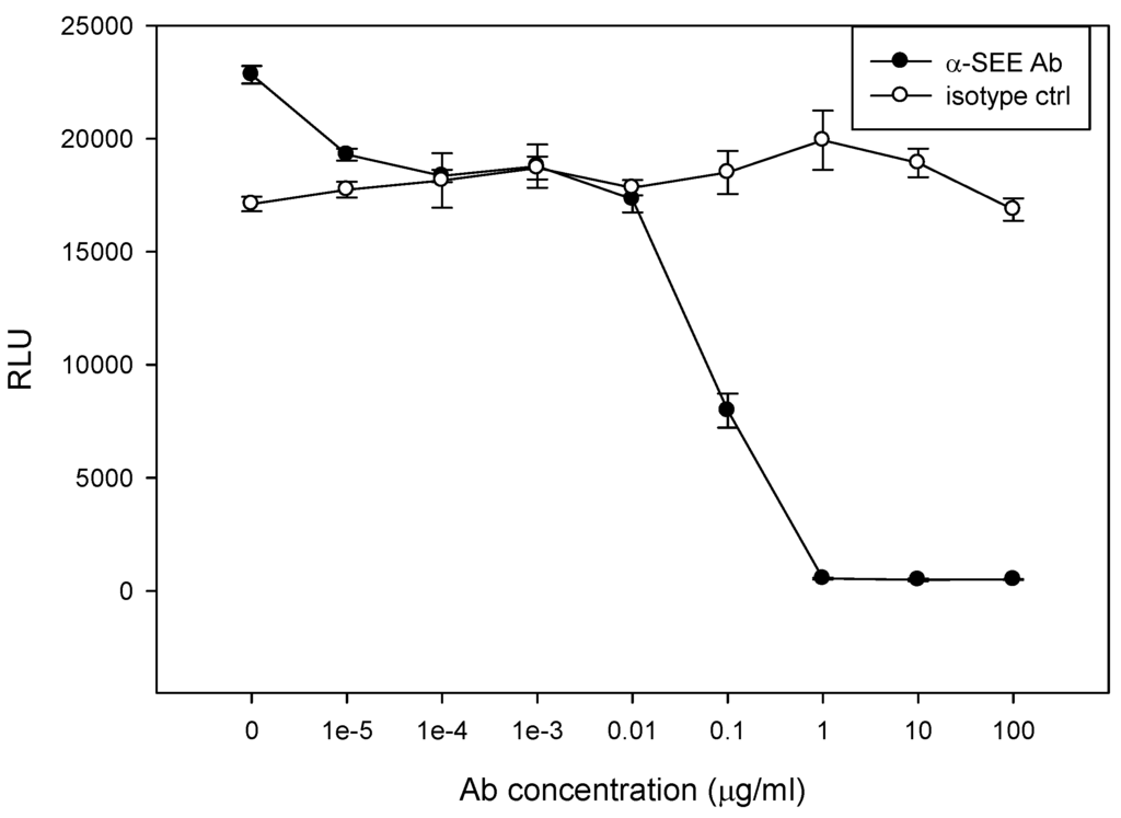
Staphylococcus aureus is a major bacterial cause of clinical infections and foodborne illnesses through its production of a group of enterotoxins (SEs) which cause gastroenteritis and also function as superantigens to massively activate T cells. In the present study, we tested Staphylococcal enterotoxin type E (SEE), which was detected in 17 of the 38 suspected staphylococcal food poisoning incidents in a British study and was the causative agent in outbreaks in France, UK and USA. The current method for detection of enterotoxin activity is an
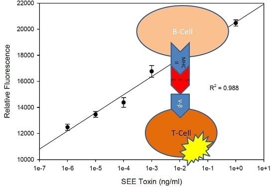
in vivo monkey or kitten bioassay; however, this expensive procedure has low sensitivity and poor reproducibility, requires many animals, is impractical to test on a large number of samples, and raises ethical concerns with regard to the use of experimental animals. The purpose of this study is to develop rapid sensitive and quantitative bioassays for detection of active SEE. We apply a genetically engineered T cell-line expressing the luciferase reporter gene under the regulation of nuclear factor of activated T-cells response element (NFAT-RE), combined with a Raji B-cell line that presents the SEE-MHC (major histocompatibility complex) class II to the engineered T cell line. Exposure of the above mixed culture to SEE induces differential expression of the luciferase gene and bioluminescence is read out in a dose dependent manner over a 6-log range. The limit of detection of biologically active SEE is 1 fg/mL which is 10
9 times more sensitive than the monkey and kitten bioassay.
Full article
►▼
Show Figures
Open AccessArticle
Review Over a 3-Year Period of European Union Proficiency Tests for Detection of Staphylococcal Enterotoxins in Food Matrices
by
Yacine Nia, Isabelle Mutel, Adrien Assere, Bertrand Lombard, Frederic Auvray and Jacques-Antoine Hennekinne
Cited by 20 | Viewed by 5847
Abstract
Staphylococcal food poisoning outbreaks are a major cause of foodborne illnesses in Europe and their notifications have been mandatory since 2005. Even though the European regulation on microbiological criteria for food defines a criterion on staphylococcal enterotoxin (SE) only in cheese and dairy
[...] Read more.
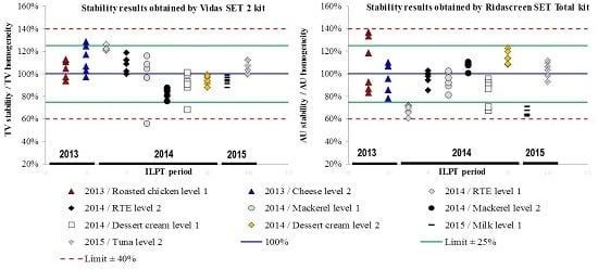
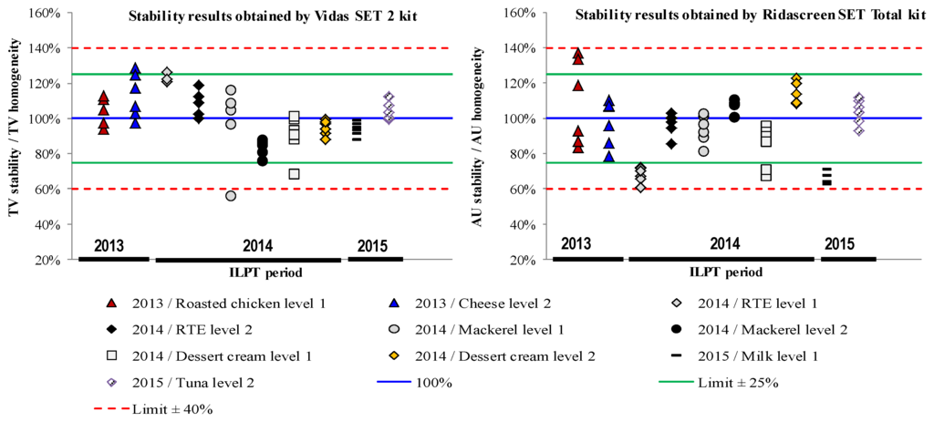
Staphylococcal food poisoning outbreaks are a major cause of foodborne illnesses in Europe and their notifications have been mandatory since 2005. Even though the European regulation on microbiological criteria for food defines a criterion on staphylococcal enterotoxin (SE) only in cheese and dairy products, European Food Safety Authority (EFSA) data reported that various types of food matrices are involved in staphylococcal food poisoning outbreaks. The European Screening Method (ESM) of European Union Reference Laboratory for Coagulase Positive Staphylococci (EURL CPS) was validated in 2011 for SE detection in food matrices and is currently the official method used for screening purposes in Europe. In this context, EURLCPS is annually organizing Inter-Laboratory Proficiency Testing Trials (ILPT) to evaluate the competency of the European countries’ National Reference Laboratories (NRLs) to analyse SE content in food matrices. A total of 31 NRLs representing 93% of European countries participated in these ILPTs. Eight food matrices were used for ILPT over the period 2013–2015, including cheese, freeze-dried cheese, tuna, mackerel, roasted chicken, ready-to-eat food, milk, and pastry. Food samples were spiked with four SE types (
i.e., SEA, SEC, SED, and SEE) at various concentrations. Homogeneity and stability studies showed that ILPT samples were both homogeneous and stable. The analysis of results obtained by participants for a total of 155 blank and 620 contaminated samples allowed for evaluation of trueness (>98%) and specificity (100%) of ESM. Further to the validation study of ESM carried out in 2011, these three ILPTs allowed for the assessment of the proficiency of the NRL network and the performance of ESM on a large variety of food matrices and samples. The ILPT design presented here will be helpful for the organization of ILPT on SE detection by NRLs or other expert laboratories.
Full article
►▼
Show Figures
Open AccessFeature PaperArticle
Rapid Detection of Escherichia coli O157 and Shiga Toxins by Lateral Flow Immunoassays
by
Jinliang Wang, Robab Katani, Lingling Li, Narasimha Hegde, Elisabeth L. Roberts, Vivek Kapur and Chitrita DebRoy
Cited by 30 | Viewed by 8913
Abstract
Shiga toxin-producing
Escherichia coli O157:H7 (STEC) cause food-borne illness that may be fatal. STEC strains enumerate two types of potent Shiga toxins (Stx1 and Stx2) that are responsible for causing diseases. It is important to detect the
E. coli O157 and Shiga toxins
[...] Read more.
Shiga toxin-producing
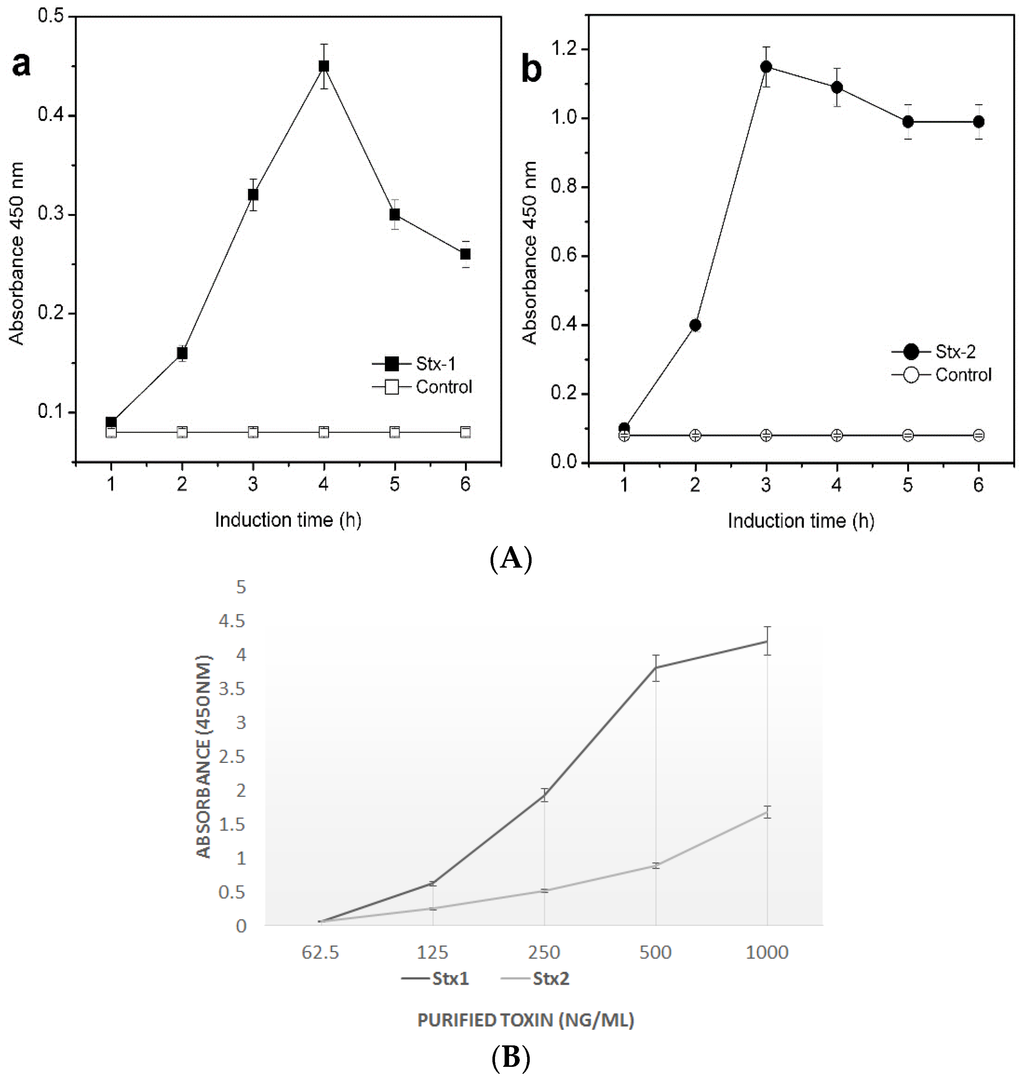
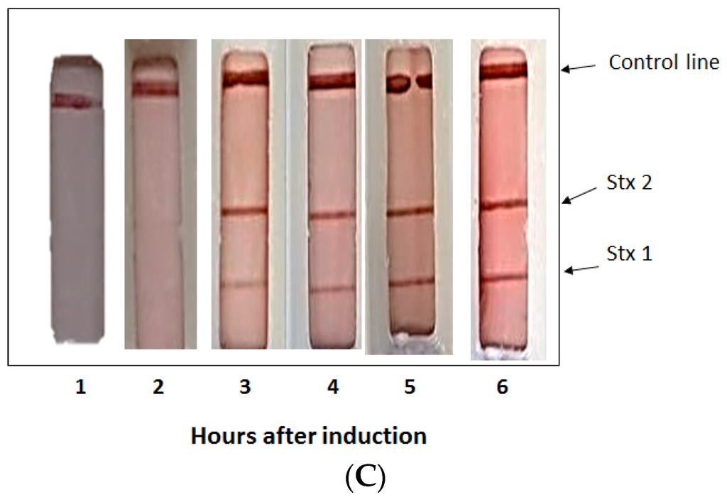
Escherichia coli O157:H7 (STEC) cause food-borne illness that may be fatal. STEC strains enumerate two types of potent Shiga toxins (Stx1 and Stx2) that are responsible for causing diseases. It is important to detect the
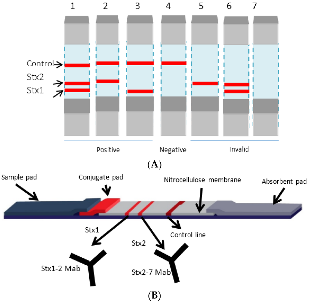
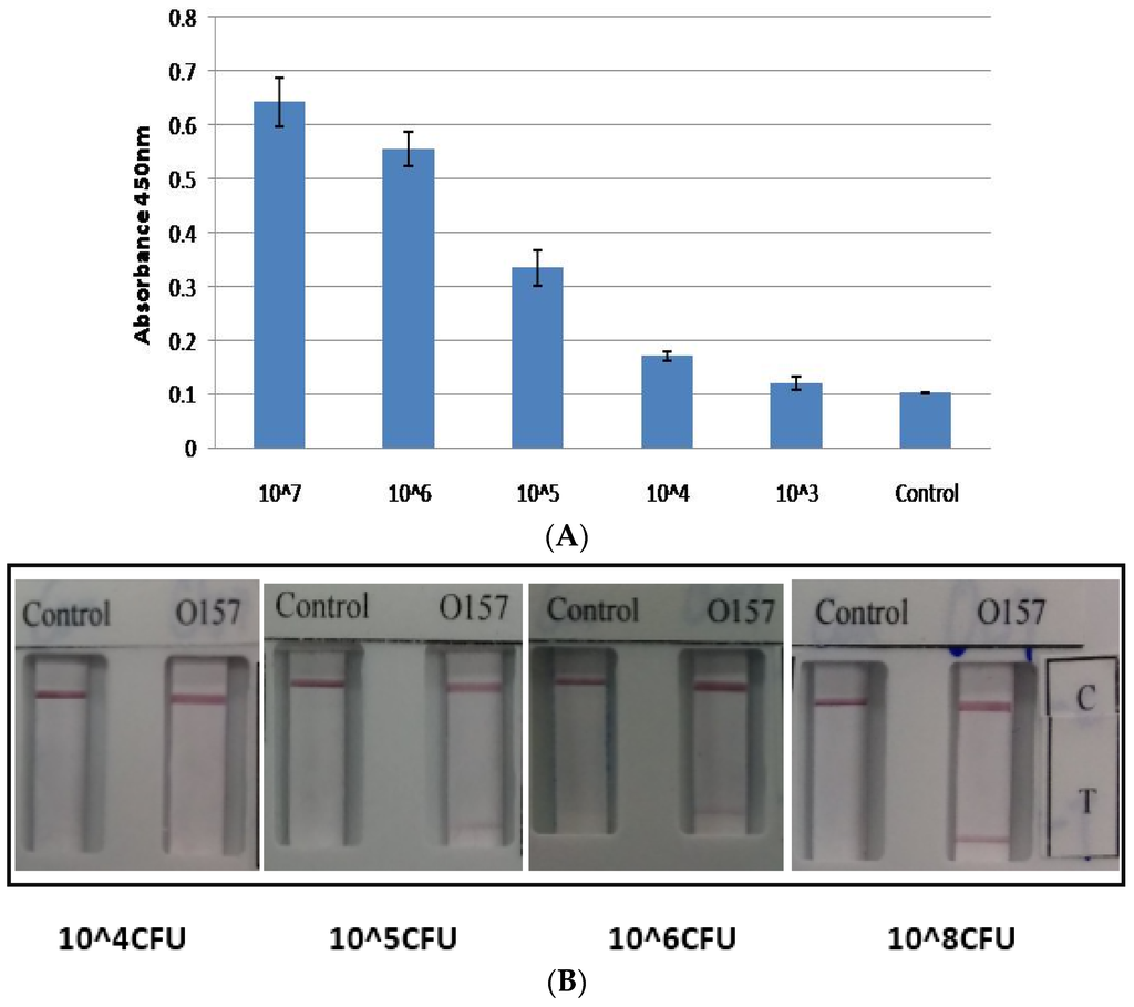
E. coli O157 and Shiga toxins in food to prevent outbreak of diseases. We describe the development of two multi-analyte antibody-based lateral flow immunoassays (LFIA); one for the detection of Stx1 and Stx2 and one for the detection of
E. coli O157 that may be used simultaneously to detect pathogenic
E. coli O157:H7. The LFIA strips were developed by conjugating nano colloidal gold particles with monoclonal antibodies against Stx1 and Stx2 and anti-lipid A antibodies to capture Shiga toxins and O157 antigen, respectively. Our results indicate that the LFIA for Stx is highly specific and detected Stx1 and Stx2 within three hours of induction of STEC with ciprofloxacin at 37 °C. The limit of detection for
E. coli O157 LFIA was found to be 10
5 CFU/mL in ground beef spiked with the pathogen. The LFIAs are rapid, accurate and easy to use and do not require sophisticated equipment or trained personnel. Following the assay, colored bands on the membrane develop for end-point detection. The LFIAs may be used for screening STEC in food and the environment.
Full article
►▼
Show Figures
Open AccessFeature PaperArticle
Rapid Microfluidic Assay for the Detection of Botulinum Neurotoxin in Animal Sera
by
Lmar Babrak, Alice Lin, Larry H. Stanker, Jeffery McGarvey and Robert Hnasko
Cited by 17 | Viewed by 6202
Abstract
Potent Botulinum neurotoxins (BoNTs) represent a threat to public health and safety. Botulism is a disease caused by BoNT intoxication that results in muscle paralysis that can be fatal. Sensitive assays capable of detecting BoNTs from different substrates and settings are essential to
[...] Read more.
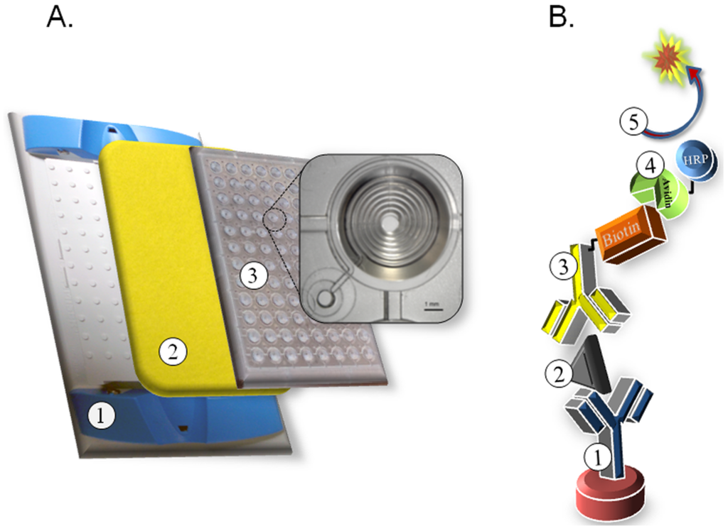
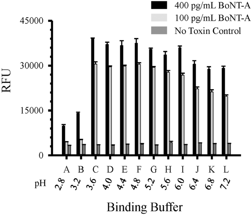
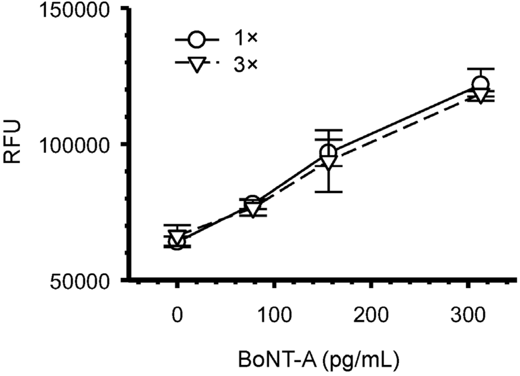
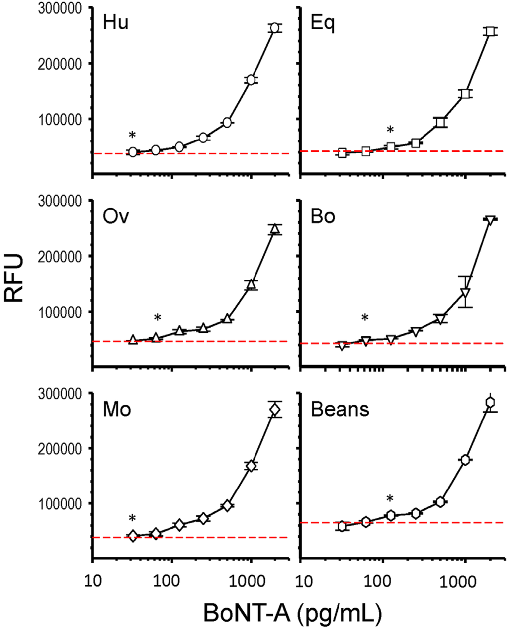
Potent Botulinum neurotoxins (BoNTs) represent a threat to public health and safety. Botulism is a disease caused by BoNT intoxication that results in muscle paralysis that can be fatal. Sensitive assays capable of detecting BoNTs from different substrates and settings are essential to limit foodborne contamination and morbidity. In this report, we describe a rapid 96-well microfluidic double sandwich immunoassay for the sensitive detection of BoNT-A from animal sera. This BoNT microfluidic assay requires only 5 μL of serum, provides results in 75 min using a standard fluorescence microplate reader and generates minimal hazardous waste. The assay has a <30 pg·mL
−1 limit of detection (LOD) of BoNT-A from spiked human serum. This sensitive microfluidic BoNT-A assay offers a fast and simplified workflow suitable for the detection of BoNT-A from serum samples of limited volume in most laboratory settings.
Full article
►▼
Show Figures
Open AccessFeature PaperArticle
Use of Monoclonal Antibodies in the Sensitive Detection and Neutralization of Botulinum Neurotoxin Serotype B
by
Luisa W. Cheng, Thomas D. Henderson, Tina I. Lam and Larry H. Stanker
Cited by 6 | Viewed by 5440
Abstract
Botulinum neurotoxins (BoNT) are some of nature’s most potent toxins. Due to potential food contamination, and bioterrorism concerns, the development of detection reagents, therapeutics and countermeasures are of urgent interest. Recently, we have developed a sensitive electrochemiluminescent (ECL) immunoassay for BoNT/B, using monoclonal
[...] Read more.
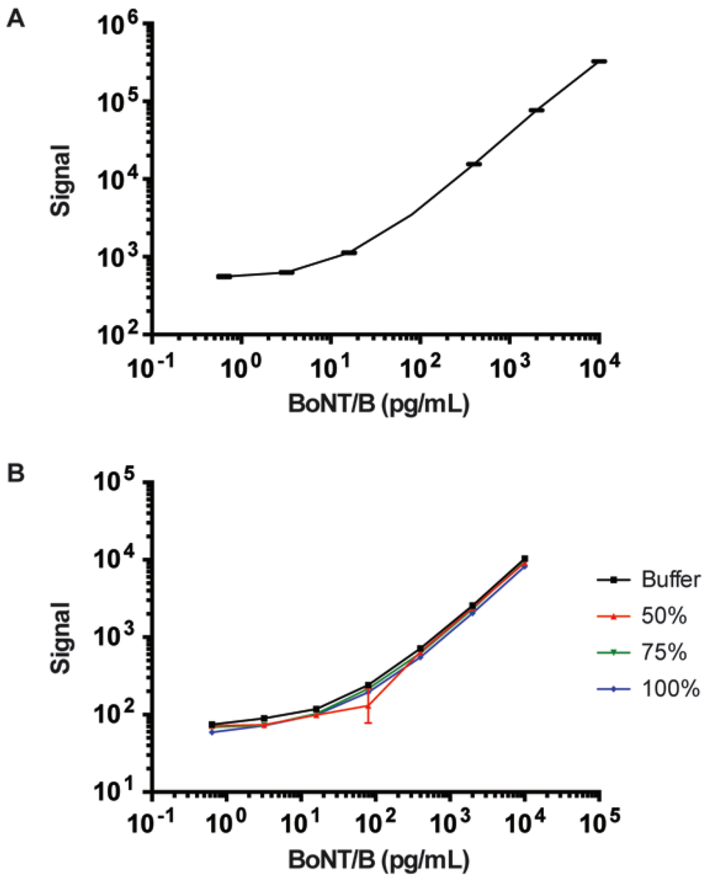
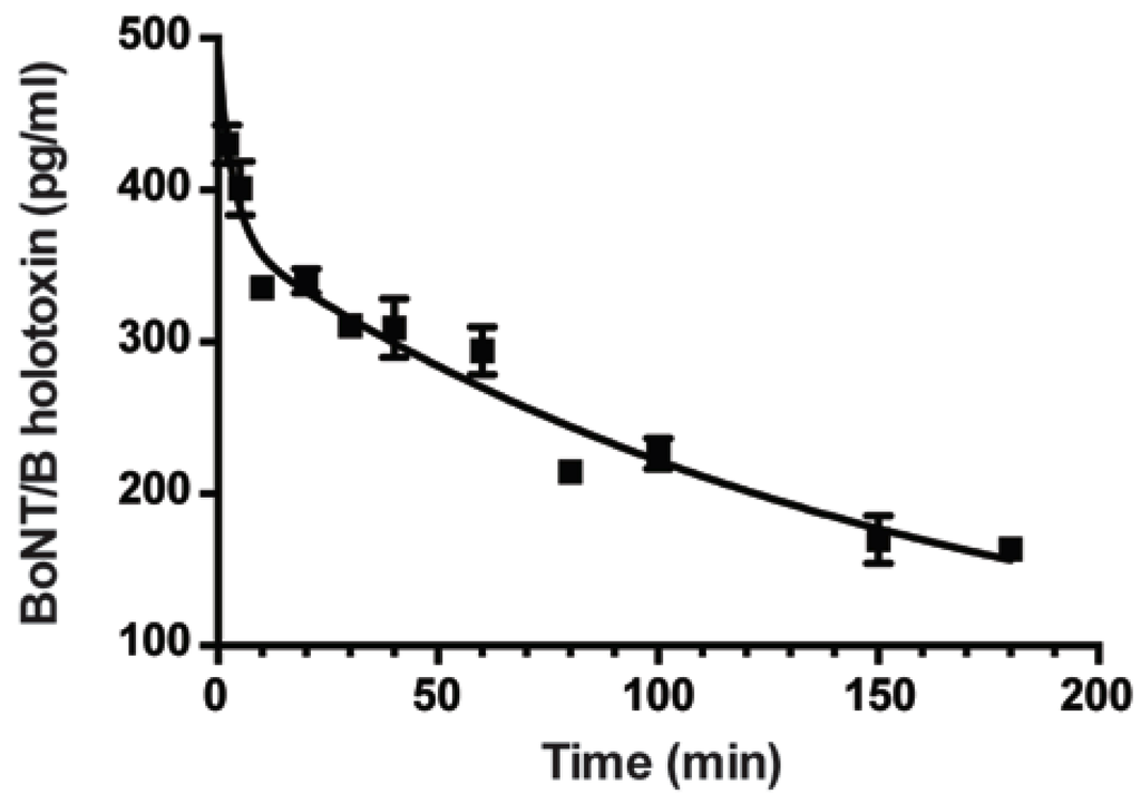
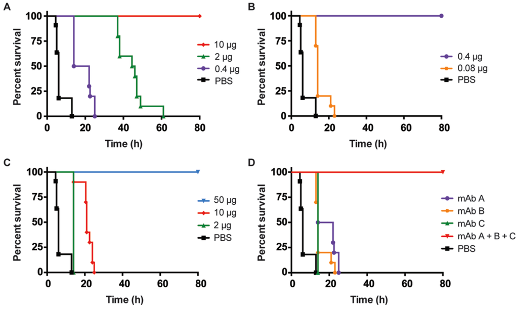
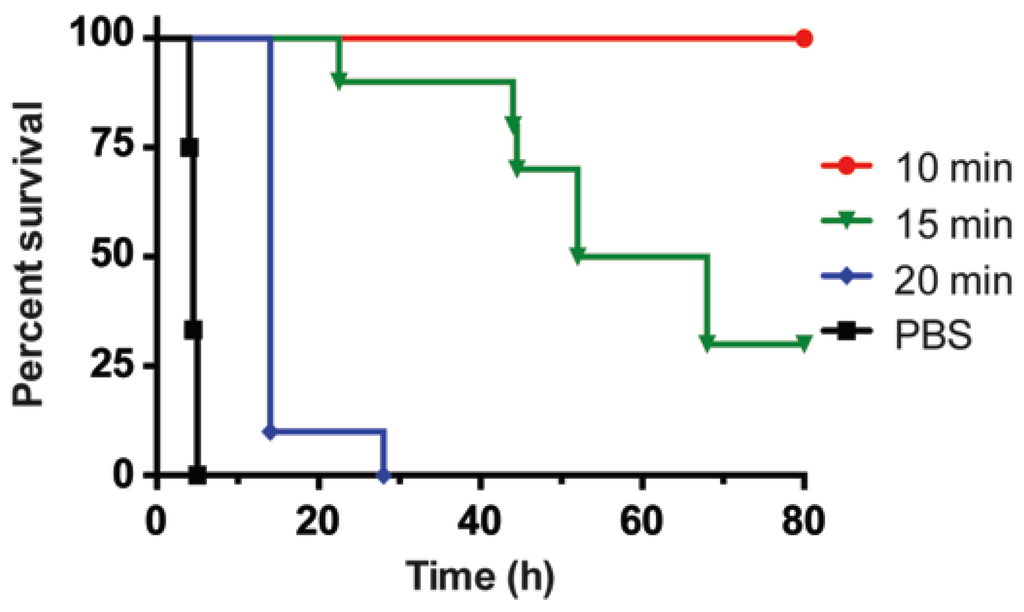
Botulinum neurotoxins (BoNT) are some of nature’s most potent toxins. Due to potential food contamination, and bioterrorism concerns, the development of detection reagents, therapeutics and countermeasures are of urgent interest. Recently, we have developed a sensitive electrochemiluminescent (ECL) immunoassay for BoNT/B, using monoclonal antibodies (mAbs) MCS6-27 and anti-BoNT/B rabbit polyclonal antibodies as the capture and detector. The ECL assay detected as little as 1 pg/mL BoNT/B in the buffer matrix, surpassing the detection sensitivities of the gold standard mouse bioassays. The ECL assay also allowed detection of BoNT/B in sera matrices of up to 100% sera with negligible matrix effects. This highly-sensitive assay allowed the determination of the biological half-lives of BoNT/B holotoxin
in vivo. We further tested the toxin neutralization potential of our monoclonal antibodies using the mouse systemic and oral intoxication models. A combination of mAbs protected mice in both pre- and post-exposure models to lethal doses of BoNT/B. MAbs were capable of increasing survival of animals when administered even 10 h post-intoxication in an oral model, suggesting a likely time for BoNT/B complexes to reach the blood stream. More sensitive detection assays and treatments against BoNT intoxication will greatly enhance efforts to combat botulism.
Full article
►▼
Show Figures
Open AccessArticle
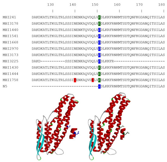
The Mutation Glu151Asp in the B-Component of the Bacillus cereus Non-Hemolytic Enterotoxin (Nhe) Leads to a Diverging Reactivity in Antibody-Based Detection Systems
by
Andrea Didier, Nadja Jeßberger, Victoria Krey, Richard Dietrich, Siegfried Scherer and Erwin Märtlbauer
Cited by 6 | Viewed by 5131
Abstract
The ability of
Bacillus cereus to cause foodborne toxicoinfections leads to increasing concerns regarding consumer protection. For the diarrhea-associated enterotoxins, the assessment of the non-hemolytic enterotoxin B (NheB) titer determined by a sandwich enzyme immunoassay (EIA) correlates best with
in vitro cytotoxicity. In
[...] Read more.
The ability of
Bacillus cereus to cause foodborne toxicoinfections leads to increasing concerns regarding consumer protection. For the diarrhea-associated enterotoxins, the assessment of the non-hemolytic enterotoxin B (NheB) titer determined by a sandwich enzyme immunoassay (EIA) correlates best with
in vitro cytotoxicity. In general, the regulation of enterotoxin expression of
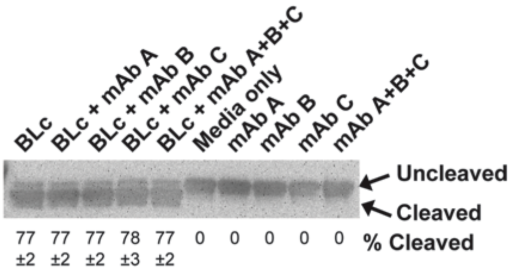
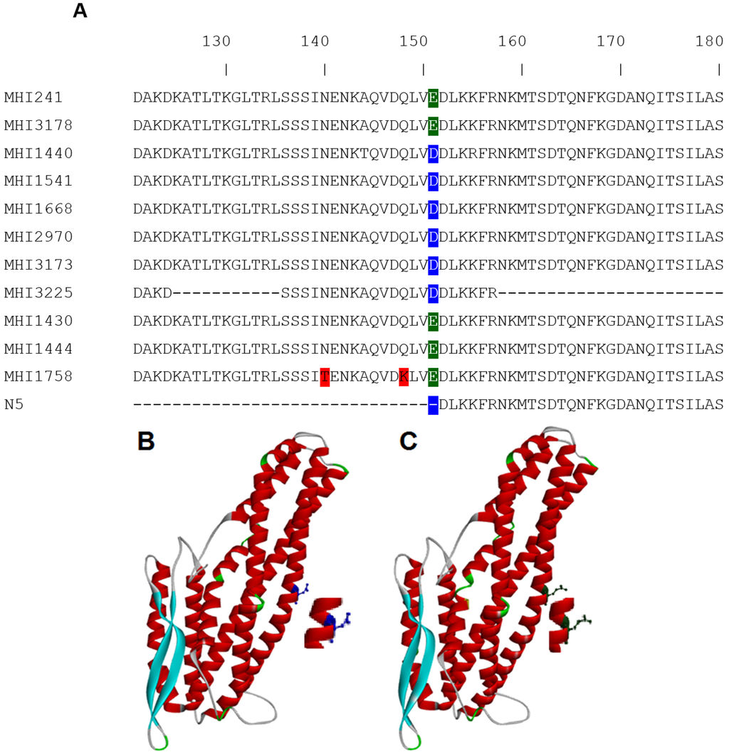
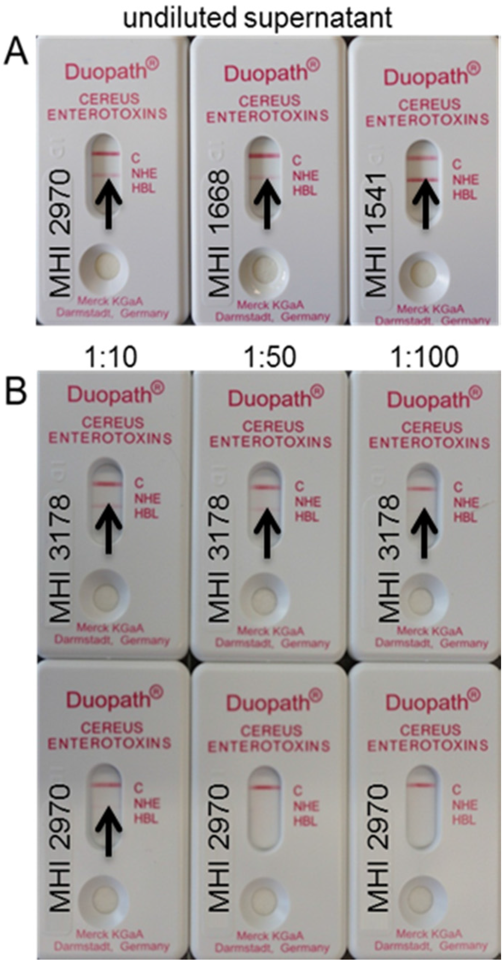
B. cereus is a coordinately-regulated process influenced by environmental, and probably also by host factors. As long as these factors are not completely understood, the currently-applied diagnostic procedures are based on indirect approaches to assess the potential virulence of an isolate. To date, sandwich EIA results serve as a surrogate marker to categorize isolates as either potentially low or highly toxic. Here, we report on a single amino acid exchange in the NheB sequence leading to an underestimation of the cytotoxic potential in a limited number of strains. During the screening of a large panel of
B. cereus isolates, six showed uncommon features with low sandwich EIA titers despite high cytotoxicity. Sequence analysis revealed the point-mutation
Glu151
Asp in the potential binding region of the capture antibody. Application of this antibody also results in low titers in an indirect EIA format and shows variable detection intensities in Western-immunoblots. A commercially-available assay based on a lateral flow device detects all strains correctly as NheB producers in a qualitative manner. In conclusion, isolates showing low NheB titers should additionally be assayed in an indirect EIA or for their
in vitro cytotoxicity to ensure a correct classification as either low or highly toxic.
Full article
►▼
Show Figures
Open AccessArticle
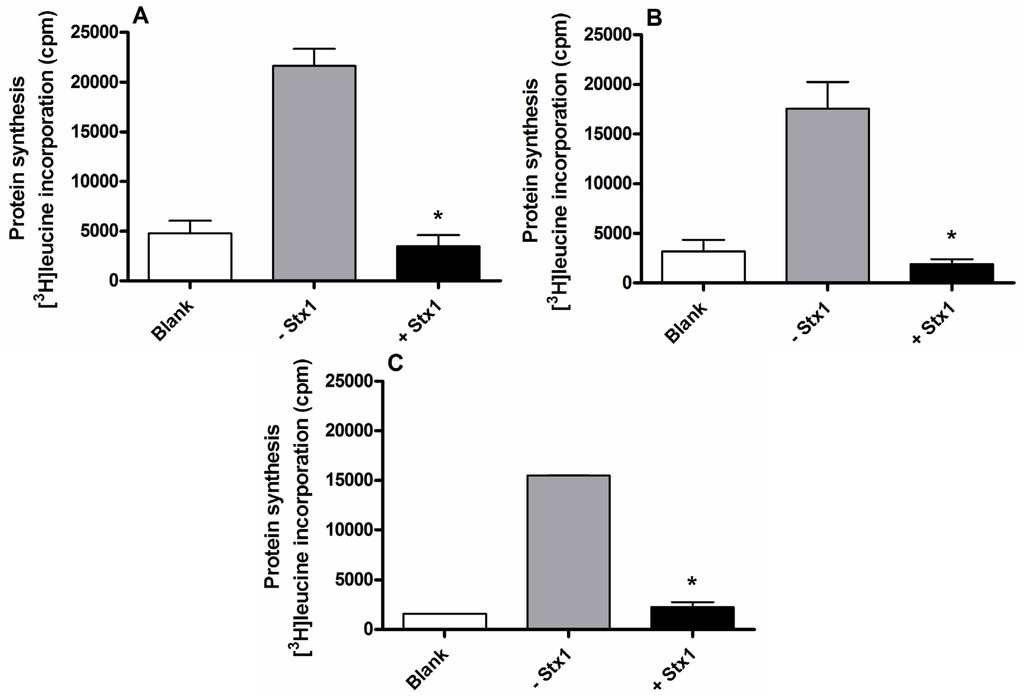
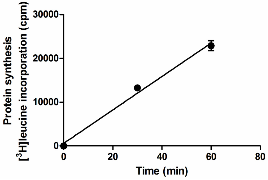
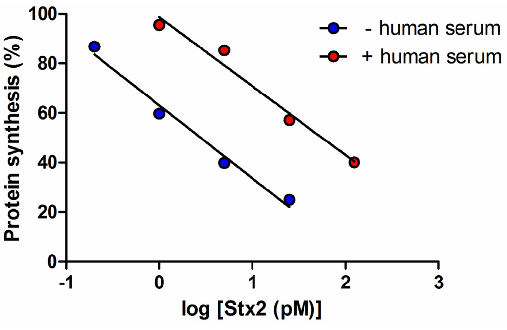
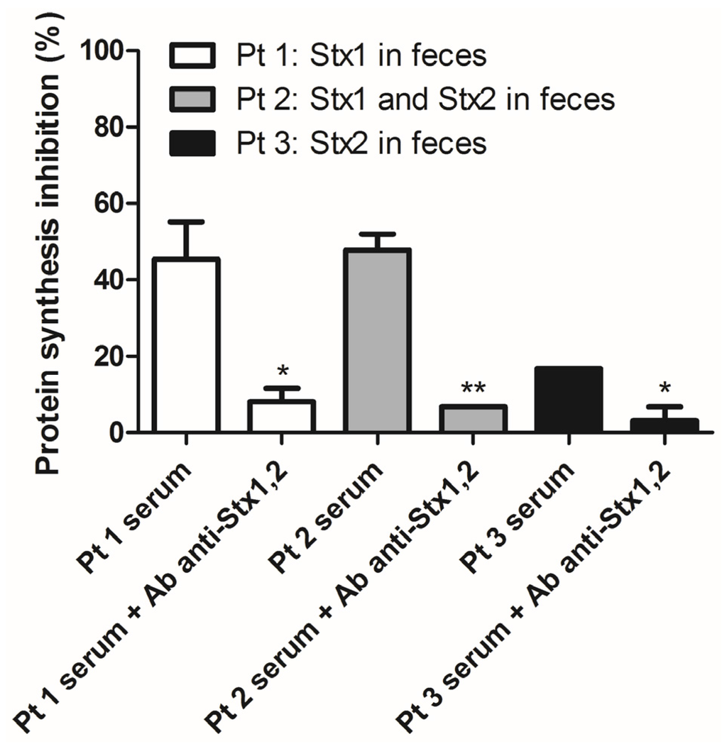
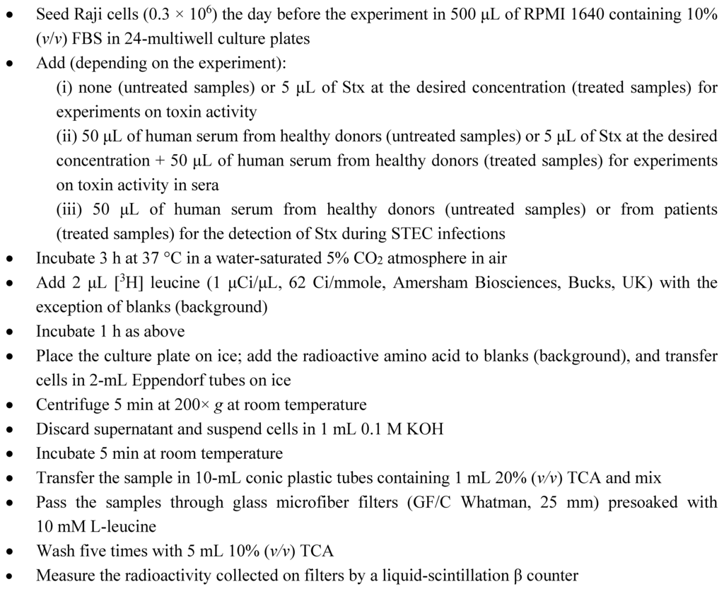
A Rapid and Sensitive Method to Measure the Functional Activity of Shiga Toxins in Human Serum
by
Valentina Arfilli, Domenica Carnicelli, Gianluigi Ardissino, Erminio Torresani, Gaia Scavia and Maurizio Brigotti
Cited by 12 | Viewed by 5003
Abstract
Shiga toxins (Stx) have a definite role in the development of hemolytic uremic syndrome in children with hemorrhagic colitis caused by pathogenic Stx-producing
Escherichia coli (STEC) strains. The dramatic effects of these toxins on the microvasculature of different organs, particularly of the kidney,
[...] Read more.
Shiga toxins (Stx) have a definite role in the development of hemolytic uremic syndrome in children with hemorrhagic colitis caused by pathogenic Stx-producing
Escherichia coli (STEC) strains. The dramatic effects of these toxins on the microvasculature of different organs, particularly of the kidney, are well known, whereas there is no consensus on the mechanism by which Stx reach the endothelia of target organs and/or indirectly injure these body sites. We hereby describe a quick (4 h), radioactive, Raji cell-based method designed for the detection of Stx in human sera. The assay monitors the translation impairment induced by these powerful inhibitors of protein synthesis, which are identified properly by neutralizing their activity with specific monoclonal antibodies. By this method, we detected for the first time the functional activity of Stx in sera of STEC-infected patients during hemorrhagic colitis. Recent research has pointed to a dynamic process of Stx-induced renal intoxication in which concurrent and interactive steps are involved. Our rapid and specific method could be useful for studying the kinetics of Stx during the natural course of STEC infection and the interplay between Stx activity in serum and Stx presence in different blood fractions (neutrophils, monocytes, platelets, leukocyte-platelet aggregates, microvesicles, lipoproteins).
Full article
►▼
Show Figures
Open AccessBrief Report
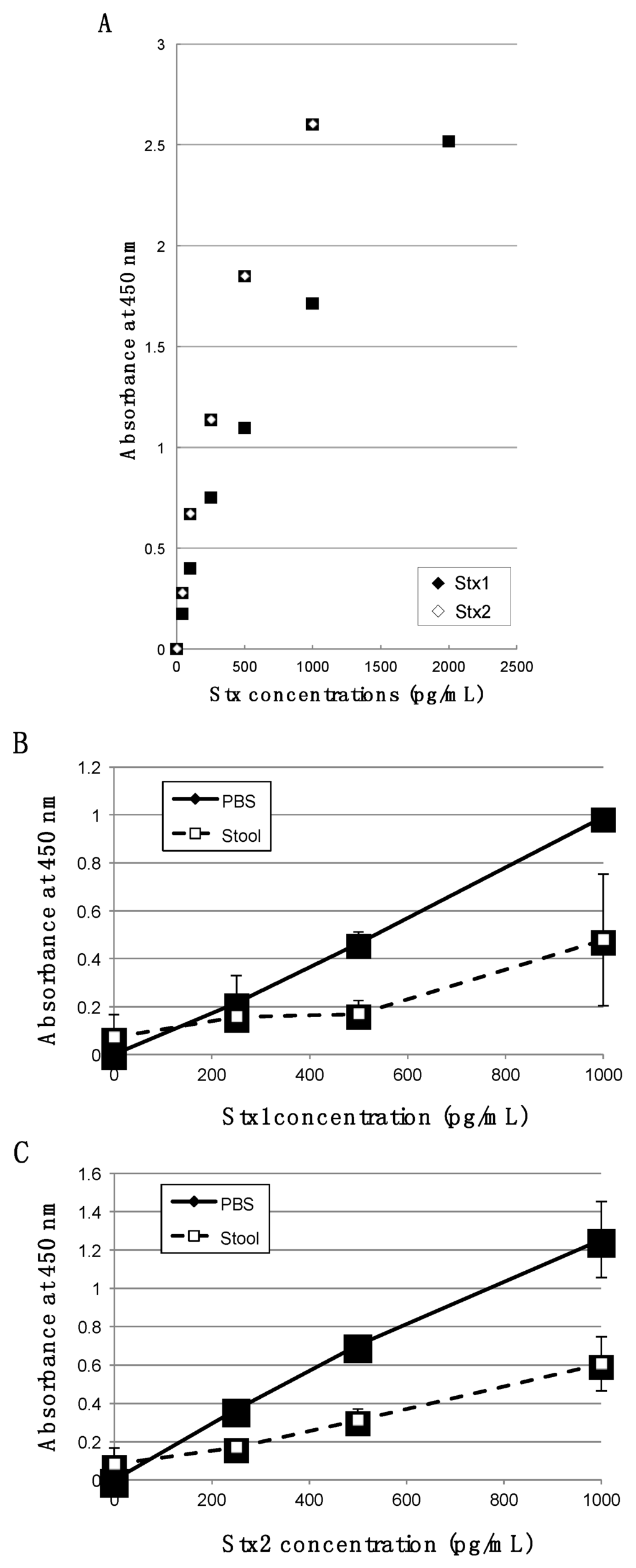
Quantitative Detection of Shiga Toxins Directly from Stool Specimens of Patients Associated with an Outbreak of Enterohemorrhagic Escherichia coli in Japan—Quantitative Shiga toxin detection from stool during EHEC outbreak
by
Eiki Yamasaki, Masanori Watahiki, Junko Isobe, Tetsutaro Sata, G. Balakrish Nair and Hisao Kurazono
Cited by 13 | Viewed by 5295
Abstract
Detection of Shiga toxins (Stx) is important for accurate diagnosis of Enterohemorrhagic
Escherichia coli infection. In this study, we quantitatively analyzed Stx protein in nine patients’ stool during an outbreak that occurred in Japan. Highly sensitive immunoassay (bead enzyme-linked immunosorbent assay (bead-ELISA)) revealed
[...] Read more.
Detection of Shiga toxins (Stx) is important for accurate diagnosis of Enterohemorrhagic
Escherichia coli infection. In this study, we quantitatively analyzed Stx protein in nine patients’ stool during an outbreak that occurred in Japan. Highly sensitive immunoassay (bead enzyme-linked immunosorbent assay (bead-ELISA)) revealed that the concentrations of toxins in stool of patients ranged from 0.71 to 10.44 ng/mL for Stx1 and 2.75 to 51.61 ng/mL for Stx2. To our knowledge, this is the first report that reveals the range of Stx protein concentrations in human stools.
Full article
►▼
Show Figures
Open AccessArticle
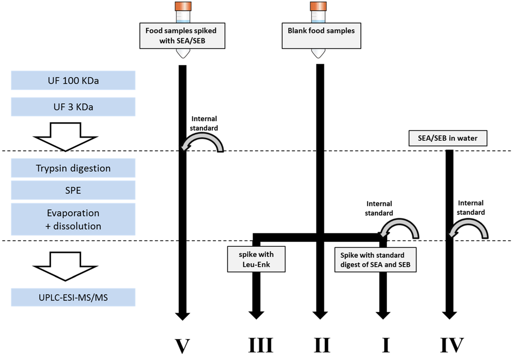
Quantitative Analysis of Staphylococcal Enterotoxins A and B in Food Matrices Using Ultra High-Performance Liquid Chromatography Tandem Mass Spectrometry (UPLC-MS/MS)
by
Aida Zuberovic Muratovic, Thomas Hagström, Johan Rosén, Kristina Granelli and Karl-Erik Hellenäs
Cited by 35 | Viewed by 6528
Abstract
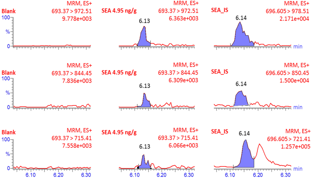
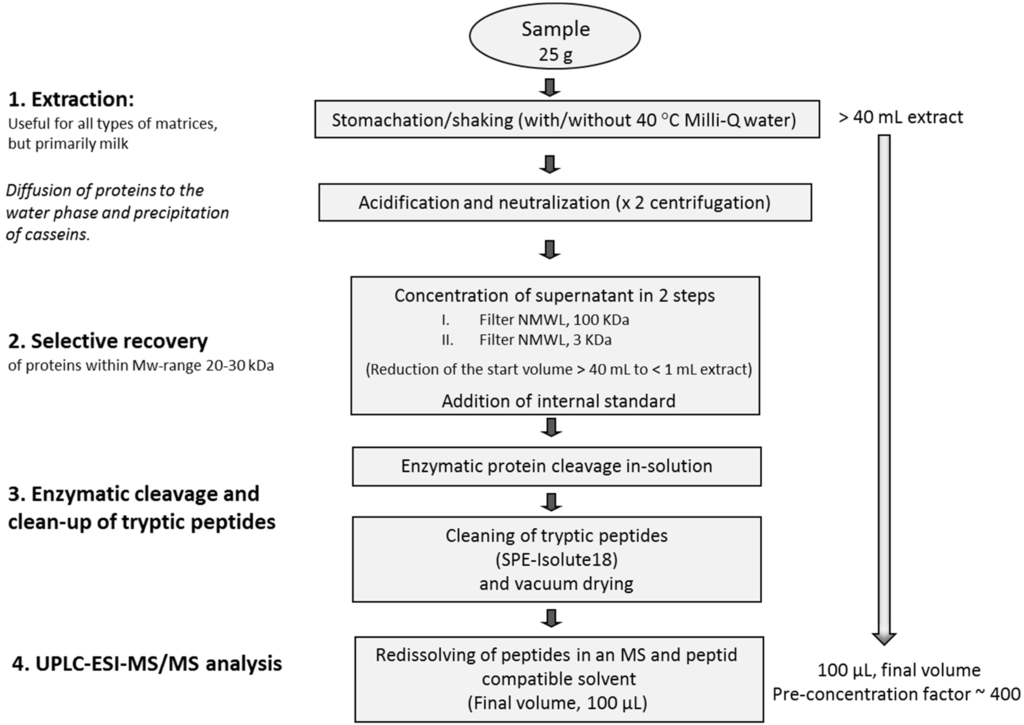
A method that uses mass spectrometry (MS) for identification and quantification of protein toxins, staphylococcal enterotoxins A and B (SEA and SEB), in milk and shrimp is described. The analysis was performed using a tryptic peptide, from each of the toxins, as the
[...] Read more.
A method that uses mass spectrometry (MS) for identification and quantification of protein toxins, staphylococcal enterotoxins A and B (SEA and SEB), in milk and shrimp is described. The analysis was performed using a tryptic peptide, from each of the toxins, as the target analyte together with the corresponding
13C-labeled synthetic internal standard peptide. The performance of the method was evaluated by analyzing spiked samples in the quantification range 2.5–30 ng/g (
R2 = 0.92–0.99). The limit of quantification (LOQ) in milk and the limit of detection (LOD) in shrimp was 2.5 ng/g, for both SEA and SEB toxins. The in-house reproducibility (RSD) was 8%–30% and 5%–41% at different concentrations for milk and shrimp, respectively. The method was compared to the ELISA method, used at the EU-RL (France), for milk samples spiked with SEA at low levels, in the quantification range of 2.5 to 5 ng/g. The comparison showed good coherence for the two methods: 2.9 (MS)/1.8 (ELISA) and 3.6 (MS)/3.8 (ELISA) ng/g. The major advantage of the developed method is that it allows direct confirmation of the molecular identity and quantitative analysis of SEA and SEB at low nanogram levels using a label and antibody free approach. Therefore, this method is an important step in the development of alternatives to the immune-assay tests currently used for staphylococcal enterotoxin analysis.
Full article
►▼
Show Figures
Open AccessArticle
Mass Spectrometric Detection of Bacterial Protein Toxins and Their Enzymatic Activity
by
Suzanne R. Kalb, Anne E. Boyer and John R. Barr
Cited by 26 | Viewed by 6498
Abstract
Mass spectrometry has recently become a powerful technique for bacterial identification. Mass spectrometry approaches generally rely upon introduction of the bacteria into a matrix-assisted laser-desorption time-of-flight (MALDI-TOF) mass spectrometer with mass spectrometric recognition of proteins specific to that organism that form a reliable
[...] Read more.
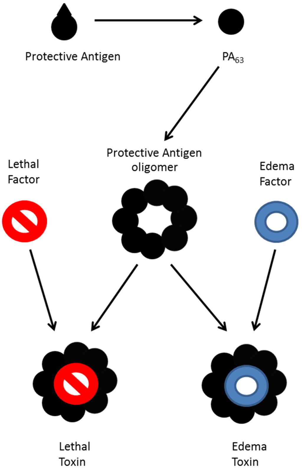
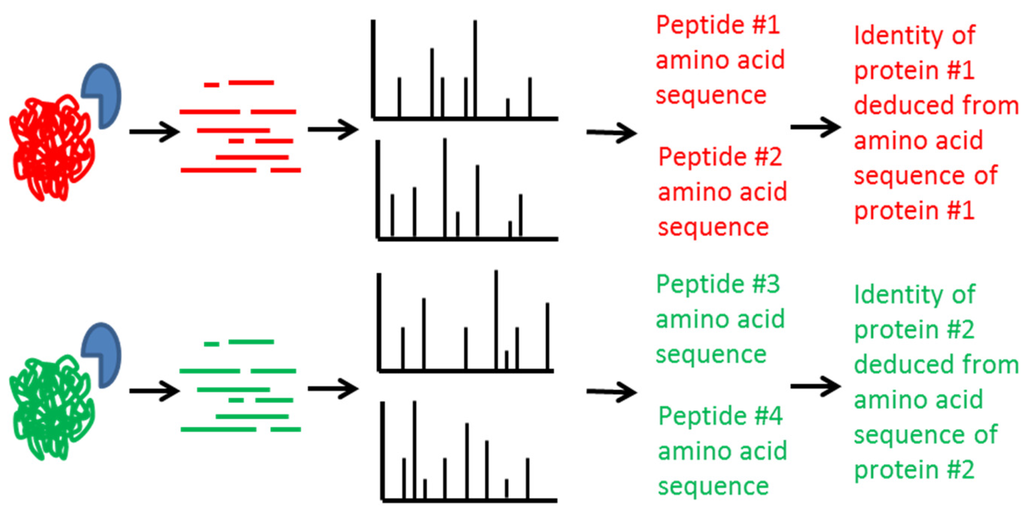
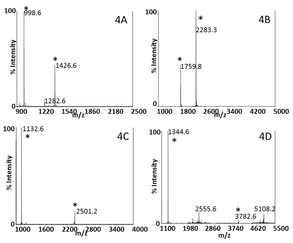
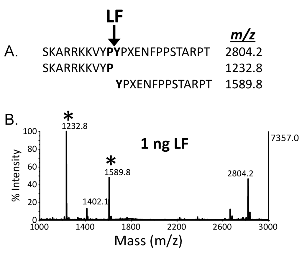
Mass spectrometry has recently become a powerful technique for bacterial identification. Mass spectrometry approaches generally rely upon introduction of the bacteria into a matrix-assisted laser-desorption time-of-flight (MALDI-TOF) mass spectrometer with mass spectrometric recognition of proteins specific to that organism that form a reliable fingerprint. With some bacteria, such as
Bacillus anthracis and
Clostridium botulinum, the health threat posed by these organisms is not the organism itself, but rather the protein toxins produced by the organisms. One such example is botulinum neurotoxin (BoNT), a potent neurotoxin produced by
C. botulinum. There are seven known serotypes of BoNT, A–G, and many of the serotypes can be further differentiated into toxin variants, which are up to 99.9% identical in some cases. Mass spectrometric proteomic techniques have been established to differentiate the serotype or toxin variant of BoNT produced by varied strains of
C. botulinum. Detection of potent biological toxins requires high analytical sensitivity and mass spectrometry based methods have been developed to determine the enzymatic activity of BoNT and the anthrax lethal toxins produced by
B. anthracis. This enzymatic activity, unique for each toxin, is assessed with detection of the toxin-induced cleavage of strategically designed peptide substrates by MALDI-TOF mass spectrometry offering unparalleled specificity. Furthermore, activity assays allow for the assessment of the biological activity of a toxin and its potential health risk. Such methods have become important diagnostics for botulism and anthrax. Here, we review mass spectrometry based methods for the enzymatic activity of BoNT and the anthrax lethal factor toxin.
Full article
►▼
Show Figures
Open AccessArticle
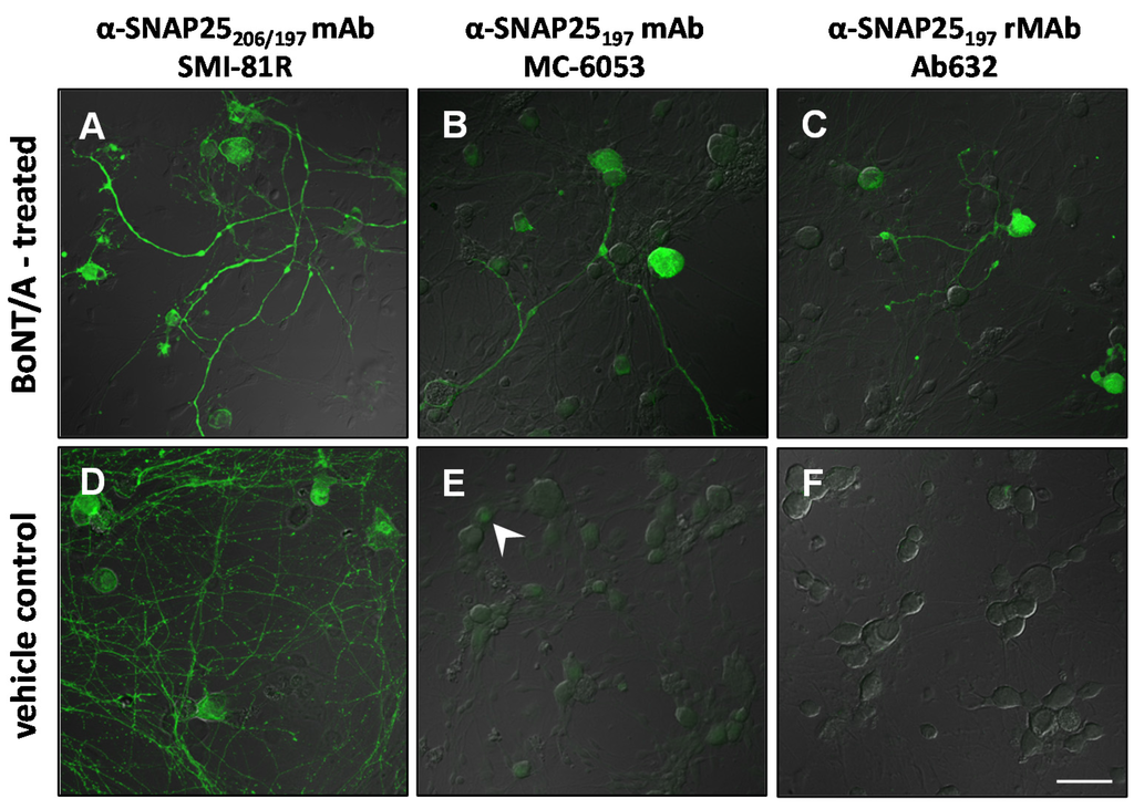
A Highly Specific Monoclonal Antibody for Botulinum Neurotoxin Type A-Cleaved SNAP25
by
Catherine Rhéaume, Brian B. Cai, Joanne Wang, Ester Fernández-Salas, K. Roger Aoki, Joseph Francis and Ron S. Broide
Cited by 20 | Viewed by 7061
Abstract
Botulinum neurotoxin type-A (BoNT/A), as onabotulinumtoxinA, is approved globally for 11 major therapeutic and cosmetic indications. While the mechanism of action for BoNT/A at the presynaptic nerve terminal has been established, questions remain regarding intracellular trafficking patterns and overall fate of the toxin.
[...] Read more.
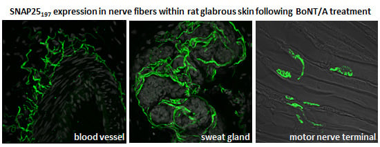
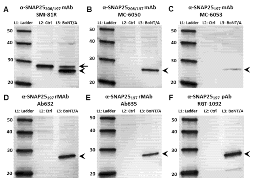
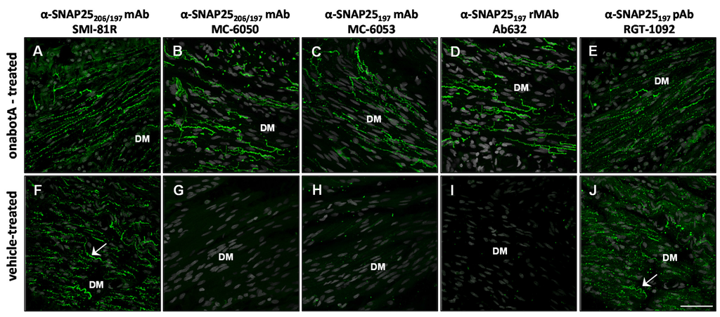
Botulinum neurotoxin type-A (BoNT/A), as onabotulinumtoxinA, is approved globally for 11 major therapeutic and cosmetic indications. While the mechanism of action for BoNT/A at the presynaptic nerve terminal has been established, questions remain regarding intracellular trafficking patterns and overall fate of the toxin. Resolving these questions partly depends on the ability to detect BoNT/A’s location, distribution, and movement within a cell. Due to BoNT/A’s high potency and extremely low concentrations within neurons, an alternative approach has been employed. This involves utilizing specific antibodies against the BoNT/A-cleaved SNAP25 substrate (SNAP25
197) to track the enzymatic activity of toxin within cells. Using our highly specific mouse monoclonal antibody (mAb) against SNAP25
197, we generated human and murine recombinant versions (rMAb) using specific backbone immunoglobulins. In this study, we validated the specificity of our anti-SNAP25
197 rMAbs in several different assays and performed side-by-side comparisons to commercially-available and in-house antibodies against SNAP25. Our rMAbs were highly specific for SNAP25
197 in all assays and on several different BoNT/A-treated tissues, showing no cross-reactivity with full-length SNAP25. This was not the case with other reportedly SNAP25
197-selective antibodies, which were selective in some, but not all assays. The rMAbs described herein represent effective new tools for detecting BoNT/A activity within cells.
Full article
►▼
Show Figures
Open AccessArticle
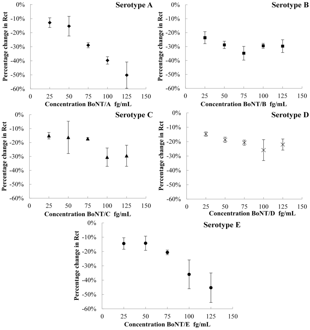
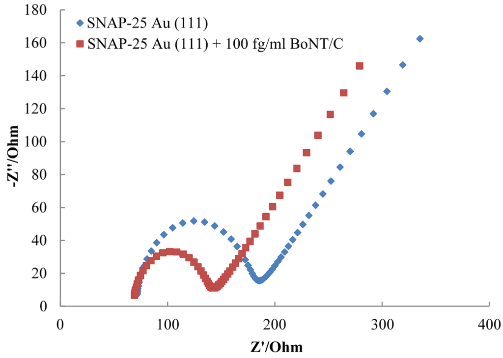
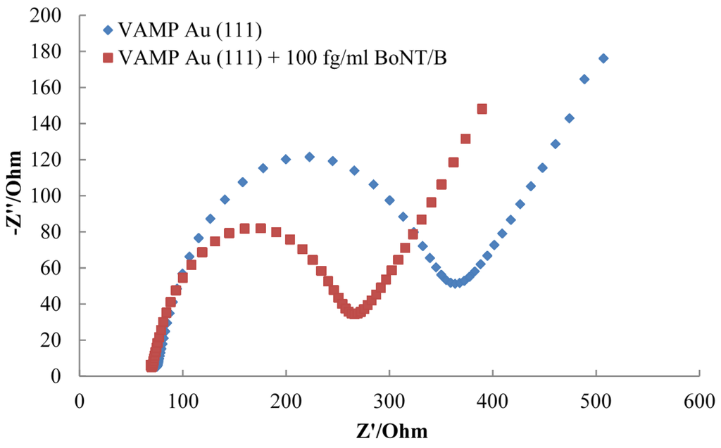
Botulinum Neurotoxin Serotypes Detected by Electrochemical Impedance Spectroscopy
by
Alison C. Savage, Nicholas Buckley, Jennifer Halliwell and Christopher Gwenin
Cited by 17 | Viewed by 7358
Abstract
Botulinum neurotoxin is one of the deadliest biological toxins known to mankind and is able to cause the debilitating disease botulism. The rapid detection of the different serotypes of botulinum neurotoxin is essential for both diagnosis of botulism and identifying the presence of
[...] Read more.
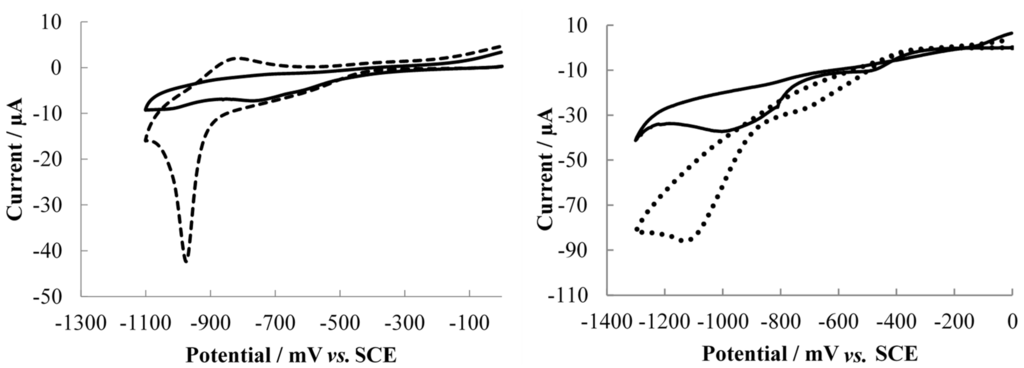
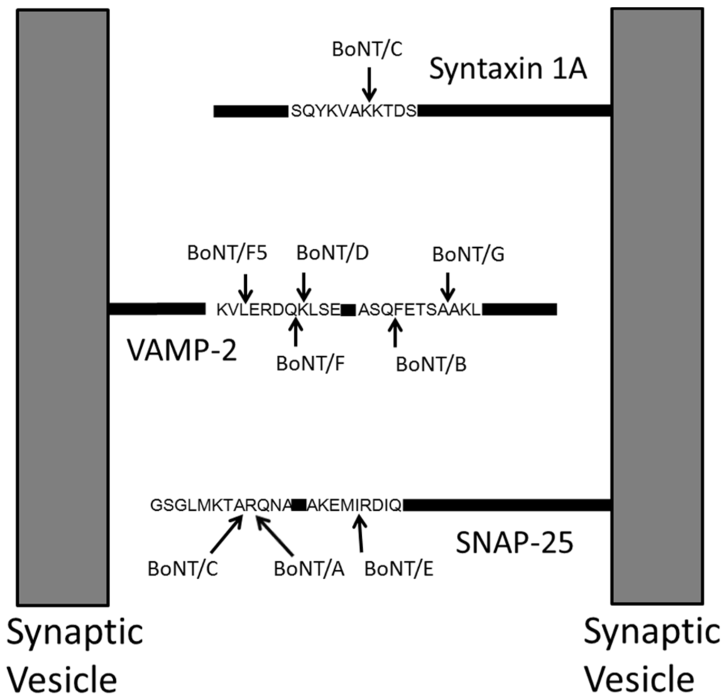
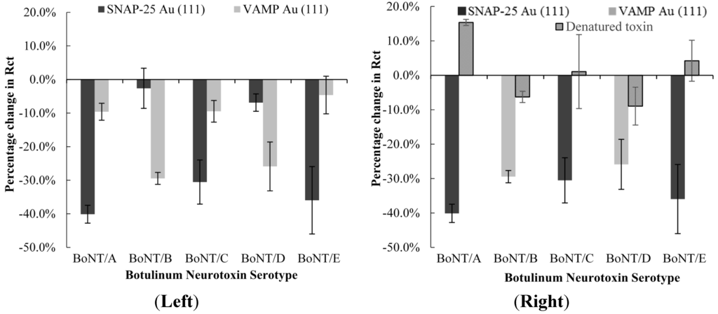
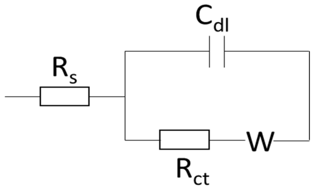
Botulinum neurotoxin is one of the deadliest biological toxins known to mankind and is able to cause the debilitating disease botulism. The rapid detection of the different serotypes of botulinum neurotoxin is essential for both diagnosis of botulism and identifying the presence of toxin in potential cases of terrorism and food contamination. The modes of action of botulinum neurotoxins are well-established in literature and differ for each serotype. The toxins are known to specifically cleave portions of the SNARE proteins SNAP-25 or VAMP; an interaction that can be monitored by electrochemical impedance spectroscopy. This study presents a SNAP-25 and a VAMP biosensors for detecting the activity of five botulinum neurotoxin serotypes (A–E) using electrochemical impedance spectroscopy. The biosensors are able to detect concentrations of toxins as low as 25 fg/mL, in a short time-frame compared with the current standard methods of detection. Both biosensors show greater specificity for their compatible serotypes compared with incompatible serotypes and denatured toxins.
Full article
►▼
Show Figures


