Article

Emulsions Stabilized with an Electrostatic Complex of Quaternized Cellulose Nanofiber and Octanoyl Gelatin and the Effect of pH Value on Their Stability

Department of Biomedical Science & Institute of Bioscience and Biotechnology, Kangwon National University, Chuncheon 24341, Republic of Korea
*
Author to whom correspondence should be addressed.
These authors contributed equally to this work.
Appl. Sci. 2024, 14(10), 4122; https://doi.org/10.3390/app14104122
Submission received: 8 October 2023 / Revised: 3 January 2024 / Accepted: 4 January 2024 / Published: 13 May 2024
(This article belongs to the Section Materials Science and Engineering)

Abstract

:
Oil/water (O/W) emulsions were prepared using the complex of quarternized cellulose nanofiber (QCNF) and octanoyl gelatin (OC−Gel) as an emulsifier, and the effect of pH value on their stability was investigated. OC−Gel was prepared through a condensation reaction, confirmed by 1H Nuclear Magnetic Resonance (NMR) and Fourier Transform Infrared (FT−IR) spectroscopy. It reduced air/water interfacial tension more effectively than unmodified gelatin. The complexation degree of OC−Gel and QCNF, measured by optical density, showed its maximum at a QCNF/OC−Gel mass ratio of 1/8 when the pH value of the medium was 7.4, and it increased in a saturated manner with increasing pH value. The signals of QCNF were found in the FT−IR and X−ray diffraction spectra of the complex, suggesting that the positively charged CNF were included in the OC−Gel−based complex. The complex formed a rough surface with smooth debris because the surface roughness of the complex aggregation reflected that of both QCNF and OC−Gel aggregation. QCNF could stabilize oil droplets to form a Pickering O/W emulsion. The complex of QCNF/OC−Gel was also a good emulsifier. QCNF and the complex were as potent as OC−Gel in emulsifying mineral oil in water. Most of the droplets fell within 5–25 µm, regardless of what the emulsifier was. The emulsion stabilized with OC−Gel increased in its oil droplet size more than two times in 20 days at all the pH vales tested (pH 3, 5, 7.4, 9), whereas the emulsion stabilized with QCNF remained almost constant in size during the same period regardless of the pH values. The droplet size of emulsion stabilized with the QCNF/OC−Gel complex did not change appreciably when the pH value was 5, 7.4, and 9. The complex seemed to act as a capsule wall and prevent the coalescence of the droplets. However, it increased dramatically due to the coalescence at pH 3, possibly because the complex could be dissolved under a strong acidic condition.

1. Introduction

Emulsions can be prepared with emulsifiers that reduce the interfacial tension and stabilize droplets of discontinuous phase [1,2,3,4]. Emulsions are required to be stable in terms of their droplet size during storage for their further use. Emulsions are destabilized following the order of droplet agglomeration, droplet coalescence, and phase separation [5,6,7,8]. The agglomeration is prevented electrostatically by choosing a charged emulsifier and sterically by selecting a flexible polymeric emulsifier, in order to achieve stable emulsions [9,10,11,12]. Even if the droplets are agglomerated, they may be stable and seldom subjected to coalescence if they are completely coated with insoluble matters (e.g., microcapsule wall) because the insoluble matters hardly allow for direct droplet−to−droplet contact [13,14,15,16]. When stimuli such as pH value change, temperature change, and light irradiation are applied to the emulsions, the physicochemical properties of the emulsifiers (e.g., the conformation and the solubility) can be changed, causing the droplets to be destabilized [17,18,19,20]. Poly(N−isopropylacrylamide−co−methacrylic acid−co−octadecyl acrylate)(P(NIPAM−MAA−ODA)) was prepared for its use as an emulsifier for the preparation of temperature− and pH−responsive emulsions. The copolymer chain could be contracted in response to heat and acidification and cause the droplets to be coalescenced and destabilized [21]. If the chain contraction takes place, the surface−activity of the copolymer and the interfacial area where a copolymer chain can stabilize may decrease. In another related study, poly(2−hydroxyethyl acrylate−co−coumaryl acrylate−co−2−ethylhexyl acrylate)(P(HEA−CA−EHA)) was used to stabilize oil droplets in water to prepare a temperature− and UV−responsive emulsion. The copolymer chain could also shrink in response to heat and the light irradiation, leading to the destabilization of the droplets [22]. Recently, a proteinoid comprising Asp, Leu, and cystamine was prepared as a disulfide emulsifier for the preparation of a reduction−responsive emulsion [23]. When the emulsion was put under a reductive condition, the droplet was destabilized because the disulfide bond of the proteinoid was broken down and lost its emulsifying ability. More recently, poly(2−hydroxyethyl acrylate−co−propyl methacryate) (P(HEA/PMA)) was prepared as a thermo−sensitive emulsifier for the preparation of Near Infrared (NIR)−responsive emulsion [24]. Upon the light irradiation, gold nanoparticles contained in the emulsion generated heat due to the surface plasmon resonance, the heat rendered the thermos−sensitive polymers shrunk, and the polymer lost its droplet−stabilizing ability.
In this study, a complex of quarternized cellulose nanofiber (QCNF) and octanoyl gelatin (OC−Gel) was used as an emulsifier for the preparation of oil/water (O/W) emulsions, and the effect of the pH value on the stability of the oil droplets was investigated. OC−Gel is negatively charged, and it can form an electrostatic complex with the positively charged QCNF when the pH value is greater than the isoelectric point. The insoluble complex can be formed on the surface of oil droplets because OC−Gel is interface−active, and it would isolate a droplet from others and prevent coalescence. When the pH value decreases below the isoelectric point (e.g., an acidic condition), OC−Gel changes to be positively charged, the complex becomes solubilized, and the droplets are subjected to coalescence (Figure 1). The pH−responsive emulsion developed in the present study would be applicable to a drug carrier that can release its cargo under an acidic condition (e.g., stomach).

2. Materials and Methods

2.1. Materials

Quaternized cellulose nanofibrils (QCNF) was purchased from cellulose lab Co. (Montreal, QC, Canada). Gelatin (type B), hexanoyl chloride, octanoyl chloride, mineral oil, trimethylamine (TEA), dimethyl sulfoxide (DMSO), deuterium oxide (D2O), 1× PBS, glycine buffer, MES buffer, and HEPES buffer were purchased from Sigma−Aldrich Chemical Co. (St. Louis, MO, USA). Ethanol was purchased from Dae Jung Chemical Co. (Siheung, Republic of Korea).

2.2. Preparation of Hydrophobicised Gelatin

Hydrophobicized gelatin (HpGel) was prepared by a condensation reaction reported elsewhere [25,26,27]. A total of 5.00 g of gelatin was dissolved in 50 mL of DMSO contained in a 100 mL of glass beaker, 0.5 mL of TEA was added to the gelatin solution, and the mixture solution was stirred at room temperature (20–23 °C) for 12 h. A fatty acid chloride (0.269 g of hexanoyl chloride, 0.325 g of octanoyl chloride) was added to the reaction mixture so that the molar ratio of the amino group of gelatin to the acid chloride group was 3:1, and it was stirred at room temperature for a further 24 h. HpGel was precipitated out by pouring the reaction mixture into 600 mL of cool ethanol contained in a 1 L beaker. The precipitate was separated by filtration and re−precipitated for purification. After being washed with the same non−solvent, HpGel was dried in a vacuum oven thermostated at 50 °C. HpGel prepared using hexanoyl chloride and octanoyl chloride were abbreviated to HC−Gel and OC−Gel, respectively.

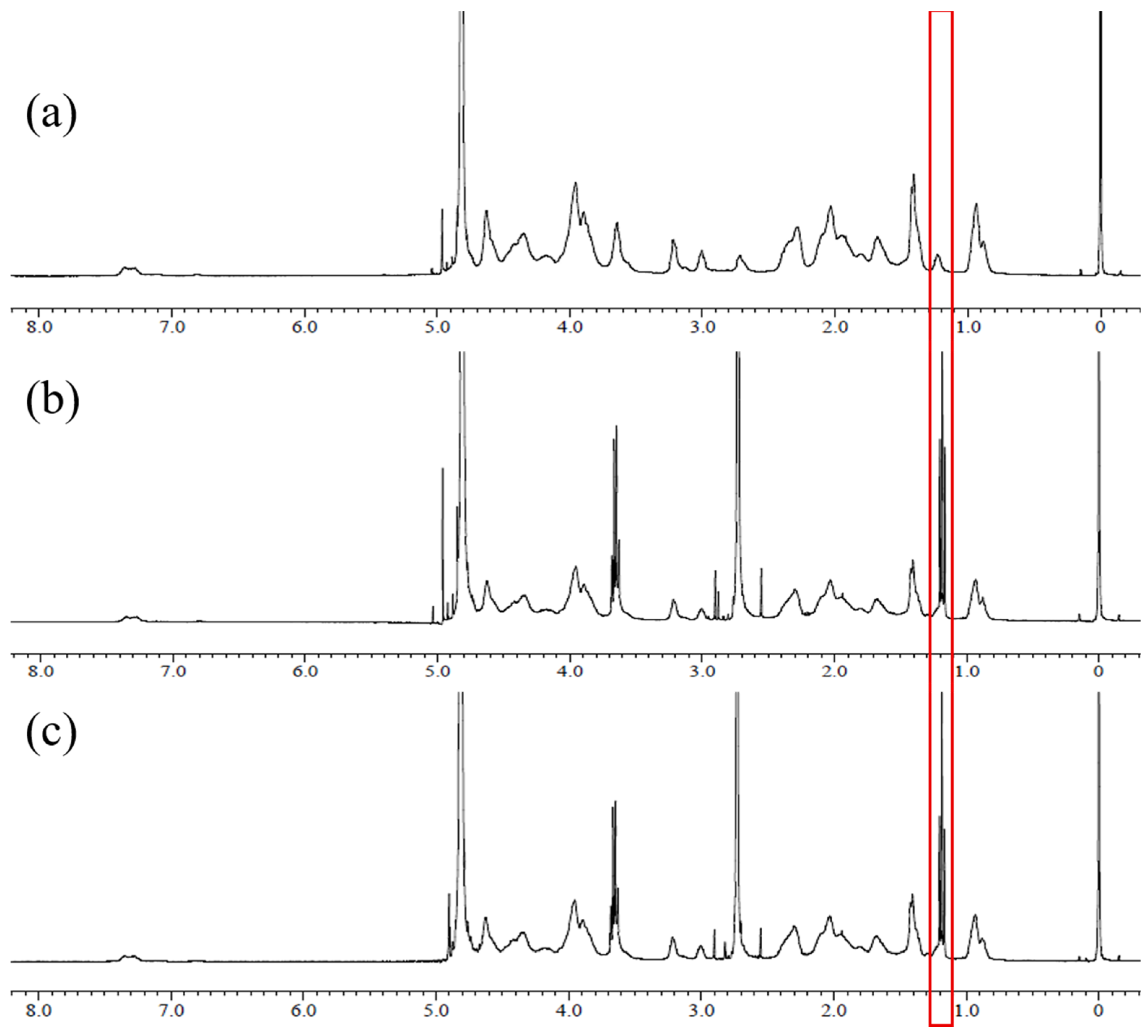
2.3. 1H NMR Spectroscopy

HC−Gel and OC−Gel were further dried in an oven thermostated at 45 °C using phosphorous pentoxide as a dying agent. Each of HC−Gel and OC−Gel was dissolved in D2O and the 1H Nuclear Magnetic Resonance (NMR) spectra were obtained using an NMR spectrophotometer (JEOL JNM− ECZ400s/L1 400 MHz spectrophotometer, Japan; located in the Central Laboratory Center of Kangwon National University). 1H NMR spectrophotometer was operated at 400 MHz 1H frequency, 8224 Hz sweep width, 1.99 s acquisition time, and 1 s recycle delay.

2.4. Measurement of Air/Water Interface Tension

Air/water interfacial tensions were determined by a ring method. Each of QCNF, Gel, HC−Gel, and OC−Gel was dissolved in PBS (10 mM, pH 7.4) so that the concentration was 1 mg/mL. Solutions of different concentrations were obtained by diluting the solutions two−fold serially with the same buffer solution. The interfacial tensions were measured at different concentrations using a tensiometer (DST 60, SEO Co., Suwon−si, Gyeonggi−do, Republic of Korea). Data were represented in a line and scatter graph using SigmaPlot 12.5 (Systat Software, San Jose, CA, USA) software.

2.5. Measurement of QCNF/HpGel Complexation Degree

QCNF was dispersed in HEPES buffer (pH 7.4) so that the concentration was 1 mg/mL. Various amounts of OC−Gel were dissolved in the QCNF suspension so that the mass ratio of QCNF to OC−Gel was 1:1 to 1:20. The pH value of the suspension medium was changed from pH 3.0 to pH 9.0 with the mass ratio of QCNF to OC−Gel kept being 1:5 and 1:8 in case the effect of the pH value on the complexation degree was investigated. The mixture suspension was rolled on a roller mixer at room temperature (20–25 °C) overnight and the optical density at 600 nm was measured on a UV spectrophotometer (7315 Spectrophotometer, JENWAY, Staordshire, UK). Data were represented in a line and scatter graph using SigmaPlot 12.5 (Systat Software, San Jose, CA, USA) software.

2.6. FT−IR Spectroscopy

Fourier Transform Infrared (FT−IR) spectroscopy was determined by a KBr method. Each of QCNF, gelatin, OC−Gel, and QCNF/OC−Gel complex was mixed with KBr, ground using a mortar and a pestle, and pressed using a press to obtain pellets. QCNF, gelatin, and OC−Gel were used as they were. The QCNF/OC−Gel complex was prepared as described previously. HEPES buffer (pH 7.4) was used as a medium, the QCNF concentration was 1 mg/mL, and the QCNF/OC−Gel mass ratio was 1:8, at which the maximum complex took place. The complex was filtered using filter paper, and it was freeze−dried. The FT−IR spectra were taken on a FT−IR spectrometer (Frontier, PerkinElmer, UK; located in Central Laboratory of Kangwon National University, Chuncheon, Korea). Data were represented in a line graph using SigmaPlot 12.5 (Systat Software, San Jose, CA, USA) software.

2.7. X−ray Diffractometry

QCNF, gelatin, OC−Gel, and the QCNF/OC−Gel complex were crystalogically examined by X−ray diffractometry using a diffractometer (X’Pert PRO MPD; PANalytical, Almelo, Netherland; in the Central Laboratory of Kangwon National University). X−ray sources were Cu Kα1 radiation (λ = 1.5506 Å) and Cu Kα2 radiation (λ = 1.5444 Å), and the Kα2/Kα1 ratio was 0.5. As the operating parameter values, 2θ range of 5–90o, divergence slit of 1.52 nm, generator voltage of 40 kV, scan range of 5, and scan step size of 0.0131303 were adopted. Data were represented in a line graph using SigmaPlot 12.5 (Systat Software, San Jose, CA, USA) software.

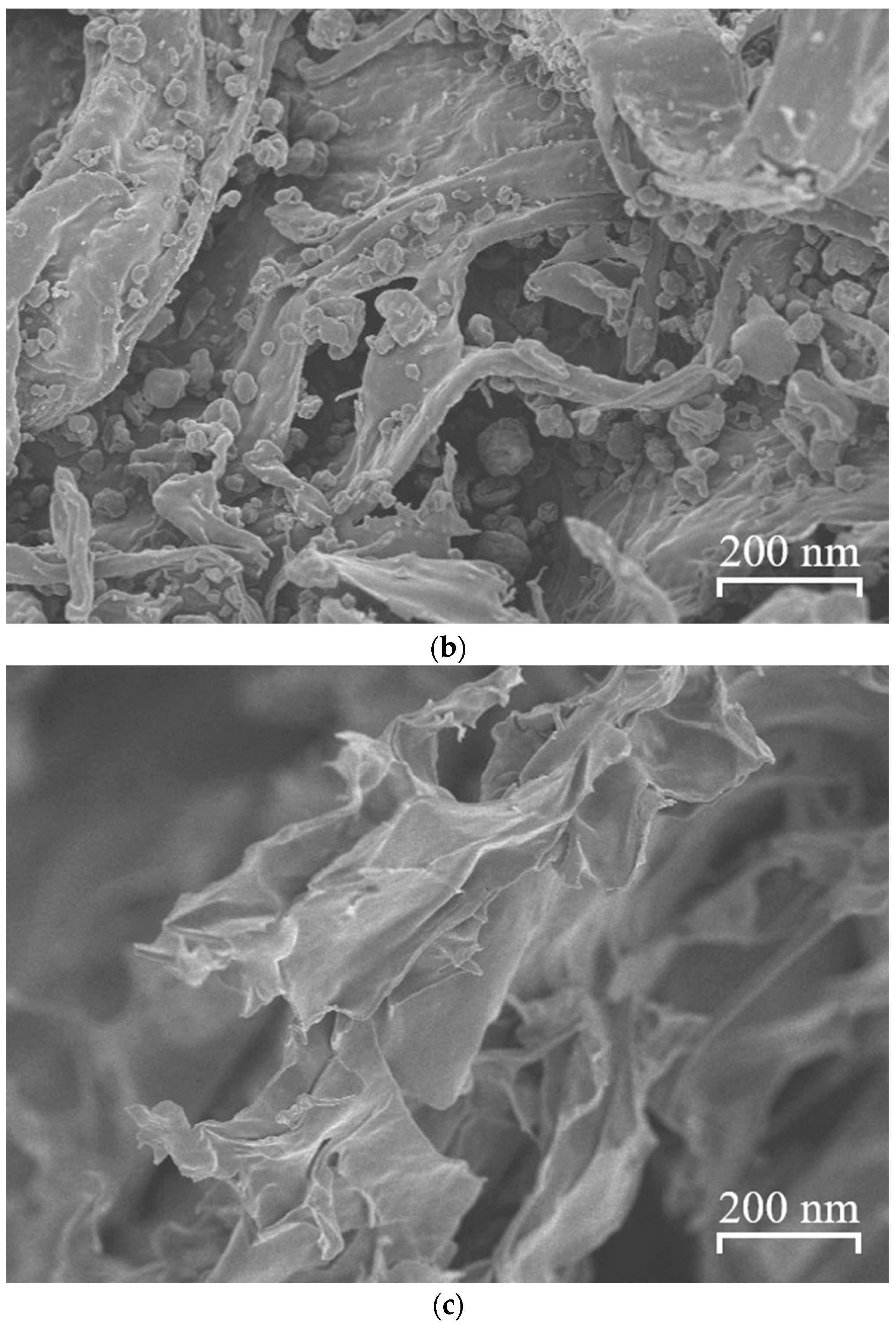
2.8. Scanning Electron Microscopy

Each OC−Gel, QCNF, and QCNF/OC−Gel complex was put on a metal stub, and it was sputtered with gold. The surface of each sample was investigated on a scanning electron microscope (SEM; JEOL JSM−7900F, Japan; located in Central Laboratory of Kangwon National University, Chuncheon, Republic of Korea), and the acceleration electron voltage was 5.0 keV.

2.9. Preparation of Emulsion Stabilized with QCNF and OC−Gel

A total of 80 mg each of QCNF and OC−Gel was dispersed or dissolved in 40 mL of distilled water. Alternatively, 71.1 mg of HpGel and 8.9 mg of QCNF were co−dispersed in 40 mL of distilled water so that the QCNF/HpGel mass ratio was 1:8, where the maximum complexation took place. The QCNF suspension, the OC−Gel solution, and the QCNF/HpGel mixture suspension were used as an aqueous phase for the preparation of the O/W emulsion.
While homogenizing the aqueous phase using a high−speed homogenizer (HG−15D, Daihan Scientific, Seoul, Republic of Korea) operating at 10,000 rpm, 10 mL of mineral oil was added to the aqueous solution in a dropwise manner over 30 s and the mixture was further homogenized for 2 min. The pH value of resulting O/W emulsion was adjusted to 3 using 1N HCl solution. The emulsions stabilized with OC−Gel, QCNF, and QCNF/OC−Gel were abbreviated to Em(OC−Gel), Em(QCNF), and Em(QCNF/OC−Gel), respectively.

2.10. Measurement of Size and Stability of Oil Droplets of Em(OC−Gel), Em(QCNF), and Em(QCNF/OC−Gel) by Microscopic Observation

The oil droplets of Em(OC−Gel), Em(QCNF), and Em(QCNF/OC−Gel) were observed on an optical microscope (CX31, OLYMPUS, Japan) with the magnification of X 400. The droplet size distributions were obtained using an image analyzer (Image−Pro Plus version 5.1, Media Cybernetics, Rockville, MD, USA), and the mean diameters were calculated by averaging the diameters based on the number of droplets [28]. The stability of the mean diameter was investigated by keeping the emulsions at room temperature (20–25 °C) for 20 days. Data were represented as mean ± standard deviation (SD). Data were represented in a line and scatter graph using SigmaPlot 12.5 (Systat Software, San Jose, CA, USA) software.

3. Results and Discussion

3.1. 1H NMR Spectroscopy

Figure 2 shows the 1H NMR spectra of Gel, HC−Gel, and OC−Gel. In the spectrum of HC−Gel, the peak of the hexanoyl group appeared around 1.2 ppm, together with the peaks of Gel. In the spectrum of OC−Gel, the peak of the octanoyl group appeared at the same position, along with the peaks of Gel. The 1H spectra revealed that the hydrophobic moieties (i.e., hexanoyl group, octanoyl group) were successfully bonded to Gel. The acid chloride group is much more reactive than the carboxyl group, and it can readily react with the functional groups (i.e., hydroxyl group and amino group) of the Gel through a condensation reaction.

3.2. Measurement of Air/Water Interface Tension

Figure 3 shows the air/water interfacial tensions at different concentrations of QCNF, Gel, HC−Gel, and OC−Gel solution. The interfacial tension of the buffer solution (i.e., PBS (10 mM, pH 7.4)) was around 71.8 dyne/cm, almost the same as that of water, 72 dyne/cm. The interfacial tension hardly changed when the concentration of QCNF increased up to 1 mg/mL, suggesting that QCNF was not interface−active. QCNF is a bundle of CNF and it has trimethyl ammonium groups on its surface. Accordingly, the fiber is polar, and it can be totally wetted with water; thus, it would hardly have an amphiphilic property. The Gel solution decreased to 64.1 dyne/cm in its interfacial tension when the concentration increased to 1 mg/mL. The decrease rate was rapid in the early stage and slow in the later stage. The saturated decrease with increasing concentration is typical of a surface−active agent solution. Gel is known to be surface−active and used as an emulsifier for oil−in−water emulsions [29,30,31]. The HC−Gel solution decreased to 64.3 dyne/cm when the concentration increased to 1 mg/mL, and its interfacial tension also decreased in a saturated manner. There was no significant difference in the interfacial tension between the Gel solution and the HC−Gel solution in the full range of concentrations tested. The hexanoyl group can provide a lipophilic property to gelatin, but it seemed to too short in its hydrocarbon chain to increase the interfacial activity of gelatin. The interfacial tension profile of the OC−Gel solution resembled those of the Gel solution and the HC−Gel solution. However, the interfacial tension of the former solution was significantly lower than those of the latter solutions. For example, the minimum interfacial tension of the OC−Gel solution was 57.5 dyne/cm, much lower than those of the Gel solution and the HC−Gel solution (i.e., 64.1 and 64.3 dyne/cm, respectively). The octyl chain of the octanoyl group is a lipophilic hydrocarbon chain, and it would be able to increase the amphiphilicity of gelatin, accounting for why OC−Gel was more interface−active than Gel and HC−Gel.

3.3. Measurement of QCNF/HpGel Complexation Degree

Figure 4a shows the optical density at 600 nm of the QCNF/OC−Gel mixture suspension (pH 7.4) at different mass ratios. The suspension was somewhat turbid when the mass ratio was 1:0 (i.e., QCNF suspension). QCNF is a fiber of cellulose whose surface is quarternized; it is not soluble but disperses well in water due to its polar surface, and it can scatter visible light, giving rise to some turbidity. The optical density increased with increasing the ratio: it showed its maximum value (ca. 1.15) at the ratio of 1:8 and then decreased to 0.74 when the ratio increased to 1:20. Gelatin (Type B) has its isoelectric point around pH 4.8 [32,33,34]. The acid chloride of octanoyl chloide can react with the amino groups of gelatin to form amide bonds. Accordingly, the number of amino groups in OC−Gel would be less than that in Gelatin; thus, the isoelectric point of the former one would be lower than that of the latter one. OC−Gel would be positively charged in the suspension whose pH value is 7.4 (much higher than the isoelectric point) and it would readily electrostatically interact with the positively charged QCNF to form complexes. This can explain why the optical density increased when the ratio increased to 1:8. The surface of QCNF will become saturated with OC−Gel as the ratio increases because the positive charge point and the surface area to accommodate OC−Gel is limited. Not to increase further in the optical density in the mass range of 1:8 to 1:20 could be ascribed to the surface saturation. The optical density rather decreased in the higher mass ratio range. OC−Gel is a kind of amphiphilic and surface−active molecule, and it would be able to disperse the QCNF/OC−Gel complex and stabilize it, accounting for a decrease in the optical density in the higher ratio range.
Figure 4b shows the optical density at 600 nm of the QCNF/OC−Gel mixture suspension at different pH values with the QCNF/OC−Gel mass ratio kept at 1:8. The QCNF suspension (i.e., control suspension) showed a low optical density (ca. 0.135) at pH 3.0, and it exhibited no significant change in the optical density in the full range of pH values tested. QCNF has no titrable and ionizable groups, the surface charge would hardly be affected by pH value change, and it would hardly aggregate regardless of the pH value due to its strong positive charge caused by the tertiary amino groups. In fact, QCNF was well dispersed in buffer solutions, and its suspension was homogeneous and stable at all the pH values tested. The QCNF/OC−Gel mixture suspension showed no appreciable change in the optical density either when the pH value was changed from 3 to 4. OC−Gel would be positively charged in the range of pH 3–4 because its isoelectric point was close to 4.8; thus, there would be no electrostatic interaction between QCNF and OC−Gel. The QCNF/OC−Gel mixture suspension markedly increased in optical density when the pH value was changed from 4 to 5. OC−Gel became negatively charged during the pH change because its isoelectric point fell within pH 4~5. Accordingly, a strong electrostatic interaction between QCNF and OC−Gel would take a place in pH 4~5 to form complexes, accounting for the rapid increase in optical density [35,36]. The optical density of the mixture suspension increased slowly when the pH value increased from 5 to 7, and thereafter it remained almost constant in range of pH 7 to 9. After the isoelectric point, the negative charge of a protein (e.g., OC−Gel) increases with increasing pH value in a saturated manner; thus, the amount of OC−Gel complexed with QCNF would increase in the same manner. This would be a reason why the optical density increased in a saturated manner in the range of pH 4~9.0

3.4. FT−IR Spectroscopy

Figure 5 shows the FT−IR spectra of QCNF, gelatin, OC−Gel, and the QCNF/OC−Gel complex. In the spectrum of QCNF, the hydroxyl group was found at 3313 and 3281 cm−1, and the methyl group of the trimethyl ammonium in 2900~2920 cm−1. In the spectrum of gelatin, the primary amino group was found at 1618 cm−1, the second amino group at 1532 cm−1, C−H bending in 1450–1600 cm−1, and C−N stretching at 1236 cm−1. In the spectrum of OC−Gel, the CH2 bending of the octanoyl group was found at 2917 cm−1, the carbonyl group of the ester bond at 1639 cm−1, the primary amino group at 1618 cm−1, the second amino group at 1532 cm−1, C−H bending in 1450–1600 cm−1, C−N stretching at 1236 cm−1, and the long alkyl chain of the octanoyl group at 1016 cm−1. Since the signals of the octanoyl group (i.e., CH2 bending signal, carbonyl group signal, long alkyl chain signal) were found together with those of gelatin, it was concluded that the octanoyl group was covalently attached to gelatin. In the spectrum of QCNF/OC−Gel complex, the signals of OC−Gel were found along with those of QCNF, suggesting that the complex was successfully formed, and it was composed of QCNF and OC−Gel.

3.5. X−ray Diffractometry

Figure 6 shows the X−ray diffraction spectra of QCNF, OC−Gel, and the QCNF/OC−Gel complex. In the spectrum of QCNF, diffraction peaks were found at 2θ of 16.36° and 22.62°, corresponding to the (110) and (200) plane of the cellulose crystal, respectively [37,38]. In the spectrum of OC−Gel, no diffraction peak was found, indicating that the hydrophobically modified gelatin was amorphous. Gelatin is an amorphous polymer. The hydrophobic moiety (i.e., octanoyl group) used for the hydrophobic modification of gelatin would be hydrophobically assembled to form a crystalline structure, but the content of the octanoyl group seemed to be too small to do that. In the spectrum of QCNF/OC−Gel complex, the characteristic diffraction peaks of QCNF were found, suggesting that QCNF was contained in the complex, and it maintained its crystalline structure after being complexed with gelatin.

3.6. Scanning Electron Microscopy

Figure 7 shows the SEM micrographs of OC−Gel, QCNF, and the QCNF/OC−Gel complex. HpGel formed a smooth surface. It can aggregate at the molecular level to form a compact and smooth surface. In fact, gelatin is known to have good film−forming properties [39,40,41]. On the contrary, QCNF formed a rough surface. It is a cellulose nanofiber (i.e., a bundle of cellulose) and can be entangled to form a broken surface. On the other hand, the QCNF/OC−Gel complex built a rough surface, having smooth leaves. HpGel complexed with QCNF and formed smooth leaf−like films around the nanofibers, forming an uneven surface.

3.7. Measurement of Size and Stability of Oil Droplets of Em(OC−Gel), Em(QCNF), and Em(QCNF/OC−Gel) by Microscopic Observation

Figure 8a shows the optical micrographs of Em(OC−Gel), Em(QCNF), and Em(QCNF/OC−Gel). Oil droplets whose diameter are a few to tens of nanometers were found in all the emulsion samples, suggesting that OC−Gel and QCNF were able to stabilize oil droplets. OC−Gel was surface−active (Figure 3); thus, it would reduce O/W interfacial tension and stabilize the oil droplets effectively. QCNF is a cellulose nanofiber whose surface is quaternized. Since the quaternized domain would be more polar than the unquaternized one, it would be amphiphilic and thus be able to stabilize oil droplets. In addition, QCNF is a kind of particle, and it might reduce O/W interfacial tension and act as an emulsifier, just like Pickering emulsions can be prepared using nanoparticles as an emulsifier [42,43,44,45]. Figure 8b shows the size distributions of the oil droplets of Em(OC−Gel), Em(QCNF), and Em(QCNF/OC−Gel). Em(OC−Gel) exhibited a unimodal distribution; most of the droplets were found in the range of 5 to 25 µm, and the mean diameter was calculated to be 14.1 ± 6.12 nm. Em(QCNF) showed a distribution somewhat deviating from a unimodal distribution; most droplets fell within the same size range as those of Em(OC−Gel), and the mean diameter was 11.2 ± 6.39 nm. Em(QCNF/OC−Gel) also showed a non−unimodal distribution; most droplets were in the same size range as those of the other emulsions, and the mean diameter was 12.3 ± 5.66 nm. Considering the mode of the size distribution, it could be said that Em(OC−Gel) was more homogeneous in size than Em(QCNF) and Em(QCNF/OC−Gel).
Figure 9 shows the time−dependent droplet size change of Em(OC−Gel), Em(QCNF), and Em(QCNF/OC−Gel) at pH 3, 5, 7.4 and 9. The droplet size of Em(OC−Gel) began to increase 3 days after preparation, and the mean diameter changed from 14.1 nm to larger than 35 nm at all the pH values tested. The droplets can undergo agglomeration when the surface potential of the droplets (e.g., Zeta potential) and the steric hindrance arisen by the emulsifier are not high enough to prevent the droplets from coming in contact with one another. In fact, OC−Gel is a kind of protein, and it has a positive or negative charge value, depending on the pH value of the medium. In nature, however, its charge is less than 20 mV and insufficient to prevent the aggregation of oil droplets [46,47]. In addition, since apolar and polar amino acid residues are in gelatin in a random sequence, there are no hydrophilic segments which are long enough to push away approaching droplets. Thus, OC−Gel would hardly exhibit the steric hindrance to prevent agglomeration. Agglomerated oil droplets can be subjected to coalescence when the O/W interfacial energy increases due to the surface migration of the emulsifier. Once the droplets contact with one another, the emulsifier (i.e., OC−Gel) can migrate laterally on the O/W interface, and the interface void of the emulsifier can be created; thus, coalescence among the oil droplets can take place to reduce the interfacial energy. Gelatin is flexible; thus, it would readily migrate on the interface when the droplets contact one another and the fluid is drained out of the thin film between contacting interfaces. On the other hand, Em(QCNF) increased from 11.2 to about 18 µm in diameter for 20 days, and the pH value had no significant effect on the size change. Since the degree of size change was much less than that of Em(OC−Gel), it was concluded that Em(QCNF) was more stable than Em(OC−Gel) in terms of droplet size. QCNF is strongly positively charged due to the trimethyl ammonium group, and its zeta potential is greater than + 30 mV. Thus, the oil droplets of Em(QCNF) would hardly agglomerate owing to the strong repulsive force developed among the droplets, accounting for why Em(QCNF) was relatively stable. In addition, QCNF is a long nanofiber, and it would be able to sterically stabilize the oil droplets due to its long length. Furthermore, even if the droplets come in close contact with one another despite the strong inter−particular repulsion and the steric stabilization, QCNF would hardly migrate on the O/W interface and keep its position because it is inflexible and rigid. This would be another reason why QCNF could stabilize the oil droplets effectively. Meanwhile, Em(QCNF/OC−Gel) slightly increased from 12.3 to about 14 µm in diameter for 20 days when the pH value was 7.4 and 9. The complexation between QCNF and OC−Gel took place when the pH value was greater than 5. The QCNF/OC−Gel complex was thought to act as a coat of oil droplets and prevent their coalescence. Upon complexation, both the positive charge of QCNF and the negative charge of OC−Gel would decrease because of the charge neutralization. Thus, when the pH value was 7.4 and 9, the inter−particular electrostatic repulsion would be weak, and the agglomeration of the droplets would readily take place. However, the insoluble complex would allow the droplets to avoid their coalescence because it can isolate the oil droplets from their environment just as the microcapsule wall did. This would be a reason why the oil size remained almost constant for 20 days at pH 7.4 and 9. When the pH value was 5, the emulsion was stable in terms of size for 12 days, but it somewhat increased in diameter during the remaining period of the experiment. When the pH value was 3, the droplet size did not change markedly for 12 days, but it increased dramatically from 14.2 to 33.3 µm. OC−Gel is positively charged under an acidic condition (e.g., pH 3) and it can hardly complex with QCNF. Thus, they would stabilize the droplets independently and individually but not by the complexed form. Besides the positive charge of OC−Gel, the intensive positive charge of QCNF would give rise to a strong inter−particular repulsion and reduce the agglomeration and the coalescence. This may explain why the emulsion was stable for 12 days even under the condition. However, the total number of collisions among the droplets will increase with time, the O/W interface without the emulsifiers would be created, and it may cause a coalescence and an increase in droplet size, accounting for why the droplet size markedly increased during the later stage.

4. Conclusions

OC−Gel, QCNF, and QCNF/OC−Gel were used as emulsifiers for the preparation of O/W emulsions, and the emulsion stability was investigated in terms of droplet size at different pH values (pH 3, 5, 7.4, 9). A hydrophobically modified gelatin (i.e., OC−Gel) was more surface−active than unmodified gelatin. When the medium pH value was 7.4 (greater than the isoelectric point of OC−Gel), the maximum complexation took place at a QCNF/OC−Gel mass ratio of 1/8. The complexation degree increased rapidly when the medium pH value increased to 6, and thereafter, it increased slowly. Therefore, it could be said that a QCNF/OC−Gel mass ratio of 1/8 and a pH value of 7.4 is the near−optimum condition for the formation of this complex. FT−IR and X−ray diffraction spectroscopy revealed that QCNF was included in the electrostatic complex. According to SEM microscopy, the surface of the QCNF/OC−Gel complex reflected both the characteristic of the surface of OC−Gel aggregation (i.e., smooth) and that of the surface of QCNF (i.e., rough).
OC−Gel, QCNF, and the QCNF/OC−Gel complex could all emulsify mineral oil in water successfully. However, they showed different stability in oil droplet size. The droplet size of Em(OC−Gel) increased more than two times in 20 days at all pH vales tested (pH 3, 5, 7.4, 9), probably because the electrostatic and steric stabilization potency of OC−Gel was relatively weak, whereas the droplet size of Em(QCNF) remained almost constant during the same period at all the pH values, suggesting that QCNF was more potent than OC−Gel. Since QCNF is strongly positively charged and is long (tens of micrometer in length), it would be able to stabilize the droplets electrostatically and sterically. On the other hand, the emulsion stabilized with the QCNF/OC−Gel complex was stable in the droplet size at all pH values except at pH 3.0, where the complex could be dissolved; thus, the droplets were subjected to coalescence. The complex seemed to play a role as a capsule wall, isolating droplets and preventing their coalescence. The pH−responsive emulsion developed in the present study would be able to be used as a drug carrier which can release its payload under an acidic condition (e.g., in the stomach).

Author Contributions

Methodology, S.C.P.; Investigation, H.K.S. and S.C.P.; Visualization, H.K.S. and S.C.P.; Supervision, J.-C.K. All authors have read and agreed to the published version of the manuscript.

Funding

This research was supported by the Basic Science Research Program through the National Research Foundation of Korea (NRF) funded by the Ministry of Education (No. 2018R1A6A1A03025582). This work was supported by a National Research Foundation of Korea (NRF) grant funded by the Korean government (MSIT) (No. 2022R1A2C2003353). This research was supported by “Regional Innovation Strategy (RIS)” through the National Research Foundation of Korea (NRF) funded by the Ministry of Education (MOE) (2022RIS−005).

Institutional Review Board Statement

Not applicable.

Informed Consent Statement

Not applicable.

Data Availability Statement

The original contributions presented in the study are included in the article, further inquiries can be directed to the corresponding author.

Conflicts of Interest

The authors declare no conflict of interest.

References

  1. Amine, C.; Dreher, J.; Helgason, T.; Tadros, T. Investigation of emulsifying properties and emulsion stability of plant and milk proteins using interfacial tension and interfacial elasticity. Food Hydrocoll. 2014, 39, 180–186. [Google Scholar] [CrossRef]
  2. Dickinson, E. Hydrocolloids as emulsifiers and emulsion stabilizers. Food Hydrocoll. 2009, 23, 1473–1482. [Google Scholar] [CrossRef]
  3. Nesterenko, A.; Drelich, A.; Lu, H.; Clausse, D.; Pezron, I. Influence of a mixed particle/surfactant emulsifier system on water-in-oil emulsion stability. Colloids Surf. A Physicochem. Eng. Asp. 2014, 457, 49–57. [Google Scholar] [CrossRef]
  4. Osborn, H.T.; Akoh, C.C. Effect of emulsifier type, droplet size, and oil concentration on lipid oxidation in structured lipid-based oil-in-water emulsions. Food Chem. 2004, 84, 451–456. [Google Scholar] [CrossRef]
  5. Cardoso-Ugarte, G.A.; Ramírez-Corona, N.; López-Malo, A.; Palou, E.; Martín-González, M.F.S.; Jiménez-Munguía, M.T. Modeling phase separation and droplet size of W/O emulsions with oregano essential oil as a function of its formulation and homogenization conditions. J. Dispers. Sci. Technol. 2018, 39, 1065–1073. [Google Scholar] [CrossRef]
  6. Ushikubo, F.Y.; Cunha, R.L. Stability mechanisms of liquid water-in-oil emulsions. Food Hydrocoll. 2014, 34, 145–153. [Google Scholar] [CrossRef]
  7. Capek, I. Degradation of kinetically-stable o/w emulsions. Adv. Colloid Interface Sci. 2004, 107, 125–155. [Google Scholar] [CrossRef] [PubMed]
  8. Wardhono, E.Y.; Zafimahova-Ratisbonne, A.; Saleh, K.; Clausse, D.; Lanoiselle, J.-L. W/O Emulsion Destabilization and Release of a Polysaccharide Entrapped in the Droplets. J. Dispers. Sci. Technol. 2016, 37, 1581–1589. [Google Scholar] [CrossRef]
  9. Burgos-Díaz, C.; Wandersleben, T.; Marqués, A.M.; Rubilar, M. Multilayer emulsions stabilized by vegetable proteins and polysaccharides. Curr. Opin. Colloid Interface Sci. 2016, 25, 51–57. [Google Scholar] [CrossRef]
  10. Maier, C.; Zeeb, B.; Weiss, J. Investigations into aggregate formation with oppositely charged oil-in-water emulsions at different pH values. Colloids Surf. B Biointerfaces 2014, 117, 368–375. [Google Scholar] [CrossRef]
  11. Bobin, M.-F.; Michel, V.; Martini, M.-C. Study of formulation and stability of emulsions with polymeric emulsifiers. Colloids Surf. A Physicochem. Eng. Asp. 1999, 152, 53–58. [Google Scholar] [CrossRef]
  12. Xu, Y.-T.; Wang, Y.-H.; Chen, F.-P.; Tang, C.-H. Whether ovalbumin performs as a particulate or polymeric emulsifier is largely determined by pH. Food Hydrocoll. 2020, 103, 105694. [Google Scholar] [CrossRef]
  13. Kang, M.K.; Kim, J.-C. Preparations and temperature- and pH-dependent release property of ethylcellulose microcapsules containing N-isopropylacrylamide copolymer. J. Appl. Polym. Sci. 2010, 118, 421–427. [Google Scholar] [CrossRef]
  14. Yari, S.; Nasirpour, A.; Fathi, M. Effect of Polymer Concentration and Acidification Time on Olive Oil Microcapsules Obtained by Complex Coacervation. Appl. Food Biotechnol. 2016, 3, 53–58. [Google Scholar] [CrossRef]
  15. Loxley, A.; Vincent, B. Preparation of Poly(methylmethacrylate) Microcapsules with Liquid Cores. J. Colloid Interface Sci. 1998, 208, 49–62. [Google Scholar] [CrossRef] [PubMed]
  16. Aghbashlo, M.; Mobli, H.; Madadlou, A.; Rafiee, S. The correlation of wall material composition with flow characteristics and encapsulation behavior of fish oil emulsion. Food Res. Int. 2012, 49, 379–388. [Google Scholar] [CrossRef]
  17. Park, S.H.; Kim, J.-C. Oxidation–Responsive Emulsions Stabilized with Poly(Vinyl Pyrrolidone-co-allyl Phenyl Sulfide). Polymers 2020, 12, 498. [Google Scholar] [CrossRef] [PubMed]
  18. Tang, J.; Quinlan, P.J.; Tam, K.C. Stimuli-responsive Pickering emulsions: Recent advances and potential applications. Soft Matter. 2015, 11, 3512–3529. [Google Scholar] [CrossRef] [PubMed]
  19. Brugger, B.; Richtering, W. Magnetic, Thermosensitive Microgels as Stimuli-Responsive Emulsifiers Allowing for Remote Control of Separability and Stability of Oil in Water-Emulsions. Adv. Mater. 2007, 19, 2973–2978. [Google Scholar] [CrossRef]
  20. Shahid, S.; Gurram, S.R.; Basavaraj, M.G. Doubly pH Responsive Emulsions by Exploiting Aggregation of Oppositely Charged Nanoparticles and Polyelectrolytes. Langmuir 2018, 34, 5060–5071. [Google Scholar] [CrossRef]
  21. Seo, S.R.; Lee, H.Y.; Kim, J.-C. Thermo- and pH-Responsiveness of Emulsions Stabilized with Acidic Thermosentive Polymers. J. Dispers. Sci. Technol. 2013, 34, 1280–1285. [Google Scholar] [CrossRef]
  22. Seo, S.R.; Kim, J.-C. Emulsions Stabilized with poly(Hydroxyethyl Acrylate-co-Coumaryl Acrylate-co-2-Ethylhexyl acrylate). J. Macromol. Sci. Part A 2013, 50, 855–860. [Google Scholar] [CrossRef]
  23. Kwon, K.; Kim, J.-C. Emulsion stabilized with disulfide proteinoid and its stability in reducing condition. J. Dispers. Sci. Technol. 2018, 39, 333–340. [Google Scholar] [CrossRef]
  24. Park, S.H.; Kim, J.-C. Oil-in-gold nanoparticle solution emulsion stabilized with amphiphilic polymers and its stability under NIR irradiation. J. Dispers. Sci. Technol. 2018, 39, 961–969. [Google Scholar] [CrossRef]
  25. Yoshizawa, K.; Taguchi, T. Bonding behavior of hydrophobically modified gelatin films on the intestinal surface. J. Bioact. Compat. Polym. 2014, 29, 560–571. [Google Scholar] [CrossRef]
  26. Chen, X.; Mizuta, R.; Fukata, N.; Taguchi, T. Design of bio-inspired adhesive surface composed of hexanoyl group-modified gelatin and silicon nanowire. Colloids Surf. B Biointerfaces 2019, 178, 111–119. [Google Scholar] [CrossRef] [PubMed]
  27. Toledano, O.; Magdassi, S. Emulsification and Foaming Properties of Hydrophobically Modified Gelatin. J. Colloid Interface Sci. 1998, 200, 235–240. [Google Scholar] [CrossRef]
  28. Liu, Y.; Shi, Z.; Zou, Y.; Yu, J.; Liu, L.; Fan, Y. Comparison of Cellulose and Chitin Nanofibers on Pickering Emulsion Stability—Investigation of Size and Surface Wettability Contribution. Int. J. Biol. Macromol. 2023, 235, 123754. [Google Scholar] [CrossRef] [PubMed]
  29. Lin, L.-H.; Chen, K.-M. Preparation and surface activity of gelatin derivative surfactants. Colloids Surf. A Physicochem. Eng. Asp. 2006, 272, 8–14. [Google Scholar] [CrossRef]
  30. Zhang, T.; Xu, J.; Zhang, Y.; Wang, X.; Lorenzo, J.M.; Zhong, J. Gelatins as emulsifiers for oil-in-water emulsions: Extraction, chemical composition, molecular structure, and molecular modification. Trends Food Sci. Technol. 2020, 106, 113–131. [Google Scholar] [CrossRef]
  31. Auweter, H.; André, V.; Horn, D.; Lüddecke, E. The function of gelatin in controlled precipitation processes of nanosize particles. J. Dispers. Sci. Technol. 1998, 19, 163–184. [Google Scholar] [CrossRef]
  32. Sewald, L.; Claaßen, C.; Götz, T.; Claaßen, M.H.; Truffault, V.; Tovar, G.E.M.; Southan, A.; Borchers, K. Beyond the Modification Degree: Impact of Raw Material on Physicochemical Properties of Gelatin Type A and Type B Methacryloyls. Macromol. Biosci. 2018, 18, 1800168. [Google Scholar] [CrossRef] [PubMed]
  33. Ji, F.; Zhou, W.; Zhang, Z.; Zhang, B. Effects of Relative Molecular Weight Distribution and Isoelectric Point on the Swelling Behavior of Gelatin Films. Front. Chem. 2022, 10, 857976. [Google Scholar] [CrossRef]
  34. Zhang, Z.; Li, G.; Shi, B. Physicochemical properties of collagen, gelatin and collagen hydrolysate derived from bovine limed split wastes. J. Soc. Leather Technol. Chem. 2006, 90, 23–28. [Google Scholar]
  35. Park, S.H.; Zhao, F.; Park, S.C.; Kim, J.C. Gelatin-Loaded Cubosomes Stabilized with Hydrophobically Modified Quaternized Cellulose Nanofiber and Their PH-Dependent Release Property. J. Biomater. Appl. 2021, 35, 1109–1118. [Google Scholar] [CrossRef]
  36. Zhao, Y.; Khalid, N.; Shu, G.; Neves, M.A.; Kobayashi, I.; Nakajima, M. Complex Coacervates from Gelatin and Octenyl Succinic Anhydride Modified Kudzu Starch: Insights of Formulation and Characterization. Food Hydrocoll. 2019, 86, 70–77. [Google Scholar] [CrossRef]
  37. Bian, H.; Yang, Y.; Tu, P.; Chen, J.Y. Value-Added Utilization of Wheat Straw: From Cellulose and Cellulose Nanofiber to All-Cellulose Nanocomposite Film. Membranes 2022, 12, 475. [Google Scholar] [CrossRef]
  38. Zhang, B.; Huang, C.; Zhao, H.; Wang, J.; Yin, C.; Zhang, L.; Zhao, Y. Eects of Cellulose Nanocrystals and Cellulose Nanofibers on the Structure and Properties of Polyhydroxybutyrate Nanocomposites. Polymers 2019, 11, 2063. [Google Scholar] [CrossRef] [PubMed]
  39. Gómez-Estaca, J.; Montero, P.; Fernández-Martín, F.; Gómez-Guillén, M.C. Physico-chemical and film-forming properties of bovine-hide and tuna-skin gelatin: A comparative study. J. Food Eng. 2009, 90, 480–486. [Google Scholar] [CrossRef]
  40. Hoque, M.S.; Benjakul, S.; Prodpran, T. Effect of heat treatment of film-forming solution on the properties of film from cuttlefish (Sepia pharaonis) skin gelatin. J. Food Eng. 2010, 96, 66–73. [Google Scholar] [CrossRef]
  41. Cao, N.; Fu, Y.; He, J. Preparation and physical properties of soy protein isolate and gelatin composite films. Food Hydrocoll. 2007, 21, 1153–1162. [Google Scholar] [CrossRef]
  42. Goi, Y.; Fujisawa, S.; Saito, T.; Yamane, K.; Kuroda, K.; Isogai, A. Dual Functions of TEMPO-Oxidized Cellulose Nanofibers in Oil-in-Water Emulsions: A Pickering Emulsifier and a Unique Dispersion Stabilizer. Langmuir 2019, 35, 10920–10926. [Google Scholar] [CrossRef]
  43. Costa, A.L.R.; Gomes, A.; Tibolla, H.; Menegalli, F.C.; Cunha, R.L. Cellulose nanofibers from banana peels as a Pickering emulsifier: High-energy emulsification processes. Carbohydr. Polym. 2018, 194, 122–131. [Google Scholar] [CrossRef] [PubMed]
  44. Wang, Y.; Wang, W.; Jia, H.; Gao, G.; Wang, X.; Zhang, X.; Wang, Y. Using Cellulose Nanofibers and Its Palm Oil Pickering Emulsion as Fat Substitutes in Emulsified Sausage. J. Food Sci. 2018, 83, 1740–1747. [Google Scholar] [CrossRef] [PubMed]
  45. Tanpichai, S.; Biswas, S.K.; Witayakran, S.; Yano, H. Optically transparent tough nanocomposites with a hierarchical structure of cellulose nanofiber networks prepared by the Pickering emulsion method. Compos. Part A Appl. Sci. Manuf. 2020, 132, 105811. [Google Scholar] [CrossRef]
  46. Ahsan, S.M.; Rao, C.M. The role of surface charge in the desolvation process of gelatin: Implications in nanoparticle synthesis and modulation of drug release. Int. J. Nanomed. 2017, 12, 795–808. [Google Scholar] [CrossRef]
  47. Lai, J.-Y.; Lu, P.-L.; Chen, K.-H.; Tabata, Y.; Hsiue, G.-H. Effect of Charge and Molecular Weight on the Functionality of Gelatin Carriers for Corneal Endothelial Cell Therapy. Biomacromolecules 2006, 7, 1836–1844. [Google Scholar] [CrossRef]
Figure 1. Schematic representation of O/W emulsion prepared using the complex of QCNF and OC−Gel as emulsifier. OC−Gel is negatively charged, and it can form an electrostatic complex with the positively charged QCNF when the pH value is greater than the isoelectric point. The insoluble complex would isolate a droplet from others and prevent droplet coalescence. When the pH value decreases below the isoelectric point (e.g., an acidic condition), OC−Gel changes to be positively charged, the complex becomes solubilized, and the droplets are subjected to coalescence.
Applsci 14 04122 g001
Figure 2. 1H NMR spectrum of Gel (a), HC−Gel (b), and OC−Gel (c).
Applsci 14 04122 g002
Figure 3. Air/water interfacial tensions at different concentrations of QCNF (●), Gel (○), HC−Gel (▼), and OC−Gel (△) solution.
Applsci 14 04122 g003
Figure 4. (a) Optical density at 600 nm of QCNF/OC−Gel mixture suspension (pH 7.4) at different mass ratios. (b) Optical density at 600 nm of QCNF/OC−Gel mixture suspension at different pH values with QCNF/OC−Gel mass ratio kept at 1:0 (●) and 1:8 (○).
Applsci 14 04122 g004
Figure 5. FT−IR spectra of QCNF (a), gelatin (b), OC−Gel (c), and QCNF/OC−Gel complex (d).
Applsci 14 04122 g005
Figure 6. (a) X−ray diffraction spectra of QCNF, (b) OC−Gel, and (c) QCNF/OC−Gel complex.
Applsci 14 04122 g006
Figure 7. (a) SEM micrographs of OC−Gel, (b) QCNF, and (c) QCNF/OC−Gel complex.
Applsci 14 04122 g007aApplsci 14 04122 g007b
Figure 8. (a) Optical micrographs of Em(OC−Gel), Em(QCNF), and Em(QCNF/OC−Gel). (b) Size distributions of oil droplets of Em(OC−Gel), Em(QCNF), and Em(QCNF/OC−Gel).
Applsci 14 04122 g008
Figure 9. (a) Time−dependent droplet size change of Em(OC−Gel), (b) Em(QCNF), and (c) Em(QCNF/OC−Gel) at pH 3 (●), 5 (○), 7.4 (▼), and 9 (△).
Applsci 14 04122 g009aApplsci 14 04122 g009b
Disclaimer/Publisher’s Note: The statements, opinions and data contained in all publications are solely those of the individual author(s) and contributor(s) and not of MDPI and/or the editor(s). MDPI and/or the editor(s) disclaim responsibility for any injury to people or property resulting from any ideas, methods, instructions or products referred to in the content.

