Article

Mathematical Evaluation of Population Changes of Lactobacillus acidophilus and Bifidobacterium animalis ssp. lactis as Free and Encapsulated Cells in Butter

1
Midwest Dairy Foods Research Center, South Dakota State University, Brookings, SD 57007, USA
2
Dairy and Food Science Department, South Dakota State University, Brookings, SD 57007, USA
3
Mathematics and Statistics Department, South Dakota State University, Brookings, SD 57007, USA
*
Author to whom correspondence should be addressed.
Fermentation 2024, 10(5), 245; https://doi.org/10.3390/fermentation10050245
Submission received: 28 March 2024 / Revised: 25 April 2024 / Accepted: 4 May 2024 / Published: 7 May 2024
(This article belongs to the Special Issue High Quality Functional Food: Potential of Probiotics 2.0)

Abstract

:
Growing butter markets, domestically and globally, provide opportunities for value-added variants of butter. Adding probiotics to butter could boosts its bioactivity; however, maintaining probiotic viability during storage is a major challenge. Mathematical analysis of probiotic population changes could help improve our understanding of how probiotics interact with butter and storage conditions. Two strains of probiotics in a 1:1 ratio as free cells or Whey Protein Hydrolysate–Maltodextrin (WPH-MD)-encapsulated cells, Lactobacillus acidophilus ATCC 4356 (LA5) and Bifidobacterium animalis ssp. lactis ATCC 27536 (BB12), were separately mixed into butter at 1% levels. Using analysis of covariance, a mathematical evaluation for probiotic population changes was performed by periodically determining viable counts, resulting in an adjusted R2 value of 0.98 and demonstrating a strong relationship between the dependent variable (log10 counts of probiotics) and independent variables (cell type, temperature of storage, and time of storage). After 21 days of storage, the number of free cells in butter dropped from 7.45 log10 CFU/g to 0.56 log10 CFU/g. On the other hand, it took 63 days for encapsulated cells to achieve 0.80 log10 CFU/g at the same temperature. The same trend persisted at −18 °C, indicating that the WPH-MD encapsulant had a protective effect.

1. Introduction

In view of the increased consumer demands for healthier food, the industry directs the development of new products in the field of healthy foods. The concept of functional foods includes foods or food ingredients that have health benefits in addition to traditional foods. Reports from FAO and WHO mention that probiotics could help improve the balance of microorganisms in the digestive system [1], and there should be at least 106–107 CFU/g minimum probiotic concentration in the final product to exert its claimed effect [2]. Fermented dairy products are generally the first choice to produce probiotic food, as they also impart health benefits naturally [3]. However, in recent years, butter demand has increased due to the physiological effects of dairy fat [4]. The major concern to incorporating probiotics in the food matrix is the viability and stability of probiotic microorganisms during processing and storage, before they reach the gastrointestinal tract [3].
Different factors, such as pH, temperature of storage, and packaging material, have been reported to affect probiotic survival, which is essential for the efficacy of probiotic products [5]. The use of oxygen-impermeable containers to prevent oxidative damage, the selection of robust probiotic strains known for their survivability, and the application of stress adaptation techniques to enhance resilience to environmental challenges are all important factors to consider. Different techniques have been used to increase the viability of probiotics in dairy products, like two-step fermentation, microencapsulation, and supplementation with prebiotics. Microencapsulation has emerged as the most effective technique, as the encapsulant acts as a protective layer for the probiotics in harsh processing conditions during processing and storage [6]. In an earlier study in our lab, it was observed that when we encapsulate probiotics with WPH-MD, it increases their survival and viability, and could be applied to dairy products rich in fat, such as butter and whipped cream [7,8]. Since butter is an emulsion of water and oil, it offers a special matrix for the addition of bacterial cultures. A staple of the human diet, butter is high in fat-soluble vitamins and saturated fatty acids. Given that these probiotic strains may have positive effects on consumer health, research into how they function in butter is highly relevant to the development of new products. LA5 and BB12 are well-established probiotic strains known for their beneficial properties, and their incorporation into foods like butter can enhance its nutritional profile [3]. By modeling the population changes of these strains, both in their free and encapsulated forms, researchers can gain valuable insights into how various factors, such as production and storage conditions, impact the viability of probiotics in butter. Understanding these dynamics is crucial for optimizing the formulation and processing of probiotic-enriched butter products, ensuring their efficacy and longevity on the market while providing consumers with enhanced health-promoting options.
The most commonly used method for the detection of viable numbers of probiotics in food is the count of colony-forming units (CFUs) in cultures on solid media, but it was found to be a very time-consuming and expensive process to regularly monitor the viable number of probiotics. A need was observed to find an alternative solution to estimate the desired number of probiotics to a specified time given a specific set of conditions like product attributes, storage temperature, and level of inoculation of probiotics during the manufacturing of products. This study aims to develop mathematical models for a better understanding of population changes of Lactobacillus acidophilus and Bifidobacterium animalis ssp. lactis and potential projection of their viable counts during storage of butter at 4 °C and −18 °C. Three independent trials, including two different treatments, were conducted: butter inoculated with free cells of probiotics, and butter inoculated with encapsulated cells of probiotics. A mathematical model that considers how bacteria (free cells vs. encapsulated) survive and grow in butter, along with how they respond to changes in temperature, can serve as an additional way to figure out how many viable counts are present at any given time during the storage of probiotic butter.

2. Materials and Methods

2.1. Materials

Raw cream was procured from the Davis Dairy Plant at South Dakota State University. Probiotic organisms Bifidobacterium animalis ssp. lactis ATCC27536 (BB12) and Lactobacillus acidophilus ATCC4356 (LA5) were activated from cryovials stored at −80 °C in the Dairy Microbiology Lab. Whey Protein Hydrolysate (WPH10) was procured from Milk Specialities Global, Eden Prairie, MN, USA, and stored at 20 °C in an airtight package. Maltodextrin was obtained from Fisher Scientific, Waltham, MA, USA, and stored at room temperature (25 °C) in an air-tight plastic container.

2.2. Methods

2.2.1. Preparation of Encapsulated Cells of Probiotics

Activation of Lactobacillus acidophilus (LA5) and Bifidobacterium animal ssp. lactis (BB12)

Commercial strains of Bifidobacterium animalis ssp. lactis ATCC 27536 (BB12) and Lactobacillus acidophilus ATCC 4356 (LA5) were cultivated in MRS (De Man, Rogosa, and Sharpe) broth (BD DifcoTM, Fisher Scientific, Waltham, MA, USA) under anaerobic jar conditions for 72 h at 37 °C using gaspak sachets (BDTM Fisher Scientific, Waltham, MA, USA). Following activation and three sub-culturing steps, cells were harvested by centrifugation at 1200× g for 20 min at 4 °C. The cells of BB12 and LA5 were obtained by removing the supernatant and then washing the mixture two or three times with an isotonic phosphate buffer solution (PBS) (pH 7.2). The optical density of cells was adjusted to 0.9–1.0 OD at 600 nm using a spectrophotometer (Spectronic 200, ThermoFisher Scientific, Madison, WI, USA) corresponding to 9–10 logs CFU/mL in phosphate buffer solution [8]. The harvested cells of BB12 and LA5 were maintained at 4 °C for inoculation.

Whey Protein Hydrolysate–Maltodextrin Solution for Spray-Drying

The method outlined by Minj et al. was followed to prepare the conjugated WPH-MD solution [9]. The WPH and maltodextrin were mixed at a rate of 5% w/v in two liters of sterile water at room temperature and mixed at 25 °C for 2 h with the help of a magnetic stirrer (Corning, Abington, MA, USA). The pH was maintained at 8.2 using 0.5 N KOH. The solution was then hydrated at 4 °C for 18 h and pH was maintained at 8.2 after hydration. After that, the solution was heated to 90 °C for 24 h in a shaking water bath, with pH readings taken every 0, 3, 5, and 8 h. After that, the conjugated solution was quickly chilled in ice water and spiked with the probiotic strains LA5 and BB12 to give a final concentration of probiotics of 9–10 logs CFU/mL. After that, both cultures were spiked at a 1:1 ratio, and the conjugated solution was kept at 4 °C until it was sprayed dry.

Drying of Whey Protein Hydrolysate–Maltodextrin-Conjugated Probiotic Solution

The spray-drying of the WPH-MD probiotic solution was carried out using an NIRO spray-dryer in the Davis Dairy Plant at South Dakota State University. The drying of the solution was performed as described by Minj et al. [9]. After the conditions were stabilized with water at a flow rate of 130 mL/min and a pressure of 385 kPa, the conjugated solution was poured into a balance tank and fed to the dryer through a feeding pump. The temperatures at the inlet and outlet were kept at 190 °C and 85 °C, respectively. After being collected from the cyclone collector, the spray-dried powder was sealed in an airtight bag and kept at 4 °C until needed.

2.2.2. Preparation of Butter with Free Cells and Encapsulated Cells of Probiotics

Manufacturing of Butter Samples

The raw cream, which had a 38% fat content, was procured from South Dakota State University’s Davis dairy plant. For manufacturing of different variants of butter, it was divided into 600 g portions each and batch-pasteurized in a shaking water bath (BS-06 Lab Companion, Billerica, MA, USA) for 30 min at 66 °C. For churning of cream, it was cooled to 10 °C. The buttermilk and butter grains were separated from the cream by hand churning. After that, two to three washings were carried out in cold, distilled water [10].

Incorporation of Free Cells and Encapsulated Cells of Probiotics in Butter

The probiotic powder with 9–10 log CFU/g was added at 1% concentration and combined with the butter grains as soon as the butter grains began to form. The encapsulated probiotic powder was worked with butter properly. In the case of free cells, the optical density (O.D.) of the cell suspension was adjusted to 0.9–1.0 O.D. or 9–10 logs CFU/mL, and it was then thoroughly mixed with butter. Following that, the probiotic butter was kept in a sterile container at 4 °C and −18 °C for further analysis [8].

2.3. Analysis

2.3.1. Viability of Probiotics

The viable probiotic counts of the butter variants were ascertained by tempering one gram of the sample at room temperature in nine milliliters of phosphate buffer solution (PBS). To adequately mix the PBS and emulsion, the tube was kept in a water bath at 45 °C for one to two minutes. Then, using a PBS solution, serial dilutions were made. LA5 was plated directly on MRS agar, while for the enumeration of BB12, MRS agar was supplemented with 0.05% L-cysteine. A gaspak system was used to incubate the plates anaerobically at 37 °C for 72 h. Matrix-Assisted Laser Desorption Ionization–Time of Flight (MALDI-TOF) was used to characterize (species identification) the representative colonies from the MRS (LA5) and MRS with L-cysteine plates (BB12).

2.3.2. Modelling of Population Change of LA5 and BB12

Probiotic populations in the butter samples were observed weekly and measured using viable plate count techniques during the storage period. Regression modeling was used to examine the connection between probiotic population change and storage conditions. When creating the regression model, predictor variables like time (in days), storage temperature (4 °C and −18 °C), and cell type (free and encapsulated cells) were taken into consideration. The prediction model for the viable counts of LA5 and BB12 over storage time at 4 °C and −18 °C was developed by analysis of covariance, where storage time (days) was taken as a continuous covariate, with two factors as cell type (free cells and encapsulated cells of LA5 and BB12) and storage temperature (4 °C and −18 °C). We also looked at the interaction of the type of cells and day. The log10 CFU/g counts of LA5 and BB12 were taken as the dependent variable. Two separate equations for the prediction of log10 CFU/g counts of LA5 and BB12 were developed after the statistical analysis. For statistical analysis, we used the GLM procedure in SAS 9.4 (SAS Institute, Cary, NC, USA).

2.3.3. Acid Value

A 50 g sample of butter was melted in an oven at 60 °C for two to three hours and then filtered through Whatman-4 filter paper to obtain clear fat. An Erlenmeyer flask containing 5 g of the thoroughly combined test proportion and 50 mL of neutral ethyl alcohol was then mixed with 0.1 mL of phenolphthalein solution. The mixture was boiled in a water bath. Subsequently, the boiling mixture was treated with 0.1 N alcoholic potassium hydroxide (KOH) and titrated until a persistent, faint pink color developed that persisted for a minimum of 10 s [8]. The equation (Equation (1)) below was used to determine the acid value:
Acid value (A) = 56.1 × N × V/W
where N is the normality of KOH, V is the volume of KOH (mL), and W is the weight of the sample (g).

2.3.4. Confocal Laser Scanning Microscopy

The effect on the microstructure of butter was evaluated using confocal laser scanning microscopy (Leica LiaChroic Stellaris 5 confocal system, Leica, Wetzlar, Germany). Two different dyes for fat and proteins were used. Nile Red was used to stain the liquid fat and Fluorescein Isothiocyanate (FITC) for protein. The dyes were dissolved in ethanol until they reached a 0.01% final concentration. After churning, the butter grains were mixed with 1 milliliter each of FITC and NR dyes. A slide that had been pre-cooled received the sample. After letting the solvents evaporate for five minutes, the samples were covered with a cover slip. For an hour, the samples were refrigerated before microstructural analysis. The wavelengths at which NR and FITC emit fluorescent light are 595–648 nm and 500–536 nm, respectively. An Ar laser was used to create microstructural images at 60× magnification. Real-time images were captured under white light while the slides were being adjusted using the Ar fluorescence laser. Images were captured using coarse and fine focus [11].

3. Results and Discussion

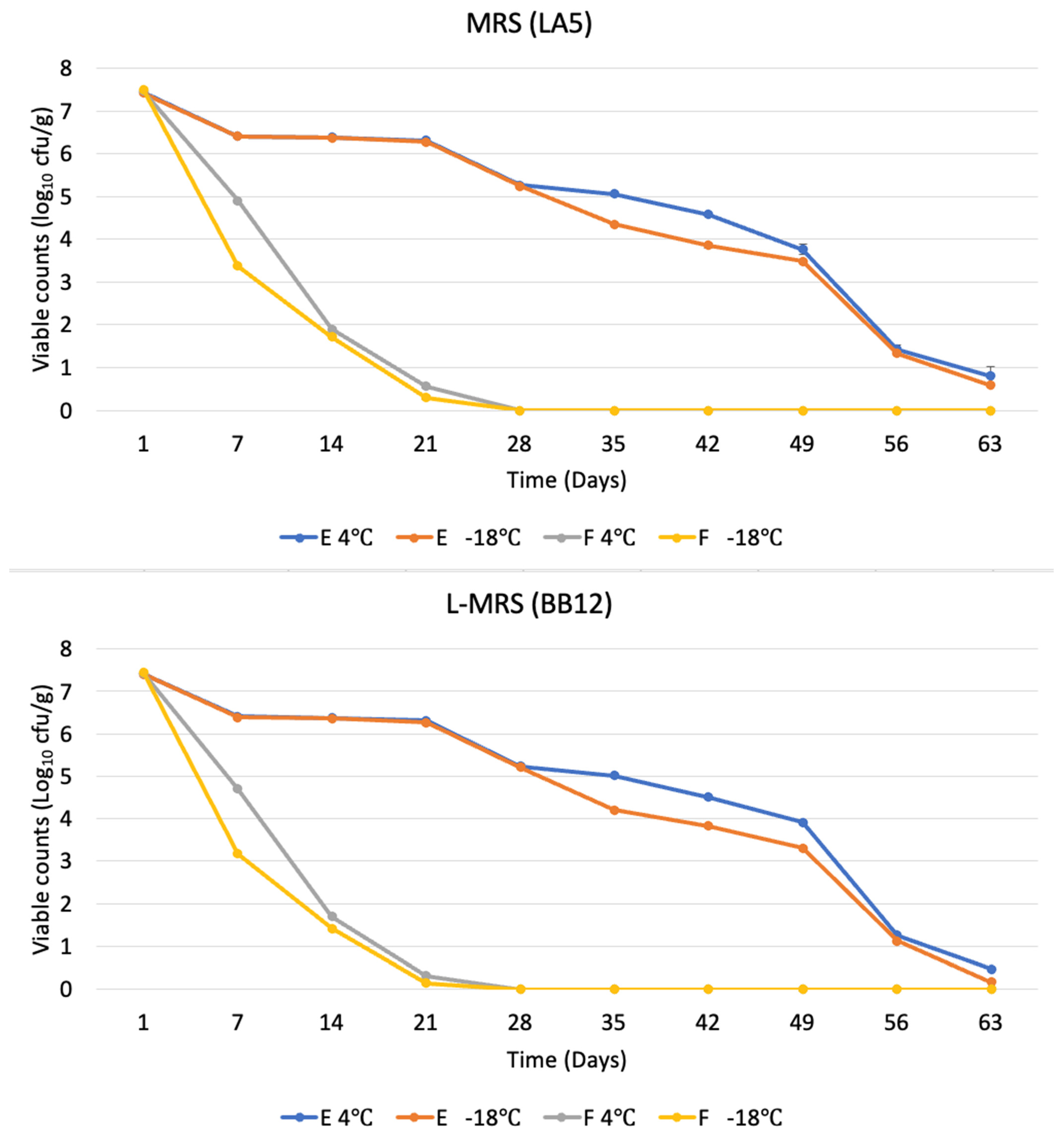
3.1. Viable Probiotic Counts of Butter

Probiotic microorganisms’ viability and metabolic activity are crucial factors to consider. Probiotics have health benefits, but they only become apparent when the number of viable cells in the product increases beyond a certain point both during production and during consumption, all while maintaining the sensory qualities of the product [12]. Butter could be used as a carrier of probiotics due to its fat-rich viscous matrix, which might act as a protectant for probiotics [8,13]. Bioactive materials like probiotics can be easily incorporated into the viscous fat structure of butter; however, encapsulation provides extra protection to the probiotics during the storage of butter. The free cells and encapsulated cells of LA5 and BB12 were incorporated at the same level with a ratio of LA5:BB12 of 1:1.
At 4 °C, the initial counts of LA5 cells for free and encapsulated cells were 7.47 ± 0.013 log10 CFU/g and 7.44 ± 0.005 log10 CFU/g, respectively, and BB12 free and encapsulated cells were 7.42 ± 0.014 log10 CFU/g and 7.42 ± 0.03 log10 CFU/g, respectively. At −18 °C, the initial counts of LA5 cells for free and encapsulated cells were 7.50 ± 0.016 log10 CFU/g and 7.41 ± 0.008 log10 CFU/g, respectively, and BB12 cells for free and encapsulated cells were 7.45 ± 0.008 log10 CFU/g and 7.40 ± 0.005 log10 CFU/g, respectively. The counts of both LA5 and BB12 started to decline after storage at both temperatures; however, the rate of decline was much faster in the variants having free cells of probiotics compared to the encapsulated cells of probiotics (Figure 1). After 21 days of storage, free cells of LA5 log10 CFU/g as well as BB12 log10 CFU/g declined below 1.0 log10 CFU/g, whereas in the case of encapsulated cells at both temperatures, the decline in LA5 and BB12 viable cells was not as sharp as free cells, because whey protein and starch might act as cryoprotectants and improve the survivability of probiotics during cold storage [14]. Similar observations were reported previously that adding starch improves the probiotic’s ability to withstand heat and storage conditions after emulsification [15]. The survival rate during freezing could also increase due to the presence of calcium and phosphate in milk proteins [16] or due to the proteins themselves acting as amphoteric molecules because of their buffering ability [17]. Several studies have demonstrated that encapsulation lengthens the shelf life of new products, preserves microorganisms, and improves/increases/enhances probiotic viability [2,18,19,20,21].
As shown by the confocal laser scanning microscopy in Section 3.4 below, the number of protein bodies increased over the storage period, which was possible as a result of hydration and disintegration of the WPH-MD encapsulant. The decline in viable counts of probiotics in butter variants with encapsulant may, however, be due to the exposure of LA5 and BB12 to the low temperature, due to disintegration of WPH-MD encapsulant [22]. The incorporation of BB-12 and (as specified earlier) LA-5, either in free or microencapsulated form, into white-brined cheese was investigated by Özer et al. [23], and they found a significant difference in the survivability of cells between free and encapsulated cells. Both probiotic strains LA5 and BB12 showed the same declining trend up to 14 days of storage, and their rate of decline was the same, whereas Erkaya [24] reported that BB12 showed a higher decline rate after 30 days of storage compared to other species’ strains [6]. Compared to Bifidobacteria, lactobacilli are generally observed to be more resistant because of their ability to adapt to a wide range of environmental conditions, including temperature, pH, and substrate availability [25,26,27]. Additionally, bifidobacteria’s growth may be inhibited when they come into contact with metabolic products made by lactic acid bacteria, which are naturally found in dairy products [28]. Bifidobacteria may be inhibited by other species within a multi-strain probiotic mixture, as evidenced by the fact that mixes of non-bifidobacterial strains are more effective than mixes containing one or more Bifidobacterium strains. More generally, antagonistic agents produced by various species can inhibit one another, as a variety of antimicrobial compounds, including bacteriocins, hydrogen peroxide, and organic acids, are produced by lactobacilli, and these antimicrobial agents may suppress bifidobacterium growth and metabolic processes. Moreover, lactobacilli may also compete with Bifidobacterium for vital nutrients [29]. Proposed causes of cell death after freezing and thawing include membrane damage and DNA denaturation [30,31]; however, an increase in intracellular solute concentration also damages proteins. It could be interpreted that the encapsulation technique potentially offers an effective means of enhancing the viability of probiotics during storage. However, there may still be such situations, such as the potential for probiotics that have been encapsulated to be exposed to low temperatures due to potential encapsulant disintegration. Taking these intricacies into account makes it imperative to create a mathematical model that could predict the probiotic population fluctuations during storage. To maximize storage conditions and extend the shelf life of probiotic-enriched butter products, a model of this kind could incorporate variables such as temperature, encapsulation efficacy, and storage time.

3.2. Modelling of LA5 and BB12

The creation of a mathematical model for the probiotic population changes in butter during storage is helpful to ensure the effectiveness and quality of the final product as well as customer satisfaction. Considering variables like shelf life and storage temperature, this kind of model enables product manufacturers to predict and control the intricate dynamics of probiotic viability over time. Strategies can be designed to maintain desired probiotic levels until the product reaches consumers and optimize storage conditions by potentially forecasting how probiotic populations change during storage. By taking this proactive approach, it may be possible to set realistic expiration dates, guaranteeing that consumers receive products with desirable probiotic counts and viability. Based on the results in Section 3.1 and the analysis of covariance, the following equations (Equations (2) and (3)) for the prediction of LA5 andBB12 log10 CFU/g can be used as follows.
Following residual analysis of multiple linear regression, the quadratic term of Day (D2) was found to be important. The estimated regression equation (Equation (2)) for the prediction of LA5 log10 CFU/g counts has an adjusted R-square of 0.97 and is given by
y ^ = 8.220 0.292 ( T ) 1.041 ( C ) 0.638 ( D ) + 0.013 ( D 2 ) + 0.621 ( C × D ) 0.014 ( C × D 2 )
The estimated regression equation (Equation (3)) for the prediction of BB12 log10 CFU/g counts has an adjusted R-square of 0.97 and is given by
y ^ = 8.223 0.329 ( T ) 1.076 ( C ) 0.673 ( D ) + 0.014 ( D 2 ) + 0.662 ( C × D ) 0.016 ( C × D 2 )
where y ^ is estimated log10 CFU/g, T is the temperature of storage (°C), C is cell type (free or encapsulated cell), D is the number of days of storage, C is 1 for encapsulated cells and 0 for free cells, and T is 1 for −18 °C and 0 for 4 °C.
The above-mentioned two equations (Equations (2) and (3)) could potentially predict the log10 CFU/g counts at a given point of time during storage at a definite temperature given the specific condition of level of inoculation, product characteristics, and storage temperature. It is also possible to estimate the days associated with a given log10 CFU/g (inverse prediction), for example, for 6 log10 counts of LA5, the estimated days are 20.25 (for T = −18 and Cell = E), 24.40 (T = 4 and Cell = E), 3.01 (T = −18 and Cell = F), and 3.58 (T = 4 and Cell = F). Confidence intervals can also be constructed [32]. These equations can potentially be used for the different Lactobacillus and Bifidobacterium strains; however, it should be considered that every strain, even within a species, has different metabolic pathways and properties, which might affect the accuracy of the mentioned equation. The strains closely related to Lactobacillus acidophilus and Bifidobacterium animalis ssp. lactis within the same species or different species could potentially be used in butter and their prediction could be performed through these equations (Equations (2) and (3)). It could be concluded that these equations can potentially enable manufacturers to optimize overall product quality, as they would be able to predict the desired probiotic counts in variable conditions as well as beyond the tested period. These equations could also be extended for different products like cream cheese, mayonnaise, or nut butter; however, the results would need to be validated for a longer storage period and under different storage conditions through specific studies. The outcome of the analysis of covariance for the prediction of LA5 and BB12 log10 CFU/g counts is presented in Figure 2.

3.3. Acid Value of Butter

The number of free acids in fats, measured in milligrams of potassium hydroxide needed to neutralize them, is known as the acid value of the fat. The acid value of butter plays a crucial role in assessing its quality and stability by revealing the concentration of free fatty acids within the product. These fatty acids result from the breakdown of triglycerides, typically due to enzymatic or hydrolytic actions. Elevated acid values signal higher levels of free fatty acids, which in turn increases the risk of rancidity, undesirable flavors, and shorter shelf life. Butter and other fats with a high water content are more likely to hydrolyze and can oxidize rapidly, acquiring a distinct bad taste and smell [33]. There was no significant increase in the acid value of the butter variants having free cells or encapsulated cells of LA5 and BB12; the reason behind that could be the low lipolytic activity of lactobacillus acidophilus and Bifidobacteria animalis ssp. lactis, which is also reported by E. Dincer et al. 2018 [34]. The acid value of butter with free and encapsulated cells is presented (Table 1). The probiotic bacteria remained after 63 days of storage, and even after the viable count test period, the acid value remained unchanged until 84 days of storage. This might suggest that the two probiotic microorganisms, LA5 and BB12, in either free or encapsulated forms and different storage conditions, have no direct relationship with the acid value of butter.

3.4. Confocal Laser Scanning Microscopy

Confocal laser scanning microscopy provides high-resolution imaging of the internal structure of butter, where we can observe the composition, homogeneity, and microstructure of butter using specific dyes for specific components of butter. Furthermore, it can provide an idea about the rheological properties of butter [35]. The results of confocal laser scanning microscopy (CLSM) applied to butter samples, utilizing FITC for binding proteins and Nile Red for visualizing the fat phase, are presented in Figure 3. The use of FITC, known for its affinity to proteins, illuminated the proteinaceous elements within the butter, producing distinct green fluorescence signals. Simultaneously, Nile Red, specific to lipid-rich structures, enhanced the visualization of the fat phase, exhibiting a vibrant red fluorescence [11]. As the probiotics were encapsulated in whey protein encapsulant, it was observed more in butter with encapsulated cells of probiotics, as they showed more green particles (protein) as compared to the butter with free cells of probiotics, as it did not have the extra protein contributed by the encapsulant. It was also observed that the number of green particles (protein) increased over the storage, and their size decreased, which might be due to the dissociation of the encapsulant, as it might have interacted with water molecules present in the butter and disintegrated after prolonged contact, resulting in more binding of FITC to protein particles [22]. This phenomenon highlights the significance of CLSM in comprehending the dynamics of encapsulation and its influence on butter’s microstructure, offering insightful information about the stability and encapsulation characteristics of butter with probiotics. However, this would need further investigation.

4. Conclusions

Butter shares a very big part in the US dairy product market, and the revenue is expected to show an annual growth rate (CAGR 2024–2029) of 17.27%, resulting in a projected market volume of USD 1.53 billion by 2029. The potential for butter to serve as a carrier for probiotics presents an exciting opportunity for the dairy industry and farmers to penetrate the functional food market. Butter, being a widely consumed dairy product, can act as an ideal vehicle for delivering probiotics, enhancing its nutritional value, and appealing to health-conscious consumers. Moreover, the inclusion of probiotics in butter can offer a convenient way for individuals to incorporate these beneficial microorganisms into their daily diet, potentially improving gut health and overall well-being.
It is generally difficult to monitor the viability of probiotic strains like BB12 and LA5 during prolonged storage of products, especially because traditional plating techniques take a long time. By quickly estimating probiotic viability, predictive equations provide a reliable solution that helps manufacturers optimize storage procedures and make well-informed decisions regarding formulation and packaging. In a highly competitive market, adopting mathematical models offers a quicker and more dependable way to maintain product quality and satisfy health-conscious consumers, even though validation against conventional methods is still crucial.
Furthermore, the development and refinement of mathematical predictive models for probiotic viability in butter open avenues for further research and innovation. Future studies could explore additional variables that may impact probiotic stability, such as the effects of different types of fats, emulsifiers, and antioxidants on probiotic survival in butter along with the studies of different probiotic strains. These models could also be expanded for a wide variety of products having a viscous matrix, and generative AI could potentially be used to further improve the efficiency of these models.

Author Contributions

Conceptualization, R.K. and S.A.; methodology, R.K. and S.A.; validation, R.K., S.A. and G.D.; formal analysis, R.K. and G.D.; investigation, R.K. and S.A.; writing—original draft preparation, R.K.; writing—review and editing, R.K., K.G., S.A. and G.D.; supervision, S.A.; project administration, S.A.; funding acquisition, S.A. All authors have read and agreed to the published version of the manuscript.

Funding

This research was funded by the Midwest Dairy Association Index number 3X1462.

Institutional Review Board Statement

Not applicable.

Informed Consent Statement

Not applicable.

Data Availability Statement

All data used to support the findings are included within.

Acknowledgments

The authors would like to acknowledge the Department of Dairy and Food Science SDSU and Agricultural Experimental Station SDSU. All authors have consented to the acknowledgment.

Conflicts of Interest

The authors declare no conflicts of interest.

References

  1. FAO/WHO. Probiotics in Food: Health and Nutritional Properties and Guidelines for Evaluation. Available online: https://www.fao.org/documents/card/en?details=7c102d95-2fd5-5b22-8faf-f0b2e68dfbb6/ (accessed on 4 October 2001).
  2. da Silva, M.N.; Tagliapietra, B.L.; dos Santos Richards, N.S.P. Encapsulation, storage viability, and consumer acceptance of probiotic butter. LWT 2021, 139, 110536. [Google Scholar] [CrossRef]
  3. Bellinazo, P.L.; Vitola, H.R.S.; Cruxen, C.E.d.S.; Braun, C.L.K.; Hackbart, H.C.d.S.; da Silva, W.P.; Fiorentini, Â.M. Probiotic butter: Viability of Lactobacillus casei strains and bixin antioxidant effect (Bixa orellana L.). J. Food Process. Preserv. 2019, 43, e14088. [Google Scholar] [CrossRef]
  4. Zacarchenco, P.B.; Spadoti, L.M.; Silva e Alves, A.T.; Zanetti, V.C.; Verruck, S. Probiotic Butter. In Probiotic Foods and Beverages: Technologies and Protocols; Springer: Berlin/Heidelberg, Germany, 2023; pp. 67–80. [Google Scholar]
  5. Terpou, A.; Papadaki, A.; Lappa, I.K.; Kachrimanidou, V.; Bosnea, L.A.; Kopsahelis, N. Probiotics in food systems: Significance and emerging strategies towards improved viability and delivery of enhanced beneficial value. Nutrients 2019, 11, 1591. [Google Scholar] [CrossRef] [PubMed]
  6. Frakolaki, G.; Giannou, V.; Kekos, D.; Tzia, C. A review of the microencapsulation techniques for the incorporation of probiotic bacteria in functional foods. Crit. Rev. Food Sci. Nutr. 2021, 61, 1515–1536. [Google Scholar] [CrossRef]
  7. Gaba, K.; Anand, S. Potential of Incorporating a Functional Probiotic Encapsulant in Whipped Cream. Fermentation 2023, 9, 928. [Google Scholar] [CrossRef]
  8. Gaba, K.; Anand, S.; Syamala, A. Development of Value-Added Butter by Incorporating Whey Protein Hydrolysate-Encapsulated Probiotics. Microorganisms 2023, 11, 1139. [Google Scholar] [CrossRef] [PubMed]
  9. Minj, S.; Anand, S. Development of a spray-dried conjugated whey protein hydrolysate powder with entrapped probiotics. J. Dairy Sci. 2022, 105, 2038–2048. [Google Scholar] [CrossRef]
  10. Ewe, J.-A.; Loo, S.-Y. Effect of cream fermentation on microbiological, physicochemical and rheological properties of L. helveticus-butter. Food Chem. 2016, 201, 29–36. [Google Scholar] [CrossRef] [PubMed]
  11. Rønholt, S.; Kirkensgaard, J.J.K.; Pedersen, T.B.; Mortensen, K.; Knudsen, J.C. Polymorphism, microstructure and rheology of butter. Effects of cream heat treatment. Food Chem. 2012, 135, 1730–1739. [Google Scholar] [CrossRef]
  12. Yilmaz-Ersan, L.; Ozcan, T.; Akpinar-Bayizit, A.; Turan, M.A.; Taskin, M.B. Probiotic cream: Viability of probiotic bacteria and chemical characterization. J. Food Process. Preser. 2017, 41, e12797. [Google Scholar] [CrossRef]
  13. Olszewska, M.; Staniewski, B.; Łaniewska-Trokenheim, Ł. Cell viability of Bifidobacterium lactis strain in long-term storage butter assessed with the plate count and fluorescence techniques. Czech J. Food Sci. 2012, 30, 421–428. [Google Scholar] [CrossRef]
  14. Dianawati, D.; Mishra, V.; Shah, N.P. Survival of Bifidobacterium longum 1941 microencapsulated with proteins and sugars after freezing and freeze drying. Food Res. Int. 2013, 51, 503–509. [Google Scholar] [CrossRef]
  15. Zanjani, M.A.K.; Ehsani, M.R.; Tarzi, B.G.; Sharifan, A. Promoting probiotics survival by microencapsualtion with Hylon starch and genipin cross-linked coatings in simulated gastro-intestinal condition and heat treatment. Iran. J. Pharm. Res. IJPR 2018, 17, 753. [Google Scholar]
  16. Ming, L.C.; Rahim, R.A.; Wan, H.Y.; Ariff, A.B. Formulation of protective agents for improvement of Lactobacillus salivarius I 24 survival rate subjected to freeze drying for production of live cells in powderized form. Food Bioprocess Techno. 2009, 2, 431–436. [Google Scholar] [CrossRef]
  17. Ugwu, S.O.; Apte, S.P. The effect of buffers on protein conformational stability. Pharm. Technol. 2004, 28, 86–109. [Google Scholar]
  18. Menezes, M.F.D.S.C.D.; Silva, T.M.D.; Etchepare, M.D.A.; Fonseca, B.D.S.; Sonza, V.P.; Codevilla, C.F.; Barin, J.S.; Silva, C.D.B.D.; Menezes, C.R.d. Improvement of the viability of probiotics (Lactobacillus acidophilus) by multilayer encapsulation. Ciênc. Rural 2019, 49, e20181020. [Google Scholar] [CrossRef]
  19. Feng, K.; Huang, R.-M.; Wu, R.-Q.; Wei, Y.-S.; Zong, M.-H.; Linhardt, R.J.; Wu, H. A novel route for double-layered encapsulation of probiotics with improved viability under adverse conditions. Food Chem. 2020, 310, 125977. [Google Scholar] [CrossRef]
  20. Yao, M.; Li, B.; Ye, H.; Huang, W.; Luo, Q.; Xiao, H.; McClements, D.J.; Li, L. Enhanced viability of probiotics (Pediococcus pentosaceus Li05) by encapsulation in microgels doped with inorganic nanoparticles. Food Hydrocoll. 2018, 83, 246–252. [Google Scholar] [CrossRef]
  21. Martín, M.J.; Lara-Villoslada, F.; Ruiz, M.A.; Morales, M.E. Microencapsulation of bacteria: A review of different technologies and their impact on the probiotic effects. Innov. Food Sci. Emerg. Technol. 2015, 27, 15–25. [Google Scholar] [CrossRef]
  22. Huang, X.; Gänzle, M.; Zhang, H.; Zhao, M.; Fang, Y.; Nishinari, K. Microencapsulation of probiotic lactobacilli with shellac as moisture barrier and to allow controlled release. J. Sci. Food Agric. 2021, 101, 726–734. [Google Scholar] [CrossRef] [PubMed]
  23. Özer, B.; Kirmaci, H.A.; Şenel, E.; Atamer, M.; Hayaloğlu, A. Improving the viability of Bifidobacterium bifidum BB-12 and Lactobacillus acidophilus LA-5 in white-brined cheese by microencapsulation. Int. Dairy J. 2009, 19, 22–29. [Google Scholar] [CrossRef]
  24. Erkaya, T.; Ürkek, B.; Doğru, Ü.; Çetin, B.; Şengül, M. Probiotic butter: Stability, free fatty acid composition and some quality parameters during refrigerated storage. Int. Dairy J. 2015, 49, 102–110. [Google Scholar] [CrossRef]
  25. Nunes, G.L.; Motta, M.H.; Cichoski, A.J.; Wagner, R.; Muller, É.I.; Codevilla, C.F.; Silva, C.d.B.d.; Menezes, C.R.d. Encapsulation of Lactobacillus acidophilus La-5 and Bifidobacterium Bb-12 by spray drying and evaluation of its resistance in simulated gastrointestinal conditions, thermal treatments and storage conditions. Ciênc. Rural 2018, 48, e20180035. [Google Scholar] [CrossRef]
  26. Holkem, A.T.; Robichaud, V.; Favaro-Trindade, C.S.; Lacroix, M. Chemopreventive properties of extracts obtained from blueberry (Vaccinium myrtillus L.) and jabuticaba (Myrciaria cauliflora Berg.) in combination with probiotics. Nutr. Cancer 2021, 73, 671–685. [Google Scholar] [CrossRef]
  27. Tripathi, M.K.; Giri, S.K. Probiotic functional foods: Survival of probiotics during processing and storage. J. Funct. Foods 2014, 9, 225–241. [Google Scholar] [CrossRef]
  28. Boylston, T.D.; Vinderola, C.G.; Ghoddusi, H.B.; Reinheimer, J.A. Incorporation of bifidobacteria into cheeses: Challenges and rewards. Int. Dairy J. 2004, 14, 375–387. [Google Scholar] [CrossRef]
  29. Chapman, C.; Gibson, G.R.; Rowland, I. Health benefits of probiotics: Are mixtures more effective than single strains? Eur. J. Nutr. 2011, 50, 1–17. [Google Scholar] [CrossRef]
  30. Alur, M.a.; Grecz, N. Mechanism of injury of Escherichiacoli by freezing and thawing. Biochem. Biophys. Res. Commun. 1975, 62, 308–312. [Google Scholar] [CrossRef]
  31. El-Kest, S.E.; Marth, E.H. Freezing of Listeria monocytogenes and other microorganisms: A review. J. Food Prot. 1992, 55, 639–648. [Google Scholar] [CrossRef]
  32. Djira, G.; Hasler, M.; Gerhard, D.; Segbehoe, L.; Schaarschmidt, F. Mratios: Ratios of coefficients in the general linear model. R Package Vers. 2020, 1, 1–46. [Google Scholar]
  33. Koczoń, P.; Gruczyńska, E.; Kowalski, B. Changes in the acid value of butter during storage at different temperatures as assessed by standard methods or by FT-IR spectroscopy. Am. J. Food Technol. 2008, 3, 154–163. [Google Scholar] [CrossRef]
  34. Dinçer, E.; Kıvanç, M. Lipolytic activity of lactic acid bacteria isolated from Turkish pastırma. Anadolu Univ. J. Sci. Technol. C-Life Sci. Biotechnol. 2018, 7, 12–19. [Google Scholar] [CrossRef]
  35. Buldo, P.; Wiking, L. The role of mixing temperature on microstructure and rheological properties of butter blends. J. Am. Oil Chem. Soc. 2012, 89, 787–795. [Google Scholar] [CrossRef]
Figure 1. Viable counts of probiotics Lactobacillus acidophilus on MRS agar (top) and Bifidobacterium animalis ssp. lactis on L-MRS agar (bottom) at 4 °C and −18 °C. F: free cells, E: encapsulated cells.
Fermentation 10 00245 g001
Figure 2. Analysis of covariance for Lactobacillus acidophilus (a) and Bifidobacterium animalis ssp. lactis (b) where the x-axis represents the log10 CFU/g. F: free cells, E: encapsulated cells; 4 and −18 are storage temperatures in °C.
Fermentation 10 00245 g002
Figure 3. Confocal laser scanning microscopy image of butter variants: control butter (a), butter with free cells of LA5 and BB12 (b), butter with encapsulated cells of probiotics at 0 days (c), 28 days (d), 56 days (e), 84 days (f). Nile red fluorescence (red) represents the fat phase and Fluorescein Isothiocyanate FITC (green) represents protein bodies.
Fermentation 10 00245 g003
Table 1. Acid value of butter stored at 4 and −18 °C with free and encapsulated cells of LA5 and BB12.
Acid Value
ControlFree CellEncapsulated Cells
Days4 °C−18 °C4 °C−18 °C4 °C−18 °C
11.246 ± 0.010 Aa1.253 ± 0.009 Aa1.262 ± 0.008 Aa1.287 ± 0.003 Aa1.264 ± 0.014 Aa1.271 ± 0.007 Aa
281.256 ± 0.010 Aa1.256 ± 0.010 Aa1.279 ± 0.009 Aa1.294 ± 0.007 Aa1.267 ± 0.011 Aa1.277 ± 0.005 Aa
561.294 ± 0.015 Aa1.254 ± 0.015 Aa1.304 ± 0.012 Aa1.275 ± 0.012 Aa1.273 ± 0.012 Aa1.275 ± 0.013 Aa
841.306 ± 0.014 Aa1.263 ± 0.014 Aa1.313 ± 0.012 Aa1.284 ± 0.010 Aa1.282 ± 0.014 Aa1.286 ± 0.013 Aa
Capital letter (A) shows a significant difference row-wise. Small letter (a) shows a significant difference column-wise.
Disclaimer/Publisher’s Note: The statements, opinions and data contained in all publications are solely those of the individual author(s) and contributor(s) and not of MDPI and/or the editor(s). MDPI and/or the editor(s) disclaim responsibility for any injury to people or property resulting from any ideas, methods, instructions or products referred to in the content.

