Article

Defining the miRnome of Saphenous Vein Smooth Muscle Cells from Patients with Type 2 Diabetes Mellitus

1
Cardiovascular Research Group, Faculty of Life Sciences, University of Bradford, Bradford BD7 1DP, UK
2
Faculty of Life Sciences, University of Bradford, Bradford BD7 1DP, UK
3
Biomedical Institute for Multimorbidity, Hull-York Medical School Centre for Biomedicine, University of Hull, Hull HU6 7RU, UK
4
Faculty of Biological Sciences, University of Leeds, Leeds LS2 9JT, UK
5
Faculty of Medicine and Health, University of Leeds, Leeds LS2 9JT, UK
*
Author to whom correspondence should be addressed.
Diabetology 2024, 5(2), 178-189; https://doi.org/10.3390/diabetology5020014
Submission received: 23 February 2024 / Revised: 16 April 2024 / Accepted: 10 May 2024 / Published: 11 May 2024
(This article belongs to the Special Issue Exclusive Papers Collection of Editorial Board Members in Diabetology)

Abstract

:
Type 2 diabetes mellitus (T2DM) patients suffer premature development of cardiovascular disease and commonly require cardiac revascularization using the autologous saphenous vein (SV). Smooth muscle cells (SMCs) are the principal cell type within the vascular wall and are dysfunctional in T2DM SV-SMCs, yet the mechanisms underpinning this are incompletely understood. The purpose of this study was to interrogate differential microRNA (miRNA) expression in SV-SMCs to enhance our understanding of T2DM SV-SMC phenotypic change. miRNA expression in primary human SV-SMCs from T2DM and non-diabetic (ND) donors was determined using an array (n = 6 each of ND and T2DM SV-SMCs). Differentially expressed miRNAs were ranked, and functional annotation of the 30 most differentially expressed miRNAs using DAVID and KEGG analysis revealed pathways related to SMC phenotype, including proliferation, migration, cytokine production and cell signaling. After selecting miRNAs known to be involved in SMC phenotypic regulation, miR-17, miR-29b-2, miR-31, miR-130b and miR-491 were further validated using qRT-PCR (n = 5 each of ND and T2DM SV-SMC), with miR-29b-2 subsequently being removed from further investigation. Potential mRNA targets were identified using mirDIP. Predicted target analysis highlighted likely dysregulation in transcription, epigenetic regulation, cell survival, intracellular signaling and cytoskeletal regulation, all of which are known to be dysfunctional in T2DM SV-SMCs. In conclusion, this paper identified four miRNAs that are dysregulated in T2DM SV-SMCs and are implicated in functional changes in the behavior of these cells. This provides a step forward in our understanding of the molecular and epigenetic regulation of vascular dysfunction in T2DM.

1. Introduction

The prevalence of type 2 diabetes mellitus (T2DM) is increasing globally at a rapid rate. In the UK, it is estimated that up to 8% of adults have either T2DM or pre-diabetes, and this pattern is repeated throughout the Western world and increasingly across Asia [1,2]. There is an urgent need to identify both early markers of T2DM and new therapeutic targets for the development of effective pharmacological management of the disease.
One of the leading causes of morbidity and mortality in T2DM patients is the premature development of cardiovascular disease with extensive and widespread atherosclerotic lesions. Patients with T2DM, therefore, frequently need cardiac revascularization using either percutaneous coronary intervention or coronary artery bypass grafting (CABG). The majority of CABG surgeries utilize the long autologous saphenous vein (SV) due to its availability and the requirement for multiple simultaneous grafts; however, patency of these vessels is poor compared to arterial grafts with only approximately 50% of venous grafts remaining patent 10 years post-surgery [3,4].
The most abundant cell type within the vascular wall is the smooth muscle cell (SMC). Whilst SMCs are responsible for maintaining vascular tone, they are also implicated in post-CABG failure through neointimal hyperplasia and progressive atherosclerosis. Ordinarily quiescent, SMCs can transiently and reversibly adopt a variety of phenotypes such as synthetic, foam cell-like, mesenchymal or osteogenic [5]. The ability of SMCs to react and dynamically change their phenotype in response to external cues is crucial in the timeframe immediately after vessel implantation. Here, SMCs must transiently switch to a proliferative synthetic phenotype in order to fully adapt to coronary circulation before reverting to a quiescent contractile phenotype for long-term bypass functionality [6].
We previously demonstrated that cultured T2DM SV-SMCs exhibit behaviors distinct from those of non-diabetic (ND) patients. Specifically, T2DM SV-SMCs possess an aberrant phenotype that is characterized by reduced proliferative capacity, increased contractile marker expression and a disorganized, fragmented cytoskeleton [7,8,9]. This could conceivably contribute to SV graft failure by inhibiting the required phenotypic switching in the post-surgery period. Furthermore, these cells display evidence of prolonged DNA damage and acquisition of senescence-associated secretory phenotype (SASP) that can perpetuate and enhance SMC dysfunction in T2DM patients. Importantly, many of these features are driven, at least in part, by a persistent upregulation of a particular microRNA (miRNA), miR-145 [10].
miRNAs are short, non-coding RNAs that typically negatively regulate target gene expression. They are promiscuous, whereby one miRNA can target multiple mRNAs simultaneously, thereby switching entire pathways on and off. miR-145 is well-recognized as a regulator of vascular SMC phenotype [11], so it is unsurprising that the persistent upregulation of this miRNA in T2DM SV-SMCs can have profound effects on phenotype. However, the contribution of other miRNAs to vascular dysfunction in T2DM SV-SMCs is currently understudied.
One of the attractive characteristics of miRNAs is that they are amenable to therapeutic manipulation. Although there are currently no approved miRNA-based drugs for treating cardiovascular diseases, there have been successful clinical trials for miR-122-5p in hepatitis C (recently reviewed in [12]). This demonstrates that the technology is already optimized and validated for therapeutic delivery. Therefore, the aim of this project was to explore the miRnome of both ND and T2DM SV-SMCs with a view to identifying potential new therapeutic avenues for preventing post-bypass graft failure.

2. Materials and Methods

2.1. Isolation and Culture of Primary Human SV-SMC

For the initial array, SMCs were isolated from surplus fragments of SV tissue following CABG at the Leeds General Infirmary or Nuffield Health Leeds Hospital (local ethical committee approval CA01/040). For miRNA validation experiments, SMCs were also isolated from SV; however, the vessels were extracted during two different clinical procedures—from surplus CABG fragments at Castle Hill Hospital, Hull (REC reference 15/NE/0138, IRAS Project ID: 170899; 3 ND and 2 T2DM donors) or following lower limb amputation at the Bradford Royal Infirmary (local ethical committee approval 16/077; 2 ND and 3 T2DM donors) with informed written patient consent. The studies conformed with the Declaration of Helsinki.
The adventitial tissue was removed using surgical scissors, and the vessel opened longitudinally. The endothelial layer was removed by scraping with a sterile blade. The remaining medial layer was diced into 2 mm3 pieces, which were placed into 25 cm2 flasks in Dulbecco’s Minimal Essential Medium (DMEM) containing 4.5 g/L glucose, 1% (v/v) penicillin–streptomycin–fungizone and 1% (v/v) L-glutamine. SMCs were cultured in 5% CO2 in air at 37 °C, and SMCs explanted over the subsequent days. Once confluent, cells were passaged using trypsin/EDTA as required.

2.2. Preparation of RNA for miRNA Array

A power calculation was performed using the mean and standard deviation of miR-145 expression in SV-SMCs [8]. The Type I/II error rate alpha was set as 0.05. This revealed that an n = 5 in each group would be 95% powered to detect a 2-fold change in expression between ND- and T2DM-SV-SMCs.
RNA was extracted from confluent, proliferating SV-SMCs from 12 patient donors. Donor characteristics (biochemical markers and current medications) can be found in our previous publication [8]. These donors were exclusively male and had a mean age of 63.7 (range 49–78; n = 6) for ND patients and 63.0 (range 50–85; n = 6) for patients with T2DM. RNA was extracted using the Aurum Total RNA Mini-Kit (Bio-Rad Laboratories Ltd., Watford, UK) according to manufacturers’ instructions, and purity and concentration were measured using a NanoDrop. From SV-SMCs from each patient donor, 2.5 μg RNA was supplied to Vertis Biotechnologie AG (Freising, Germany) for miRNA array analysis.

2.3. miRNA Array

The miRNA array was conducted by Vertis Biotechnologie AG. Briefly, miRNA fractions with a length between 19 and 29 nucleotides were isolated from each RNA sample using the Caliper LabCip XT apparatus. These were polyA-tailed, reverse transcribed, and the resultant cDNA was amplified to a concentration of 10–20 ng/μL using high-fidelity DNA polymerase. This was run through a 6% polyacrylamide gel and cDNA in the size range of 160–180 bp, which was eluted and sequenced on an Illumina HiSeq 2000 system.

2.4. Identification of Differentially Expressed miRNAs

Spliced Transcripts Alignment to a Reference (STAR) v. 2.7 [13] was used to align reads to the Gencode version 29 human reference genome (GRCh38.p12). A complete read was created by stitching together separate seeds and clustering based on proximity to a set of anchor seeds. Depending on the best alignment for the read, the seeds were stitched together, and a score was generated based on factors such as indels and mismatches.
MMQUANT v. 13 [14] was used to count multi-mapping reads in an unbiased way to quantify miRNA expression. Following this, differential expression analysis was carried out using DESeq2 v. 3.18 [15], which is a tool used specifically for the normalization, visualization and differential analysis of RNA-seq data.
Differentially expressed miRNAs were sorted based on their p value and fold changes. The Database for Annotation, Visualization and Integrated Discovery ‘DAVID’ bioinformatics software (version 6.8) was used to identify the biological meaning of the top 30 differentially expressed miRNA using a set of functional annotation tools [16,17]. Functional annotation clustering was used to identify common biological processes, cellular compartments and molecular functions alongside KEGG pathways.

2.5. Validation of miRNA Array Targets

RNA was isolated from confluent, proliferating SV-SMCs from a further 10 patient donors (n = 5 ND and n = 5 T2DM). Both donor groups were 80% male and 20% female. They had a mean age of 61.8 (range 39–74) for ND SV-SMCs and 66.4 (range 63–72) for T2DM SV-SMCs. Current medication relevant to the vascular system can be found in Table 1.
RNA was isolated using the BioRad Aurum Total RNA Mini-Kit. Following DAVID and KEGG analysis and a comprehensive literature search of the involvement of these miRNAs in SMC regulation, the expression of five selected miRNAs was quantified using specific commercially available TaqMan® MicroRNA assays.
Predicted targets of differentially expressed miRNAs were performed using the mirDIP microRNA data integration portal (Version 5.3.0.2, Database version 5.2.3.1) using default settings (very high score class) for the four validated dysregulated miRNAs [18]. Predicted mRNA targets were then filtered according to the number of miRNAs of which they were predicted targets. All mRNAs that could be targeted by two or more miRNAs were probed for their functions to suggest the most likely dysregulated pathways in T2DM SV-SMCs.

2.6. Statistical Analysis

Throughout the manuscript, n refers to the number of patient donors for each SMC population. Short-listing of differentially expressed miRNAs from the array was analyzed using a Wald test. For the validation data, differences between ND and T2DM SV-SMCs were analyzed using an unpaired Mann–Whitney test. Significance was considered as p < 0.05.

3. Results

3.1. Initial Identification of Differentially Expressed miRNAs

The microRNA array identified 776 miRNAs in total. Of these, 177 were down-regulated in T2DM-SMCs by 0.5-fold or more, 274 were upregulated by more than 2-fold in T2DM-SMC, and 325 had limited changes (Figure 1a; Supplementary File S1). From these data, we performed functional annotation for the 30 miRNAs that had the most significant differential expression (Figure 1b). As is expected for miRNAs, the most common pathways related to them were gene silencing by miRNA, RISC complex, mRNA binding involved in post-transcriptional gene silencing and mRNA 3′-UTR binding. However, other pathways related to SMC phenotypic change were also highlighted in significantly over-expressed terms. These included negative regulation of cell migration (5 miRNAs), negative regulation of vascular endothelial growth factor production (2 miRNAs), negative regulation of cytokine production involved in the inflammatory response (2 miRNAs), both negative and positive regulation of SMC proliferation (2 miRNAs for each), negative regulation of the extracellular signal-regulated kinase (ERK) cascade (2 miRNAs), and negative regulation of the transforming growth factor beta (TGFβ) signaling pathway (2 miRNAs).

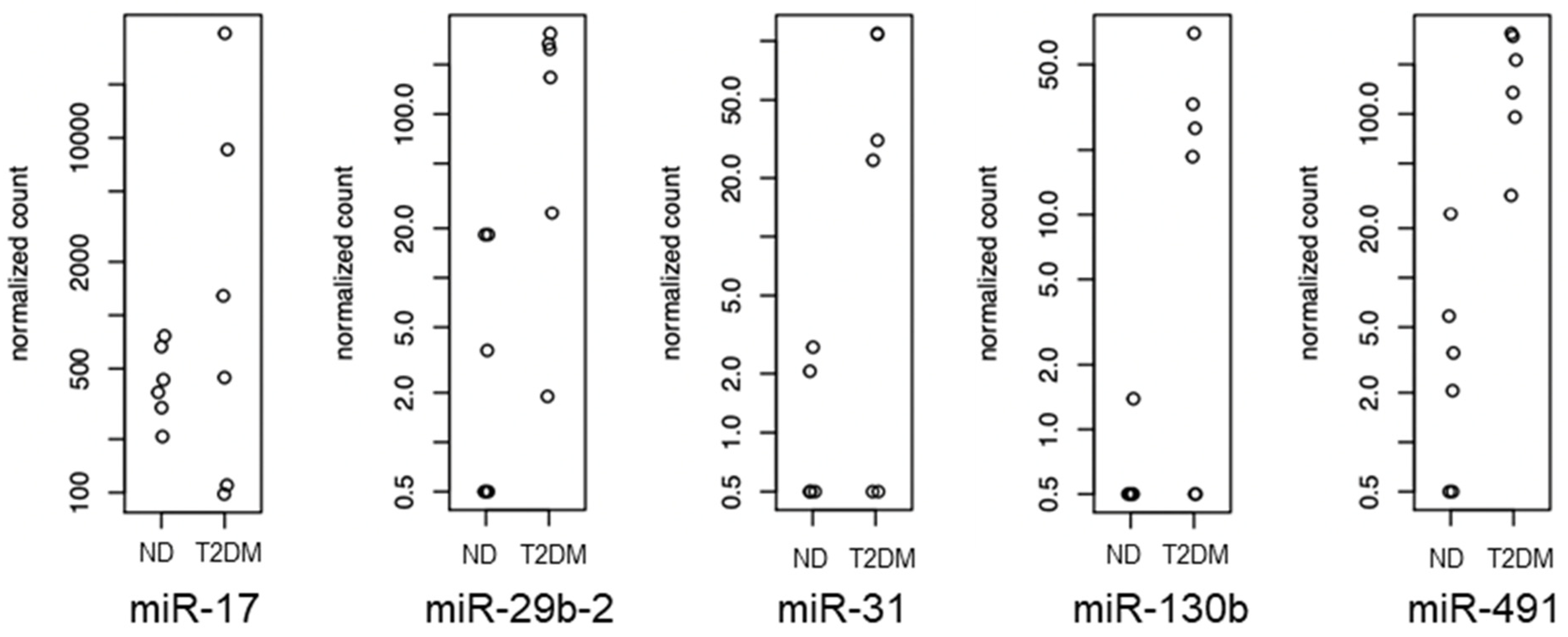
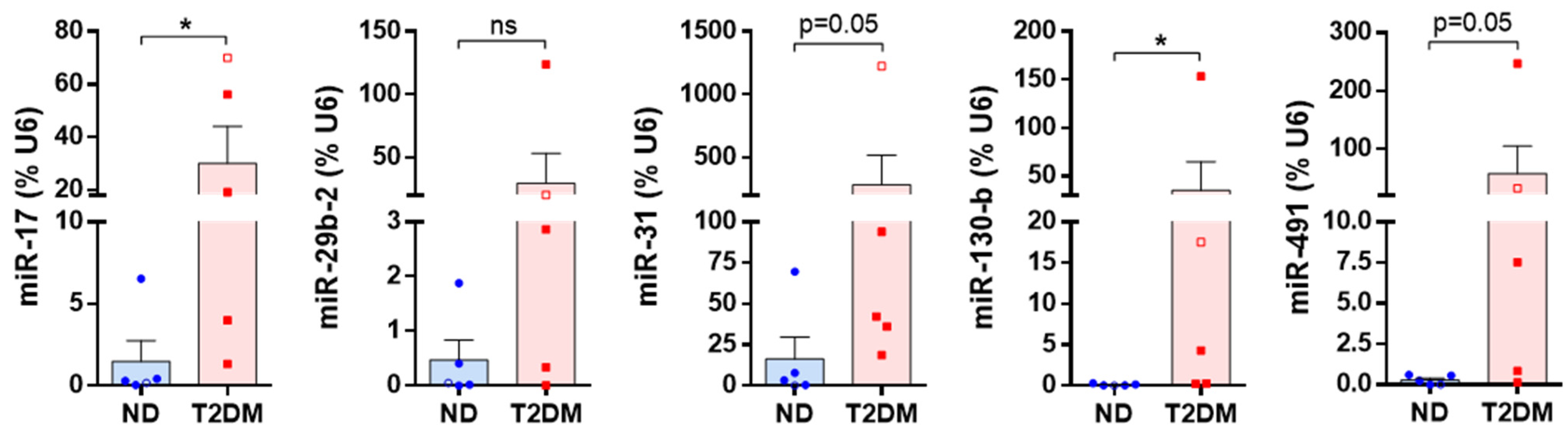
3.2. Differentially Expressed miRNAs

A literature search was conducted for the top 30 differentially expressed miRNAs identified in this study. Studies linking each miRNA with vascular SMC function were noted. Top candidate miRNAs were selected based on differential expression p value below 0.005, log fold-change greater than 1.5× (direction independent) and presence in one or more over-represented GO terms in the DAVID analysis. Using this method, we narrowed down our 30 miRNAs to five top candidates to be further validated (Table 2, Figure 2).
The results from the array were then independently verified using validated TaqMan miRNA assays, with each assay performed on an additional five samples of both ND and T2DM SV-SMCs (Figure 3). Of these, 2/5 ND donors and 3/5 T2DM donors were undergoing lower limb amputations, with the remainder having CABG surgery as noted in the methods. Regardless of surgical procedure, all SMCs were cultured from intact SV tissue. Variability between SMCs and different donors was high for all miRNAs. Even with this variability, the expression of miR-17 and miR-130b were significantly higher in T2DM-SMCs. Both miR-31 and miR-491 had a ranked p value equal to 0.05, whereas miR-29b-2 was not significantly different in ND or T2DM SV-SMCs. miR-29b-2 was thus eliminated from further downstream analysis.

3.3. Identification of Potential Functional Outcomes of Differentially Expressed miRNAs

To understand the potential impact of the upregulation of these four miRNAs on SMC phenotype and function, we performed a predicted target analysis using mirDIP. This yielded 1476 predicted mRNA-miRNA interactions (Supplementary File S2). To determine pathways or cell functions that were likely dysregulated in T2DM SV-SMC, we first filtered this gene list for mRNAs that were targeted by two or more of our miRNAs of interest, which yielded 62 mRNAs. We identified the proposed functions of each of these genes and conducted a thematic review that revealed four major clusters—regulation of transcription and translation (including epigenetic regulation), cell growth and survival, cell signaling (predominantly GPCR, growth factor, small GTPases and MAPK signaling), and regulation of the cytoskeleton and vesicular transport (Figure 4).

4. Discussion

To our knowledge, this is the first report to examine the differential miRnome in human SV-SMCs cultured from patients with and without a diagnosis of T2DM. The use of miRNA microarrays has been established for over 20 years [38] and provides a high throughput, cost-effective method to identify differences in known human miRNAs [39]. We identified persistent upregulation of miR-17, miR-31, miR-130b and miR-491, which are likely to be responsible, at least in part, for the aberrant phenotype observed in T2DM-SV-SMCs. This phenotype is independent of the patient’s gender or the method of glycemic control (Supplementary File S3). T2DM-SV-SMCs display a rhomboid morphology with a disrupted cytoskeleton [7,9], RhoA expression is inhibited post-translationally [9], and the DNA damage response is persistently activated [10]. Moreover, T2DM SV-SMCs exhibit a low proliferative rate with increased senescence and SASP, with no change in apoptosis [7,9,10]. Taken together, these cells present a phenotype that is neither classically differentiated (contractile) nor dedifferentiated (synthetic), but rather, they present features of both. miR-145 is enriched in SMCs, and its upregulation in T2DM SV-SMCs is responsible, at least in part, for many of their phenotypic characteristics [8,10,11]. However, it would be naïve to assume it was a single miRNA that was dysregulated in these cells, and by using bioinformatic approaches, we have now identified a further four miRNAs that could contribute to the phenotype that T2DM SV-SMCs possess.
Our present data indicated that miR-17 was significantly upregulated in T2DM SV-SMCs. Within the literature, the influence of miR-17 on SMC phenotype is species and context-dependent. It is associated with a contractile phenotype in both murine and rodent SMCs treated with the pro-proliferative platelet-derived growth factor-BB (PDGF-BB; [19,24]) and in human aortic SMCs treated with thrombospondin [20]; however, it can also promote dedifferentiation to a synthetic phenotype in rodent SMCs treated with oxidized LDL [21] and in a human aortic vascular SMCs cell line in response to inflammatory stimuli [23]. Furthermore, miR-17 is upregulated in the plasma of atherosclerosis patients and can promote atherosclerosis in mice [22]. Given the apparent complexity of its influence on SMC phenotype, over-expression and knockdown studies in ND and T2DM SV-SMCs, respectively, would be beneficial for elucidating exactly which features, if any, of the T2DM SV-SMC phenotype miR-17 is responsible for.
miR-29b-2 appears to be mostly associated with a contractile phenotype. It reduced proliferation and migration in human aortic SMCs [26], and inhibiting miR-29b-2 promoted dedifferentiation in human intracranial aneurysm SMCs [27]. It can target Kruppel-like factor 4 (KLF4; a key protein involved in SMC dedifferentiation) [25] and can also target TGFβ [26], a growth factor that can cause ND SV-SMCs to transiently adopt a T2DM SV-SMC phenotype [8]. However, our miR-29b-2 validation experiments were inconclusive, with T2DM SV-SMC expression being higher in only two out of the five T2DM-SMC RNA samples and substantial overlap in the remaining samples. Due to this, we removed miR-29b-2 from our subsequent predicted target pathways.
In our present study, miR-31 was upregulated with a p value equal to 0.05. miR-31 is more commonly associated with promoting a synthetic phenotype. This is maintained across species, with key pro-migratory roles in rodent aortic SMCs and the rodent A7r5 cell line [32] and a pro-proliferative role in both rodent SMCs [29] and human aortic and internal thoracic artery SMCs [28,30]. This is likely due to it targeting both myocardin and alpha smooth muscle actin (α-SMA), both of which are contractile markers [30,31]. It can also target RhoA [40]. Whilst all SMCs were isolated from the SV, the SMCs used in the initial array and the subsequent validation were isolated from different patients at two different locations and undergoing two different procedures (CABG at Castle Hill Hospital and lower limb amputation at Bradford Royal Infirmary); however, the number of donors from each procedure was split evenly across the ND and T2DM groups and thus would not be responsible for differences in expression. Given that miR-31 was significantly upregulated in the initial microarray and then achieved a p value equal to 0.05 in the TaqMan validation experiments, we propose that this is likely to be a true difference, and so retained it in our predicted target analysis.
Our data showed that miR-130b was significantly upregulated in T2DM SV-SMCs. The role of miR-130b on SMC phenotype is unclear and again appears to be context-dependent. Similar to miR-17, it is downregulated in response to thrombospondin in human aortic SMCs [20], which would suggest a contractile role, and, similar to miR-29b-2, it can target KLF4 [33]. However, whilst miR-130b inhibition has no impact on SMC proliferation, it does significantly inhibit migration, which suggests a pro-synthetic role [34]. Moreover, it has also been shown to modulate RhoA activity and expression in non-SMC cell types [41,42]. Subsequent experiments similar to those proposed for miR-17 would be of benefit in confirming the exact role of miR-130b in T2DM SV-SMC phenotypic deregulation.
The final miRNA identified was miR-491, which was upregulated in T2DM-SMCs with a p value equal to 0.05. miR-491 is more closely aligned with a contractile SMC phenotype through promoting contractile marker expression such as α-SMA, calponin and smoothelin [35]. miR-491 inhibits human SMC proliferation and migration, though this appears to be accompanied by a pro-apoptotic role [35,36]. It is also downregulated in both human plaque tissue and small hypertensive arteries, both of which typically contain synthetic SMCs [35,37]. It can also be induced by TGFβ in non-vascular cells, which is of interest given the phenotypic change that TGFβ imparts on T2DM-SMCs [8].
We used target prediction software to highlight what particular SMC functions could be altered in T2DM-SMCs as a result of differential miRNA expression. This gave four main groups—transcription, translation and epigenetics; cell cycle and apoptosis; cell signaling; and the cytoskeleton that could be altered in response to persistent upregulation of miR-17, -31, -130b and -491. Our previous studies, as well as the current one, have demonstrated that the T2DM SV-SMC phenotype is maintained by epigenetic mechanisms [7,8,10]. These cells were isolated from the body, maintained and expanded under identical culture conditions to those from ND patients for up to 6 months, yet their aberrant phenotype was maintained across passaging [9]. This is indicative of metabolic memory, where SMCs ‘remember’ the in vivo environment and maintain that phenotype accordingly, irrespective of the removal of ‘diabetic’ stimuli.
T2DM SV-SMCs proliferate more slowly than ND SV-SMCs [7,9]. All four of our identified targets can modulate mRNAs related to the cell cycle and cell survival; however, whether they are pro- or anti-proliferative, or indeed a mix of the two, remains to be determined. One of the most common signaling pathways highlighted by the analysis was that of small GTPases, which include RhoA signaling. Both guanine exchange factors (which promote RhoA activity) and GTPase activating proteins (which inhibit RhoA activity) are targets of the four miRNAs, as is TGFβ receptor 2, which could contribute to the T2DM SV-SMC phenotype via our previously published inhibition of RhoA [9] and susceptibility to TGFβ signaling [8].
There are a number of limitations to this study. Although it was sufficiently powered, the expression of miRNAs in SMCs from different donors displayed a high variability, which limits the detection of significant differences, and higher n numbers may have strengthened the findings. Thus, detecting significant changes in miR-17 and miR-130b from multiple patient donors, as we have performed here, is notable. Further information on disease duration, glycemic control and comorbidities would also enrich our understanding of the different factors that may influence miRNA expression. The potential mRNA targets have been identified on an in silico basis and would require further experiments at the protein level to confirm whether they are indeed targets in this particular cell type. Finally, non-array-based sequencing may have allowed us to identify more potential targets.

5. Conclusions

The data presented in this manuscript represent a comprehensive analysis of differentially expressed miRNAs in clinically relevant SV-SMCs from patients with or without a diagnosis of diabetes. We identified miR-17 and miR-130b as being significantly upregulated in T2DM SV-SMCs and suggest that miR-31 and miR-491 are also likely to play a role in SMC dysfunction in T2DM. Whilst acknowledging the caveats detailed above, our analysis of miR-17, miR-31, miR-130b and miR-491 represents an important advance in understanding the human T2DM SV-SMC phenotype. miR-17 and miR-130b, in particular, are worthy of further research examining the stimuli within the diabetic milieu that induce their upregulation (for example, hyperglycemia, hyperinsulinemia, proinflammatory cytokines). Assessing the mRNAs that they target and the downstream functional impact this has on protein levels and biological function would be logical further steps. This preliminary study may open up new avenues for additional research to inform future treatment of vascular disease in this vulnerable patient population.

Supplementary Materials

The following supporting information can be downloaded at: https://www.mdpi.com/article/10.3390/diabetology5020014/s1, Supplementary File S1 (miRNA microarray results); Supplementary File S2 (mirDIP predicted targets); Supplementary File S3 (spread cell area variability according to gender and method of glycemic control).

Author Contributions

Conceptualization, I.C.W., N.A.T., K.E.P. and K.R.-S.; Data curation, A.H. and A.T.; Formal analysis, A.H., A.T. and K.R.-S.; Funding acquisition, T.M.P., K.E.P. and K.R.-S.; Investigation, A.H., Y.A.-A. and S.Q.; Methodology, K.E.P., A.T. and K.R.-S.; Project administration, K.E.P. and K.R.-S.; Resources, T.M.P., I.O.B., K.E.P. and K.R.-S.; Software, A.H. and A.T.; Supervision, M.J.T., I.C.W., N.A.T., K.E.P., A.T. and K.R.-S.; Validation, A.H., Y.A.-A., S.Q. and K.R.-S.; Visualization, A.H. and K.R.-S.; Writing—original draft, A.H. and K.R.-S.; Writing—review and editing, T.M.P., I.O.B., I.C.W., N.A.T., K.E.P., A.T. and K.R.-S. All authors have read and agreed to the published version of the manuscript.

Funding

This work was supported in part by The British Heart Foundation (CH/92005) and a project grant from the Hull and East Riding Cardiac Trust Fund (to T.M.P.).

Institutional Review Board Statement

The study was conducted in accordance with the Declaration of Helsinki and approved by the Local Ethics Committee of the University of Bradford (16/077; 13 July 2016), the Local Ethics Committee of the University of Leeds (CA/01/040, 23 March 2016) and the Local Ethics Committee of the Hull-York Medical School (REC reference 15/NE/0138, IRAS Project ID: 170899).

Informed Consent Statement

Informed consent was obtained from all subjects involved in the study.

Data Availability Statement

The original contributions presented in the study are included in the article/Supplementary Materials; further inquiries can be directed to the corresponding author.

Acknowledgments

We acknowledge the support of Ethical Tissue, the University of Bradford, for the organization, consenting and collecting of vascular tissue from the Bradford Royal Infirmary, David O’Regan (consultant cardiothoracic surgeon) for providing saphenous vein tissue from the Leeds General Infirmary and Mahmoud Loubani (consultant cardiothoracic surgeon) for providing tissue from Castle Hill Hospital, Hull.

Conflicts of Interest

The authors declare no conflict of interest. The funders had no role in the design of the study; in the collection, analyses, or interpretation of data; in the writing of the manuscript; or in the decision to publish the results.

References

  1. Iacobucci, G. One in 10 UK adults could have diabetes by 2030, warns charity. Bmj 2021, 375, n2453. [Google Scholar] [CrossRef] [PubMed]
  2. Khan, M.A.B.; Hashim, M.J.; King, J.K.; Govender, R.D.; Mustafa, H.; Al Kaabi, J. Epidemiology of Type 2 Diabetes—Global Burden of Disease and Forecasted Trends. J. Epidemiol. Glob. Health 2020, 10, 107–111. [Google Scholar] [CrossRef] [PubMed]
  3. Caliskan, E.; de Souza, D.R.; Böning, A.; Liakopoulos, O.J.; Choi, Y.H.; Pepper, J.; Gibson, C.M.; Perrault, L.P.; Wolf, R.K.; Kim, K.B.; et al. Saphenous vein grafts in contemporary coronary artery bypass graft surgery. Nat. Rev. Cardiol. 2020, 17, 155–169. [Google Scholar] [CrossRef] [PubMed]
  4. Bolanle, I.O.; Riches-Suman, K.; Loubani, M.; Williamson, R.; Palmer, T.M. Revascularisation of type 2 diabetics with coronary artery disease: Insights and therapeutic targeting of O-GlcNAcylation. Nutr. Metab. Cardiovasc. Dis. 2021, 31, 1349–1356. [Google Scholar] [CrossRef]
  5. Riches-Suman, K.; Hussain, A. Identifying and targeting the molecular signature of smooth muscle cells undergoing early vascular ageing. Biochim. Biophys. Acta Mol. Basis Dis. 2022, 1868, 166403. [Google Scholar] [CrossRef] [PubMed]
  6. Wadey, K.; Lopes, J.; Bendeck, M.; George, S. Role of smooth muscle cells in coronary artery bypass grafting failure. Cardiovasc. Res. 2018, 114, 601–610. [Google Scholar] [CrossRef] [PubMed]
  7. Madi, H.A.; Riches, K.; Warburton, P.; O’Regan, D.J.; Turner, N.A.; Porter, K.E. Inherent differences in morphology, proliferation, and migration in saphenous vein smooth muscle cells cultured from nondiabetic and Type 2 diabetic patients. Am. J. Physiol. Cell Physiol. 2009, 297, C1307-1317. [Google Scholar] [CrossRef] [PubMed]
  8. Riches, K.; Alshanwani, A.R.; Warburton, P.; O’Regan, D.J.; Ball, S.G.; Wood, I.C.; Turner, N.A.; Porter, K.E. Elevated expression levels of miR-143/5 in saphenous vein smooth muscle cells from patients with Type 2 diabetes drive persistent changes in phenotype and function. J. Mol. Cell Cardiol. 2014, 74, 240–250. [Google Scholar] [CrossRef] [PubMed]
  9. Riches, K.; Warburton, P.; O’Regan, D.J.; Turner, N.A.; Porter, K.E. Type 2 diabetes impairs venous, but not arterial smooth muscle cell function: Possible role of differential RhoA activity. Cardiovasc. Revasc Med. 2014, 15, 141–148. [Google Scholar] [CrossRef]
  10. Hemmings, K.E.; Riches-Suman, K.; Bailey, M.A.; O’Regan, D.J.; Turner, N.A.; Porter, K.E. Role of MicroRNA-145 in DNA Damage Signalling and Senescence in Vascular Smooth Muscle Cells of Type 2 Diabetic Patients. Cells 2021, 10, 919. [Google Scholar] [CrossRef] [PubMed]
  11. Riches-Suman, K. Diverse roles of microRNA-145 in regulating smooth muscle (dys)function in health and disease. Biochem. Soc. Trans. 2021, 49, 353–363. [Google Scholar] [CrossRef] [PubMed]
  12. Laggerbauer, B.; Engelhardt, S. MicroRNAs as therapeutic targets in cardiovascular disease. J. Clin. Investig. 2022, 132. [Google Scholar] [CrossRef] [PubMed]
  13. Dobin, A.; Davis, C.A.; Schlesinger, F.; Drenkow, J.; Zaleski, C.; Jha, S.; Batut, P.; Chaisson, M.; Gingeras, T.R. STAR: Ultrafast universal RNA-seq aligner. Bioinformatics 2013, 29, 15–21. [Google Scholar] [CrossRef] [PubMed]
  14. Zytnicki, M. mmquant: How to count multi-mapping reads? BMC Bioinform. 2017, 18, 411. [Google Scholar] [CrossRef]
  15. Love, M.I.; Huber, W.; Anders, S. Moderated estimation of fold change and dispersion for RNA-seq data with DESeq2. Genome Biol. 2014, 15, 550. [Google Scholar] [CrossRef]
  16. Huang da, W.; Sherman, B.T.; Lempicki, R.A. Systematic and integrative analysis of large gene lists using DAVID bioinformatics resources. Nat. Protoc. 2009, 4, 44–57. [Google Scholar] [CrossRef]
  17. Huang da, W.; Sherman, B.T.; Lempicki, R.A. Bioinformatics enrichment tools: Paths toward the comprehensive functional analysis of large gene lists. Nucleic Acids Res. 2009, 37, 1–13. [Google Scholar] [CrossRef]
  18. Leitão, A.L.; Costa, M.C.; Enguita, F.J. A guide for miRNA target prediction and analysis using web-based applications. Methods Mol. Biol. 2014, 1182, 265–277. [Google Scholar] [CrossRef] [PubMed]
  19. Li, W.; Deng, P.; Wang, J.; Li, Z.; Zhang, H. MiR-17 Knockdown Promotes Vascular Smooth Muscle Cell Phenotypic Modulation Through Upregulated Interferon Regulator Factor 9 Expression. Am. J. Hypertens. 2020, 33, 1119–1126. [Google Scholar] [CrossRef]
  20. Maier, K.G.; Ruhle, B.; Stein, J.J.; Gentile, K.L.; Middleton, F.A.; Gahtan, V. Thrombospondin-1 differentially regulates microRNAs in vascular smooth muscle cells. Mol. Cell Biochem. 2016, 412, 111–117. [Google Scholar] [CrossRef]
  21. Shen, J.; Wei, W.; Wang, X.; Yang, J.; Lu, L.; Lv, X.; Xue, X. Proliferation of Vascular Smooth Muscle Cells under ox-LDL Is Regulated by Alismatis rhizoma Decoction via InhibitingERK1/2 and miR-17∼92a Cluster Activation. Evid. Based Complement. Altern. Med. 2020, 2020, 7275246. [Google Scholar] [CrossRef] [PubMed]
  22. Wang, H.; He, F.; Liang, B.; Jing, Y.; Zhang, P.; Liu, W.; Zhao, H. p53-Dependent LincRNA-p21 Protects Against Proliferation and Anti-apoptosis of Vascular Smooth Muscle Cells in Atherosclerosis by Upregulating SIRT7 via MicroRNA-17-5p. J. Cardiovasc. Transl. Res. 2021, 14, 426–440. [Google Scholar] [CrossRef] [PubMed]
  23. Yang, D.; Sun, C.; Zhang, J.; Lin, S.; Zhao, L.; Wang, L.; Lin, R.; Lv, J.; Xin, S. Proliferation of vascular smooth muscle cells under inflammation is regulated by NF-κB p65/microRNA-17/RB pathway activation. Int. J. Mol. Med. 2018, 41, 43–50. [Google Scholar] [CrossRef] [PubMed]
  24. Yu, T.; Wang, T.; Kuang, S.; Zhao, G.; Zhou, K.; Zhang, H. A microRNA-17-5p/homeobox B13 axis participates in the phenotypic modulation of vascular smooth muscle cells. Mol. Med. Rep. 2021, 24, 731. [Google Scholar] [CrossRef]
  25. Cushing, L.; Costinean, S.; Xu, W.; Jiang, Z.; Madden, L.; Kuang, P.; Huang, J.; Weisman, A.; Hata, A.; Croce, C.M.; et al. Disruption of miR-29 Leads to Aberrant Differentiation of Smooth Muscle Cells Selectively Associated with Distal Lung Vasculature. PLoS Genet. 2015, 11, e1005238. [Google Scholar] [CrossRef] [PubMed]
  26. Li, L.; Ren, S.; Hao, X.; Zhen, Z.; Ji, L.; Ji, H. MicroRNA-29b inhibits human vascular smooth muscle cell proliferation via targeting the TGF-β/Smad3 signaling pathway. Exp. Ther. Med. 2021, 21, 492. [Google Scholar] [CrossRef] [PubMed]
  27. Sun, L.; Zhao, M.; Zhang, J.; Lv, M.; Li, Y.; Yang, X.; Liu, A.; Wu, Z. MiR-29b Downregulation Induces Phenotypic Modulation of Vascular Smooth Muscle Cells: Implication for Intracranial Aneurysm Formation and Progression to Rupture. Cell Physiol. Biochem. 2017, 41, 510–518. [Google Scholar] [CrossRef] [PubMed]
  28. Huang, S.; Chen, Z.; Wu, W.; Wang, M.; Wang, R.; Cui, J.; Li, W.; Wang, S. MicroRNA-31 promotes arterial smooth muscle cell proliferation and migration by targeting mitofusin-2 in arteriosclerosis obliterans of the lower extremitie. Exp. Ther. Med. 2018, 15, 633–640. [Google Scholar] [CrossRef] [PubMed]
  29. Liu, X.; Cheng, Y.; Chen, X.; Yang, J.; Xu, L.; Zhang, C. MicroRNA-31 regulated by the extracellular regulated kinase is involved in vascular smooth muscle cell growth via large tumor suppressor homolog 2. J. Biol. Chem. 2011, 286, 42371–42380. [Google Scholar] [CrossRef] [PubMed]
  30. Wang, J.; Yan, C.H.; Li, Y.; Xu, K.; Tian, X.X.; Peng, C.F.; Tao, J.; Sun, M.Y.; Han, Y.L. MicroRNA-31 controls phenotypic modulation of human vascular smooth muscle cells by regulating its target gene cellular repressor of E1A-stimulated genes. Exp. Cell Res. 2013, 319, 1165–1175. [Google Scholar] [CrossRef] [PubMed]
  31. Yang, K.; Ren, J.; Li, X.; Wang, Z.; Xue, L.; Cui, S.; Sang, W.; Xu, T.; Zhang, J.; Yu, J.; et al. Prevention of aortic dissection and aneurysm via an ALDH2-mediated switch in vascular smooth muscle cell phenotype. Eur. Heart J. 2020, 41, 2442–2453. [Google Scholar] [CrossRef] [PubMed]
  32. Zhou, B.; Wu, N.; Yan, Y.; Wu, L.L.; Zhu, G.Q.; Xiong, X.Q. Angiotensin II-induced miR-31-5p upregulation promotes vascular smooth muscle cell proliferation and migration. Exp. Cell Res. 2022, 419, 113303. [Google Scholar] [CrossRef] [PubMed]
  33. Huang, Z.; Hu, J.; Xu, J.; Wang, H.; Dai, L. microRNA-130b May Induce Cerebral Vasospasm after Subarachnoid Hemorrhage via Modulating Kruppel-like Factor 4. Mol. Cell Biol. 2023, 43, 301–316. [Google Scholar] [CrossRef] [PubMed]
  34. Kim, Y.; Yu, N.; Jang, Y.E.; Lee, E.; Jung, Y.; Lee, D.J.; Taylor, W.R.; Jo, H.; Kim, J.; Lee, S.; et al. Conserved miR-370-3p/BMP-7 axis regulates the phenotypic change of human vascular smooth muscle cells. Sci. Rep. 2023, 13, 2404. [Google Scholar] [CrossRef] [PubMed]
  35. Ding, H.; Pan, Q.; Qian, L.; Hu, C. Differentially expressed mRNAs and their upstream miR-491-5p in patients with coronary atherosclerosis as well as the function of miR-491-5p in vascular smooth muscle cells. Korean J. Physiol. Pharmacol. 2022, 26, 183–193. [Google Scholar] [CrossRef] [PubMed]
  36. He, Z.; Wang, Y.; He, Q.; Chen, M. microRNA-491-5p protects against atherosclerosis by targeting matrix metallopeptidase-9. Open Med. 2020, 15, 492–500. [Google Scholar] [CrossRef] [PubMed]
  37. Wang, Y.; Camargo, L.L.; Beatie, W.; Mcbride, M.; Montezano, A.C.; Touyz, R.M. MicroRNA profile in human vascular smooth muscle cells from hypertensive subjects: Focus on oxidative and endoplasmic reticulum stress. J. Hypertens. 2021, 39, e249. [Google Scholar] [CrossRef]
  38. Miska, E.A.; Alvarez-Saavedra, E.; Townsend, M.; Yoshii, A.; Sestan, N.; Rakic, P.; Constantine-Paton, M.; Horvitz, H.R. Microarray analysis of microRNA expression in the developing mammalian brain. Genome Biol. 2004, 5, R68. [Google Scholar] [CrossRef] [PubMed]
  39. Ouyang, T.; Liu, Z.; Han, Z.; Ge, Q. MicroRNA Detection Specificity: Recent Advances and Future Perspective. Anal. Chem. 2019, 91, 3179–3186. [Google Scholar] [CrossRef] [PubMed]
  40. Chen, Z.; Liu, S.; Xia, Y.; Wu, K. MiR-31 Regulates Rho-Associated Kinase-Myosin Light Chain (ROCK-MLC) Pathway and Inhibits Gastric Cancer Invasion: Roles of RhoA. Med. Sci. Monit. 2016, 22, 4679–4691. [Google Scholar] [CrossRef] [PubMed]
  41. Chen, P.; Zeng, Z.; Wang, J.; Cao, W.; Song, C.; Lei, S.; Li, Y.; Ren, Z. Long noncoding RNA LINC00857 promotes pancreatic cancer proliferation and metastasis by regulating the miR-130b/RHOA axis. Cell Death Discov. 2022, 8, 198. [Google Scholar] [CrossRef] [PubMed]
  42. Danielson, L.S.; Guijarro, M.V.; Menendez, S.; Higgins, B.; Sun, Q.; Mittal, K.; Popiolek, D.A.; Overholtzer, M.; Palmer, G.D.; Hernando, E. MiR-130b modulates the invasive, migratory, and metastatic behavior of leiomyosarcoma. PLoS ONE 2023, 18, e0278844. [Google Scholar] [CrossRef] [PubMed]
Figure 1. Differentially expressed miRNAs: (a) Volcano plot demonstrating the magnitude and direction of fold change in T2DM SV-SMCs versus ND SV-SMCs and the significance of data. n = 6 each for ND and T2DM SV-SMCs. Red symbols highlight the miRNAs that progressed for further analysis. (b) Functional annotation clustering of the main biological processes, cellular compartments and molecular functions were performed for the 30 miRNAs, which had the most significant change in expression using DAVID software.
Diabetology 05 00014 g001
Figure 2. Expression of miR-17, miR-29b-2, miR-31, miR-130b and miR-491 from the miRNA microarray. The normalized counts from the miRNA microarray for the 5 selected miRNAs from the original microarray were visualized to show inter-patient variation. n = 6 for both ND and T2DM SV-SMCs.
Diabetology 05 00014 g002
Figure 3. Validation of differentially expressed miRNAs. The expression of candidate miRNAs was quantified in further SV-SMCs using commercially available and validated TaqMan assays for miR-17, miR-29b-2, miR-31, miR-130b and miR-491. Data are expressed as a percentage of the housekeeper small nucleolar RNA U6. All data n = 5 in each group (female donors indicated by open symbols), * p < 0.05, ns = not significant.
Diabetology 05 00014 g003
Figure 4. Predicted SMC pathways that are likely dysregulated in T2DM. The functions of mRNAs that are predicted to be targeted by two or more of miR-17, miR-31, miR-130b and miR-491 were identified and grouped. This suggests that epigenetic regulation, cell cycle, cell signaling and the cytoskeleton are potentially defective in T2DM SV-SMCs.
Diabetology 05 00014 g004
Table 1. Cardiovascular medications taken by SV donors for miRNA validation experiments.
Medication ClassND DonorsT2DM Donors
ACE inhibitors3/53/5
Beta blockers3/53/5
Diuretics1/52/5
Nitrates3/51/5
Statins4/53/5
Table 2. Candidate miRNAs selected for further validation: Candidate miRNAs were selected for further experiments by considering the fold differences in the expression of miRNAs in T2DM SV-SMC, the significance of the difference, and references that support the involvement of these miRNAs in SMC biology. Arrow refers to direction of change in T2DM SV-SMC.
miRNAFold difference in T2DM SV-SMC vs. ND SV-SMCp ValueSupporting
References
miR-17↑ 1.90.003747[19,20,21,22,23,24]
miR-29b-2↑ 4.70.000386[25,26,27]
miR-31↑ 2.60.002209[28,29,30,31,32]
miR-130b↑ 1.50.000452[20,33,34]
miR-491↑ 5.00.000382[35,36,37]
Disclaimer/Publisher’s Note: The statements, opinions and data contained in all publications are solely those of the individual author(s) and contributor(s) and not of MDPI and/or the editor(s). MDPI and/or the editor(s) disclaim responsibility for any injury to people or property resulting from any ideas, methods, instructions or products referred to in the content.

