


Bioengineering 2024, 11(5), 484; https://doi.org/10.3390/bioengineering11050484 (registering DOI) - 12 May 2024
Abstract
►
Show Figures
Osteoporosis is a complex endocrine disease characterized by a decline in bone mass and microstructural integrity. It constitutes a major global health problem. Recent progress in the field of artificial intelligence (AI) has opened new avenues for the effective diagnosis of osteoporosis via
[...] Read more.
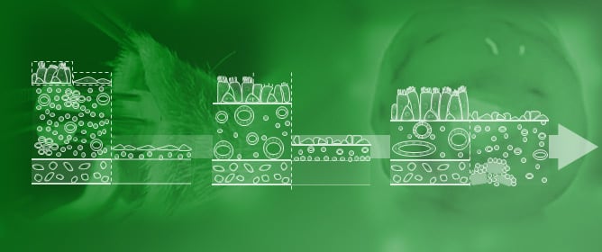
Osteoporosis is a complex endocrine disease characterized by a decline in bone mass and microstructural integrity. It constitutes a major global health problem. Recent progress in the field of artificial intelligence (AI) has opened new avenues for the effective diagnosis of osteoporosis via radiographs. This review investigates the application of AI classification of osteoporosis in radiographs. A comprehensive exploration of electronic repositories (ClinicalTrials.gov, Web of Science, PubMed, MEDLINE) was carried out in adherence to the Preferred Reporting Items for Systematic Reviews and Meta-Analyses 2020 statement (PRISMA). A collection of 31 articles was extracted from these repositories and their significant outcomes were consolidated and outlined. This encompassed insights into anatomical regions, the specific machine learning methods employed, the effectiveness in predicting BMD, and categorizing osteoporosis. Through analyzing the respective studies, we evaluated the effectiveness and limitations of AI osteoporosis classification in radiographs. The pooled reported accuracy, sensitivity, and specificity of osteoporosis classification ranges from 66.1% to 97.9%, 67.4% to 100.0%, and 60.0% to 97.5% respectively. This review underscores the potential of AI osteoporosis classification and offers valuable insights for future research endeavors, which should focus on addressing the challenges in technical and clinical integration to facilitate practical implementation of this technology.
Full article

Graphical abstract


