
NeuroSci 2024, 5(2), 169-183; https://doi.org/10.3390/neurosci5020012 - 11 May 2024
Abstract
►
Show Figures
Common Spatial Pattern (CSP) has been recognized as a standard and powerful method for the identification of Electroencephalography (EEG)-based Motor Imagery (MI) tasks when implementing brain–computer interface (BCI) systems towards the motor rehabilitation of lost movements. The combination of BCI systems with robotic
[...] Read more.
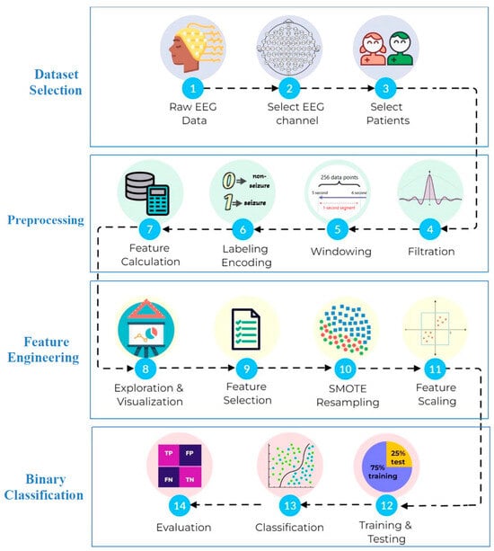
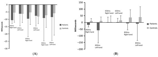
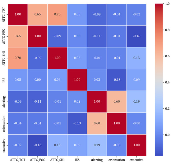
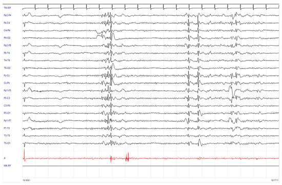
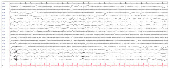
Common Spatial Pattern (CSP) has been recognized as a standard and powerful method for the identification of Electroencephalography (EEG)-based Motor Imagery (MI) tasks when implementing brain–computer interface (BCI) systems towards the motor rehabilitation of lost movements. The combination of BCI systems with robotic systems, such as upper limb exoskeletons, has proven to be a reliable tool for neuromotor rehabilitation. Therefore, in this study, the effects of temporal and frequency segmentation combined with layer increase for spatial filtering were evaluated, using three variations of the CSP method for the identification of passive movement vs. MI+passive movement. The passive movements were generated using a left upper-limb exoskeleton to assist flexion/extension tasks at two speeds (high—85 rpm and low—30 rpm). Ten healthy subjects were evaluated in two recording sessions using Linear Discriminant Analysis (LDA) as a classifier, and accuracy (ACC) and False Positive Rate (FPR) as metrics. The results allow concluding that the use of temporal, frequency or spatial selective information does not significantly (

Figure 1


