Article

Sulfonated Azocalix[4]arene-Modified Metal–Organic Framework Nanosheets for Doxorubicin Removal from Serum

by
Xiao-Min Cao
1,
Yuan-Qiu Cheng
1,2,
Meng-Meng Chen
1,2,
Shun-Yu Yao
1,2,
An-Kang Ying
1,2,
Xiu-Zhen Wang
1,
Dong-Sheng Guo
1,2,3,* and
Yue Li
1,4,*
1
College of Chemistry, Nankai University, Tianjin 300071, China
2
State Key Laboratory of Elemento-Organic Chemistry, Key Laboratory of Functional Polymer Materials (Ministry of Education), Frontiers Science Center for New Organic Matter, Collaborative Innovation Center of Chemical Science and Engineering (Tianjin), Nankai University, Tianjin 300071, China
3
Xinjiang Key Laboratory of Novel Functional Materials Chemistry, College of Chemistry, and Environmental Sciences, Kashi University, Kashi 844000, China
4
Key Laboratory of Advanced Energy Materials Chemistry (Ministry of Education), Nankai University, Tianjin 300071, China
*
Authors to whom correspondence should be addressed.
Nanomaterials 2024, 14(10), 864; https://doi.org/10.3390/nano14100864
Submission received: 17 April 2024 / Revised: 8 May 2024 / Accepted: 14 May 2024 / Published: 16 May 2024
(This article belongs to the Section Nanocomposite Materials)

Abstract

:
Chemotherapy is one of the most commonly used methods for treating cancer, but its side effects severely limit its application and impair treatment effectiveness. Removing off-target chemotherapy drugs from the serum promptly through adsorption is the most direct approach to minimize their side effects. In this study, we synthesized a series of adsorption materials to remove the chemotherapy drug doxorubicin by modifying MOF nanosheets with sulfonated azocalix[4]arenes. The strong affinity of sulfonated azocalix[4]arenes for doxorubicin results in high adsorption strength (Langmuir adsorption constant = 2.45–5.73 L mg−1) and more complete removal of the drug. The extensive external surface area of the 2D nanosheets facilitates the exposure of a large number of accessible adsorption sites, which capture DOX molecules without internal diffusion, leading to a high adsorption rate (pseudo-second-order rate constant = 0.0058–0.0065 g mg−1 min−1). These adsorbents perform effectively in physiological environments and exhibit low cytotoxicity and good hemocompatibility. These features make them suitable for removing doxorubicin from serum during “drug capture” procedures. The optimal adsorbent can remove 91% of the clinical concentration of doxorubicin within 5 min.

Graphical Abstract

1. Introduction

Cancer has become one of the leading causes of death globally [1,2]. Chemotherapy remains one of the most common and powerful cancer treatments [3]. However, nearly all chemotherapy drugs have low selectivity. While inhibiting cancer cell growth, chemotherapy drugs are also toxic to healthy cells, which triggers various side effects [4,5,6,7]. For example, doxorubicin (DOX), which is widely used in the treatment of various hematological and solid tumor malignancies [8,9], could cause cardiomyopathy, congestive heart failure, and kidney damage [10,11,12,13,14]. The trade-off between therapy effectiveness and side effects must be considered in determining treatment regimens and dosages, ultimately impacting patient survival. Although various localized drug delivery approaches, such as transarterial chemoembolization (TACE) [15,16], have been developed, a significant portion of the drug can still pass through the tumor and flow through the body with the bloodstream. The implementation of the “drug capture” or “ChemoFilter” technique, which involves removing chemotherapy drugs before they enter systemic circulation, represents a direct strategy for addressing this issue [17,18]. Since the key to this technology lies in the adsorbent used, various adsorption materials targeting chemotherapy drugs have been developed [19,20,21,22,23]. For example, Grubbs and co-workers synthesized an adsorption material based on DNA and iron oxide particles that could remove 98% of DOX from human serum within 10 min, and the effectiveness of the material was proven in a porcine model [19]. Sheikhi et al. reported a cellulose-based nanoadsorbent for capturing DOX. This adsorbent could adsorb 6000 mg of DOX per 1 mg, demonstrating its high adsorption capacity [23].
Besides capacity, adsorption strength and rate are also important parameters for characterizing the performance of adsorbents. A higher adsorption strength leads to more thorough removal of the chemotherapy drug at equilibrium and less severe side effects. Introducing units with a strong binding affinity to the adsorbate is an effective strategy to enhance adsorption strength. It was reported that sulfonated azocalix[4]arene (SAC4A), a macrocyclic compound with a deep cavity, has a strong affinity for forming a host–guest complex with DOX (Ka = 6.47 × 10⁶ M−1) [24,25,26,27]. Thus, it offers a good option as a binding site for the development of DOX-targeted adsorbents. On the other hand, the adsorption rate determines the actual quantity of drug removed within the time it takes for the blood to pass through the adsorption device. The majority of current adsorbents are porous materials [28,29,30,31]. Adsorbate molecules must diffuse through the channels before coming into contact with the binding sites, which significantly impedes the adsorption kinetics. Using adsorbent materials with a large external surface area can expose a significant quantity of accessible adsorption sites on the surface, thereby addressing this issue. Two-dimensional metal–organic framework (MOF) nanosheets have unique advantages in this context. Their ultrathin sheet-like morphology maximizes the external surface area, and their flexible composition and modifiability make them easy to adjust according to the structure and properties of the targeted substance [32,33,34]. The potential of MOF nanosheets has been intensively investigated in recent years [35,36,37,38].
In this research, through the modification of the nanosheets of NUS-8, a stable Hf-based MOF with 2D connection, with SAC4A, a series of adsorption materials were prepared for the capture of DOX. Up to 86% of the adsorption capacities of these materials could be achieved within 5 min. Their Langmuir adsorption constants range from 2.45 to 5.73 L mg−1, indicating that their adsorption strength is higher than that of the reported adsorbents. One of these materials was screened and successfully utilized for removing DOX from an actual serum sample.

2. Materials and Methods

2.1. Materials and Apparatus

The chemicals used in the experiments, including hafnium chloride (HfCl4), 1,3,5-Tri(4-carboxyphenyl)benzene (H3BTB), 2′-amino-5′-(4-carboxyphenyl)-[1,1′:3′,1″-terphenyl]-4,4″-dicarboxylic acid (H3BTB-NH2), 1,3,5-tris(4,4,5,5-tetramethyl-1,3,2-dioxaborolan-2-yl)benzene, methyl 4-iodo-3-nitrobenzoate, palladium acetate (Pd(AcO)2), tetrabutylammonium bromide ((Bu)4NBr), tripotassium phosphate (K3PO4), iron, sodium bicarbonate (NaHCO3), lithium hydroxide hydrate (LiOH·H2O), 2-(7-aza-1H-benzotriazole-1-yl)-1,1,3,3-tetramethyluronium hexafluorophosphate (HATU), N,N-diisopropylethylamine (DIPEA), sodium carbonate (Na2CO3), calix[4]arene (C4A), 4-aminobenzenesulfonic acid, 4-aminobenzoic acid, sodium nitrite (NaNO2), sodium sulfate (Na2SO4), 4-aminobenzenesulfonic acid, sodium acetate trihydrate (CH3COONa·3H2O), hydrochloride (DOX), sodium chloride (NaCl), calcium chloride (CaCl2), bovine serum albumin (BSA), and Triton X-100, were purchased from Aladdin and used without further purification unless otherwise noted. 1,3,5-tris (2-amino-4-carboxyphenyl) benzenebenzene (H3BTB-3NH2), SAC4A, and monocarboxyl- and sulfonate-modified azocalix[4]arene (CSAC4A) were synthesized following literature procedures (Figures S1−S3) [39,40,41]. H3BTB-3NH2 was synthesized through the Suzuki coupling reaction between 1,3,5-tris(4,4,5,5-tetramethyl-1,3,2-dioxaborolan-2-yl)benzene and 3-amino-4-bromobenzoic acid. SAC4A was prepared from the diazo-coupling reaction between C4A and 4-aminobenzenesulfonic acid. CSAC4A was synthesized using two steps of a diazo-coupling reaction, in which 4-aminobenzoic acid and 4-aminobenzenesulfonic acid were connected to C4A in sequence.
1H NMR measurements were carried out on a Bruker 400 MHz NMR spectrometer, and all chemical shifts are reported in parts per million (ppm). Powder X-ray diffraction (PXRD) patterns were recorded on a Rigaku D/max-2500 diffractometer with Cu Kα radiation (λ = 0.15406 nm) at 40 kV and 100 mA. The 2θ range was from 2° to 50° with a 0.02° increment and a scan speed of 15°·min−1. Scanning electron microscopy (SEM) images were recorded on a TESCAN MIRA LMS scanning electron microscope. High-resolution transmission electron microscopy (HRTEM) data were collected using a FEI Talos F200S transmission electron microscope. Atomic force microscopy (AFM) images were observed by a Bruker Dimension Icon scanning electron microscope. Inductively coupled plasma optical emission spectrometry (ICP-OES) measurements were acquired on an Agilent 5110. N2 adsorption–desorption analysis was performed using a Micromeritics APSP 2460 surface area and porosity analyzer. The samples were degassed under vacuum at 120 °C for 12 h before measurement. Thermogravimetric analysis (TGA) was conducted using the Netzsch STA2500 Regulus and Netzsch STA449F3 Thermogravimetric Analyzer, with a heating rate of 10 °C/min from 30 °C to 80 °C under atmospheric air. Fourier transform infrared spectroscopy (FTIR) spectra were recorded on a Thermo Scientific Nicolet iS20 Spectrometer using KBr pellets dispersed with sample powders in the range of 4000–400 cm−1. X-ray photoelectron spectroscopy (XPS) data were measured on a Thermo ESCALAB 250XI equipped with an Al source anode X-ray gun. Absorption spectra (UV-vis) were collected under a Shimadzu UV-2450 spectrophotometer.

2.2. Synthesis of MOF Nanosheets

NUS-8 nanosheets were synthesized following a reported method with little modification [42]. HfCl4 (56 mg, 0.176 mmol) and 1,3,5-benzenetribenzoic acid (H3BTB) (50 mg, 0.114 mmol) were dissolved in a mixed solvent of DMF (8 mL), deionized water (1 mL), and formic acid (4 mL). The solution was placed in a 20 mL Pyrex vial, ultrasonicated for 5 min, and then heated at 120 °C for 48 h. After cooling to room temperature, the resulting white solid was centrifuged, washed with DMF (5 × 6 mL), solvent-exchanged with dichloromethane (5 × 6 mL) and methanol (5 × 6 mL) in sequence, and dried under vacuum at 120 °C for 24 h.
The synthesis of NUS-8-NH2 and NUS-8-3NH2 nanosheets followed a similar process to that of NUS-8, with the only difference being the replacement of the ligand by 2′-amino-5′-(4-carboxyphenyl)-[1,1′:3′,1″-terphenyl]-4,4″-dicarboxylic acid (H3BTB-NH2) and H3BTB-3NH2, respectively.

2.3. Modification of MOF Nanosheets by SAC4A/CSAC4A

The SAC4A/CSAC4A loaded adsorbents were synthesized through the post-synthesis modification of MOF nanosheets [43]. In the synthesis of SAC4A-loaded NUS-8 nanosheets (N@SAC4A), as-prepared NUS-8 nanosheets (50 mg) were dispersed in DMF (10 mL) by 30 min of ultrasonication. Then, a solution of SAC4A (50 mg) in DMF (10 mL) was added to the dispersion. The mixture was ultrasonicated for 3 h and then stirred at room temperature for 48 h. The resulting light orange solid product was collected, washed by DMF and CH2Cl2, solvent-exchanged with methanol for 24 h, and vacuum-dried at 100 °C for 24 h.
CSAC4A-linked NUS-8-NH2 (NA-CSAC4A) was synthesized by the following procedures: CSAC4A (50 mg) and N,N-diisopropylethylamine (DIPEA, 14.5 µL) were dissolved in 10 mL of DMF, stirred at room temperature for 30 min, and further stirred for 1 h after the addition of 2-(7-aza-1H-benzotriazole-1-yl) -1,1,3,3-tetramethyluronium hexafluorophosphate (HATU, 20.2 mg). The obtained solution was added to the suspension of NUS-8-NH2 (50 mg in 10 mL DMF). After 48 h of reaction, the light orange solid product formed was centrifuged, washed by DMF and CH2Cl2, solvent-exchanged with methanol for 24 h, and then vacuum-dried at 100 °C for 24 h.
The synthetic procedure for CSAC4A-linked NUS-8-3NH2 was similar to that of NA-CSAC4A, with the exception that the substrate was replaced by NUS-8-3NH2, and the addition amounts of CSAC4A, DIPEA, and HATU were changed to 100 mg, 29 µL, and 40.4 mg, respectively.

2.4. Adsorption Experiments

These experiments were performed with procedures similar to those described in [44,45]. A specific amount of adsorbent was immersed in a 5 mL aqueous solution of DOX and stirred for 24 h to achieve sorption equilibrium. The influence of adsorbent dosage on the adsorption process was investigated using 1 to 4 mg of adsorbent, while keeping the initial DOX concentration constant at 50 µM. Several solutions of DOX with concentrations ranging from 10 to 110 µM were mixed with 1.5 mg adsorbent to test the impact of the initial adsorbate concentration. To investigate the kinetics of the adsorption process, the experiment was carried out on a suspension with an initial volume of 50 mL, 15 mg of adsorbent, and 50 µM of DOX. At different time intervals, 3 mL of the suspension was sampled and analyzed.
The adsorption performance was also studied in the presence of various interfering substances. DOX solutions with pH values adjusted to 4–8 were used to assess the impact of solution pH. NaCl (80–190 mM) and CaCl2 (3–90 mM) were selected to mimic ion interference in the serum, while BSA (15–55 mg mL−1), the primary protein component in the blood, was tested as the protein interference (physiological levels of Na+, Ca2+, and BSA are approximately 140 mM, 3 mM, and 35 mg mL−1, respectively).
After the adsorption process, the adsorbent was separated by filtration. The residual concentration of DOX in the supernatant was quantified using UV-Vis spectroscopy at 480 nm. The removal efficiency (R) and adsorption amount (qt) were calculated by the following equations:
R ( % ) = C 0 C t C 0 × 100 %
q t = ( C 0 C t ) V M m
where C0 and Ct are the concentrations of DOX before and after the adsorption process, respectively; V is the volume of the DOX solution; M is the molecular weight of DOX; and m is the mass of the adsorbent.

2.5. Cytotoxicity Tests

As a reported method [46], the Cell Counting Kit-8 (CCK-8) assay was used to evaluate the cytotoxicity of N3A-CSAC4A. Human umbilical vein endothelial cells (HUVECs) were seeded into a 96-well plate at a density of 5000 cells per well and cultured in EGM-2 culture medium at 37 °C with 5% CO2 for 24 h. After the cells adhered completely, the culture medium was changed to a fresh medium with N3A-CSAC4A (0.1–1.2 mg/mL), and the cells were incubated for another 24 h. Following the addition of CCK-8 (20 μL) and a further 4 h incubation, the absorbance at 450 nm was recorded using a microplate reader.

2.6. Hemolysis Assay

As a reported method [46], the hemolysis assay was performed to evaluate the blood compatibility of N3A-CSAC4A. Fresh blood extracted from the abdominal aorta of mice was stored in an anticoagulant tube containing heparin. PBS was added at a volume about 10 times that of the blood, and the mixture was centrifuged at 3000 rpm for 5 min to wash the red blood cells (RBCs) three times. The RBCs were diluted to a concentration of 2% with PBS and then packed into microcentrifuge tubes. Then, the RBCs (200 μL) were incubated with 800 μL of N3A-CSAC4A (0.05–1.5 mg mL−1) at 37 °C for 4 h. We used PBS as the negative control and Triton X-100 (0.1%) as the positive control. After centrifugation, the supernatant (100 μL) was pipetted into a 96-well plate for absorbance testing at 545 nm using a microplate reader.

2.7. DOX Capture from Serum

As a typical procedure [47], the serum used in this experiment was extracted from the blood of mice, and the initial DOX solution was diluted to a clinical concentration of 86 μM by serum. N3A-CSAC4A (20 mg) was dispersed in 30 mL of the above DOX solution using 5 min of sonication and then stirred at room temperature. At different intervals, 3 mL of the suspension was sampled. After the adsorbent was separated, the absorbance at 480 nm was measured using UV-Vis spectroscopy.

3. Results and Discussion

3.1. Adsorbent Design

NUS-8, which is constructed by the metal nodes of Hf6 clusters and the ligand of H3BTB, was selected as the substrate for the adsorbents due to its superior stability compared to other reported 2D MOF nanosheets [48]. Two protocols were tried to load SAC4A onto NUS-8 nanosheets (Scheme 1). The first one involves non-covalent binding. According to the hard–soft acid–base (HSAB) theory, the phenolic hydroxyl O atoms on the lower rim of SAC4A are hard bases and thus have a high affinity for the Hf6 clusters (hard acid) of NUS-8. To enhance the anchoring strength, we also attempted covalent bonding. To form an amide bond between SAC4A and MOF, one of the sulfo groups on the upper rim of the SAC4A molecule is replaced by a carboxyl group (the product is denoted as CSAC4A), and an amino group is prelocated on the H3BTB ligand. Two aminated ligands, H3BTB-NH2 and H3BTB-3NH2, with different substituent positions and numbers, were tested to screen for the adsorbent with the best performance.

3.2. Characterization

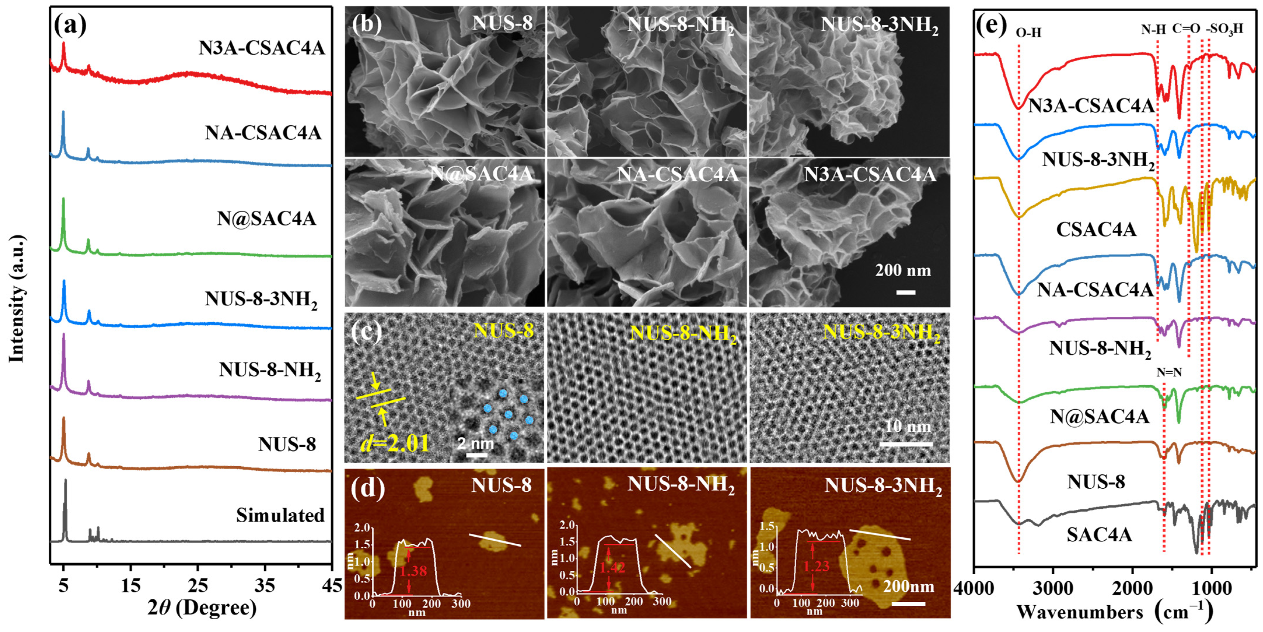
MOF nanosheets of NUS-8, NUS-8-NH2, and NUS-8-3NH2 were synthesized through the solvothermal reaction of HfCl4 with H3BTB, H3BTB-NH2 and H3BTB-3NH2, respectively. Powder X-ray diffractometry (PXRD) was first used to examine the crystallinity of the products. The recorded pattern closely matched the simulated pattern from the reported structure of NUS-8 [42,48], confirming its successful synthesis and high phase purity (Figure 1a). The PXRD patterns of NUS-8-NH2 and NUS-8-3NH2 are similar to those of NUS-8, indicating their isomorphic structures. Scanning electron microscopy (SEM) and transmission electron microscopy (TEM) images (Figure 1b and Figure S4) showed that these MOF samples present 2D layered morphologies. Hexagonal honeycomb patterns were observed in the high-resolution TEM (HR-TEM) images, confirming their long-range ordered crystalline structures. The pore size is approximately 2 nm, consistent with the backbone structures of these MOFs. The thicknesses of MOF nanosheets were measured using atomic force microscopy (AFM). The AFM images show the ultrathin structures with thicknesses of 1.38, 1.42, and 1.23 nm for NUS-8, NUS-8-NH2, and NUS-8-3NH2, respectively (Figure 1d). All three samples exhibited good chemical stability. The immersion of these samples in several common solvents for 48 h did not cause any significant changes in their PXRD patterns (Figure S5).
By loading SAC4A on MOF nanosheets through non-covalent and covalent interactions, three adsorbents were prepared. The PXRD patterns of N@SAC4A (SAC4A loaded NUS-8), NA-CSAC4A (CSAC4A-linked NUS-8-NH2), and N3A-CSAC4A (CSAC4A-linked NUS-8-3NH2) matched well with their precursors, indicating that the crystallinities of MOF nanosheets were not affected. The SEM images and TEM images revealed that the SAC4A/CSAC4A-modified products also exhibit lamellar morphologies (Figure 1b,c and Figure S4). All of these results confirm that the introduction of SAC4A/CSAC4A does not break the backbone of MOF precursors. The three modified samples exhibited absorption peaks at 1596, 1120, and 1035 cm−1 in the FT-IR spectra, which could be assigned to the stretching vibrations of N=N, S=O, and S−O, respectively, confirming the successful introduction of SAC4A/CSAC4A. In the spectrum of N@SAC4A, the intensity of the O−H stretching vibration around 3430 cm−1 was significantly reduced compared to that of NUS-8, suggesting the substitution of the OHs on the Hf6 clusters by SAC4A. The vibrations of N−H and C=O at 1680 and 1275 cm−1 in the spectra of NA-CSAC4A and N3A-CSAC4A confirm the formation of an amide bond between CSAC4A and the aminated ligand. The –NH2 groups of precursors NUS-8-NH2 and NUS-8-3NH2 do not give prominent peaks in the spectra because their absorption is overlapped by that of OH.
To further verify the introduction of SAC4A/CSAC4A, we conducted X-ray photoelectron spectroscopy (XPS) analysis (Figure S6). The S2p spectra of three SAC4A/CSAC4A-modified MOF samples are nearly identical, with two deconvoluted peaks at 167.4 and 168.5 eV attributed to the C−S and S=O bonds, respectively. This suggests that these samples contain the sulfur element from the macrocycle modifier. In the spectra of NUS-8-NH2 and NUS-8-3NH2, the N1s band can be resolved into two peaks at 400.2 and 401 eV, which are assigned to the C−N and N−H bonds, respectively. For NA-CSAC4A and N3A-CSAC4A, the N 1s spectrum displays three different chemical environments corresponding to N=N, C−N, and N−H bonds. The C−N/N−H area ratios of these two samples are higher than those of the corresponding precursors, suggesting the conversion of amino groups to amide bonds.
Inductively coupled plasma optical emission spectrometry (ICP-OES) was used to analyze the elemental compositions of SAC4A/CSAC4A-modified MOF samples. Since S and Hf are specific to the macrocycle modifier and MOF substrate, respectively, the loading amount of SAC4A/CSAC4A could be calculated from the ratio between these two elements. As shown in Table S1, the CSAC4A loading amount of NA-CSAC4A (9.26 wt%) was obviously higher than the SAC4A loading amount of N@SAC4A (7.53 wt%), demonstrating that covalent bonding is a more effective method for the functionalization of MOFs. N3A-CSAC4A further increased the loading amount to 16.69 wt%, which indicates that the increase in anchoring sites is beneficial for the modification. We also altered the addition quantity of CSAC4A in the synthesis of N3A-CSAC4A and observed that it has a positive correlation with CSAC4A loading amount.
Upon the introduction of SAC4A/CSAC4A, the Brunauer–Emmett–Teller (BET) surface areas of NUS-8, NUS-8-NH2, and NUS-8-3NH2 decreased from 634, 440, and 259 m2 g−1 to 451, 398, and 241 m2 g−1, respectively. This reduction reveals that some of the modifier molecules enter into the pores of the nanosheets (Figure S7).

3.3. Adsorbent Studies

To test the removal efficiency for DOX, three MOF nanosheet precursors and their modified products with different dosages (ranging from 0.2 to 0.8 mg mL−1) were individually incubated in DOX solutions (50 µM) for 24 h. Each adsorbent decreased the concentration of DOX in a dose-dependent manner (Figure 2a). The adsorption percentages of three MOF precursors followed the order of NUS-8-NH2 < NUS-8-3NH2 < NUS-8. The highest adsorption capacity of NUS-8 could be explained by its large surface area (Figure S8). The superior performance of NUS-8-NH2 to NUS-8-3NH2 may be due to its less positive zeta potential, which weakens the electrostatic repulsion with the positively charged DOX (Figure S9). All three SAC4A/CSAC4A-modified MOF samples gave higher adsorption percentages than the corresponding precursors, which could be attributed to the strong complexation ability of the macrocycle introduced. It is expected that the DOX molecule enters the cavity of SAC4A/CSAC4A and forms a host–guest complex on the MOF surface. The increased amplitudes of adsorption amounts are in accordance with the SAC4A/CSAC4A loading amounts in these samples (Figure S10). With the addition of 0.3 mg mL−1 of N3A-CSAC4A, the adsorption percentage of DOX could reach 90%, which is 2.7 times higher than that of NUS-8-3NH2. The further increase in macrocycle loading through the enhancement of CSAC4A concentration in the synthesis system was proven to be unnecessary. When the modifier amount was doubled, the obtained product only gave a 3% increase in the adsorption percentage of DOX (Figures S12−S14).
The DOX removal capabilities of these samples were further examined under different initial concentrations of DOX. As shown in Figure 2c, for all six adsorbents, the adsorbed amount of DOX per unit weight of adsorbent increased as the initial concentration changed from 10 to 70 µM. However, the removal percentage showed an opposite trend, which can be attributed to the depletion of adsorption sites (Figure S11). The adsorbed amount hardly changed when the DOX initial concentration was above 70 µM, indicating saturation had been reached.
To illustrate the thermodynamic information of the adsorption process, we tested the Langmuir (Equation (3)) and Freundlich (Equation (4)) adsorption isotherms to fit the equilibrium adsorption data:
q e = K L Q max C e 1 + K L C e ,
q e = K F C e 1 / n ,
In these equations, qe is the amount of DOX adsorbed at equilibrium, Qmax is the maximum adsorption capacity, Ce is the concentration of DOX at equilibrium, KL is the Langmuir constant, KF is the Freundlich constant, and n is the exponential factor of the Freundlich isotherm. The fitting curves of DOX adsorption are presented in Figure 2d, and the isotherm parameters of the two models are listed in Table 1.
For the three MOF nanosheets, the correlation coefficients (R2s) of the Freundlich isotherm model (0.9454–0.9732) are higher than the corresponding values of the Langmuir isotherm model (0.8271–0.9201), indicating that the Freundlich isotherm model is more suitable for describing the adsorption process. Their Qmaxs showed a positive correlation with the surface area, suggesting that DOX molecules are adsorbed onto the external surface of MOF nanosheets. In contrast to the cases of precursors, the Langmuir isotherm model of SAC4A/CSAC4A-modified products gave greater R2s (0.9636–0.9871) compared to the Freundlich isotherm model (0.7256–0.9605). The experimental maximum adsorption capacities were 70.5, 63.7, and 121.7 mg g−1 for N@SAC4A, NA-CSAC4A, and N3A-CSAC4A, respectively, which are close to the experimentally measured maximum adsorption capacities (69.3, 63.6, and 122.1 mg g−1; the slightly larger values of the measured adsorption capacities of N@SAC4A and NA-CSAC4A than the corresponding Qmaxs could be attributed to experimental error). These all indicate that the process of adsorption of DOX by SAC4A/CSAC4A-modified MOFs can be more accurately represented by the Langmuir isotherm model. Since the Langmuir isotherm model is generally associated with the homogeneity and the Freundlich isotherm is related to the heterogeneity of the adsorbent [49,50], the fitting results indicate that the macrocycle moieties offer uniform adsorption sites for DOX. The Qmaxs of SAC4A/CSAC4A-modified MOF nanosheets were generally higher than those of precursors. Among all the tested samples, N3A-CSAC4A gave the highest Qmax of 122.1 mg g−1, which is consistent with its highest macrocycle loading amount.
A more remarkable change upon the loading of SAC4A/CSAC4A is the enhancement of adsorption strength. The KLs of three macrocycle-modified MOF nanosheet adsorbents, which indicate their affinity towards the adsorbate, range from 2.45 to 5.73 L mg−1. Compared with the precursors, the KLs of two covalently modified samples showed an increase of an order of magnitude, while the non-covalent sample also increased by two times. Table 2 presents a comparison between the KLs of our samples and those reported in recent studies. Obviously, compared with reported adsorbents, our samples exhibit the highest affinity toward DOX. The highest affinity of our adsorbents would lead to the most complete removal of the drug after adsorption.
Then, the kinetics of the adsorption process was examined. As shown in Figure 3a, all three SAC4A/CSAC4A-modified MOF nanosheet adsorbents exhibited a sharp increase in adsorption quantity within the first 5 min, reaching 78–86% of the maximum value. This performance competes with the highest efficiency observed among reported adsorbents [23]. The fast adsorption process could be attributed to the ultrathin nanosheet morphology of these samples, which exposes the modified macrocycle moieties on the external surface to directly capture DOX. We used pseudo-first-order (Equation (5)), pseudo-second-order (Equation (6)), and intraparticle diffusion (Equation (7)) models to fit the temporal change in the adsorbed amount of DOX:
ln ( q e q t ) = ln q e k 1 t ,
t q t = 1 k 2 q e 2 + 1 q e t ,
q t = k id t 0.5 + C
where qe and qt are the quantities of DOX adsorbed at equilibrium and time t; k1, k2, and kid represent the rate constants of pseudo-first-order, pseudo-second-order, and intraparticle diffusion equations, respectively; and C is the thickness constant of the boundary layer.
The fitting graphs of the three models are presented in Figure 3b–d, and the corresponding parameters are listed in Table 3. The R2s of the pseudo-second-order model for all three modified adsorbents reach 0.99, and the fitted values of qes from this model closely align with the experimental results. These all demonstrate that the pseudo-second-order model provides the best fit for the adsorption process of DOX. Typically, pseudo-second-order adsorption kinetics suggest a chemisorption mechanism. When the DOX molecules reached the surface of modified MOF nanosheets, they promptly bonded with the SAC4A/CSAC4A moieties through host–guest complexation. The uniform environment of the SAC4A/CSAC4A moiety, along with its high affinity and 1:1 stoichiometry in forming a guest–host complex with DOX, enables the capture of DOX, demonstrating the feature of chemosorption.

3.4. Application to the DOX Capture from Serum

Because N3A-CSAC4A showed the best adsorption performance in the above experiments, the following investigations focused on this adsorbent.
The normal pH value of human serum ranges from 7.35 to 7.45, and various substances, such as ions and proteins, are present in serum. To evaluate the potential of N3A-CSAC4A in drug capture, a series of experiments was conducted to examine its adsorption performance under interference conditions. N3A-CSAC4A was first exposed to a series of DOX solutions whose pH values were adjusted to 4–8 (Figure 4a). It was observed that the adsorption percentage of DOX increased as the pH of the solution changed from 4 to 7 and then remained stable at around 90%, indicating that N3A-CSAC4A can achieve the optimum effect at normal serum pH. The reduction in adsorption percentage at lower pH values could be explained by the electrostatic interaction between the adsorbent (its zeta potential changed from −39.1 to 14.4 mV as the pH decreased from 8 to 4, Figure S15) and the positively charged DOX. Then, the adsorption efficiency of N3A-CSAC4A was tested in the presence of Na+, Ca2+, and BSA (Figure 4b and Figure S16). The results showed that under normal concentrations of Na+ and Ca2+ in serum, the adsorption efficiency is close to that of the blank control. With the increase in BSA concentration, the adsorption percentage even showed a slight rise. These results prove that the complex components of serum cannot impair the adsorption performance of N3A-CSAC4A.
In chemotherapy drug capture, the adsorbent is immobilized and comes into direct contact with blood, which imposes stringent requirements for its cytotoxicity and hemocompatibility. To assess the cytotoxicity of N3A-CSAC4A, various concentrations of its suspensions were incubated with human umbilical vein endothelial cells (HUVECs, Figure 4c). Even though the concentration of N3A-CSAC4A increased to 1.2 mg mL−1, the cell viability could still be maintained above 90% after 24 h of incubation. This result indicates that the cytotoxicity of N3A-CSAC4A is negligible. In the hemolytic study, the red blood cells were incubated with varying concentrations of N3A-CSAC4A, and the hemolysis ratio was measured with Triton X-100 as a positive control (Figure 4d). N3A-CSAC4A at a concentration of 1.5 mg mL−1 only gave a hemolysis ratio of 1.59 ± 0.75%, indicating that this adsorbent is non-toxic to erythrocytes.
Encouraged by the good adsorption performance of N3A-CSAC4A in a physiological environment, as well as its good biocompatibility and hemocompatibility, we used it to capture DOX from real serum. DOX was added to serum to a clinical concentration of 86 μM, and its concentration change was monitored after the addition of 0.67 mg mL−1 N3A-CSAC4A (Figure 5). A rapid DOX removal process was observed in the initial 5 min, with the adsorption percentage surging to 91%. After this period, the concentration decline of DOX slowed down, with the majority of DOX (97%) being adsorbed within 60 min. These results demonstrate that N3A-CSAC4A could be used as an efficient and rapid adsorbent for the removal of DOX from serum.

4. Conclusions

In summary, we developed a series of adsorbents by modifying MOF nanosheets with SAC4A/CSAC4A. The incorporated SAC4A/CSAC4A moieties serve as strong binding sites for DOX, and the extensive external surface of the MOF nanosheet substrate facilitates contact with the adsorbate, thereby expediting the adsorption kinetics. Our experiments demonstrated that, compared with non-covalent modification, covalent bonding is a more efficient approach for functionalizing MOF nanosheets, giving products with increased stability, a higher modifier loading amount, and enhanced adsorption capacity. The developed adsorbent exhibited favorable attributes such as strong anti-interference capability, low cytotoxicity, and good hemocompatibility, enabling its successful application in DOX capture from real serum samples. The results of this study suggest that, by utilizing abundant data on host–guest recognition of macrocycles, the functionalization of MOF nanosheets with macrocycles is an adaptable strategy for fabricating targeted adsorbents.

Supplementary Materials

The following supporting information can be downloaded at https://www.mdpi.com/article/10.3390/nano14100864/s1: Figure S1: 1H NMR spectrum of H3BTB-3NH2; Figure S2: 1H NMR spectrum of SAC4A; Figure S3: 1H NMR spectrum of CSAC4A; Figure S4: TEM images of MOF nanosheet precursors and SAC4A/CSAC4A-modified products; Figure S5: PXRD patterns of the samples of (a) NUS-8, (b) NUS-8-NH2, and (c) NUS-8-3NH2 after the immersion in various solvents for 24 h; Figure S6: XPS spectra of (a) N@SAC4A, (b) NA-CSAC4A, (c) N3A-CSAC4A and their MOF nanosheet precursors; Figure S7: N2 sorption isotherms of SAC4A/CSAC4A-modified and unmodified MOF nanosheets; Figure S8: Relevance between the adsorption performance and specific surface area of SAC4A/CSAC4A-modified and unmodified MOF nanosheets; Figure S9: ζ-potentials of N@SAC4A, NA-CSAC4A, N3A-CSAC4A, and their MOF nanosheet precursors; Figure S10: Change in DOX removal percentage with the loading amount of SAC4A/CSAC4A on the adsorbent; Figure S11: Changes in DOX removal percentage with initial DOX concentration; Figure S12: DOX removal by NUS-8-3NH2 and the N3A-CSAC4A samples prepared with different CSAC4A addition amounts, CDOX,0 = 50 μM. In the synthesis of N3A-CSAC4A samples, the addition amount of CSAC4A was changed from 100 mg to 50 and 200 mg, and the addition amounts of DIPEA and HATU were adjusted according to that of CSAC4A; Figure S13: Adsorbed amounts of DOX by NUS-8-3NH2 and the N3A-CSAC4A samples prepared with different CSAC4A addition amounts, 0.3 mg mL−1 adsorbent, CDOX,0 = 10–110 μM; Figure S14: DOX removal percentages at different time intervals by NUS-8-3NH2 and the N3A-CSAC4A samples prepared with different CSAC4A addition amounts; Figure S15: ζ-potentials of N3A-CSAC4A at different pH values; Figure S16: DOX removal by N3A-CSAC4A in the presence of Na+ (from left to right: 80–190 mM), Ca2+ (from left to right: 3–90 mM), and BSA (from left to right: 15–55 mM); Table S1: ICP-OES results and the calculated loading amounts of SAC4A/CSAC4.

Author Contributions

Conceptualization, D.-S.G.; data curation, S.-Y.Y.; formal analysis, X.-Z.W.; funding acquisition, D.-S.G. and Y.L.; investigation, X.-M.C.; methodology, Y.-Q.C.; project administration, D.-S.G.; supervision, Y.L.; validation, M.-M.C.; visualization, A.-K.Y.; writing—original draft, X.-M.C.; writing—review and editing, Y.L. All authors have read and agreed to the published version of the manuscript.

Funding

This work was supported by the NSFC (22376104 and U20A20259).

Data Availability Statement

Data are contained within the article.

Conflicts of Interest

The authors declare no conflicts of interest. The funders had no role in the design of the study; in the collection, analyses, or interpretation of data; in the writing of the manuscript; or in the decision to publish the results.

References

  1. Dizon, D.S.; Kamal, A.H. Cancer statistics 2024: All hands on deck. CA Cancer J. Clin. 2024, 74, 8–9. [Google Scholar] [CrossRef] [PubMed]
  2. Wang, Y.; Yan, Q.; Fan, C.; Mo, Y.; Wang, Y.; Li, X.; Liao, Q.; Guo, C.; Li, G.; Zeng, Z.; et al. Overview and countermeasures of cancer burden in China. Sci. China Life Sci. 2023, 66, 2515–2526. [Google Scholar] [CrossRef] [PubMed]
  3. Behranvand, N.; Nasri, F.; Zolfaghari Emameh, R.; Khani, P.; Hosseini, A.; Garssen, J.; Falak, R. Chemotherapy: A double-edged sword in cancer treatment. Cancer Immunol. Immun. 2022, 71, 507–526. [Google Scholar] [CrossRef] [PubMed]
  4. Falzone, L.; Bordonaro, R.; Libra, M. SnapShot: Cancer chemotherapy. Cell 2023, 186, 1816. [Google Scholar] [CrossRef]
  5. Dong, Z.; Hu, H.; Yu, X.; Tan, L.; Ma, C.; Xi, X.; Li, L.; Wang, L.; Zhou, M.; Chen, T.; et al. Novel frog skin-derived peptide Dermaseptin-PP for lung cancer treatment: In vitro/vivo evaluation and anti-tumor mechanisms study. Front. Chem. 2020, 8, 476. [Google Scholar] [CrossRef] [PubMed]
  6. Wang, X.; He, X.; Liu, C.; Zhao, W.; Yuan, X.; Li, R. Progress and perspectives of platinum nanozyme in cancer therapy. Front. Chem. 2022, 10, 1092747. [Google Scholar] [CrossRef] [PubMed]
  7. Alderton, G. Alleviating side effects. Science 2020, 370, 1287–1288. [Google Scholar]
  8. Bonadonna, G.; Monfardini, S.; De Lena, M.; Fossati-Bellani, F.; Beretta, G. Phase I and preliminary phase II evaluation of adriamycin (NSC 123127). Cancer Res. 1970, 30, 2572–2582. [Google Scholar] [PubMed]
  9. Zhang, L.; Wang, S.; Yang, Z.; Hoshika, S.; Xie, S.; Li, J.; Chen, X.; Wan, S.; Li, L.; Benner, S.A.; et al. An aptamer-nanotrain assembled from six-letter DNA delivers doxorubicin selectively to liver cancer cells. Angew. Chem. Int. Ed. 2020, 59, 663–668. [Google Scholar] [CrossRef]
  10. Li, S.; Li, F.; Wan, D.; Chen, Z.; Pan, J.; Liang, X.-J. A micelle-based stage-by-stage impelled system for efficient doxorubicin delivery. Bioact. Mater. 2023, 25, 783–795. [Google Scholar] [CrossRef]
  11. Wu, L.; Wang, L.; Du, Y.; Zhang, Y.; Ren, J. Mitochondrial quality control mechanisms as therapeutic targets in doxorubicin-induced cardiotoxicity. Trends Pharmacol. Sci. 2023, 44, 34–49. [Google Scholar] [CrossRef] [PubMed]
  12. She, G.; Du, J.C.; Wu, W.; Pu, T.T.; Zhang, Y.; Bai, R.Y.; Zhang, Y.; Pang, Z.D.; Wang, H.F.; Ren, Y.J.; et al. Hippo pathway activation mediates chemotherapy-induced anti-cancer effect and cardiomyopathy through causing mitochondrial damage and dysfunction. Theranostics 2023, 13, 560–577. [Google Scholar] [CrossRef] [PubMed]
  13. Zhou, Y.; Kong, X.; Zhao, P.; Yang, H.; Chen, L.; Miao, J.; Zhang, X.; Yang, J.; Ding, J.; Guan, Y. Peroxisome proliferator-activated receptor-α is renoprotective in doxorubicin-induced glomerular injury. Kidney Int. 2011, 79, 1302–1311. [Google Scholar] [CrossRef] [PubMed]
  14. Zhang, T.-X.; Zhang, Z.-Z.; Yue, Y.-X.; Hu, X.-Y.; Huang, F.; Shi, L.; Liu, Y.; Guo, D.-S. A general hypoxia-responsive molecular container for tumor-targeted therapy. Adv. Mater. 2020, 32, 1908435. [Google Scholar] [CrossRef]
  15. Auer, T.A.; Müller, L.; Schulze, D.; Anhamm, M.; Bettinger, D.; Steinle, V.; Haubold, J.; Zopfs, D.; Santos, D.P.D.; Eisenblätter, M.; et al. CT-guided high-dose-rate brachytherapy versus transarterial chemoembolization in patients with unresectable hepatocellular carcinoma. Radiology 2024, 310, e232044. [Google Scholar] [CrossRef] [PubMed]
  16. Hong, S.; Choi, W.S.; Purushothaman, B.; Koh, J.; Kim, H.-C.; Chung, J.W.; Song, J.M.; Choi, J.W. Drug delivery in transarterial chemoembolization of hepatocellular carcinoma: Ex vivo evaluation using transparent tissue imaging. Acta Biomater. 2022, 154, 523–535. [Google Scholar] [CrossRef]
  17. Patel, A.S.; Saeed, M.; Yee, E.J.; Yang, J.; Lam, G.J.; Losey, A.D.; Lillaney, P.V.; Thorne, B.; Chin, A.K.; Malik, S. Development and validation of endovascular chemotherapy filter device for removing high-dose doxorubicin: Preclinical study. J. Med. Devices 2014, 8, 0410081–410088. [Google Scholar] [CrossRef]
  18. Vogl, T.J.; Naguib, N.N.; Nour-Eldin, N.-E.A.; Rao, P.; Emami, A.H.; Zangos, S.; Nabil, M.; Abdelkader, A. Review on transarterial chemoembolization in hepatocellular carcinoma: Palliative, combined, neoadjuvant, bridging, and symptomatic indications. Eur. J. Radiol. 2009, 72, 505–516. [Google Scholar] [CrossRef] [PubMed]
  19. Blumenfeld, C.M.; Schulz, M.D.; Aboian, M.S.; Wilson, M.W.; Moore, T.; Hetts, S.W.; Grubbs, R.H. Drug capture materials based on genomic DNA-functionalized magnetic nanoparticles. Nat. Commun. 2018, 9, 2870. [Google Scholar] [CrossRef]
  20. Chen, X.C.; Oh, H.J.; Yu, J.F.; Yang, J.K.; Petzetakis, N.; Patel, A.S.; Hetts, S.W.; Balsara, N.P. Block copolymer membranes for efficient capture of a chemotherapy drug. ACS Macro Lett. 2016, 5, 936–941. [Google Scholar] [CrossRef]
  21. Yee, C.; McCoy, D.; Yu, J.; Losey, A.; Jordan, C.; Moore, T.; Stillson, C.; Oh, H.J.; Kilbride, B.; Roy, S.; et al. Endovascular ion exchange chemofiltration device reduces off-target doxorubicin exposure in a hepatic intra-arterial chemotherapy model. Radiol-Imag. Cancer 2019, 1, e190009. [Google Scholar] [CrossRef] [PubMed]
  22. Su, G.A.; Wadsworth, O.J.; Muller, H.S.; Archer, W.R.; Hetts, S.W.; Schulz, M.D. Polymer-nucleobase composites for chemotherapy drug capture. J. Mater. Chem. B 2023, 11, 8449–8455. [Google Scholar] [CrossRef]
  23. Young, S.A.; Muthami, J.; Pitcher, M.; Antovski, P.; Wamea, P.; Murphy, R.D.; Haghniaz, R.; Schmidt, A.; Clark, S.; Khademhosseini, A.; et al. Engineering hairy cellulose nanocrystals for chemotherapy drug capture. Mater. Today Chem. 2022, 23, 100711. [Google Scholar] [CrossRef] [PubMed]
  24. Yue, Y.-X.; Lin, Y.-L.; Chen, M.-M.; Tian, H.-W.; Ma, R.; Wang, Z.-H.; Chen, F.-Y.; Pan, Y.-C.; Guo, D.-S. Azocalixarenes: A scaffold of universal excipients with high efficiency. Sci. Chi. Chem. 2024, 67, 1697–1706. [Google Scholar] [CrossRef]
  25. Hu, X.-Y.; Fu, R.; Guo, D.-S. Hypoxia-responsive host–guest drug delivery system. Acc. Mater. Res. 2023, 4, 925–938. [Google Scholar] [CrossRef]
  26. Pan, Y.-C.; Tian, J.-H.; Guo, D.-S. Molecular recognition with macrocyclic receptors for application in precision medicine. Acc. Chem. Res. 2023, 56, 3626–3639. [Google Scholar] [CrossRef]
  27. Li, J.-J.; Rong, R.-X.; Yang, Y.; Hu, Z.-Y.; Hu, B.; Zhao, Y.-Y.; Li, H.-B.; Hu, X.-Y.; Wang, K.-R.; Guo, D.-S. Triple targeting host–guest drug delivery system based on lactose-modified azocalix[4]arene for tumor ablation. Mater. Horiz. 2023, 10, 1689–1696. [Google Scholar] [CrossRef]
  28. Reis, B.; Pfefferkorn, K.; Borchert, K.B.L.; Gohl, S.; Zimmermann, P.; Steinbach, C.; Kohn, B.D.; Scheler, U.; Reuter, U.; Pohl, D.; et al. Conjugated microporous polymer hybrid microparticles for enhanced applicability in silica-boosted diclofenac adsorption. Small Struct. 2023, 4, 2200385. [Google Scholar] [CrossRef]
  29. Chen, R.; Zheng, F.; Li, J.; Liu, Y.; Zhou, F.; Sun, H.; Yang, Q.; Zhang, Z.; Ren, Q.; Bao, Z. Aperture fine-tuning in cage-like metal-organic frameworks via molecular valve strategy for efficient hexane isomer separation. Small Struct. 2024, 5, 2300302. [Google Scholar] [CrossRef]
  30. Duan, F.; Liu, X.; Qu, D.; Li, B.; Wu, L. Polyoxometalate-based ionic frameworks for highly selective CO2 capture and separation. CCS Chem. 2021, 3, 2676–2687. [Google Scholar] [CrossRef]
  31. Xiong, Y.-Y.; Chen, C.-X.; Pham, T.; Wei, Z.-W.; Forrest, K.A.; Pan, M.; Su, C.-Y. Dynamic spacer installation of multifunctionalities into metal-organic frameworks for spontaneous one-step ethylene purification from a ternary C2-hydrocarbons mixture. CCS Chem. 2024, 6, 241–254. [Google Scholar] [CrossRef]
  32. Jiang, Z.W.; Zou, Y.C.; Zhao, T.T.; Zhen, S.J.; Li, Y.F.; Huang, C.Z. Controllable synthesis of porphyrin-based 2D lanthanide metal-organic frameworks with thickness-and metal-node-dependent photocatalytic performance. Angew. Chem. Int. Ed. 2020, 59, 3300–3306. [Google Scholar] [CrossRef] [PubMed]
  33. Zhuang, L.; Ge, L.; Liu, H.; Jiang, Z.; Jia, Y.; Li, Z.; Yang, D.; Hocking, R.K.; Li, M.; Zhang, L.; et al. A surfactant-free and scalable general strategy for synthesizing ultrathin two-dimensional metal-organic framework nanosheets for the oxygen evolution reaction. Angew. Chem. Int. Ed. 2019, 58, 13565–13572. [Google Scholar] [CrossRef] [PubMed]
  34. Xue, B.; Geng, X.; Cui, H.; Chen, H.; Wu, Z.; Chen, H.; Li, H.; Zhou, Z.; Zhao, M.; Tan, C.; et al. Size engineering of 2D MOF nanosheets for enhanced photodynamic antimicrobial therapy. Chin. Chem. Lett. 2023, 34, 108140. [Google Scholar] [CrossRef]
  35. Ahmad, N.; Younus, H.A.; Chughtai, A.H.; Van Hecke, K.; Khattak, Z.A.; Gaoke, Z.; Danish, M.; Verpoort, F. Synthesis of 2D MOF having potential for efficient dye adsorption and catalytic applications. Catal. Sci. Technol. 2018, 8, 4010–4017. [Google Scholar] [CrossRef]
  36. Zhang, M.; Qi, Z.; Feng, Y.; Guo, B.; Hao, Y.; Xu, Z.; Zhang, L.; Sun, D. A 2D porous pentiptycene-based MOF for efficient detection of Ba2+ and selective adsorption of dyes from water. Inorg. Chem. Front. 2018, 5, 1314–1320. [Google Scholar] [CrossRef]
  37. Zhang, L.; Fang, W.-X.; Wang, C.; Dong, H.; Ma, S.-H.; Luo, Y.-H. Porous frameworks for effective water adsorption: From 3D bulk to 2D nanosheets. Inorg. Chem. Front. 2021, 8, 898–913. [Google Scholar] [CrossRef]
  38. Qin, P.; Zhu, S.; Mu, M.; Gao, Y.; Cai, Z.; Lu, M. Constructing cactus-like mixed dimensional MOF@MOF as sorbent for extraction of bisphenols from environmental water. Chin. Chem. Lett. 2023, 34, 108620. [Google Scholar] [CrossRef]
  39. Zhang, Y.-B.; Furukawa, H.; Ko, N.; Nie, W.; Park, H.J.; Okajima, S.; Cordova, K.E.; Deng, H.; Kim, J.; Yaghi, O.M. Introduction of functionality, selection of topology, and enhancement of gas adsorption in multivariate metal organic framework-177. J. Am. Chem. Soc. 2015, 137, 2641–2650. [Google Scholar] [CrossRef]
  40. Lu, L.; Zhu, S.; Liu, X.; Xie, Z.; Yan, X. Highly selective chromogenic ionophores for the recognition of chromium(III) based on a water-soluble azocalixarene derivative. Anal. Chim. Acta 2005, 535, 183–187. [Google Scholar] [CrossRef]
  41. Yao, S.-Y.; Yue, Y.-X.; Ying, A.-K.; Hu, X.-Y.; Li, H.-B.; Cai, K.; Guo, D.-S. An antitumor dual-responsive host-guest supramolecular polymer based on hypoxia-cleavable azocalix[4]arene. Angew. Chem. Int. Ed. 2023, 62, e202213578. [Google Scholar] [CrossRef] [PubMed]
  42. Cao, L.; Lin, Z.; Peng, F.; Wang, W.; Huang, R.; Wang, C.; Yan, J.; Liang, J.; Zhang, Z.; Zhang, T.; et al. Self-supporting metal-organic layers as single-site solid catalysts. Angew. Chem. Int. Ed. 2016, 55, 4962–4966. [Google Scholar] [CrossRef]
  43. He, W.; Lv, D.; Guan, Y.; Yu, S. Post-synthesis modification of metal–organic frameworks: Synthesis, characteristics, and applications. J. Mater. Chem. A 2023, 11, 24519–24550. [Google Scholar] [CrossRef]
  44. Weng, X.; Ma, L.; Guo, M.; Su, Y.; Dharmarajan, R.; Chen, Z. Removal of doxorubicin hydrochloride using Fe3O4 nanoparticles synthesized by euphorbia cochinchinensis extract. Chem. Eng. J. 2018, 353, 482–489. [Google Scholar] [CrossRef]
  45. Ghafoori, M.; Cheraghi, M.; Sadr, M.K.; Lorestani, B.; Sobhanardakani, S. Magnetite graphene oxide modified with β-cyclodextrin as an effective adsorbent for the removal of methotrexate and doxorubicin hydrochloride from water. Environ. Sci. Pollut. Res. 2022, 29, 35012–35024. [Google Scholar] [CrossRef] [PubMed]
  46. Zhang, X.; Huang, H.; Lang, X.; Chen, Z.; Zeng, H.; Chang, Y.; Nie, Y. A 3D-printed PCL/PEI/DNA bioactive scaffold for chemotherapy drug capture in vivo. Int. J. Biol. Macromol. 2023, 236, 123942. [Google Scholar] [CrossRef]
  47. Krishnamoorthy, S.; Grubbs, R.H. Aldehyde-functionalized magnetic particles to capture off-target chemotherapeutic agents. ACS Omega 2020, 5, 29121–29126. [Google Scholar] [CrossRef]
  48. Hu, Z.; Mahdi, E.M.; Peng, Y.; Qian, Y.; Zhang, B.; Yan, N.; Yuan, D.; Tan, J.-C.; Zhao, D. Kinetically controlled synthesis of two-dimensional Zr/Hf metal-organic framework nanosheets via a modulated hydrothermal approach. J. Mater. Chem. A 2017, 5, 8954–8963. [Google Scholar] [CrossRef]
  49. Kumari, P.; Alka; Kumar, S.; Nisa, K.; Kumar Sharma, D. Efficient system for encapsulation and removal of paraquat and diquat from aqueous solution: 4-sulfonatocalix[n]arenes and its magnetite modified nanomaterials. J. Environ. Chem. Eng. 2019, 7, 103130. [Google Scholar] [CrossRef]
  50. Aouada, F.A.; Pan, Z.; Orts, W.J.; Mattoso, L.H. Removal of paraquat pesticide from aqueous solutions using a novel adsorbent material based on polyacrylamide and methylcellulose hydrogels. J. Appl. Polym. Sci. 2009, 114, 2139–2148. [Google Scholar] [CrossRef]
  51. Barzegarzadeh, M.; Amini-Fazl, M.S. Ultrasound-assisted high-performance removal of doxorubicin using functionalized graphene oxide-Fe3O4 magnetic nano-sorbent. J. Polym. Environ. 2023, 31, 177–192. [Google Scholar] [CrossRef]
  52. Cai, W.; Guo, M.; Weng, X.; Zhang, W.; Chen, Z. Adsorption of doxorubicin hydrochloride on glutaric anhydride functionalized Fe3O4 @ SiO2 magnetic nanoparticles. Mat. Sci. Eng. C-Mater. 2019, 98, 65–73. [Google Scholar] [CrossRef] [PubMed]
  53. Abbasi, M.A.; Ali, Z.; Qamar, Z.; Shahzad, K.; Siddiqui, H.K.; Atif, M.; Ali, Z.; Khalid, W. Phase pure synthesis of lanthanum doped bismuth ferrite nanostructures for the adsorption of doxorubicin. Ceram. Int. 2021, 47, 14390–14398. [Google Scholar] [CrossRef]
Scheme 1. Schematic illustration of the preparation of SAC4A/CSAC4A-modified MOF nanosheet adsorbents.
Nanomaterials 14 00864 sch001
Figure 1. (a) PXRD patterns, (b) SEM images, (c) TEM lattice fringes, (d) AFM images, and (e) FT-IR spectra of MOF nanosheet precursors and SAC4A/CSAC4A-modified products.
Nanomaterials 14 00864 g001
Figure 2. (a) DOX removal under the addition of different masses of adsorbents, CDOX,0 = 50 μM. (b) Removal percentages of DOX under the adsorbent mass of 1.5 mg. (c) Adsorbed amounts of DOX in the systems with 0.3 mg mL−1 adsorbent and CDOX,0 = 10–110 μM. (d) Isotherms for the adsorption of DOX (solid line: Langmuir model, dash line: Freundlich model).
Nanomaterials 14 00864 g002
Figure 3. (a) DOX removal percentages at different time intervals. Adsorption kinetic plots of DOX are fitted by (b) pseudo-first-order, (c) pseudo-second-order, and (d) intraparticle diffusion models.
Nanomaterials 14 00864 g003
Figure 4. Effect of (a) pH, (b) cations, and BSA on DOX removal efficiency by N3A-CSAC4A. Assessment of the (c) cytotoxicity and (d) hemocompatibility of N3A-CSAC4A.
Nanomaterials 14 00864 g004
Figure 5. Removal of DOX by N3A-CSAC4A from serum.
Nanomaterials 14 00864 g005
Table 1. Isotherm parameters of adsorbents for DOX adsorption.
AdsorbentsLangmuir IsothermsFreundlich Isotherms
Qmax (mg g−1)KL (L mg−1)R2KF (mg L1/n μmol−1/n g−1)1/nR2
N3A-CSAC4A122.12.450.987187.190.0930.7256
NA-CSAC4A63.62.860.980950.000.0630.9593
N@SAC4A69.35.730.963655.900.0600.9605
NUS-8-3NH234.20.1560.82717.220.3440.9454
NUS-8-NH219.00.2300.90915.050.3080.9779
NUS-853.52.160.920133.810.1240.9732
Table 2. Comparison of the adsorption performance with literature results.
AdsorbentsQmax (mg g−1)KL (L mg−1)Ref.
Fe3O4 NPs32.00.0724[44]
GO–Fe3O4–PH250.50.006261[51]
GO@Fe3O4@β-CD204.50.201[45]
Fe3O4@SiO2-Glu71.940.6406[52]
La-doped BFO625.50.011737[53]
SAC4A/CSAC4A-modified MOF nanosheets63.6–122.12.45–5.73This work
Table 3. The kinetic parameters of adsorbents for DOX adsorption.
AdsorbentsPseudo-First-OrderPseudo-Second-OrderIntraparticle Diffusion
qe (mg g−1)k1 (1 min−1)R2qe (mg g−1)k2 (g mg−1 min−1)R2kid (mg g−1·min−0.5)C (mg g−1)R2
N3A-CSAC4A9.090.00900.933984.80.00580.99970.699373.900.8964
NA-CSAC4A7.780.01050.957046.80.00650.99930.563437.860.8893
N@SAC4A9.860.01070.928747.90.00600.99890.770336.660.8992
NUS-8-3NH24.750.11760.950419.40.00800.99640.316214.120.9638
NUS-8-NH24.830.00600.93858.810.01220.98550.33983.870.9114
NUS-815.340.01050.966137.70.00270.99531.054920.890.9431
Disclaimer/Publisher’s Note: The statements, opinions and data contained in all publications are solely those of the individual author(s) and contributor(s) and not of MDPI and/or the editor(s). MDPI and/or the editor(s) disclaim responsibility for any injury to people or property resulting from any ideas, methods, instructions or products referred to in the content.