
The Three-Dimensional Criteria of Developmental Dysplasia of the Hip Using the Functional Pelvic Plane Is More Useful Than That Using the Anterior Pelvic Plane

Abstract
:1. Introduction
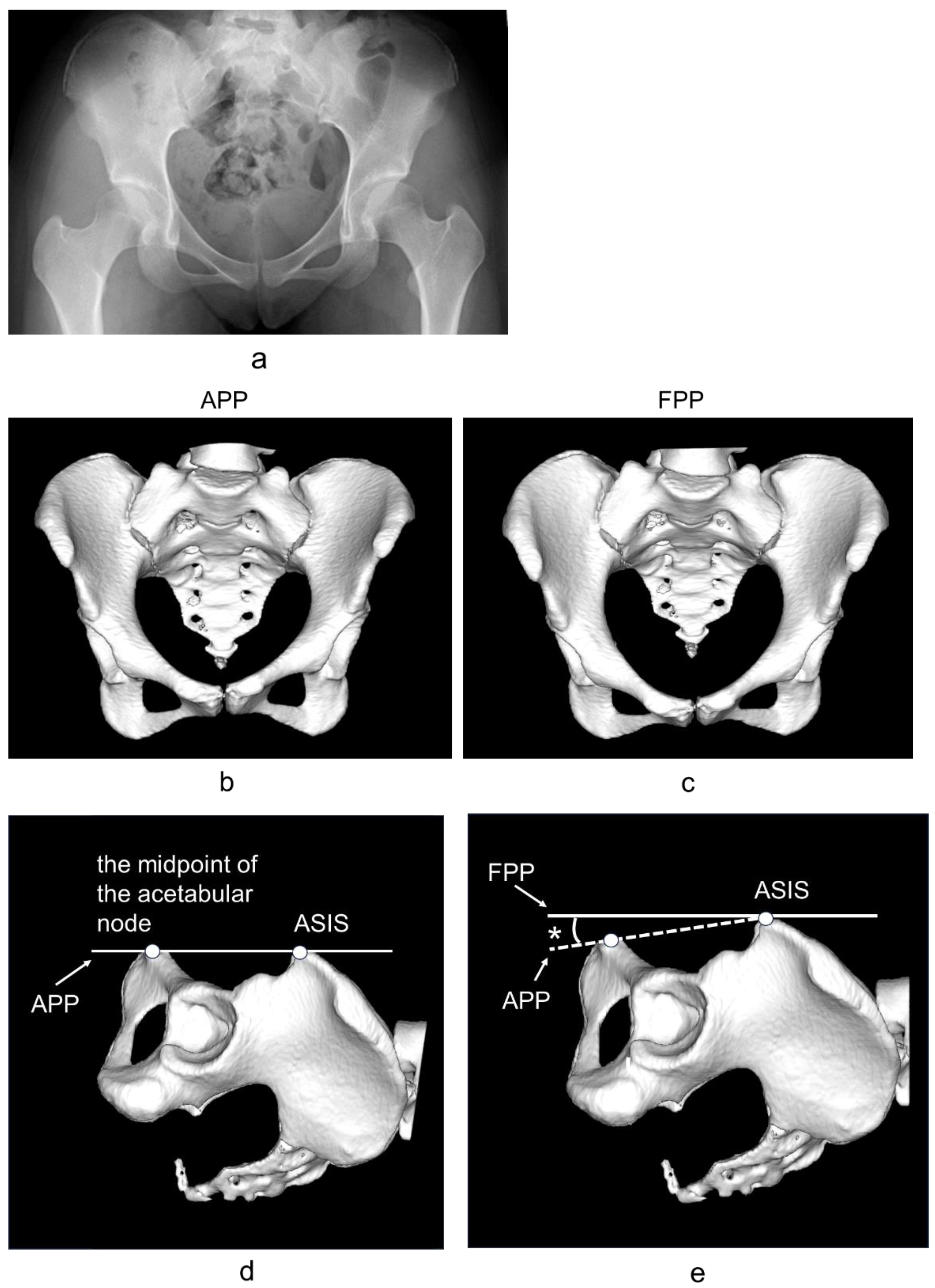
2. Materials and Methods
2.1. Participants
2.2. CT
2.3. Statistical Analysis
3. Results
4. Discussion
5. Limitations
6. Conclusions
Author Contributions
Funding
Institutional Review Board Statement
Informed Consent Statement
Data Availability Statement
Acknowledgments
Conflicts of Interest
References
- Murphy, S.B.; Ganz, R.; Müller, M.E. The prognosis in untreated dysplasia of the hip: A study of radiographic factors that predict outcome. J. Bone Jt. Surg. Am. 1995, 77, 985–989. [Google Scholar] [CrossRef] [PubMed]
- Albinana, J.; Dolan, L.A.; Spratt, K.F.; Morcuende, J.; Meyer, M.D.; Weinstein, S.L. Acetabular dysplasia after treatment for developmental dysplasia of the hip.Implications for secondary procedures. J. Bone Jt. Surg. Br. 2004, 86, 876–886. [Google Scholar] [CrossRef]
- Nakamura, S.; Ninomiya, S.; Nakamura, T. Primary osteoarthritis of the hip joint in Japan. Clin. Orthop. Relat. Res. 1989, 241, 190–196. [Google Scholar] [CrossRef]
- Takeyama, A.; Naito, M.; Shiramizu, K.; Kiyama, T. Prevalence of femoroacetabular impingement in Asian patients with osteoarthritis of the hip. Int. Orthop. 2009, 33, 1229–1232. [Google Scholar] [CrossRef]
- Hipp, J.A.; Sugano, N.; Millis, M.B.; Murphy, S.B. Planning acetabular redirection osteotomies based on joint contact pressures. Clin. Orthop. Relat. Res. 1999, 364, 134–143. [Google Scholar] [CrossRef]
- Jacobsen, S.; Sonne-Holm, S.; Søballe, K.; Gebuhr, P.; Lund, B. Hip dysplasia and osteoarthrosis: A survey of 4151 subjects from the osteoarthrosis substudy of the Copenhagen City Heart Study. Acta Orthop. 2005, 76, 149–158. [Google Scholar] [CrossRef]
- Wiberg, G. Studies on dysplastic acetabulum and congenital subluxation of the hip joint with special reference to the complication of osteoarthritis. Acta Chir. Scand. 1939, 83 (Suppl. 58), 33. [Google Scholar]
- Sharp, I.K. Acetabular dysplasia. The acetabular angle. J. Bone Jt. Surg. Br. 1961, 43, 268–272. [Google Scholar] [CrossRef]
- Anda, S.; Terjesen, T.; Kvistad, K.A.; Svenningsen, S. Acetabular angles and femoral anteversion in dysplastic hips in adults: CT investigation. J. Comput. Assist. Tomogr. 1991, 15, 115–120. [Google Scholar] [CrossRef] [PubMed]
- Chegini, S.; Beck, M.; Ferguson, S.J. The effects of impingement and dysplasia on stress distributions in the hip joint during sitting and walking: A finite element analysis. J. Orthop. Res. 2009, 27, 195–201. [Google Scholar] [CrossRef]
- Yoshimura, N.; Campbell, L.; Hashimoto, T.; Kinoshita, H.; Okayasu, T.; Wilman, C.; Coggon, D.; Croft, P.; Cooper, C. Acetabular dysplasia and hip osteoarthritis in Britain and Japan. Br. J. Rheumatol. 1998, 37, 1193–1197. [Google Scholar] [CrossRef]
- Schmitz, M.R.; Murtha, A.S.; Clohisy, J.C.; ANCHOR Study Group. Developmental dysplasia of the hip in adolescents and young adults. J. Am. Acad. Orthop. Surg. 2020, 28, 91–101. [Google Scholar] [CrossRef]
- Beltran, L.S.; Rosenberg, Z.S.; Mayo, J.D.; De Tuesta, M.D.; Martin, O.; Neto, L.P.; Bencardino, J.T. Imaging evaluation of developmental hip dysplasia in the young adult. AJR Am. J. Roentgenol. 2013, 200, 1077–1088. [Google Scholar] [CrossRef]
- Reiman, M.P.; Décary, S.; Mathew, B.; Reiman, C.K. Accuracy of clinical and imaging tests for the diagnosis of hip dysplasia and instability: A systematic review. J. Orthop. Sports Phys. Ther. 2019, 49, 87–97. [Google Scholar] [CrossRef]
- Lequesne, M.G.; Laredo, J.D. The faux profil (oblique view) of the hip in the standing position. Contribution to the evaluation of osteoarthritis of the adult hip. Ann. Rheum. Dis. 1998, 57, 676–681. [Google Scholar] [CrossRef]
- Ito, H.; Matsuno, T.; Hirayama, T.; Tanino, H.; Yamanaka, Y.; Minami, A. Three-dimensional computed tomography analysis of non-osteoarthritic adult acetabular dysplasia. Skeletal Radiol. 2009, 38, 131–139. [Google Scholar] [CrossRef]
- Flaviu, M.; Adrian, G.; Tiberiu, B. Integration of Three-dimensional Technologies in Orthopedics: A Tool for Preoperative Planning of Tibial Plateau Fractures. Acta Inf. Med. 2020, 28, 278–282. [Google Scholar]
- Papagelopoulos, P.J.; Savvidou, O.D.; Koutsouradis, P.; Chloros, G.D.; Bolia, I.K.; Sakellariou, V.I.; Kontogeorgakos, V.A.; Mavrodontis, I.I.; Mavrogenis, A.F.; Diamantopoulos, P. Three-dimensional technologies in orthopedics. Orthopedics 2018, 41, 12–20. [Google Scholar] [CrossRef]
- Wong, K.C. 3D-printed patient-specific applications in orthopedics. Orthop. Res. Rev. 2016, 8, 57–66. [Google Scholar] [CrossRef]
- Flaviu, M.; Adrian, G.; Tiberiu, B. Structured Integration and Alignment Algorithm: A Tool for Personalized Surgical Treatment of Tibial Plateau Fractures. J. Pers. Med. 2021, 11, 190. [Google Scholar] [CrossRef]
- Imai, N.; Suzuki, H.; Nozaki, A.; Hirano, Y.; Endo, N. Correlation of tilt of the anterior pelvic plane angle with anatomical pelvic tilt and morphological configuration of the acetabulum in patients with developmental dysplasia of the hip: A cross-sectional study. J. Orthop. Surg. Res. 2019, 14, 323. [Google Scholar] [CrossRef]
- Nozaki, A.; Imai, N.; Funayama, K.; Horigome, Y.; Suzuki, H.; Minato, I.; Kobayashi, K.; Kawashima, H. Accuracy of ZedView, the software for three-dimensional measurement and preoperative planning: A basic study. Medicina 2023, 59, 1030. [Google Scholar] [CrossRef]
- Murtha, P.E.; Hafez, M.A.; Jaramaz, B.; DiGioia, A.M. Variations in acetabular anatomy with reference to total hip replacement. J. Bone Jt. Surg. Br. 2008, 90, 308–313. [Google Scholar] [CrossRef]
- Miyasaka, D.; Ito, T.; Imai, N.; Suda, K.; Minato, I.; Dohmae, Y.; Endo, N. Three-dimensional assessment of femoral head coverage in normal and dysplastic hips: A novel method. Acta Med. Okayama 2014, 68, 277–284. [Google Scholar]
- Janzen, D.L.; Aippersbach, S.E.; Munk, P.L.; Sallomi, D.F.; Garbuz, D.; Werier, J.; Duncan, C.P. Three-dimensional CT measurement of adult acetabular dysplasia: Technique, preliminary results in normal subjects, and potential applications. Skeletal Radiol. 1998, 27, 352–358. [Google Scholar] [CrossRef]
- Tönnis, D. Congenital Dysplasia and Dislocation of the Hip in Children and Adults; Springer: Berlin, Germany, 1987. [Google Scholar]
- Tanifuji, O.; Sato, T.; Kobayashi, K.; Mochizuki, T.; Koga, Y.; Yamagiwa, H.; Omori, G.; Endo, N. Three-dimensional in vivo motion analysis of normal knees using single-plane fluoroscopy. J. Orthop. Sci. 2011, 16, 710–718. [Google Scholar] [CrossRef]
- Anda, S.; Svenningsen, S.; Dale, L.G.; Benum, P. The acetabular sector angle of the adult hip determined by computed tomography. Acta Radiol. Diagn. 1986, 27, 443–447. [Google Scholar] [CrossRef]
- Nishihara, S.; Sugano, N.; Nishii, T.; Ohzono, K.; Yoshikawa, H. Measurements of Pelvic Flexion Angle Using Three-Dimensional Computed Tomography. Clin. Orthop. Relat. Res. 2003, 411, 140–151. [Google Scholar] [CrossRef]
- Gala, L.; Clohisy, J.C.; Beaulé, P.E. Hip Dysplasia in the young adult. J. Bone Jt. Surg. Am. 2016, 98, 63–73. [Google Scholar] [CrossRef]
- Damen, J.; van Rijn, R.M.; Emans, P.J.; Hilberdink, W.K.; Wesseling, J.; Oei, E.H.; Bierma-Zeinstra, S.M. Prevalence and development of hip and knee osteoarthritis according to American College of Rheumatology criteria INTHE CHECK cohort. Arthritis Res. Ther. 2019, 21, 4. [Google Scholar] [CrossRef]
- Zarringam, D.; Saris, D.B.F.; Bekkers, J.E.J. Identification of early prognostic factors for knee and hip arthroplasty; a long-term follow-up of the CHECK cohort. J. Orthop. 2020, 19, 41–45. [Google Scholar] [CrossRef] [PubMed]
- Herfkens, J.; van Buuren, M.M.; Riedstra, N.S.; Verhaar, J.A.; Mascarenhas, V.V.; Agricola, R. Adding false-profile radiographs improves detection of developmental dysplasia of the hip, data from the CHECK cohort. J. Hip Preserv. Surg. 2022, 9, 3–9. [Google Scholar] [CrossRef] [PubMed]
- Chadayammuri, V.; Garabekyan, T.; Jesse, M.K.; Pascual-Garrido, C.; Strickland, C.; Milligan, K.; Mei-Dan, O. Measurement of lateral acetabular coverage: A comparison between CT and plain radiography. JHPS 2015, 2, 392–400. [Google Scholar] [CrossRef] [PubMed]
- Miyasaka, D.; Sakai, Y.; Ibuchi, S.; Suzuki, H.; Imai, N.; Endo, N. Sex- and age-specific differences in femoral head coverage and acetabular morphology among healthy subjects-derivation of normal ranges and thresholds for abnormality. Skeletal Radiol. 2017, 46, 523–531. [Google Scholar] [CrossRef]
- Murphy, S.B.; Kijewski, P.K.; Millis, M.B.; Harless, A. Acetabular dysplasia in the adolescent and young adult. Clin. Orthop. Relat. Res. 1990, 261, 214–223. [Google Scholar] [CrossRef]


| APP | Total (n = 340) | Normal (n = 238) | DDH (n = 102) |
|---|---|---|---|
| ACEA (°) * | 50.6 ± 15.2 (−16.9~88.9) | 56.9 ± 10.1 (29.6~88.9) | 36.7 ± 8.5 (−16.9~60.3) |
| PCEA (°) * | 99.4 ± 16.0 (30.4~136.8) | 100.9 ± 14.2 (61.3~136.6) | 96.1 ± 14.2 (30.4~136.8) |
| LCEA (°) * | 26.0 ± 4.2 (−13.5~59.8) | 32.0 ± 7.8 (0.4~59.8) | 13.2 ± 7.4 (−13.5~38.8) |
| ARO (°) * | 10.3 ± 9.1 (−12.5~42.2) | 6.3 ± 5.8 (−12.5~36.3) | 19.1 ± 5.5 (−12.5~42.2) |
| AASA (°) * | 54.6 ± 10.5 (21.7~82.3) | 59.2 ± 8.0 (25.2~82.3) | 44.7 ± 6.7 (21.7~62.9) |
| PASA(°) * | 94.6 ± 8.6 (54.1~124.5) | 95.8 ± 9.1 (54.1~124.5) | 91.8 ± 9.1 (54.1~111.9) |
| FPP | Total (n = 340) | Normal (n = 238) | DDH (n = 102) |
| ACEA (°) * | 54.5 ± 13.7 (−9.4~89.2) | 60.1 ± 8.5 (28.1~89.2) | 42.4 ± 7.6 (−9.4~61.5) |
| PCEA (°) * | 95.4 ± 16.3 (26.3~142.1) | 97.8 ± 14.8 (53.7~142.1) | 90.4 ± 14.7 (26.3~128.7) |
| LCEA (°) * | 26.4 ± 12.5 (−13.8~78.5) | 32.3 ± 8.6 (7.4~78.5) | 13.6 ± 9.2 (−13.8~51.8) |
| ARO (°) * | 9.8 ± 8.8 (−12.4~41.9) | 6.0 ± 5.8 (−12.4~32.5) | 17.9 ± 5.8 (−12.4~41.9) |
| AASA (°) * | 55.6 ± 10.5 (29.2~80.4) | 60.3 ± 7.5 (16.0~63.9) | 45.6 ± 7.1 (16.0~80.4) |
| PASA (°) * | 93.0 ± 9.5 (12.1~125.6) | 94.5 ± 8.7 (72.5~125.6) | 89.7 ± 9.3 (12.1~109.2) |
| APP | COV | Sensitivity | 1-Specificity |
|---|---|---|---|
| ACEA | 49.3 | 0.788 | 0.229 |
| LCEA | 22.2 | 0.931 | 0.152 |
| ARO | 10.2 | 0.848 | 0.199 |
| AASA | 51.4 | 0.892 | 0.210 |
| FPP | COV | Sensitivity | 1-Specificity |
| ACEA | 52.4 | 0.844 | 0.257 |
| LCEA | 24.5 | 0.861 | 0.067 |
| ARO | 10.3 | 0.781 | 0.217 |
| AASA | 53.1 | 0.861 | 0.181 |
| APP | FPP | |||||
|---|---|---|---|---|---|---|
| AUC | p-Value | 95% CI | AUC | p-Value | 95% CI | |
| ACEA | 0.867 | <0.001 | 0.826~0.908 | 0.880 | <0.001 | 0.841~0.919 |
| PCEA | 0.560 | 0.046 | 0.502~0.618 | 0.602 | 0.004 | 0.502~0.670 |
| LCEA | 0.941 | <0.001 | 0.917~0.965 | 0.952 | <0.001 | 0.928~0.976 |
| ARO | 0.896 | <0.001 | 0.858~0.934 | 0.876 | <0.001 | 0.835~0.917 |
| AASA | 0.868 | <0.001 | 0.830~0.906 | 0.905 | <0.001 | 0.868~0.942 |
| PASA | 0.635 | <0.001 | 0.573~0.695 | 0.634 | <0.001 | 0.572~0.696 |
| APP | Degrees of Freedom | Odds Ratio | 95% CI | p-Value |
|---|---|---|---|---|
| ARO | 339 | −0.028 | −0.031~−0.025 | <0.001 |
| LCEA | 339 | −0.001 | −0.014~−0.005 | <0.001 |
| FPP | Degrees of Freedom | Odds Ratio | 95% CI | p-Value |
| AASA | 339 | −0.029 | −0.032~−0.026 | <0.001 |
| LCEA | 339 | −0.013 | −0.017~−0.008 | <0.001 |
| APP | Intraobserver | Interobserver | ||||
|---|---|---|---|---|---|---|
| MAD (n = 340) | ICC | 95% CI | MAD (n = 340) | ICC | 95% CI | |
| ACEA * | 1.3 ± 1.2 (0.0~5.3) | 0.937 | 0.922~0.949 | 2.4 ± 1.9 (0.1~7.8) | 0.856 | 0.810~0.901 |
| PCEA * | 2.0 ± 1.8 (0.0~7.6) | 0.892 | 0.867~0.912 | 3.1 ± 2.3 (0.2~8.7) | 0.821 | 0.788~0.854 |
| LCEA * | 1.3 ± 1.4 (0.0~6.8) | 0.933 | 0.918~0.946 | 3.3 ± 2.2 (0.0~8.8) | 0.813 | 0.769~0.849 |
| ARO * | 1.1 ± 1.1 (0.0~4.0) | 0.966 | 0.957~0.972 | 2.4 ± 2.0 (0.1~7.9) | 0.854 | 0.823~0.881 |
| AASA * | 0.6 ± 0.5 (0.0~1.9) | 0.971 | 0.964~0.977 | 1.4 ± 1.5 (0.1~8.8) | 0.928 | 0.918~0.939 |
| PASA * | 1.0 ± 0.9 (0.0~5.0) | 0.963 | 0.954~0.970 | 1.6 ± 1.4 (0.0~8.0) | 0.921 | 0.903~0.937 |
| FPP | Intraobserver | Interobserver | ||||
| MAD (n = 340) | ICC | 95% CI | MAD (n = 340) | ICC | 95% CI | |
| ACEA * | 1.3 ± 1.0 (0.0~4.8) | 0.944 | 0.931~0.954 | 1.9 ± 12.0 (0.1~7.4) | 0.908 | 0.886~0.925 |
| PCEA * | 2.1 ± 1.7 (0.0~5.9) | 0.882 | 0.856~0.908 | 2.3 ± 1.9 (0.1~6.9) | 0.861 | 0.805~0.913 |
| LCEA * | 1.5 ± 1.5 (0.0~7.3) | 0.934 | 0.918~0.946 | 2.3 ± 2.0 (0.0~7.9) | 0.859 | 0.759~0.919 |
| ARO * | 0.9 ± 1.2 (0.0~5.3) | 0.952 | 0.941~0.961 | 1.8 ± 1.6 (0.0~6.0) | 0.909 | 0.888~0.926 |
| AASA * | 1.0 ± 0.7 (0.0~3.0) | 0.967 | 0.959~0.974 | 1.5 ± 1.2 (0.2~5.0) | 0.928 | 0.912~0.942 |
| PASA * | 1.0 ± 0.8 (0.0~3.9) | 0.966 | 0.957~0.972 | 1.3 ± 1.1 (0.0~4.5) | 0.943 | 0.929~0.954 |
Disclaimer/Publisher’s Note: The statements, opinions and data contained in all publications are solely those of the individual author(s) and contributor(s) and not of MDPI and/or the editor(s). MDPI and/or the editor(s) disclaim responsibility for any injury to people or property resulting from any ideas, methods, instructions or products referred to in the content. |
© 2024 by the authors. Licensee MDPI, Basel, Switzerland. This article is an open access article distributed under the terms and conditions of the Creative Commons Attribution (CC BY) license (https://creativecommons.org/licenses/by/4.0/).
Share and Cite
Ibuchi, S.; Imai, N.; Horigome, Y.; Suzuki, H.; Kawashima, H. The Three-Dimensional Criteria of Developmental Dysplasia of the Hip Using the Functional Pelvic Plane Is More Useful Than That Using the Anterior Pelvic Plane. J. Clin. Med. 2024, 13, 2536. https://doi.org/10.3390/jcm13092536
Ibuchi S, Imai N, Horigome Y, Suzuki H, Kawashima H. The Three-Dimensional Criteria of Developmental Dysplasia of the Hip Using the Functional Pelvic Plane Is More Useful Than That Using the Anterior Pelvic Plane. Journal of Clinical Medicine. 2024; 13(9):2536. https://doi.org/10.3390/jcm13092536
Chicago/Turabian StyleIbuchi, Shinya, Norio Imai, Yoji Horigome, Hayato Suzuki, and Hiroyuki Kawashima. 2024. "The Three-Dimensional Criteria of Developmental Dysplasia of the Hip Using the Functional Pelvic Plane Is More Useful Than That Using the Anterior Pelvic Plane" Journal of Clinical Medicine 13, no. 9: 2536. https://doi.org/10.3390/jcm13092536