1. Introduction
In China, the dry root of
Scutellaria baicalensis has been widely used as a traditional medicine for treating various diseases. Our ancestors had applied the herb for thousands of years. The recent studies indicated that it has protective effects against cerebral ischemia [
1,
2]. Baicalin (BA) is the main constituent of
Scutellaria baicalensis [
3]. The previous studies demonstrated that BA has a variety of biological activities, such as curing bacterial meningitis, and infectious brain edema [
4,
5], it also has the anti-tumor effect [
6]. Some studies also indicated that, BA had specific radical scavenging activity and protective effects, which can protect the brain from ischemia–reperfusion injury in rats [
7,
8,
9,
10,
11]. In addition, BA also has the protective effects on neurons subjecting to oxygen–glucose deprivation and reperfusion injury.
BA has been proved to exhibit so many pharmacological effects on treatment of cerebral ischemia-reperfusion injury, however, the low lipid and water solubility of BA has poor penetration of the blood–brain barrier (BBB), which is the primary obstacle to the delivery of therapeutic drugs to the brain. Thus, the clinical application of BA has been severely limited for the treatment of the brain disease [
12]. Therefore, it is necessary for us to develop a novel drug delivery system to improve the solubility of BA. Liposomes, an effective nanometer-scale drug delivery system which can carry hydrophilic and lipophilic as well as amphoteric drug molecules entrapped either in the core or in the liposome bilayer [
13,
14]. A number of studies showed the liposome carrier could improve the solubility of drugs and change the in vivo distribution of entrapped drugs.
The objective of this study was to develop a novel baicalin-loaded liposome (BA-LP) formulation in order to improve the drug lipophilicity and further to enhance the drug-concentration in the brain tissues. The morphology, size, zeta potential, encapsulation efficiency, and the in vitro release of BA-LP have been investigated. Because the drug is used to treat diseases and only patient is the ultimate consumer of drug. It is very important to study the pharmacokinetics of drugs under the disease states. Middle cerebral artery occlusion (MCAO) is a kind of cerebrovascular disease model in rat [
15]. In the pharmacokinetics and biodistribution studies, a simple, rapid, accurate and reliable method was established for determination of BA in rat plasma and tissues. Then, the pharmacokinetic characteristics of BA or BA-LP on the MCAO model have been investigated, and the experiment on the normal animal group has also been done for comparison.
3. Discussion
Since BA has been proved to exhibit so many pharmacological effects on treatment of cerebral ischemia-reperfusion injury, however, similar like most components obtained from the traditional Chinese herbs, BA possesses low lipid and water solubility, which causes low permeation into biological membranes. Therefore, improving the ability of BA to pass through the BBB is the precondition for the clinical application of BA.
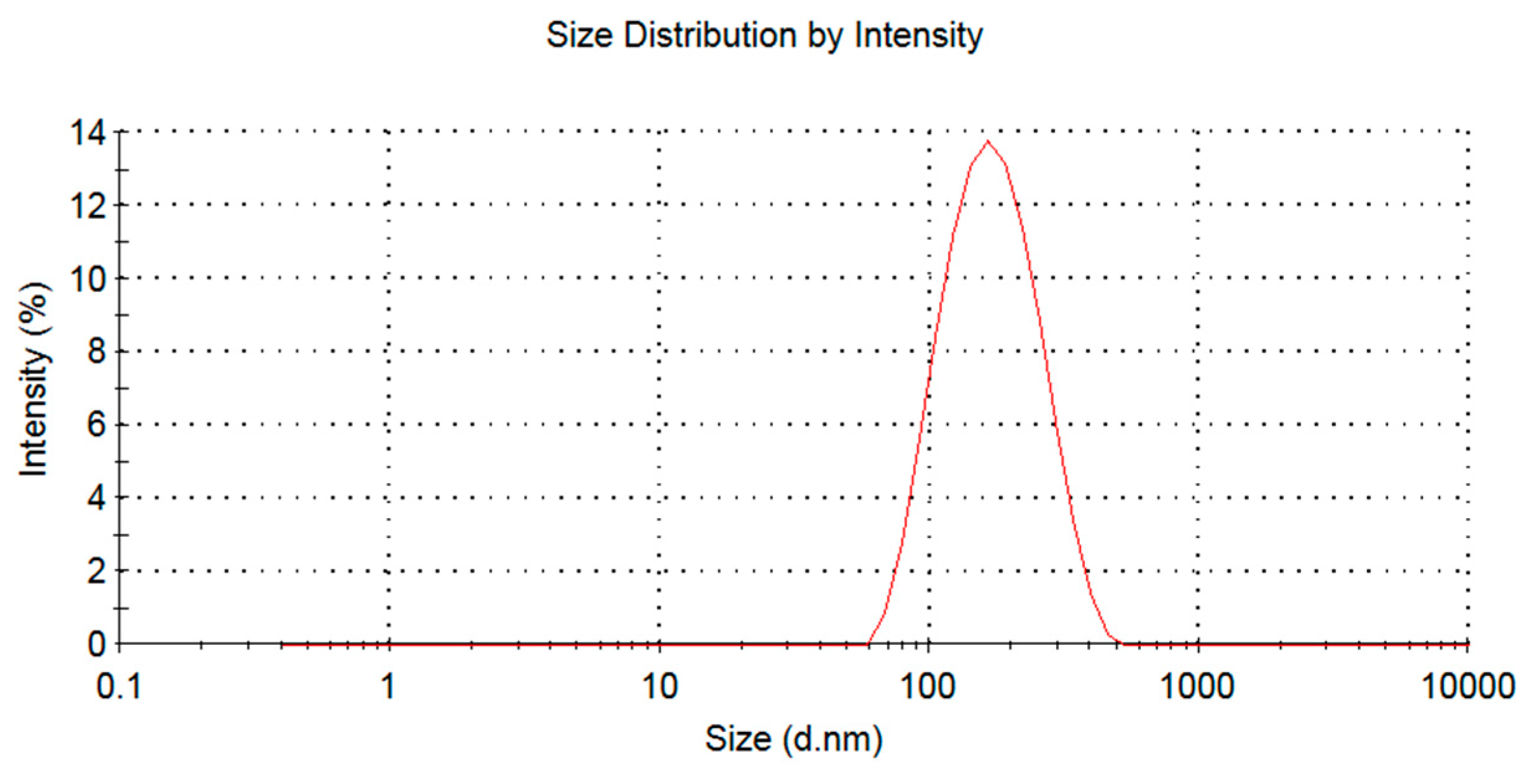
Currently, liposomes have been considered as one of the most promising drug delivery carriers, it can improve drug solubility and have the potential of the targeting effect, thus to increase the therapeutic index. Therefore, in previous study, we applied the Box-Behnken design to optimize the preparation process of BA lipid system. The optimal prescription was as follows: the proportion of phospholipid drugs 3.81:1, phospholipid cholesterol ratio 5.70:1, hydration volume 1.02 mL, ultrasonic power 60 W, ultrasonic time 10 min, ultrasonic temperature 20 ± 5 °C. The resulting BA-LP was round in shape, with a particle size of 160–190 nm. The zeta potential of BA-LP was −5.7 mV, which indicated that there were a number of negative charges on the surface of BA-LP. The entrapment efficiency of BA-LP was 42 ± 1%.
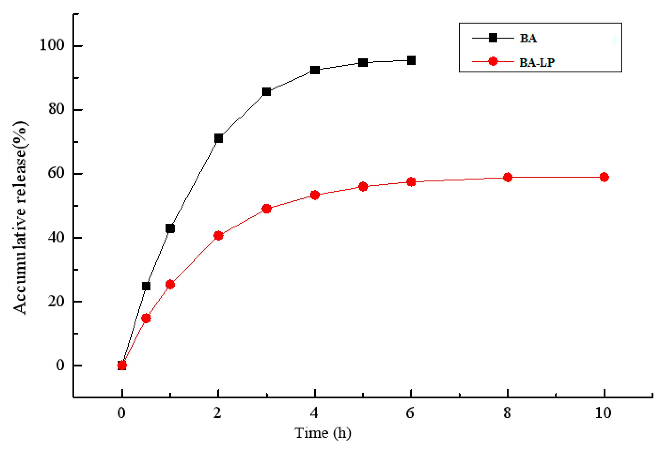
The in vitro release study of BA-LP indicated that the first 4 h was an initial burst-release phase, and the in vitro accumulative release rate within 4 h was 53.35%, Therefore, the release rate became slow, demonstrating a typical sustained and prolonged drug-release behavior. The zero order kinetics, first-order kinetics, biphasic kinetics, Niebergull and Higuchi models were applied to simulate in vitro release of BA-LP. The in vitro drug-release kinetic model of BA-LP fit well with the biphasic dynamic model equation: Q = 1 − (60.12 e0.56t − 59.08 e0.0014t) (r = 0.999). Therefore, it was speculated that the sustained -release property of BA-LP can improve the clinical therapeutic effect of BA.
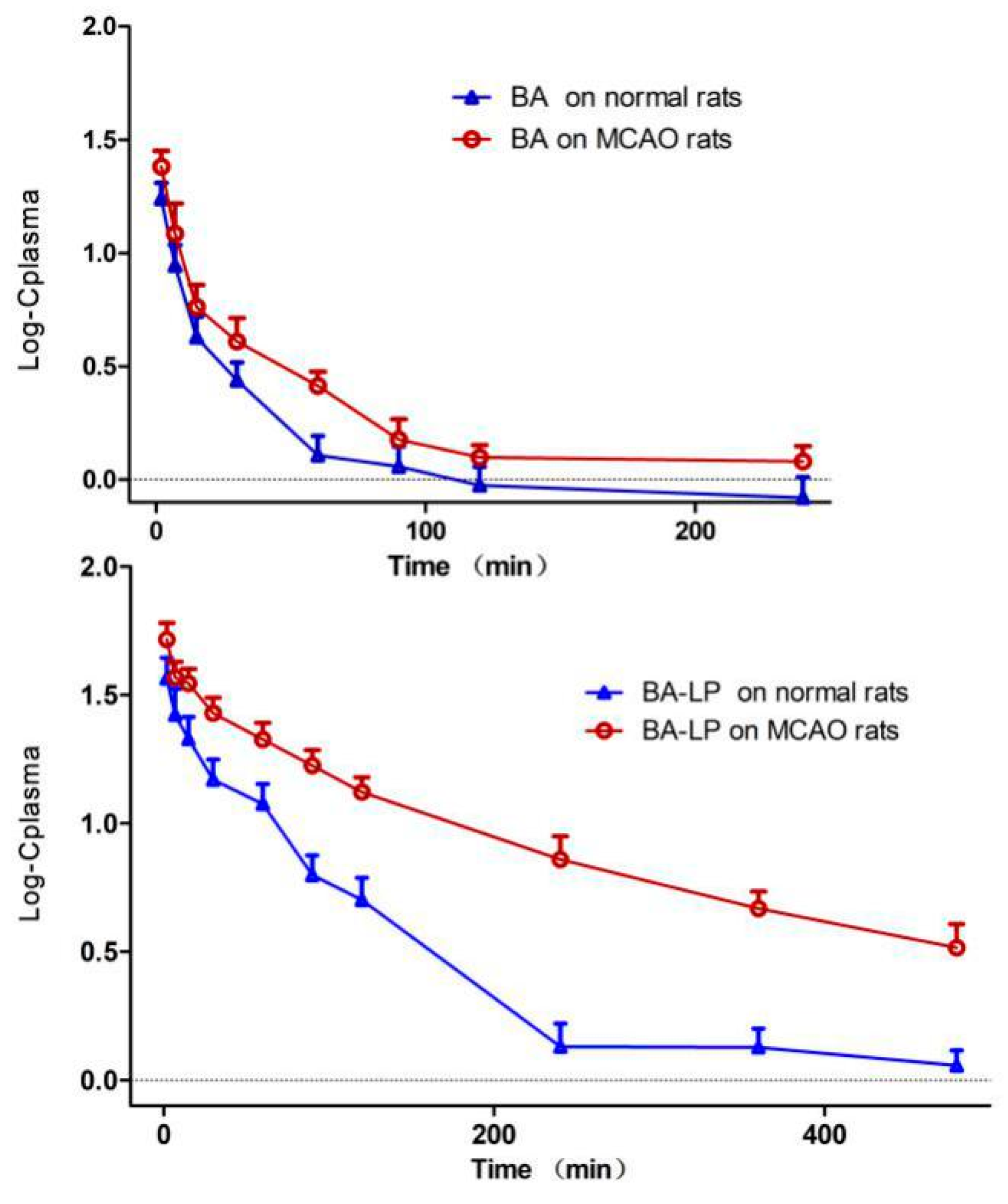
Generally, most of the pharmacokinetic studies were performed on healthy animals, which could ignore the effect of pathology on pharmacokinetics. It is valuable to study the pharmacokinetic profile based on pathological animals, because the body state of animal is closer to clinic. In our study, we selected the MCAO model to simulate the state of stroke. The transient focal ischemia model is minimal harmful and has been used widely in scientific research. The pharmacokinetic study of BA in liposome form on MCAO model rats has never been reported before.
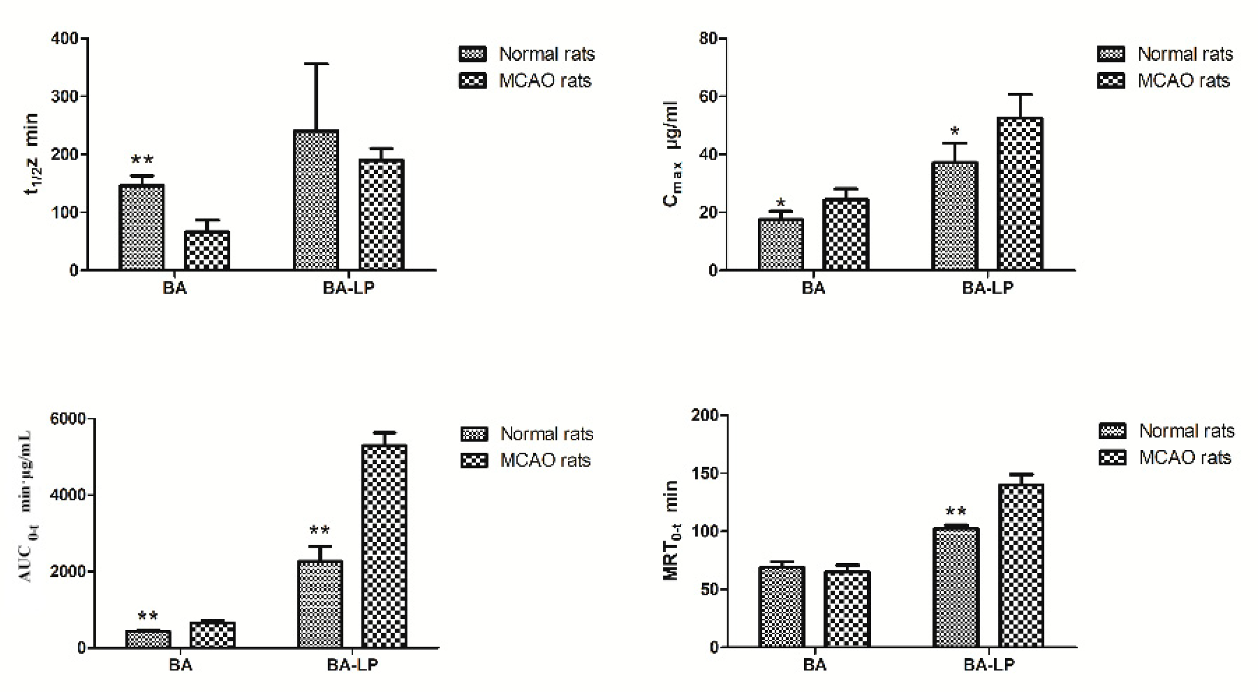
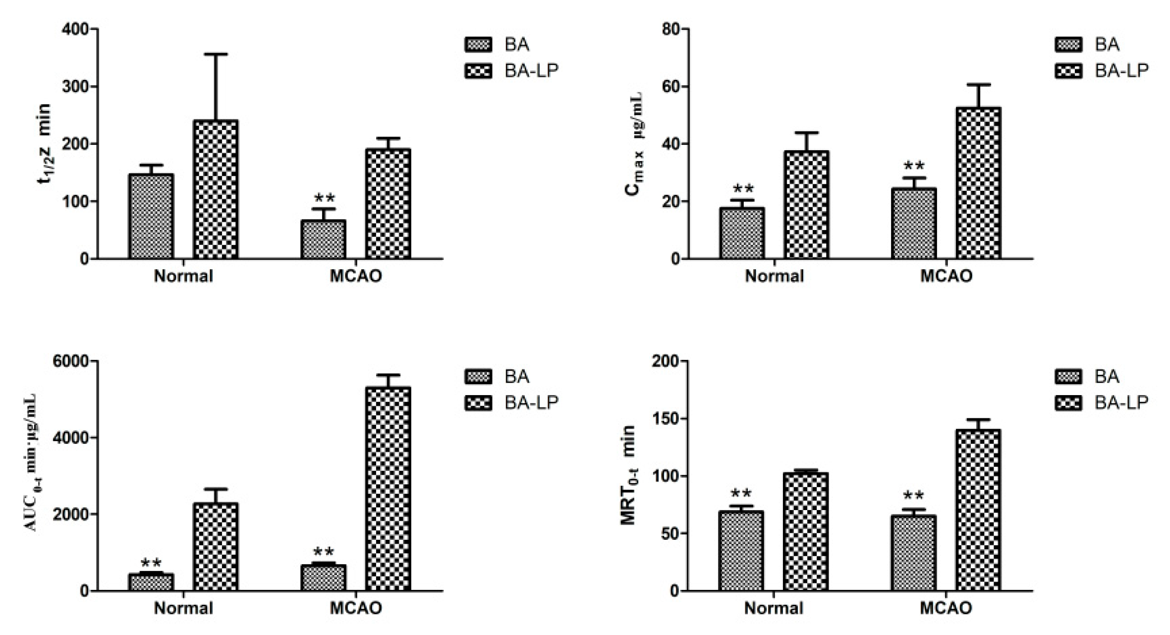
The effect of stroke on disposition and absorption of BA was investigated, the parameters between the groups of MCAO model and normal rats were compared as well. In the MCAO group, bigger values of C
max and AUC
0–t were obtained, which indicated that it may enhance the speed of distribution or reduce the elimination. This result is consistent with a previous pharmacokinetic study [
15], in which, BA had a better absorption effect in the pathologic condition. These results proved the rationality of using BA in cerebrovascular disease, which would improve the therapeutic efficacy.
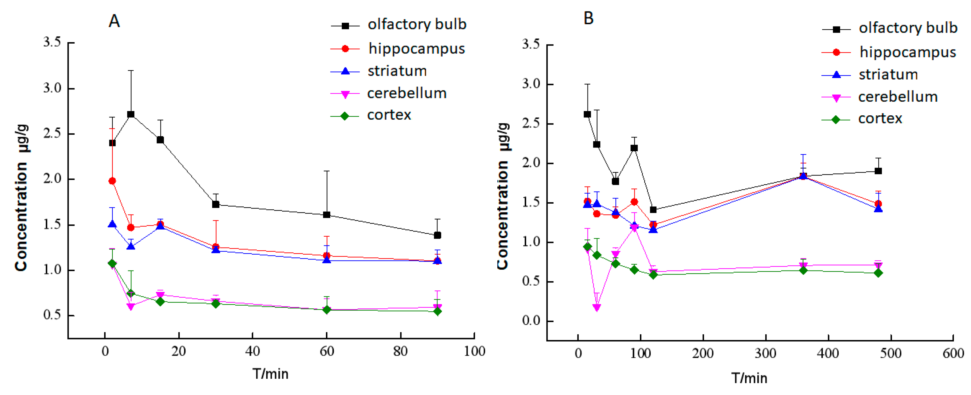
In the brain tissuse, the distribution of drugs in rats with the MCAO model was greatly increased both with BA and BA-LP. It suggested that stroke could increase the permeability of BBB by increasing the number of endothelial caveolae and transcytosis rate and disrupting tight junction as reported, which may enhance the transportation of drug. The vascular pathway may also play an important role in drug delivery under pathological conditions. The brain tissues are rich with microvascular. Stroke may also affect the permeability there, even when occlusion happened in middle cerebral artery [
16,
17].
In the pharmacokinetics study on the normal and MCAO rats, in plasma, the Cmax and AUC0–t values were significantly greater with BA-LP than BA. In addition, the other pharmacokinetic parameters were also significantly improved as evidenced by the 1.64-fold increase in the t1/2z and a 4.34-fold reduction in the renal clearance rate with a 1.49-fold increase in the MRT0–t on the normal animal model; a 2.87-fold increase in the t1/2z and a 8.08-fold reduction in the renal clearance rate with a 2.14-fold increase in the MRT0–t on the normal animal model; this improvement in the pharmacokinetic parameters could potentially lead to the prolonged the retention time of BA in vivo and thereby improve the therapeutic efficacy of BA.
The tissue distribution behavior of BA was significantly altered in the case of BA-LP administrated in comparison with BA. The concentrations of BA in the heart, liver, spleen, lungs and brain were all increased after administrated BA-LP compared with BA in both normal model and MCAO model, but decreased in kidneys. The target parameters results indicated the similar differences between BA and BA-LP. However, the mechanism of biodistribution after oral administration needs to be clarified by further studies. In addition, it’s worth mentioning that the toxicity studies of BA show that BA has very little toxicity to normal epithelial and normal peripheral blood and myeloid cells [
18]. These results indicate that the liposomes for BA is helped to pass through the BBB, and may be applicable to cerebrovascular disease.
4. Materials and Methods
4.1. Materials and Reagents
BA reference substance was obtained from the National Institute for the Control of Biological and Pharmaceutical Drugs (Beijing, China), Batch No. 110715-201006; Soybean lecithin was obtained from Shanghai Ivet Medical Technology Co., Ltd. (Shanghai, China), Batch No. SY–SI-160802; Cholesterol was purchased from Shanghai Ivet Medical Technology Co., Ltd. (Shanghai, China), Batch No. B40936; Quercetin reference substance was provided by Chengdu Man Site Biotechnology Co., Ltd. (Chengdu, China), Batch No. MUST-16111114; Methanol (High Performance Liquid Chromatography grade) was from Tedia Company (Darmstadt, Germany); Double distilled water used in this study was prepared in a laboratory double distilled water purification system.
4.2. Preparation of BA-LP Liposomes
BA-LP was prepared by the method of reverse evaporation in our previous research, briefly, phospholipid (100 mg), cholesterol (17.5 mg) were dissolved into ethanol (18 mL). BA (25 mg) was dissolved into PBS (pH 7.0) at 5 mg/mL, and then injected the solution into the organic phase, and was disrupted in ultrasonic homogenizer for 10 min at 20 °C, after 10 min of disrupting in ultrasonic homogenizer at 20 °C, the organic solvent was evaporated on a rotary evaporator under reduced pressure to obtain the colloid. The resulting colloid was dissolved by the addition of PBS (pH 7.0) to obtain the BA-LP solution [
19,
20,
21].
4.3. Physicochemical Characterization of BA-LP
The morphologies of BA-LP were examined by transmission electron microscopy (TEM). The BA-LP samples were diluted with PBS (pH 7.0) and dropped on a Formvar®-coated copper grid and then were air-dried for 1 min at room temperature. The particle size and zeta potential of BA-LP was measured with a Malvern Zetasizer 3000 (Malvern Instruments Ltd., Malvern, UK). The samples of BA-LP were diluted with the physiological saline (1:20) before measurement.
4.4. Entrapment Efficiency Study
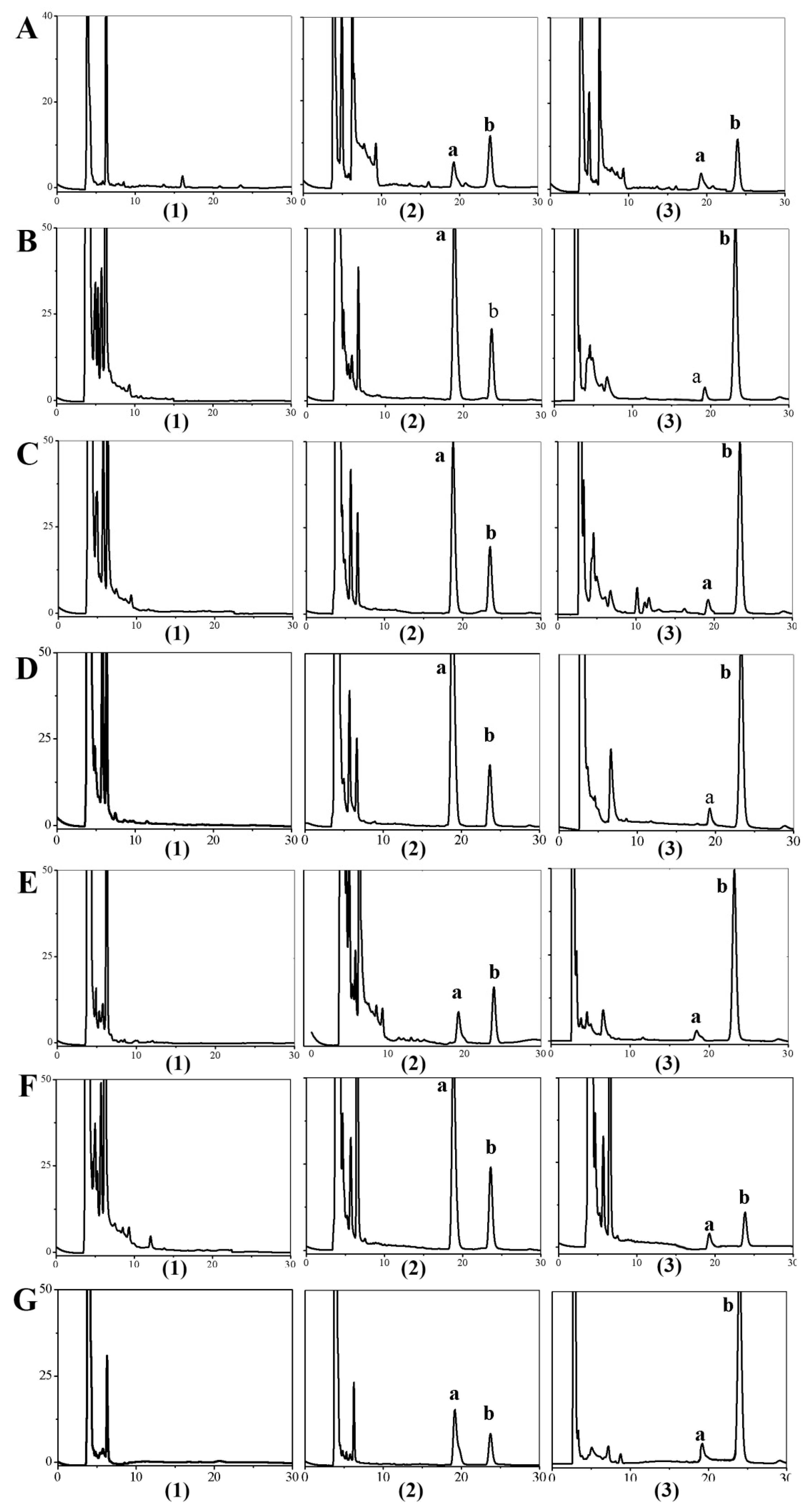
The BA content of BA-LP was detected with HPLC. An HPLC assay was carried out by using a reverse-phase C18 column (XTerra® MS, 4.6250 mm, 4.6 × 250 mm, Waters, Dublin, Ireland), the mobile phase was 59% double-distilled water, 41% methanol, 0.2% phosphoric acid (v/v) at a flow rate of 1 mL/min and a detection wavelength of 276 nm and the injection volume was 20 μL.
The entrapment efficiency (EE) of BA-LP was measured using the dialysis method (dialysis membrane with a molecular weight cut-off of 10,000). The BA-LP sample was placed into the dialysis membrane bag and dialysed against phosphate-bufferd saline (pH 7.4) for 7 h by gentle shaking. After dialysis, free drug (W
f) was determined by HPLC, as established in this study. An aliquot of 0.2 mL of BA-LP was vortex-mixed with 0.8 mL of methanol for 5 min and then was filtrated through a 0.22 μm hydrophobic Millipore membrane (Tengjin Experimental Equipment Company, Tianjin, China), and the total drug contents (W
total) was determined by HPLC. The entrapment efficiency was calculated by the following equation:
where EE% was the entrapment efficiency, W
f was the amount of free BA in the BA-LP sample, and W
total was the total amount of BA in BA-LP sample.
4.5. In Vitro Release Study
The in vitro release studies of BA-LP vs. BA solution were performed by using the dialysis bag method (molecular weight cutoff [MWCO] 10,000 Da), The PBS (pH 7.0) has been chosen as a release medium, 2 mL of BA-LP and free-BA solution was placed into dialysis bags and tightly sealed respectively. And then, the test bags were immersed in release medium at a stirring rate of 80 rpm and at 37 °C. At predetermined time points of 0.5, 1, 2, 3, 4, 6, 8, 10 h, 3 mL dissolution media was withdrawn, which was complemented with 3 mL of fresh release medium, at 37 °C, to maintain the same volume. The sample solution was centrifuged at 10,000 rpm for 10 min, and the supernatant was then injected into the HPLC system. This study was repeated three times and the result was expressed as mean ± standard deviation (SD) The in vitro release behaviors were plotted and fitted using different release dynamic models [
22,
23,
24,
25].
4.6. Pharmacokinetics and Tissue Distribution Studies
4.6.1. HPLC Analyses
Chromatographic Conditions
The HPLC system (Agilent 1260, Agilent Technologies, Santa Clara, CA, USA) consisted of a XTerra® MS C18 analytical column (4.6 mm × 150 mm, 5.0 µm, Waters, Dublin, Ireland), a pump (G1311B quaternary pump, Agilent), a UV detector and an automatic injector (G1329B, Agilent). The mobile phrase was methanol–water–phosphoric acid (41:59:0.2, v/v/v) at a flow rate of 1.0 mL/min with linear isocratic elution. The detector operated at 276 nm. The injection volume was 20 μL and the column temperature was 35 °C.
Calibration Curve
The BA reference standard and IS (meletin) were accurate weighed and dissolved in methanol, and then diluted to appropriate concentration ranges for the establishment of calibration curves in rat plasma and tissue homogenate. The concentration of stock solutions of reference substance and IS were 409.4 μg/mL and 155.28 μg/mL, respectively. The baicalin reference standard solutions at six different concentrations were prepared by spiking 300 μL blank plasma and tissue homogenate with appropriate volumes of the standard stock solution. The below were the baicalin reference standard solutions’ concentrations at plasma and tissue homogenate: the plasma were 0.11, 0.22, 5.46, 27.29, 40.94, 54.58 μg/mL, the heart were 0.054, 0.11, 1.09, 5.46, 10.92, 13.64 μg/mL, the liver were 0.054, 0.11, 1.09, 5.46, 10.92, 13.64 μg/mL, the spleen were 0.01, 0.02, 5.46, 27.29, 40.94, 54.58 μg/mL, the lung were 0.054, 0.11, 1.09, 2.73, 4.09, 5.46 μg/mL, the kidney were 0.054, 0.11, 5.46, 13.64, 21.83, 27.29 μg/mL, and the brain were 0.054, 0.11, 5.46, 13.64, 21.83, 27.29 μg/mL. BA in plasma and tissues were assayed according to a modified HPLC method. The residue was dissolved in 200 μL of methanol, centrifuged at 12,000 rpm for 10 min, and 20 μL of the solution was injected into an HPLC system [
26,
27].
4.6.2. Animals
Sprague-Dawley rats (280–320 g) were from the Laboratory Animal Center of Jiangxi University of Traditional Chinese Medicine (Nanchang, Jiangxi, China) and were maintained at the temperature of 20 ± 2 °C on a 12-h light–dark cycle, with relative humidity of 50–60% and with free access to food and water. They were fasted for 12 h before intravenous administration. All procedures involving rats were in compliance with the ethical recommendations and guidelines for the care of laboratory animals and this animals experiment was approved by the Jiangxi University of Traditional Chinese Medicine Animal Ethical Experimentation Committee (2016KL-027).
4.6.3. The MCAO Model
The MCAO model rats were induced according to the method of Longa et al. (1989) with minor modifications. Briefly, after isolating the right common carotid artery (CCA), the external carotid artery (ECA), and the internal carotid artery (ICA), the ECA and CCA were ligated, and then a 0.26-mm polylysine-coated nylon monofilament was inserted through the ICA to occlude the middle cerebral artery (MCA) in the brain, followed by a reperfusion 2 h later [
28,
29].
4.6.4. Animal Experiment
The rats used in this study were randomly divided into four groups with five animals at each time point. Groups 1 and 2 were used normal rats and received the intravenous BA and BA-LP at a dose of 18 mg/kg, respectively. Groups 3 and 4 were the MCAO rats with the intravenous BA and BA-LP at a dose of 18 mg/kg. After dosing for 2, 7, 15, 30, 60, 90, 120, 240, 360 and 480 min, blood was collected from the femoral artery with polyethylene pipes. Then, the blood samples were centrifuged at 1.0 × 10
4 g for 10 min to separate plasma. The supernates were collected as plasma samples [
30,
31]. The animals were decapitated immediately after blood was collected at each time point. Then, the heart, liver, spleen, lung and kidney were isolated, and the skull was cut open and the olfactory bulb, cortex, striatum, hippocampus and cerebellum were carefully excised. After weighing, the organ samples were homogenized with normal saline in a 1:2 ratio (
w/
w), except striatum, hippocampus both in a 1:4 ratio (
w/
w) and olfactory bulb in a 1:5 ratio (
w/
w). All the plasma and the homogenate of each tissue were treated similar: A 300 μL aliquot of the plasma or tissue homogenates was added to 0.06 mL of 1 mol/L hydrochloric acid, 80 μL of IS working solution (155.28 μg/mL) and then mixed vigorously for 1 min. Samples were deproteinised by adding 1.2 mL of acetonitrile, followed by vigorous mixing for 4 min and centrifugation at 1.0 × 10
4 g for 10 min. Then, the supernatants were evaporated to dryness under a stream of nitrogen at 35 °C and stored for up to 24 h in a freezer (−80 °C) until HPLC analysis.
4.6.5. Data Analysis
In vivo pharmacokinetic evaluation: All data obtained were subsequently calculated using the software program Phoenix WinNonlin 6.3 (Pharsight Corporation, Mountain View, CA, USA). A non-compartmental model was chosen to calculate the pharmacokinetic parameters of half-life (t1/2z), area under the concentration-time curve (AUC0–t), apparent volume of distribution (Vz), peak concentration (Cmax), the mean residence time, clearance rate (CLz), etc. The statistical significances of t1/2z, Cmax, AUC0–t and MRT0–t between the MCAO and normal rats and between BA and BA-LP groups were evaluated using the t test, and a value of p < 0.05 was considered statistically significant.


