by
Tomoki Kimura 1,* and Taiho Kambe 2,*
1
Department of Life Science, Faculty of Science and Engineering, Setsunan University, Neyagawa, Osaka 572-8508, Japan
2
Division of Integrated Life Science, Graduate School of Biostudies, Kyoto University, Kyoto 606-8502, Japan
Int. J. Mol. Sci. 2016, 17(3), 336; https://doi.org/10.3390/ijms17030336 - 4 Mar 2016
Cited by 313 | Viewed by 22764
Abstract
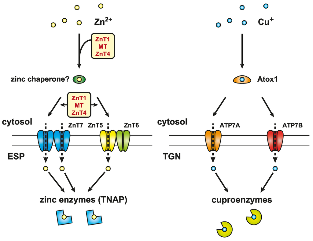
Around 3000 proteins are thought to bind zinc in vivo, which corresponds to ~10% of the human proteome. Zinc plays a pivotal role as a structural, catalytic, and signaling component that functions in numerous physiological processes. It is more widely used as
[...] Read more.
Around 3000 proteins are thought to bind zinc in vivo, which corresponds to ~10% of the human proteome. Zinc plays a pivotal role as a structural, catalytic, and signaling component that functions in numerous physiological processes. It is more widely used as a structural element in proteins than any other transition metal ion, is a catalytic component of many enzymes, and acts as a cellular signaling mediator. Thus, it is expected that zinc metabolism and homeostasis have sophisticated regulation, and elucidating the underlying molecular basis of this is essential to understanding zinc functions in cellular physiology and pathogenesis. In recent decades, an increasing amount of evidence has uncovered critical roles of a number of proteins in zinc metabolism and homeostasis through influxing, chelating, sequestrating, coordinating, releasing, and effluxing zinc. Metallothioneins (MT) and Zrt- and Irt-like proteins (ZIP) and Zn transporters (ZnT) are the proteins primarily involved in these processes, and their malfunction has been implicated in a number of inherited diseases such as acrodermatitis enteropathica. The present review updates our current understanding of the biological functions of MTs and ZIP and ZnT transporters from several new perspectives.
Full article
(This article belongs to the Special Issue Metalloproteins)
▼
Show Figures
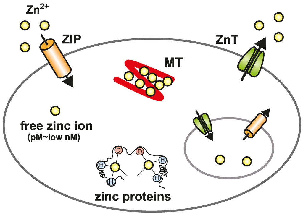
Figure 1

