
Transcriptome and Proteome Analysis Revealed Key Pathways Regulating Final Stage of Oocyte Maturation of the Turkey (Meleagris gallopavo)

 , , ,
, , ,  , ,
, , .png) , , , and
, , , and
Abstract
:1. Introduction
2. Results
2.1. Sequencing Results
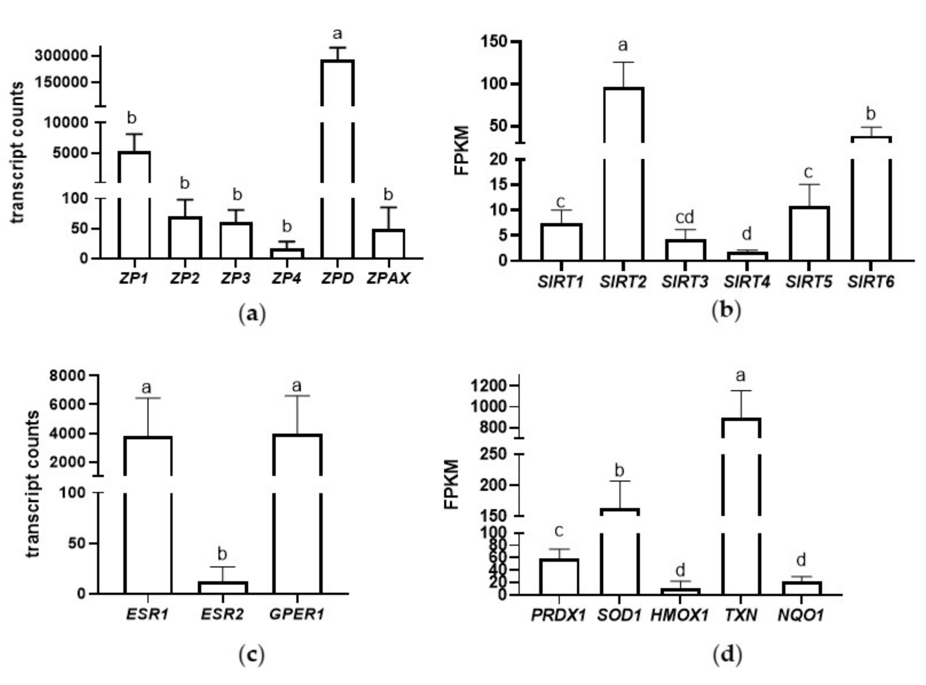
2.2. Transcriptome of Inner Perivitelline Layer: Functional Annotation and Pathway Analysis
2.3. Proteome of Inner Perivitelline Layer: Functional Annotation and Pathway Analysis
2.4. Phylogenetic Analysis of Bird ZP Glycoproteins
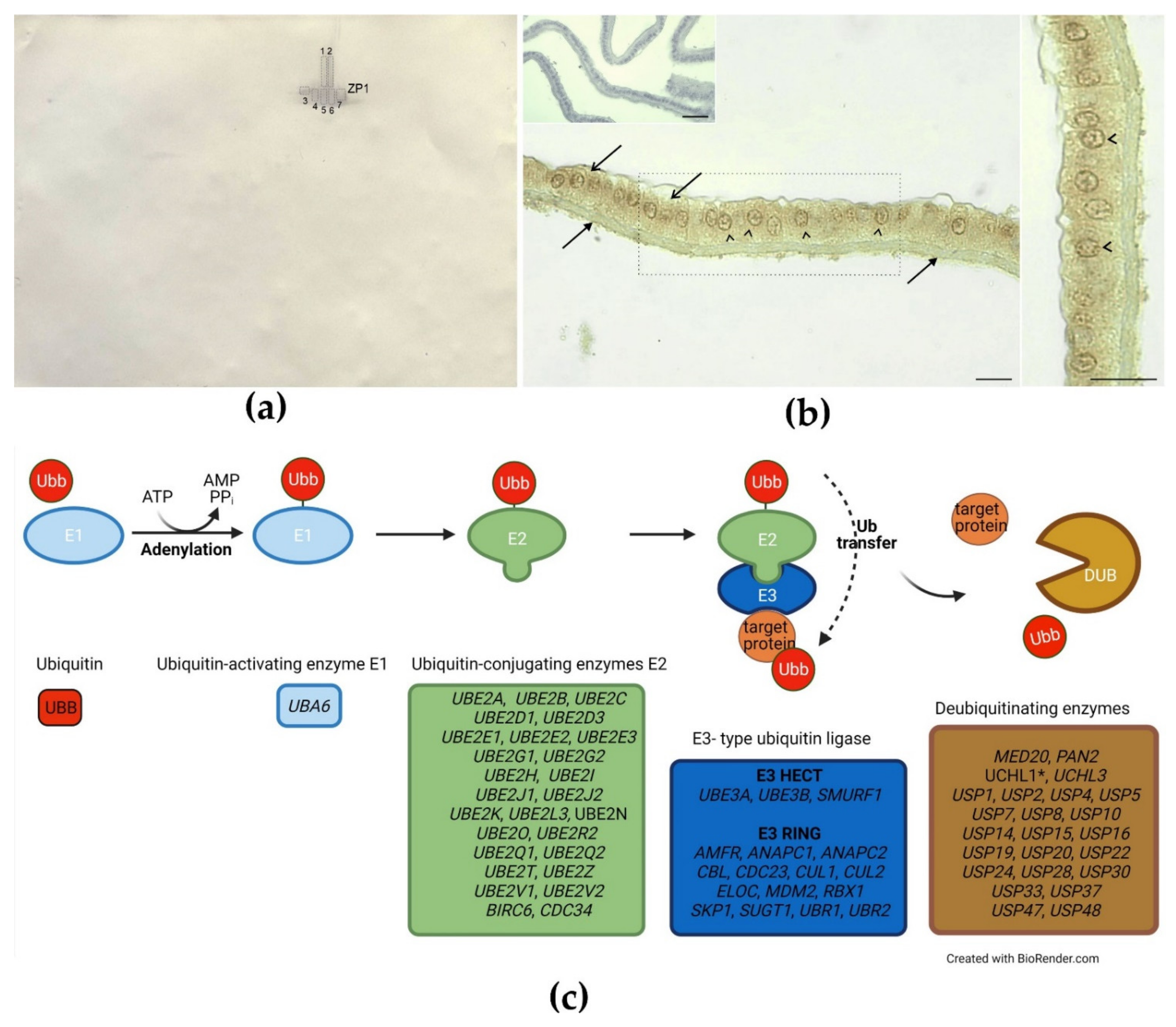
2.5. Identification of Ubiquitinated Proteins and Immunohistochemical Detection of Ubiquitin in the IPVL and the GC layer
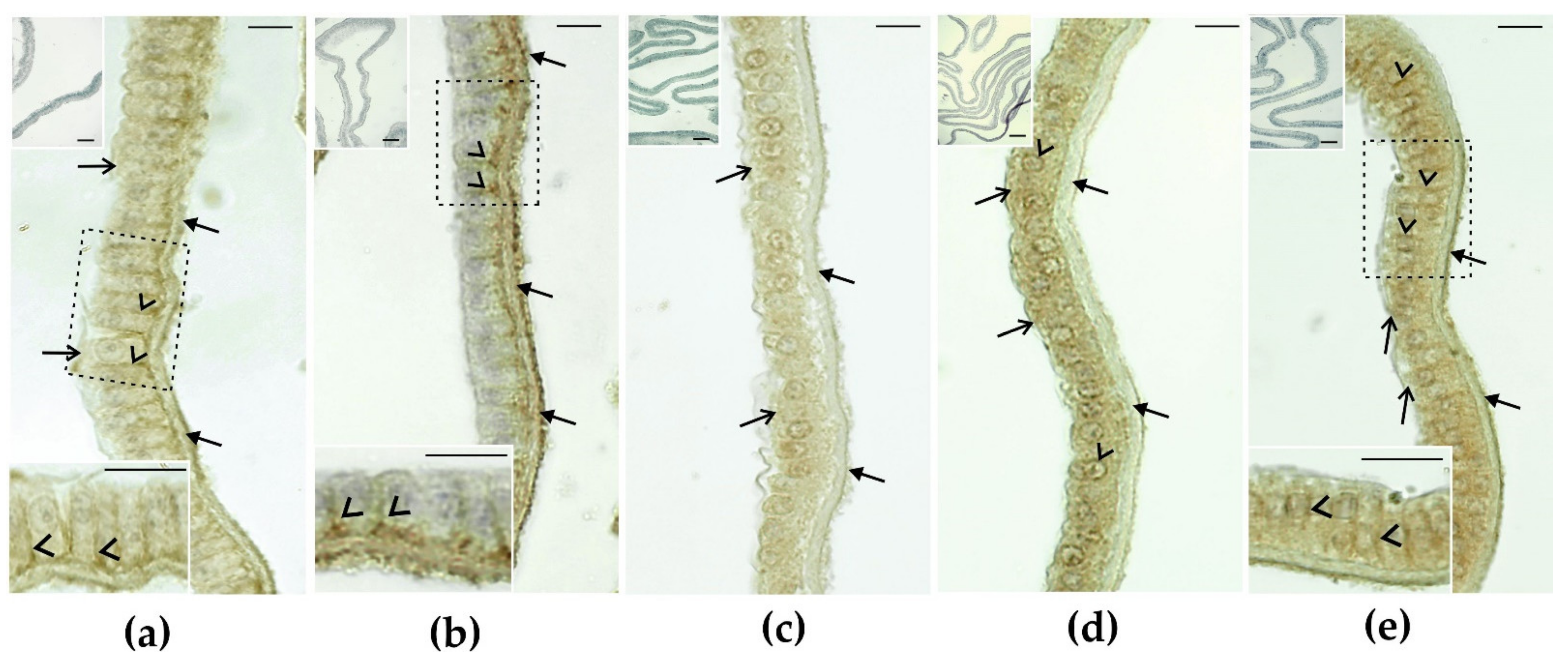
2.6. Immunohistochemical Detection of Cell Junction Proteins in GCs Surrounding the IPVL
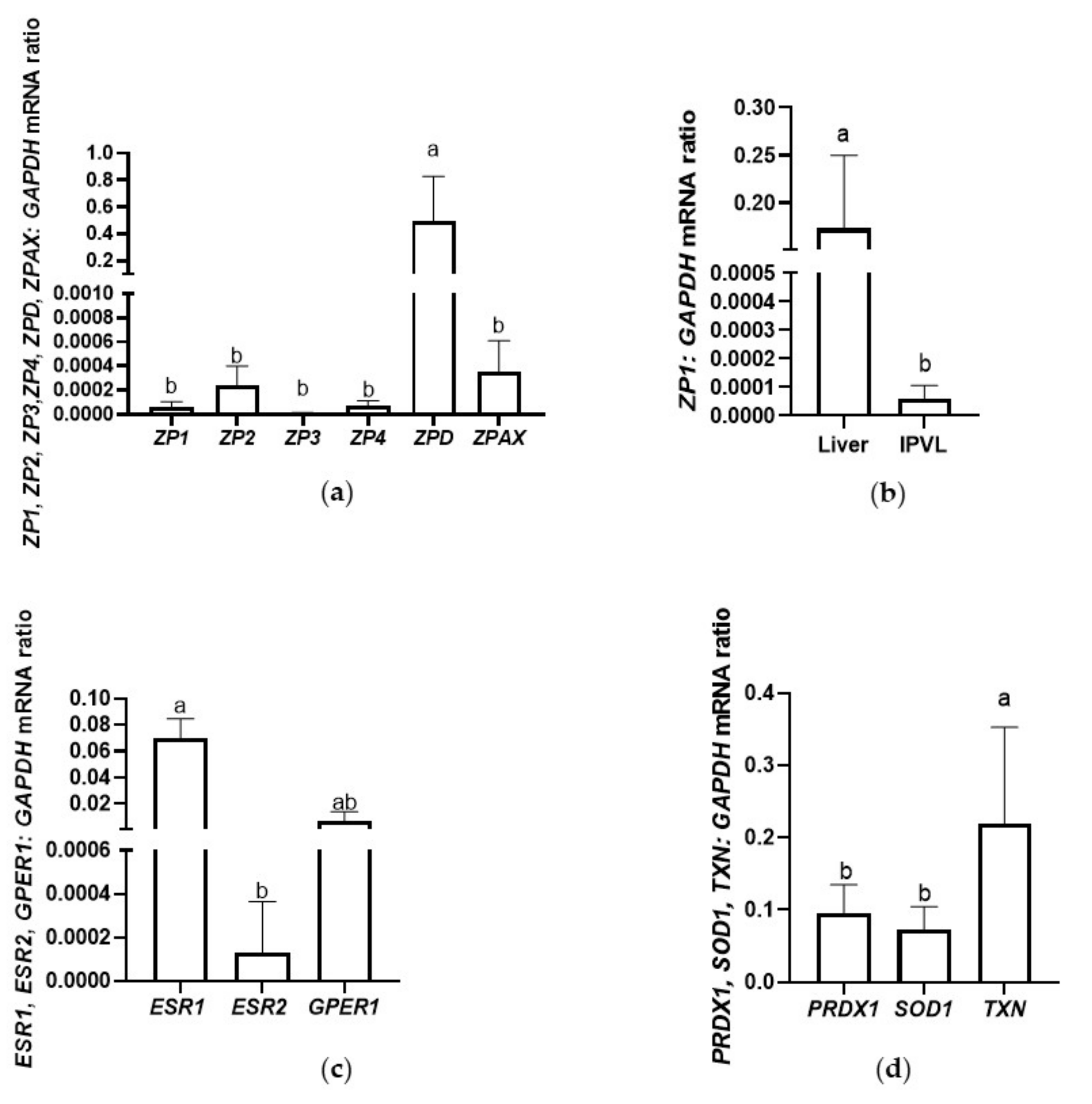
2.7. Validation of Selected Genes by Quantitative Real-Time Reverse Transcriptase-Polymerase Chain Reaction (qRT-PCR)
3. Discussion
3.1. ZP Glycoproteins
3.2. Ubiquitination
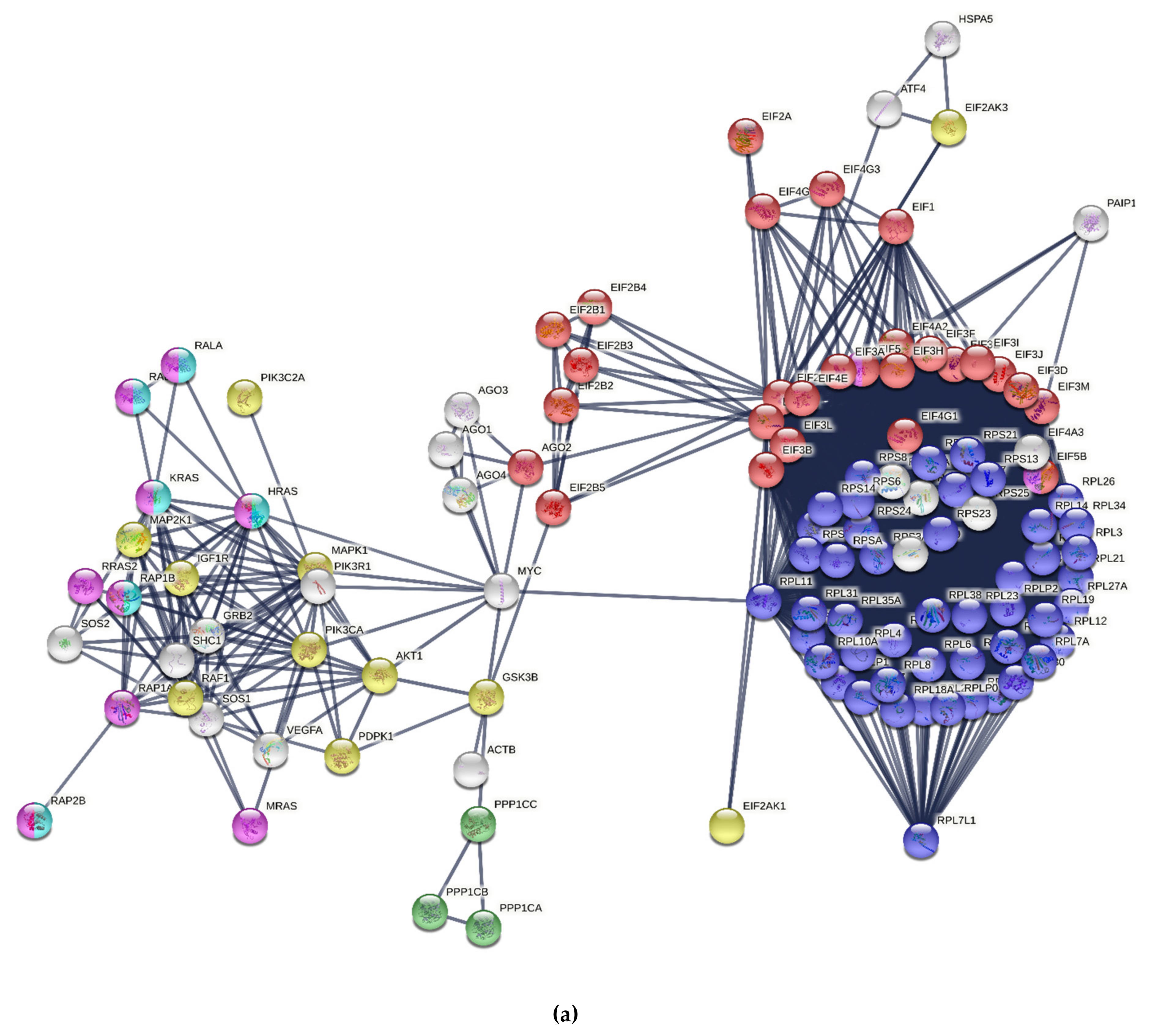
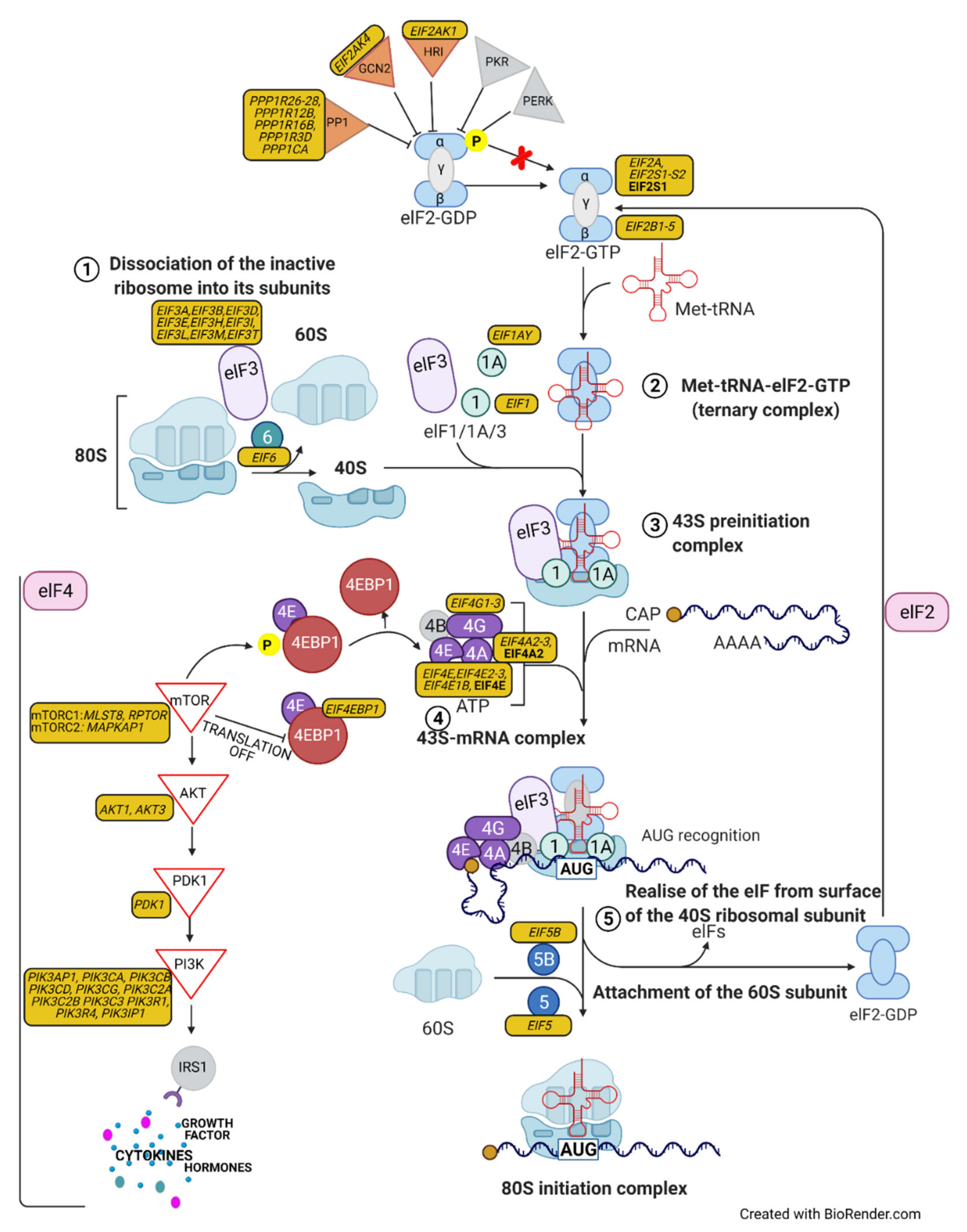
3.3. Protein Synthesis
3.4. Sirtuin Signaling Pathway
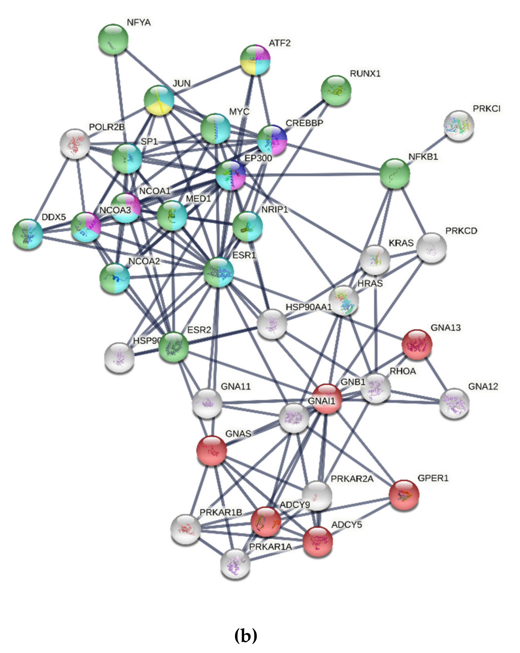
3.5. Estrogen Receptor Signaling
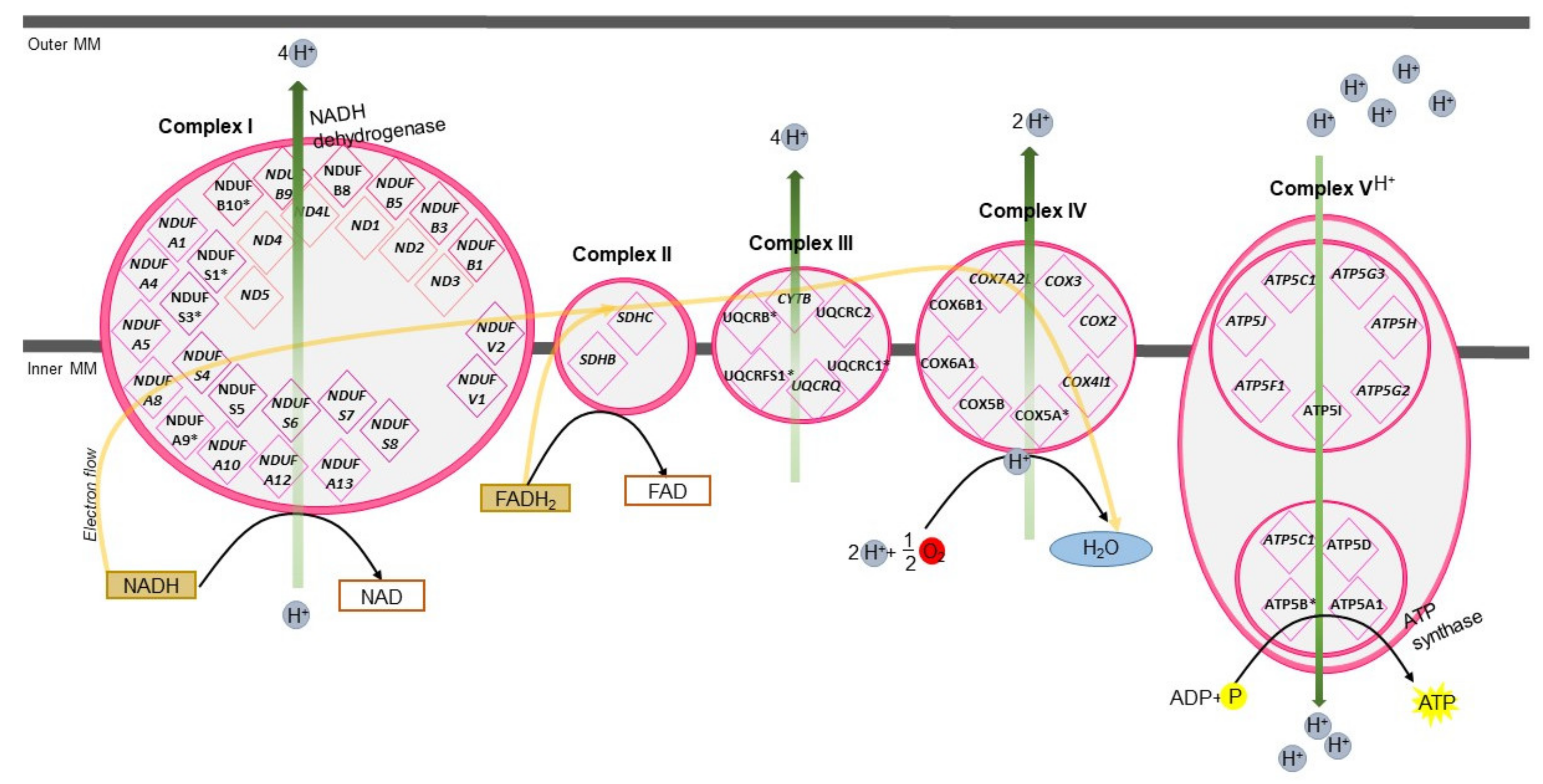
3.6. Mitochondrial Oxidative Phosphorylation
3.7. The Cellular Response against Oxidative Stress
3.8. Cell-Cell Junction—Intercellular Interactions of GCs
4. Materials and Methods
4.1. Birds, Housing, and Tissue Collection
4.2. Tissue Morphology
4.3. RNA Isolation and the Evaluation of RNA Integrity
4.4. Library Preparation and Sequencing Procedures
4.5. Quality Control and Mapping Process
4.6. 2DE and Protein Identification by MALDI TOF/TOF
4.7. Functional Analysis
4.8. Phylogenetic Analysis
4.9. Immunohistochemistry
4.10. Identification of Ubiquitinated Proteins in the IPVL
4.11. Quantitative Real-Time Reverse Transcriptase PCR
Statistical Analysis
5. Conclusions
Supplementary Materials
Author Contributions
Funding
Institutional Review Board Statement
Informed Consent Statement
Data Availability Statement
Acknowledgments
Conflicts of Interest
References
- Okumura, H. Avian Egg and Egg Coat. Chem. Biol. Pteridines Folates 2017, 1001, 75–90. [Google Scholar]
- Ichikawa, Y.; Matsuzaki, M.; Hiyama, G.; Mizushima, S.; Sasanami, T. Sperm-egg interaction during fertilization in birds. J. Poult. Sci. 2016, 53, 173–180. [Google Scholar] [CrossRef] [PubMed] [Green Version]
- Bausek, N.; Waclawek, M.; Schneider, W.J.; Wohlrab, F. The major chicken egg envelope protein ZP1 is different from ZPB and is synthesized in the liver. J. Biol. Chem. 2000, 275, 28866–28872. [Google Scholar] [CrossRef] [PubMed] [Green Version]
- Sasanami, T.; Pan, J.; Doi, Y.; Hisada, M.; Kohsaka, T.; Toriyama, M. Secretion of egg envelope protein ZPC after C-terminal proteolytic processing in quail granulosa cells. Eur. J. Biochem. 2002, 269, 2223–2231. [Google Scholar] [CrossRef]
- Sasanami, T.; Pan, J.; Mori, M. Expression of perivitelline membrane glycoprotein ZP1 in the liver of Japanese quail (Coturnix japonica) after in vivo treatment with diethylstilbestrol. J. Steroid. Biochem. Mol. Biol. 2003, 84, 109–116. [Google Scholar] [CrossRef]
- Okumura, H.; Kohno, Y.; Iwata, Y.; Mori, H.; Aoki, N.; Sato, C.; Kitajima, K.; Nadano, D.; Matsuda, T. A newly identified zona pellucida glycoprotein, ZPD, and dimeric ZP1 of chicken egg envelope are involved in sperm activation on sperm-egg interaction. Biochem. J. 2004, 384, 191–199. [Google Scholar] [CrossRef] [Green Version]
- Takeuchi, Y.; Nishimura, K.; Aoki, N.; Adachi, T.; Sato, C.; Kitajima, K.; Matsuda, T. A 42-kDa glycoprotein from chicken egg-envelope, an avian homolog of the ZPC family glycoproteins in mammalian zona pellucida. Its first identification, cDNA cloning and granulosa cell-specific expression. Eur. J. Biochem. 1999, 260, 736–742. [Google Scholar] [CrossRef] [Green Version]
- Sato, T.; Kinoshita, M.; Kansaku, N.; Tahara, K.; Tsukada, A.; Ono, H.; Yoshimura, T.; Dohra, H.; Sasanami, T. Molecular characterization of egg envelope glycoprotein ZPD in the ovary of Japanese quail (Coturnix japonica). Reproduction 2009, 137, 333–343. [Google Scholar] [CrossRef] [Green Version]
- Rodler, D.; Sasanami, T.; Sinowatz, F. Assembly of the inner perivitelline layer, a homolog of the mammalian zona pellucida: An immunohistochemical and ultrastructural study. Cells Tissues Organs. 2012, 195, 330–339. [Google Scholar] [CrossRef] [Green Version]
- Rodler, D. Histochemical detection of glycoconjugates in the inner perivitelline layer of Japanese quail (Coturnix japonica). Anat. Histol. Embryol. 2011, 40, 441–449. [Google Scholar] [CrossRef]
- Goudet, G.; Mugnier, S.; Callebaut, I.; Monget, P. Phylogenetic analysis and identification of pseudogenes reveal a progressive loss of zona pellucida genes during evolution of vertebrates. Biol. Reprod. 2008, 78, 796–806. [Google Scholar] [CrossRef] [Green Version]
- Johnson, A.L. Ovarian follicle selection and granulosa cell differentiation. Poult. Sci. 2015, 94, 781–785. [Google Scholar] [CrossRef]
- Elis, S.; Batellier, F.; Couty, I.; Balzergue, S.; Martin-Magniette, M.L.; Monget, P.; Blesbois, E.; Govoroun, M.S. Search for the genes involved in oocyte maturation and early embryo development in the hen. BMC Genomics 2008, 9, 110. [Google Scholar] [CrossRef] [Green Version]
- Machell, N.H.; Blaschuk, O.W.; Farookhi, R. Developmental expression and distribution of N- and E-cadherin in the rat ovary. Biol. Reprod. 2000, 63, 797–804. [Google Scholar] [CrossRef] [Green Version]
- Gilchrist, R.B.; Ritter, L.J.; Armstrong, D.T. Oocyte-somatic cell interactions during follicle development in mammals. Anim. Reprod. Sci. 2004, 82–83, 431–446. [Google Scholar] [CrossRef]
- Słowińska, M.; Pardyak, L.; Liszewska, E.; Judycka, S.; Bukowska, J.; Dietrich, M.A.; Paukszto, Ł.; Jastrzębski, J.; Kozłowski, K.; Kowalczyk, A.; et al. Characterization and biological role of cysteine-rich venom protein belonging to CRISPs from turkey seminal plasma. Biol. Reprod. 2021, 104, 1302–1321. [Google Scholar] [CrossRef]
- Yin, L.; Yu, L.; Zhang, L.; Ran, J.; Li, J.; Yang, C.; Jiang, X.; Du, H.; Hu, X.; Liu, Y. Transcriptome analysis reveals differentially expressed genes and pathways for oviduct development and defense in prelaying and laying hens. Am. J. Reprod. Immunol. 2019, 82, e13159. [Google Scholar] [CrossRef]
- Yin, Z.; Lian, L.; Zhu, F.; Zhang, Z.H.; Hincke, M.; Yang, N.; Hou, Z.C. The transcriptome landscapes of ovary and three oviduct segments during chicken (Gallus gallus) egg formation. Genomics 2020, 112, 243–251. [Google Scholar] [CrossRef]
- Sah, N.; Kuehu, D.L.; Khadka, V.S.; Deng, Y.; Jha, R.; Wasti, S.; Mishra, B. RNA sequencing-based analysis of the magnum tissues revealed the novel genes and biological pathways involved in the egg-white formation in the laying hen. BMC Genomics 2021, 22, 318. [Google Scholar] [CrossRef]
- Jonchère, V.; Réhault-Godbert, S.; Hennequet-Antier, C.; Cabau, C.; Sibut, V.; Cogburn, L.A.; Nys, Y.; Gautron, J. Gene expression profiling to identify eggshell proteins involved in physical defense of the chicken egg. BMC Genomics 2010, 11, 57. [Google Scholar] [CrossRef] [Green Version]
- Zhang, F.; Yin, Z.T.; Zhang, J.F.; Zhu, F.; Hincke, M.; Yang, N.; Hou, Z.C. Integrating transcriptome, proteome and QTL data to discover functionally important genes for duck eggshell and albumen formation. Genomics 2020, 112, 3687–3695. [Google Scholar] [CrossRef]
- Sun, Y.; Wu, Q.; Pan, J.; Li, T.; Liu, L.; Chen, D.; Zhang, X.; Chen, H.; Li, Y.; Lin, R. Identification of differentially expressed genes and signalling pathways in the ovary of higher and lower laying ducks. Br. Poult. Sci. 2020, 61, 609–614. [Google Scholar] [CrossRef]
- Chen, X.; Sun, X.; Chimbaka, I.M.; Qin, N.; Xu, X.; Liswaniso, S.; Xu, R.; Gonzalez, J.M. Transcriptome analysis of ovarian follicles reveals potential pivotal genes associated with increased and decreased rates of chicken egg production. Front. Genet. 2021, 12, 622751. [Google Scholar] [CrossRef]
- Poyatos Pertiñez, S.; Wilson, P.W.; Icken, W.; Cavero, D.; Bain, M.M.; Jones, A.C.; Dunn, I.C. Transcriptome analysis of the uterus of hens laying eggs differing in cuticle deposition. BMC Genomics 2020, 21, 516. [Google Scholar] [CrossRef]
- Elis, S.; Blesbois, E.; Couty, I.; Balzergue, S.; Martin-Magniette, M.L.; Batellierc, F.; Govoroun, M.S. Identification of germinal disk region derived genes potentially involved in hen fertility. Mol. Reprod. Dev. 2009, 76, 1043–1055. [Google Scholar] [CrossRef]
- Wang, Y.; Chen, Q.; Liu, Z.; Guo, X.; Du, Y.; Yuan, Z.; Guo, M.; Kang, L.; Sun, Y.; Jiang, Y. Transcriptome analysis on single small yellow follicles reveals that Wnt4 is involved in chicken follicle selection. Front. Endocrinol. 2017, 8, 317. [Google Scholar] [CrossRef] [Green Version]
- Chen, Q.; Wang, Y.; Liu, Z.; Guo, X.; Sun, Y.; Kang, L.; Jiang, Y. Transcriptomic and proteomic analyses of ovarian follicles reveal the role of VLDLR in chicken follicle selection. BMC Genomics 2020, 21, 486. [Google Scholar] [CrossRef]
- Ghanem, K.; Johnson, A.L. Proteome profiling of chicken ovarian follicles immediately before and after cyclic recruitment. Mol. Reprod. Dev. 2021. [Google Scholar] [CrossRef]
- Mann, K. Proteomic analysis of the chicken egg vitelline membrane. Proteomics 2008, 8, 2322–2332. [Google Scholar] [CrossRef]
- Zhou, Y.; Qiu, N.; Mine, Y.; Meng, Y.; Keast, R.; Zhu, C. Quantitative comparative proteomic analysis of chicken egg vitelline membrane proteins during high-temperature storage. J. Agric. Food Chem. 2020, 68, 9816–9825. [Google Scholar] [CrossRef]
- Wang, H.; Qiu, N.; Mine, Y.; Sun, H.; Meng, Y.; Bin, L.; Keast, R. Quantitative comparative integrated proteomic and phosphoproteomic analysis of chicken egg yolk proteins under diverse storage temperatures. J. Agric. Food Chem. 2020, 68, 1157–1167. [Google Scholar] [CrossRef] [PubMed]
- Guérin-Dubiard, C.; Pasco, M.; Mollé, D.; Désert, C.; Croguennec, T.; Nau, F. Proteomic analysis of hen egg white. J. Agric. Food Chem. 2006, 54, 3901–3910. [Google Scholar] [CrossRef] [PubMed]
- Kinoshita, M.; Rodler, D.; Sugiura, K.; Matsushima, K.; Kansaku, N.; Tahara, K.; Tsukada, A.; Ono, H.; Yoshimura, T.; Yoshizaki, N.; et al. Zona pellucida protein ZP2 is expressed in the oocyte of Japanese quail (Coturnix japonica). Reproduction 2010, 139, 359–371. [Google Scholar] [CrossRef] [PubMed] [Green Version]
- Serizawa, M.; Kinoshita, M.; Rodler, D.; Tsukada, A.; Ono, H.; Yoshimura, T.; Kansaku, N.; Sasanami, T. Oocytic expression of zona pellucida protein ZP4 in Japanese quail (Coturnix japonica). Anim. Sci. J. 2011, 82, 227–235. [Google Scholar] [CrossRef] [PubMed] [Green Version]
- Benson, A.P.; Christensen, V.L.; Fairchild, B.D.; Davis, A.J. The mRNA for zona pellucida proteins B1, C and D in two genetic lines of turkey hens that differ in fertility. Anim. Reprod. Sci. 2009, 111, 149–159. [Google Scholar] [CrossRef]
- Sasanami, T.; Ohtsuki, M.; Hanafy, A.M.; Mori, M. Accumulation of ZP1 and ZPC in quail perivitelline membrane during follicular development. J. Poult. Sci. 2004, 41, 289–297. [Google Scholar] [CrossRef] [Green Version]
- Pan, J.; Sasanami, T.; Kono, Y.; Matsuda, T.; Mori, M. Effects of testosterone on production of perivitelline membrane glycoprotein ZPC by granulosa cells of Japanese quail (Coturnix japonica). Biol. Reprod. 2001, 64, 310–316. [Google Scholar] [CrossRef] [Green Version]
- Gupta, S.K.; Bhandari, B.; Shrestha, A.; Biswal, B.K.; Palaniappan, C.; Malhotra, S.S.; Gupta, N. Mammalian zona pellucida glycoproteins: Structure and function during fertilization. Cell Tissue Res. 2012, 349, 665–678. [Google Scholar] [CrossRef]
- Lefièvre, L.; Conner, S.J.; Salpekar, A.; Olufowobi, O.; Ashton, P.; Pavlovic, B.; Lenton, W.; Afnan, M.; Brewis, I.A.; Monk, M.; et al. Four zona pellucida glycoproteins are expressed in the human. Hum. Reprod. 2004, 19, 1580–1586. [Google Scholar] [CrossRef] [Green Version]
- Kumar, D.; Bansal, G.; Narang, A.; Basak, T.; Abbas, T.; Dash, D. Integrating transcriptome and proteome profiling: Strategies and applications. Proteomics 2016, 16, 2533–2544. [Google Scholar] [CrossRef]
- Litscher, E.S.; Wassarman, P.M. Evolution, structure, and synthesis of vertebrate egg-coat proteins. Trends. Dev. Biol. 2014, 8, 65–76. [Google Scholar]
- Wu, T.; Cheng, Y.; Liu, Z.; Tao, W.; Zheng, S.; Wang, D. Bioinformatic analyses of zona pellucida genes in vertebrates and their expression in Nile tilapia. Fish. Physiol. Biochem. 2018, 44, 435–449. [Google Scholar] [CrossRef]
- Lindsay, L.L.; Wallace, M.A.; Hedrick, J.L. A hatching enzyme substrate in the Xenopus laevis egg envelope is a high molecular weight ZPA homolog. Dev. Growth Differ. 2001, 43, 305–313. [Google Scholar] [CrossRef] [Green Version]
- Sasanami, T.; Sugiura, K.; Tokumoto, T.; Yoshizaki, N.; Dohra, H.; Nishio, S.; Mizushima, S.; Hiyama, G.; Matsuda, T. Sperm proteasome degrades egg envelope glycoprotein ZP1 during fertilization of Japanese quail (Coturnix japonica). Reproduction 2012, 144, 423–431. [Google Scholar] [CrossRef] [Green Version]
- Labas, V.; Grasseau, I.; Cahier, K.; Gargaros, A.; Harichaux, G.; Teixeira-Gomes, A.P.; Alves, S.; Bourin, M.; Gérard, N.; Blesbois, E. Qualitative and quantitative peptidomic and proteomic approaches to phenotyping chicken semen. J. Proteomics 2015, 112, 313–335. [Google Scholar] [CrossRef]
- Słowińska, M.; Paukszto, Ł.; Jastrzębski, J.P.; Bukowska, J.; Kozłowski, K.; Jankowski, J.; Ciereszko, A. Transcriptome analysis of turkey (Meleagris gallopavo) reproductive tract revealed key pathways regulating spermatogenesis and post-testicular sperm maturation. Poult. Sci. 2020, 99, 6094–6118. [Google Scholar] [CrossRef]
- Sutovsky, P.; Manandhar, G.; McCauley, T.C.; Caamaño, J.N.; Sutovsky, M.; Thompson, W.E.; Day, B.N. Proteasomal interference prevents zona pellucida penetration and fertilization in mammals. Biol. Reprod. 2004, 71, 1625–1637. [Google Scholar] [CrossRef] [Green Version]
- Xuan, B.; Li, Z.C.; Wang, Q.Y.; Xu, M.; Chen, X.; Jin, Y. Inhibition of PSMD4 alters ZP1 ubiquitination state and sperm-oocyte-binding ability in pigs. Reprod. Domest. Anim. 2018, 53, 688–694. [Google Scholar] [CrossRef]
- Koyanagi, S.; Hamasaki, H.; Sekiguchi, S.; Hara, K.; Ishii, Y.; Kyuwa, S.; Yoshikawa, Y. Effects of ubiquitin C-terminal hydrolase L1 deficiency on mouse ova. Reproduction 2012, 143, 271–279. [Google Scholar] [CrossRef] [Green Version]
- Sekiguchi, S.; Kwon, J.; Yoshida, E.; Hamasaki, H.; Ichinose, S.; Hideshima, M.; Kuraoka, M.; Takahashi, A.; Ishii, Y.; Kyuwa, S.; et al. Localization of ubiquitin C-terminal hydrolase L1 in mouse ova and its function in the plasma membrane to block polyspermy. Am. J. Pathol 2006, 169, 1722–1729. [Google Scholar] [CrossRef] [Green Version]
- Vasudevan, S.; Seli, E.; Steitz, J.A. Metazoan oocyte and early embryo development program: A progression through translation regulatory cascades. Genes Dev. 2006, 20, 138–146. [Google Scholar] [CrossRef] [Green Version]
- Rattan, S.I.S. Protein synthesis and regulation in eukaryotes. In Principles of Medical Biology, 1st ed.; Bittar, E., Bittar, N., Eds.; Elsevier: Amsterdam, The Netherlands, 1995; Volume 4, pp. 247–263. [Google Scholar] [CrossRef]
- Schmitt, A.; Nebreda, A.R. Signalling pathways in oocyte meiotic maturation. J. Cell Sci. 2002, 115, 2457–2459. [Google Scholar] [CrossRef]
- Ernst, E.H.; Grøndahl, M.L.; Grund, S.; Hardy, K.; Heuck, A.; Sunden, L.; Franks, S.; Andersen, C.Y.; Villesen, P.; Lykke-Hartmann, K. Dormancy and activation of human oocytes from primordial and primary follicles: Molecular clues to oocyte regulation. Hum. Reprod. 2017, 32, 1684–1700. [Google Scholar] [CrossRef]
- Ruebel, M.L.; Schall, P.Z.; Midic, U.; Vincent, K.A.; Goheen, B.; VandeVoort, C.A.; Latham, K.E. Transcriptome analysis of rhesus monkey failed-to-mature oocytes: Deficiencies in transcriptional regulation and cytoplasmic maturation of the oocyte mRNA population. Mol. Hum. Reprod. 2018, 24, 478–494. [Google Scholar] [CrossRef]
- Romasko, E.J.; Amarnath, D.; Midic, U.; Latham, K.E. Association of maternal mRNA and phosphorylated EIF4EBP1 variants with the spindle in mouse oocytes: Localized translational control supporting female meiosis in mammals. Genetics 2013, 195, 349–358. [Google Scholar] [CrossRef] [Green Version]
- Tatone, C.; Di Emidio, G.; Barbonetti, A.; Carta, G.; Luciano, A.M.; Falone, S.; Amicarelli, F. Sirtuins in gamete biology and reproductive physiology: Emerging roles and therapeutic potential in female and male infertility. Hum. Reprod. Update 2018, 24, 267–289. [Google Scholar] [CrossRef]
- Xu, D.; Jiang, X.; He, H.; Liu, D.; Yang, L.; Chen, H.; Wu, L.; Geng, G.; Li, Q. SIRT2 functions in aging, autophagy, and apoptosis in post-maturation bovine oocytes. Life Sci. 2019, 232, 116639. [Google Scholar] [CrossRef]
- Ma, R.; Zhang, Y.; Zhang, L.; Han, J.; Rui, R. Sirt1 protects pig oocyte against in vitro aging. Anim. Sci. J. 2015, 86, 826–832. [Google Scholar] [CrossRef]
- Zeng, J.; Jiang, M.; Wu, X.; Diao, F.; Qiu, D.; Hou, X.; Wang, H.; Li, L.; Li, C.; Ge, J.; et al. SIRT4 is essential for metabolic control and meiotic structure during mouse oocyte maturation. Aging Cell 2018, 17, e12789. [Google Scholar] [CrossRef]
- Xiao, C.; Kim, H.S.; Lahusen, T.; Wang, R.H.; Xu, X.; Gavrilova, O.; Jou, W.; Gius, D.; Deng, C.X. SIRT6 deficiency results in severe hypoglycemia by enhancing both basal and insulin-stimulated glucose uptake in mice. J. Biol. Chem. 2010, 285, 36776–36784. [Google Scholar] [CrossRef] [Green Version]
- Li, Y.; Miao, Y.; Chen, J.; Xiong, B. SIRT6 Maintains redox homeostasis to promote porcine oocyte maturation. Front. Cell Dev. Biol. 2021, 9, 625540. [Google Scholar] [CrossRef] [PubMed]
- Yoshimura, Y.; Barua, A. Female Reproductive System and Immunology. In Avian Reproduction; Springer: Singapore, 2017; Volume 1001, pp. 33–57. [Google Scholar]
- Bondesson, M.; Hao, R.; Lin, C.Y.; Williams, C.; Gustafsson, J.Å. Estrogen receptor signaling during vertebrate development. Biochim. Biophys. Acta. 2015, 1849, 142–151. [Google Scholar] [CrossRef] [PubMed] [Green Version]
- Kamiyoshi, M.; Niwa, T.; Tanaka, K. Nuclear estrogen receptor bindings in granulosa cells and estradiol-17 beta contents in follicular membranes of the ovary of the hen during the ovulatory cycle. Gen. Comp. Endocrinol 1986, 61, 428–435. [Google Scholar] [CrossRef]
- Jing, R.; Gu, L.; Li, J.; Gong, Y. A transcriptomic comparison of theca and granulosa cells in chicken and cattle follicles reveals ESR2 as a potential regulator of CYP19A1 expression in the theca cells of chicken follicles. Comp. Biochem Physiol Part. D Genomics Proteomics 2018, 27, 40–53. [Google Scholar] [CrossRef]
- Fuentes, N.; Silveyra, P. Estrogen receptor signaling mechanisms. Adv. Protein Chem. Struct. Biol. 2019, 116, 135–170. [Google Scholar] [CrossRef]
- Yi, P.; Wang, Z.; Feng, Q.; Pintilie, G.D.; Foulds, C.E.; Lanz, R.B.; Ludtke, S.J.; Schmid, M.F.; Chiu, W.; O’Malley, B.W. Structure of a biologically active estrogen receptor-coactivator complex on DNA. Mol. Cell 2015, 57, 1047–1058. [Google Scholar] [CrossRef] [Green Version]
- Fujimoto, N.; Kitamura, S. Effects of environmental estrogenic chemicals on AP1 mediated transcription with estrogen receptors alpha and beta. J. Steroid Biochem. Mol. Biol. 2004, 88, 53–59. [Google Scholar] [CrossRef]
- O’Lone, R.; Frith, M.C.; Karlsson, E.K.; Hansen, U. Genomic targets of nuclear estrogen receptors. Mol. Endocrinol. 2004, 18, 1859–1875. [Google Scholar] [CrossRef]
- Kim, D.; Johnson, A.L. Differentiation of the granulosa layer from hen prehierarchal follicles associated with follicle-stimulating hormone receptor signaling. Mol. Reprod. Dev. 2018, 85, 729–737. [Google Scholar] [CrossRef]
- Pang, Y.; Thomas, P. Role of G protein-coupled estrogen receptor 1, GPER, in inhibition of oocyte maturation by endogenous estrogens in zebrafish. Dev. Biol. 2010, 342, 194–206. [Google Scholar] [CrossRef] [Green Version]
- Peyton, C.; Thomas, P. Involvement of epidermal growth factor receptor signaling in estrogen inhibition of oocyte maturation mediated through the G protein-coupled estrogen receptor (Gper) in zebrafish (Danio rerio). Biol Reprod. 2011, 85, 42–50. [Google Scholar] [CrossRef]
- Sagata, N. Meiotic metaphase arrest in animal oocytes: Its mechanisms and biological significance. Trends Cell Biol. 1996, 6, 22–28. [Google Scholar] [CrossRef]
- Yang, S.Y.; Lee, H.J.; Lee, H.C.; Hwang, Y.S.; Park, Y.H.; Ono, T.; Han, J.Y. The dynamic development of germ cells during chicken embryogenesis. Poult. Sci. 2018, 97, 650–657. [Google Scholar] [CrossRef]
- Van Blerkom, J.; Davis, P.W.; Lee, J. ATP content of human oocytes and developmental potential and outcome after in-vitro fertilization and embryo transfer. Hum. Reprod 1995, 10, 415–424. [Google Scholar] [CrossRef]
- Smith, L.C.; Alcivar, A.A. Cytoplasmic inheritance and its effects on development and performance. J. Reprod. Fertil. Suppl. 1993, 48, 31–43. [Google Scholar] [CrossRef]
- Yu, Y.; Dumollard, R.; Rossbach, A.; Lai, F.A.; Swann, K. Redistribution of mitochondria leads to bursts of ATP production during spontaneous mouse oocyte maturation. J. Cell Physiol 2010, 224, 672–680. [Google Scholar] [CrossRef] [Green Version]
- Van Blerkom, J.; Davis, P.; Thalhammer, V. Regulation of mitochondrial polarity in mouse and human oocytes: The influence of cumulus derived nitric oxide. Mol. Hum. Reprod 2008, 14, 431–444. [Google Scholar] [CrossRef]
- Van Blerkom, J. Mitochondrial function in the human oocyte and embryo and their role in developmental competence. Mitochondrion 2011, 11, 797–813. [Google Scholar] [CrossRef]
- Goud, A.P.; Goud, P.T.; Diamond, M.P.; Gonik, B.; Abu-Soud, H.M. Reactive oxygen species and oocyte aging: Role of superoxide, hydrogen peroxide, and hypochlorous acid. Free Radic. Biol. Med. 2008, 44, 1295–1304. [Google Scholar] [CrossRef] [Green Version]
- Lord, T.; Aitken, R.J. Oxidative stress and ageing of the post-ovulatory oocyte. Reproduction 2013, 146, R217–R227. [Google Scholar] [CrossRef] [Green Version]
- Nakamura, Y.; Yamagata, Y.; Sugino, N.; Takayama, H.; Kato, H. Nitric oxide inhibits oocyte meiotic maturation. Biol. Reprod. 2002, 67, 1588–1592. [Google Scholar] [CrossRef] [PubMed] [Green Version]
- Halbleib, J.M.; Nelson, W.J. Cadherins in development: Cell adhesion, sorting, and tissue morphogenesis. Genes Dev. 2006, 20, 3199–3214. [Google Scholar] [CrossRef] [PubMed] [Green Version]
- Takai, Y.; Ikeda, W.; Ogita, H.; Rikitake, Y. The immunoglobulin-like cell adhesion molecule nectin and its associated protein afadin. Annu Rev. Cell Dev. Biol. 2008, 24, 309–342. [Google Scholar] [CrossRef] [PubMed]
- Wang, C.; Roy, S.K. Expression of E-cadherin and N-cadherin in perinatal hamster ovary: Possible involvement in primordial follicle formation and regulation by follicle-stimulating hormone. Endocrinology 2010, 151, 2319–2330. [Google Scholar] [CrossRef] [Green Version]
- Smith, S.R.; Fulton, N.; Collins, C.S.; Welsh, M.; Bayne, R.A.; Coutts, S.M.; Childs, A.J.; Anderson, R.A. N- and E-cadherin expression in human ovarian and urogenital duct development. Fertil. Steril. 2010, 93, 2348–2353. [Google Scholar] [CrossRef]
- Mora, J.M.; Fenwick, M.A.; Castle, L.; Baithun, M.; Ryder, T.A.; Mobberley, M.; Carzaniga, R.; Franks, S.; Hardy, K. Characterization and significance of adhesion and junction-related proteins in mouse ovarian follicles. Biol. Reprod. 2012, 86, 1–14. [Google Scholar] [CrossRef]
- Bothun, A.M.; Woods, D.C. Dynamics of WNT signaling components in the human ovary from development to adulthood. Histochem. Cell Biol. 2019, 151, 115–123. [Google Scholar] [CrossRef]
- Schuster, M.K.; Schmierer, B.; Shkumatava, A.; Kuchler, K. Activin A and follicle-stimulating hormone control tight junctions in avian granulosa cells by regulating occludin expression. Biol. Reprod. 2004, 70, 1493–1499. [Google Scholar] [CrossRef] [Green Version]
- Gonzalez-Mariscal, L.; Betanzos, A.; Nava, P.; Jaramillo, B.E. Tight junction proteins. Prog. Biophys. Mol. Biol. 2003, 81, 1–44. [Google Scholar] [CrossRef]
- Fanning, A.S.; Anderson, J.M. Zonula occludens-1 and -2 are cytosolic scaffolds that regulate the assembly of cellular junctions. Ann. N. Y. Acad. Sci. 2009, 1165, 113–120. [Google Scholar] [CrossRef] [Green Version]
- Russell, D.L.; Gilchrist, R.B.; Brown, H.M.; Thompson, J.G. Bidirectional communication between cumulus cells and the oocyte: Old hands and new players? Theriogenology 2016, 86, 62–68. [Google Scholar] [CrossRef] [Green Version]
- Yoshimura, Y.; Okamoto, T.; Tamura, T. Ultrastructural changes of oocyte and follicular wall during oocyte maturation in the Japanese quail (Coturnix coturnix japonica). J. Reprod. Fertil. 1993, 97, 189–196. [Google Scholar] [CrossRef]
- Goodenough, D.A.; Goliger, J.A.; Paul, D.L. Connexins, connexons, and intercellular communication. Annu. Rev. Biochem. 1996, 65, 475–502. [Google Scholar] [CrossRef]
- Kumar, N.M.; Gilula, N.B. The gap junction communication channel. Cell 1996, 84, 381–388. [Google Scholar] [CrossRef] [Green Version]
- Gittens, J.E.I.; Mhawi, A.A.; Lidington, D.; Ouellette, Y.; Kidder, G.M. Functional analysis of gap junctions in ovarian granulosa cells: Distinct role for connexin43 in early stages of folliculogenesis. Am. J. Physiol. Physiol. 2003, 284, C880–C887. [Google Scholar] [CrossRef] [Green Version]
- Lee, S.; Hiradate, Y.; Hoshino, Y.; Ko, Y.G.; Tanemura, K.; Sato, E. Localization and quantitative analysis of Cx43 in porcine oocytes during in vitro maturation. Zygote 2016, 24, 364–370. [Google Scholar] [CrossRef]
- Robker, R.L.; Hennebold, J.D.; Russell, D.L. Coordination of ovulation and oocyte maturation: A good egg at the right time. Endocrinology 2018, 159, 3209–3218. [Google Scholar] [CrossRef]
- Bilinska, B.; Hejmej, A.; Kotula-Balak, M. Preparation of testicular samples for histology and immunohistochemistry. Methods. Mol. Biol. 2018, 1748, 17–36. [Google Scholar] [CrossRef]
- Ewels, P.; Magnusson, M.; Lundin, S.; Käller, M. MultiQC: Summarize analysis results for multiple tools and samples in a single report. Bioinformatics 2016, 32, 3047–3048. [Google Scholar] [CrossRef] [Green Version]
- Dobin, A.; Davis, C.A.; Schlesinger, F.; Drenkow, J.; Zaleski, C.; Jha, S.; Batut, P.; Chaisson, M.; Gingeras, T.R. STAR: Ultrafast universal RNA-seq aligner. Bioinformatics 2013, 29, 15–21. [Google Scholar] [CrossRef]
- Pertea, M.; Pertea, G.M.; Antonescu, C.M.; Chang, T.C.; Mendell, J.T.; Salzberg, S.L. StringTie enables improved reconstruction of a transcriptome from RNA-seq reads. Nat. Biotechnol. 2015, 33, 290–295. [Google Scholar] [CrossRef] [PubMed] [Green Version]
- Pertea, M.; Kim, D.; Pertea, G.M.; Leek, J.T.; Salzberg, S.L. Transcript-level expression analysis of RNA-seq experiments with HISAT, StringTie and Ballgown. Nat. Protoc. 2016, 11, 1650–1667. [Google Scholar] [CrossRef] [PubMed]
- Bradford, M.M. A rapid and sensitive method for the quantitation of microgram quantities of protein utilizing the principle of protein-dye binding. Anal. Biochem. 1976, 72, 248–254. [Google Scholar] [CrossRef]
- Słowińska, M.; Nynca, J.; Arnold, G.J.; Fröhlich, T.; Jankowski, J.; Kozłowski, K.; Mostek, A.; Ciereszko, A. Proteomic identification of turkey (Meleagris gallopavo) seminal plasma proteins. Poult. Sci. 2017, 96, 3422–3435. [Google Scholar] [CrossRef]
- Słowińska, M.; Nynca, J.; Bąk, B.; Wilde, J.; Siuda, M.; Ciereszko, A. 2D-DIGE proteomic analysis reveals changes in haemolymph proteome of 1-dayold honey bee (Apis mellifera) workers in response to infection with Varroa destructor mites. Apidologie 2019, 50, 632–656. [Google Scholar] [CrossRef] [Green Version]
- Kramer, A.; Green, J.; Pollard, J., Jr.; Tugendreich, S. Causal analysis approaches in Ingenuity Pathway Analysis. Bioinformatics 2014, 30, 523–530. [Google Scholar] [CrossRef]
- Szklarczyk, D.; Gable, A.L.; Lyon, D.; Junge, A.; Wyder, S.; Huerta-Cepas, J.; Simonovic, M.; Doncheva, N.T.; Morris, J.H.; Bork, P.; et al. STRING v11: Protein-protein association networks with increased coverage, supporting functional discovery in genome-wide experimental datasets. Nucleic Acids Res. 2019, 47(D7), D607–D613. [Google Scholar] [CrossRef] [Green Version]
- Katoh, K.; Misawa, K.; Kuma, K.; Miyata, T. MAFFT: A novel method for rapid multiple sequence alignment based on fast Fourier transform. Nucleic Acids Res. 2002, 30, 3059–3066. [Google Scholar] [CrossRef] [Green Version]
- Capella-Gutiérrez, S.; Silla-Martínez, J.M.; Gabaldón, T. trimAl: A tool for automated alignment trimming in large-scale phylogenetic analyses. Bioinformatics 2009, 25, 1972–1973. [Google Scholar] [CrossRef]
- Huelsenbeck, J.P.; Ronquist, F. MRBAYES: Bayesian inference of phylogenetic trees. Bioinformatics 2001, 17, 754–755. [Google Scholar] [CrossRef] [Green Version]
- Kumar, S.; Stecher, G.; Li, M.; Knyaz, C.; Tamura, K. MEGA X: Molecular evolutionary genetics analysis across computing platforms. Mol. Biol. Evol. 2018, 35, 1547–1549. [Google Scholar] [CrossRef]
- Pardyak, L.; Kaminska, A.; Brzoskwinia, M.; Hejmej, A.; Kotula-Balak, M.; Jankowski, J.; Ciereszko, A.; Bilinska, B. Differential expression of cell-cell junction proteins in the testis, epididymis, and ductus deferens of domestic turkeys (Meleagris gallopavo) with white and yellow semen. Poult. Sci. 2020, 99, 555–566. [Google Scholar] [CrossRef]
- Słowińska, M.; Bukowska, J.; Hejmej, A.; Bilińska, B.; Kozłowski, K.; Jankowski, J.; Ciereszko, A. Hepatocyte growth factor activator is a potential target proteinase for Kazal-type inhibitor in turkey (Meleagris gallopavo) seminal plasma. Theriogenology 2015, 84, 425–436.e3. [Google Scholar] [CrossRef]
- Słowińska, M.; Liszewska, E.; Dietrich, G.J.; Ciereszko, A. Characterization of proacrosin/acrosin system after liquid storage and cryopreservation of turkey semen (Meleagris gallopavo). Theriogenology 2012, 78, 1065–1077. [Google Scholar] [CrossRef]
- Luque-Garcia, J.L.; Zhou, G.; Spellman, D.S.; Sun, T.T.; Neubert, T.A. Analysis of electroblotted proteins by mass spectrometry: Protein identification after Western blotting. Mol. Cell Proteomics 2008, 7, 308–314. [Google Scholar] [CrossRef] [Green Version]
- Zhao, S.; Fernald, R.D. Comprehensive algorithm for quantitative real-time polymerase chain reaction. J. Comput. Biol. 2005, 12, 1047–1064. [Google Scholar] [CrossRef]


| IPVL Samples | O1 | O2 | O3 | O4 | O5 | O6 |
|---|---|---|---|---|---|---|
| row reads | 1.03 × 10⁸ | 1.18 × 10⁸ | 1.05 × 10⁸ | 1.16 × 10⁸ | 1.2 × 10⁸ | 1.01 × 10⁸ |
| trimmed reads | 1.02 × 10⁸ | 1.16 × 10⁸ | 1.03 × 10⁸ | 1.14 × 10⁸ | 1.19 × 10⁸ | 0.99 × 10⁸ |
| mapped | 82307430 | 93758986 | 87048822 | 96081072 | 99511690 | 83720988 |
| uniquely mapped | 80362334 | 91783236 | 84612194 | 93744112 | 97406022 | 81650560 |
| mapped to coding regions (%) | 41.90 | 40.75 | 43.94 | 44.59 | 43.72 | 43.38 |
| mapped to UTR (%) | 7.18 | 7.20 | 5.07 | 5.22 | 5.38 | 5.47 |
| mapped to introns (%) | 5.30 | 5.14 | 7.13 | 7.61 | 7.70 | 7.51 |
| mapped to intergenic regions (%) | 45.62 | 46.90 | 43.85 | 42.58 | 43.20 | 43.64 |
| multi-mapped | 1,940,566 | 1,969,616 | 2,431,930 | 2,333,492 | 2,102,302 | 2,065,832 |
| too many loci | 4530 | 6134 | 4698 | 3468 | 3366 | 4596 |
| p Value | No Molecules | Functional Analysis | p Value | No Molecules |
|---|---|---|---|---|
| Trancriptome | Ingeniuty Canonical Pathways | Proteome | ||
| 1.05 × 1028 | 142 | Protein Ubiquitination Pathway | 9.18 × 10⁸ | 15 |
| 1.35 × 1026 | 121 | EIF2 Signaling | 1.43 × 10⁵ | 11 |
| 7.81 × 10²⁰ | 86 | Regulation of eIF4 and p70S6K Signaling | 1.66 × 10⁴ | 8 |
| 6.05 × 10¹⁸ | 128 | Sirtuin Signaling Pathway | 6.00 × 10¹¹ | 11 |
| 8.65 × 10¹⁷ | 137 | Estrogen Receptor Signaling | 1.57 × 10³ | 10 |
| 9.21 × 10¹⁷ | 86 | Mitochondrial Dysfunction | 1.89 × 10¹⁸ | 22 |
| 4.68 × 10¹⁵ | 89 | NRF2-mediated Oxidative Stress Response | 6.02 × 10¹² | 17 |
| 1.32 × 10¹⁴ | 95 | mTOR Signaling | 4.83 × 10³ | 7 |
| 9.55 × 10¹¹ | 54 | Oxidative Phosphorylation | 1.20 × 10¹⁸ | 19 |
| 8.33 × 10⁶ | 31 | Remodeling of Epithelial Adherens Junctions | 9.18 × 10¹¹ | 10 |
| 1.80 × 10⁵ | 22 | BAG2 Signaling Pathway | 3.05 × 10¹⁰ | 9 |
| Gluconeogenesis I | 4.50 × 10⁹ | 7 | ||
| 4.38 × 10⁷ | 79 | Actin Cytoskeleton Signaling | 3.62 × 10⁸ | 14 |
| Molecular and Cellular Function | ||||
| 7.31 × 10¹⁰–3.36 × 1070 | 760 | Protein Synthesis | 2.91 × 10⁵–1.47 × 10¹⁶ | 68 |
| 1.86 × 10¹⁴–2.18 × 1068 | 376 | RNA Post-Transcriptional Modification | ||
| 9.96 × 10¹⁰–1.18 × 1053 | 1728 | Cell Death and Survival | 5.64 × 10⁵–4.34 × 1024 | 127 |
| 1.32 × 10¹⁸–1.68 × 1040 | 380 | Protein Degradation | ||
| 8.39 × 10¹⁰–4.77 × 1034 | 1048 | Molecular Transport | ||
| Post-Transcriptional Modification | 5.05 × 10⁵–2.93 × 10²² | 43 | ||
| Protein Folding | 5.05 × 10⁵–4.02 × 10²¹ | 22 | ||
| Cellular Compromise | 1.71 × 10⁶–2.20 × 10¹⁵ | 46 | ||
| Physiological System Development and Function | ||||
| 1.15 × 10¹²–6.47 × 1059 | 1242 | Organismal Survival | 9.44 × 10¹⁰–9.44 × 10¹⁰ | 76 |
| 1.81 × 10¹⁰–3.72 × 1024 | 1012 | Embryonic Development | 4.43 × 10⁷–8.27 × 10⁹ | 23 |
| 3.60 × 10¹⁰–3.72 × 1024 | 1413 | Organismal Development | 3.55 × 10⁵–8.27 × 10⁹ | 55 |
| 1.86 × 10¹¹–3.72 × 1024 | 371 | Tissue Morphology | ||
| 5.78 × 10¹⁰–1.06 × 10¹⁹ | 542 | Connective Tissue Development and Function | ||
| Tissue Development | 1.13 × 10⁵–6.47 × 1059 | 52 | ||
| Organ Development | 2.73 × 10⁶–8.27 × 10⁹ | 30 | ||
| Score | Top Networks | Score | ||
| 28 | 35 | Molecular Transport, RNA Post-Transcriptional Modification, RNA Trafficking | ||
| 28 | 35 | Protein Synthesis. RNA Damage and Repair | ||
| 28 | 35 | Post-Translational Modification, Protein Degradation, Protein Synthesis | ||
| Cellular Assembly and Organization, RNA Post-Transcriptional Modification | 57 | 29 | ||
| Cell Death and Survival, Drug Metabolism. Small Molecule Biochemistry | 46 | 25 | ||
| Gene Expression, Post-Translational Modification, Protein Folding | 39 | 22 | ||
| Cell Death and Survival, Gene Expression Protein Synthesis | 32 | 19 | ||
Publisher’s Note: MDPI stays neutral with regard to jurisdictional claims in published maps and institutional affiliations. |
© 2021 by the authors. Licensee MDPI, Basel, Switzerland. This article is an open access article distributed under the terms and conditions of the Creative Commons Attribution (CC BY) license (https://creativecommons.org/licenses/by/4.0/).
Share and Cite
Słowińska, M.; Paukszto, Ł.; Pardyak, L.; Jastrzębski, J.P.; Liszewska, E.; Wiśniewska, J.; Kozłowski, K.; Jankowski, J.; Bilińska, B.; Ciereszko, A. Transcriptome and Proteome Analysis Revealed Key Pathways Regulating Final Stage of Oocyte Maturation of the Turkey (Meleagris gallopavo). Int. J. Mol. Sci. 2021, 22, 10589. https://doi.org/10.3390/ijms221910589
Słowińska M, Paukszto Ł, Pardyak L, Jastrzębski JP, Liszewska E, Wiśniewska J, Kozłowski K, Jankowski J, Bilińska B, Ciereszko A. Transcriptome and Proteome Analysis Revealed Key Pathways Regulating Final Stage of Oocyte Maturation of the Turkey (Meleagris gallopavo). International Journal of Molecular Sciences. 2021; 22(19):10589. https://doi.org/10.3390/ijms221910589
Chicago/Turabian StyleSłowińska, Mariola, Łukasz Paukszto, Laura Pardyak, Jan P. Jastrzębski, Ewa Liszewska, Joanna Wiśniewska, Krzysztof Kozłowski, Jan Jankowski, Barbara Bilińska, and Andrzej Ciereszko. 2021. "Transcriptome and Proteome Analysis Revealed Key Pathways Regulating Final Stage of Oocyte Maturation of the Turkey (Meleagris gallopavo)" International Journal of Molecular Sciences 22, no. 19: 10589. https://doi.org/10.3390/ijms221910589