
Individual and Binary Mixture Toxicity of Five Nanoparticles in Marine Microalga Heterosigma akashiwo

 ,
,  ,
,  and
and 
Abstract
:1. Introduction
2. Results
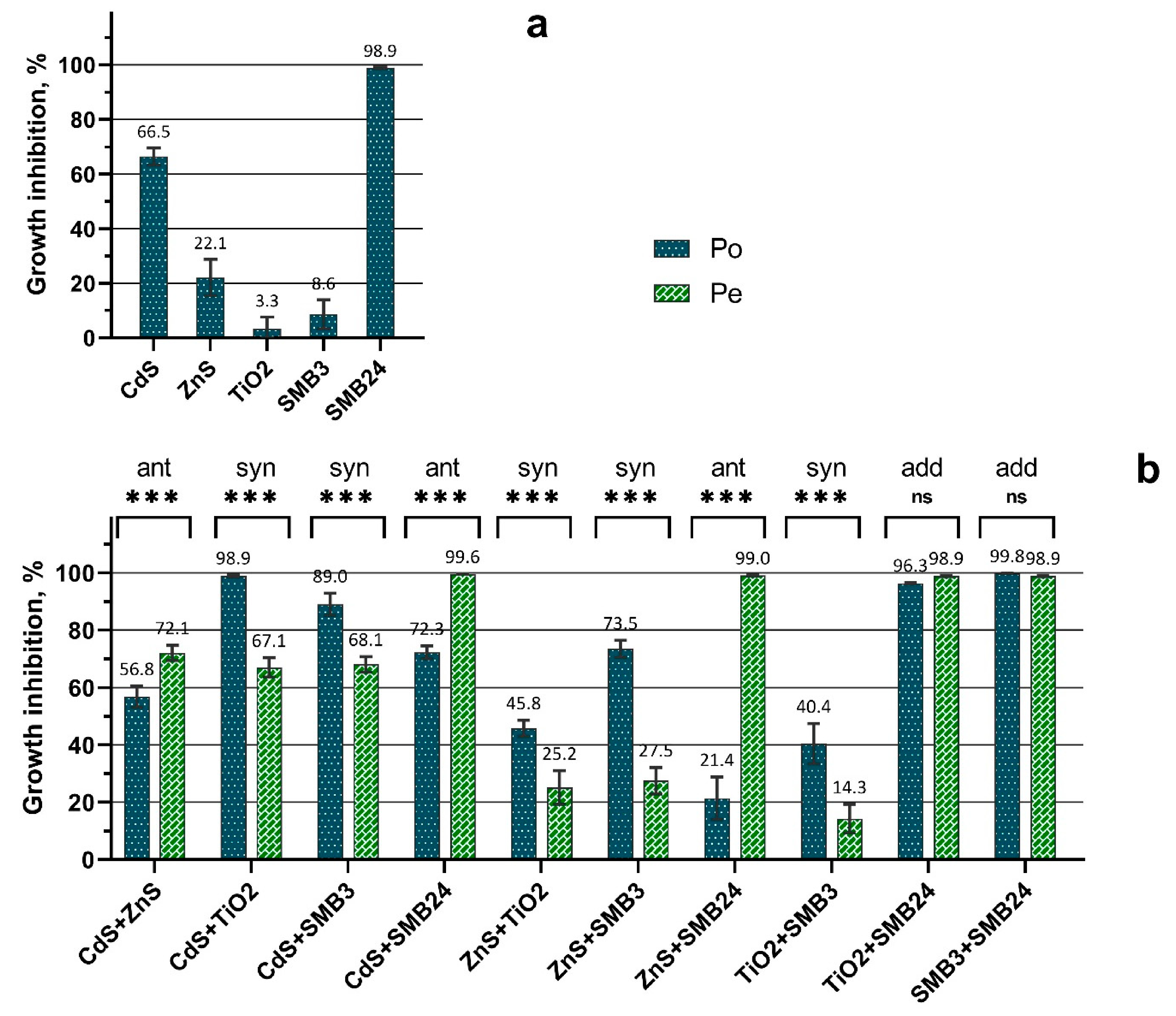
2.1. First Stage of the Bioassay: Single Nanoparticle Assessment
2.2. Second Stage of the Bioassay: Binary Mixture Assessment
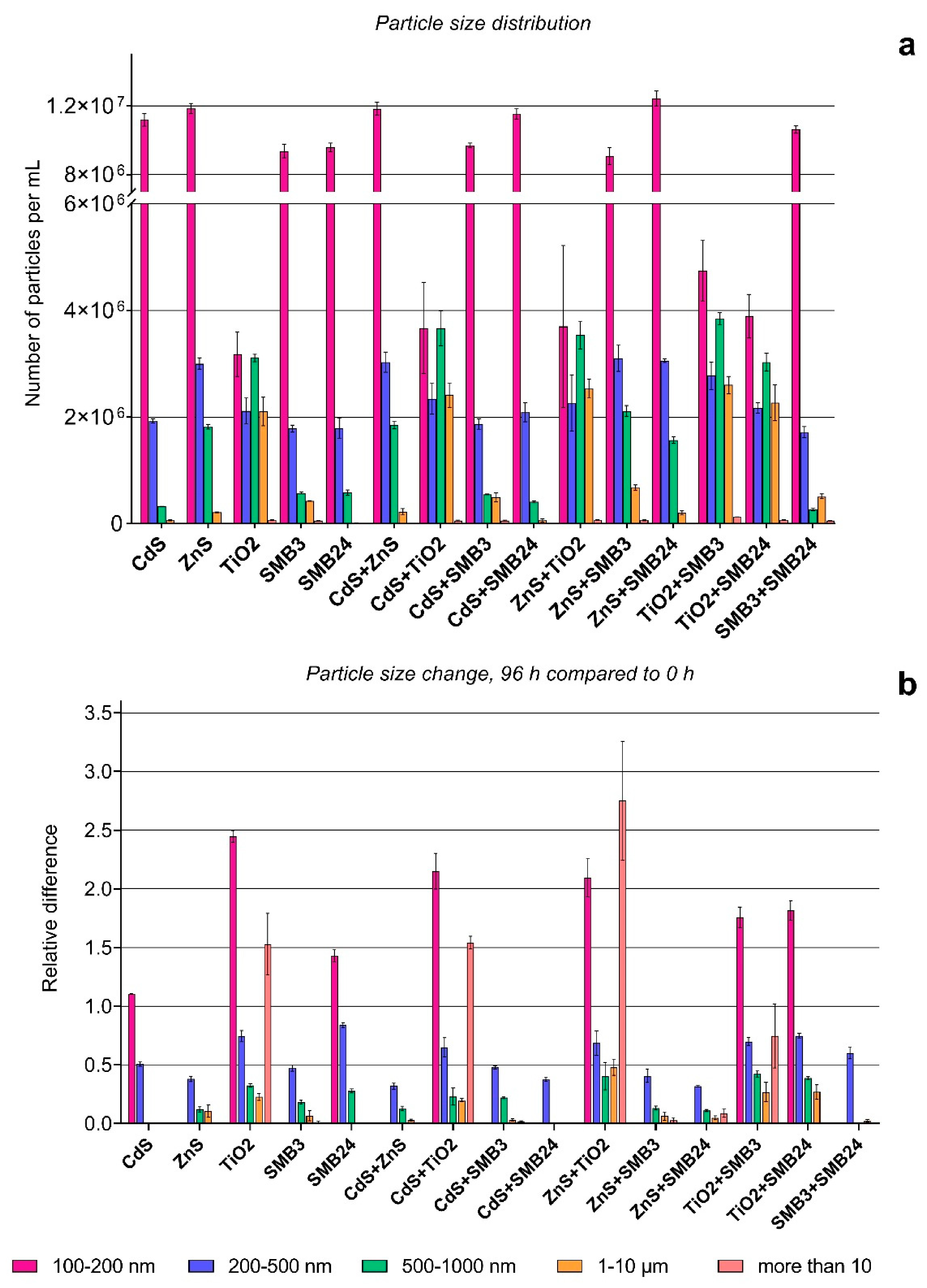
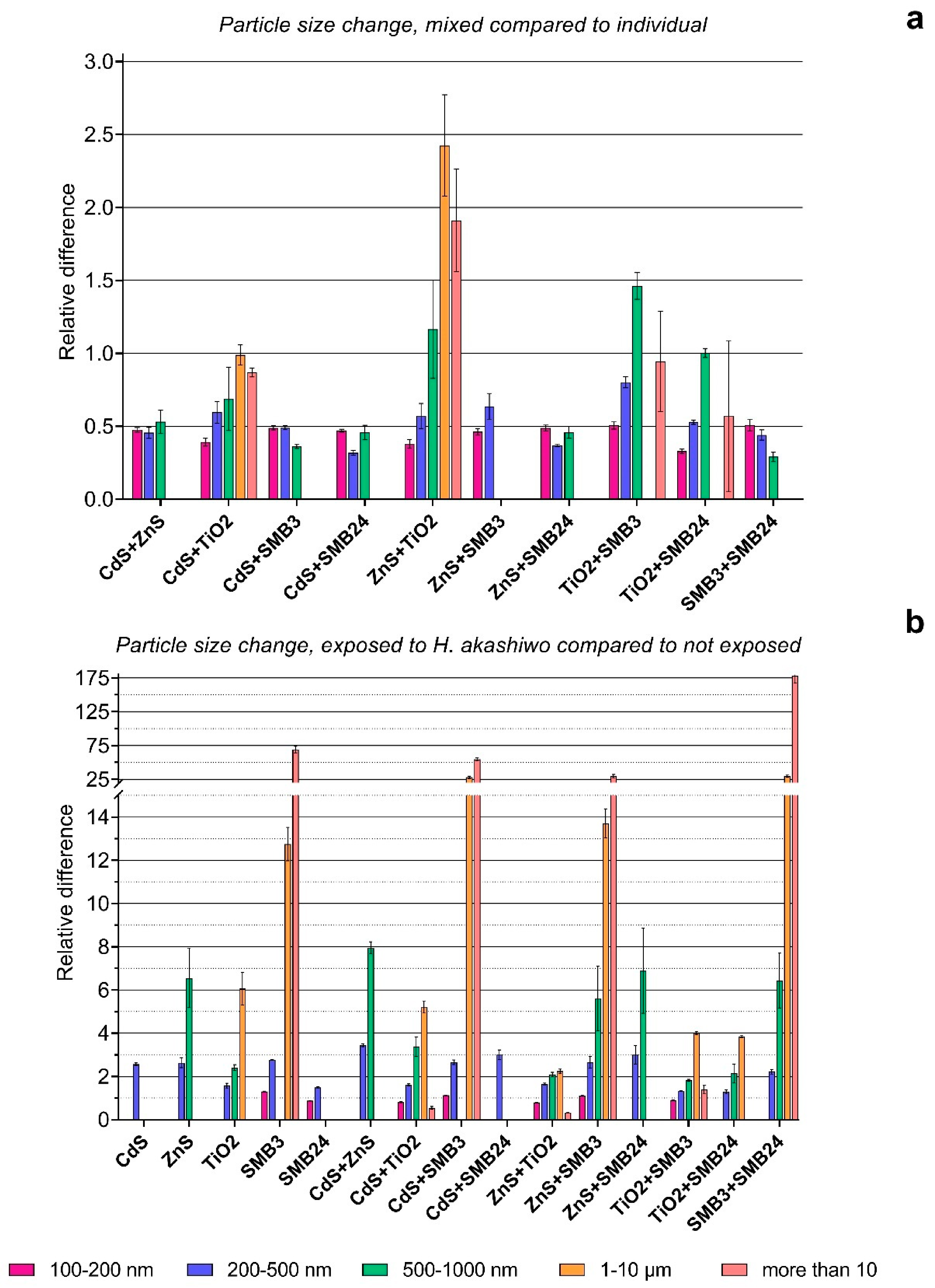
2.3. Particle Transformation Assessment
2.4. Summary
3. Discussion
4. Materials and Methods
4.1. Nanoparticles
4.2. Microalga Culture
4.3. Exposure Protocol
4.4. Cell Count, Staining, and Measurement Protocols
4.5. The Estimation of Particle Size Distribution and Particle Size Changes after Exposure
4.6. Statistical Analysis
5. Conclusions
Author Contributions
Funding
Institutional Review Board Statement
Informed Consent Statement
Conflicts of Interest
References
- Giese, B.; Klaessig, F.; Park, B.; Kaegi, R.; Steinfeldt, M.; Wigger, H.; von Gleich, A.; Gottschalk, F. Risks, Release and Concentrations of Engineered Nanomaterial in the Environment. Sci. Rep. 2018, 8, 1565. [Google Scholar] [CrossRef] [PubMed]
- Book, F.; Backhaus, T. Aquatic ecotoxicity of manufactured silica nanoparticles: A systematic review and meta-analysis. Sci. Total Environ. 2021, 806, 150893. [Google Scholar] [CrossRef] [PubMed]
- Keller, A.A.; McFerran, S.; Lazareva, A.; Suh, S. Global life cycle releases of engineered nanomaterials. J. Nanopart. Res. 2013, 15, 1692. [Google Scholar] [CrossRef]
- Yu, S.; Shen, M.; Li, S.; Fu, Y.; Zhang, D.; Liu, H.; Liu, J. Aggregation kinetics of different surface-modified polystyrene nanoparticles in monovalent and divalent electrolytes. Environ. Pollut. 2019, 255, 113302. [Google Scholar] [CrossRef]
- Fubini, B.; Ghiazza, M.; Fenoglio, I. Physico-chemical features of engineered nanoparticles relevant to their toxicity. Nanotoxicology 2010, 4, 347–363. [Google Scholar] [CrossRef]
- Tang, Y.L.; Xin, H.J.; Yang, F.; Long, X. A historical review and bibliometric analysis of nanoparticles toxicity on algae. J. Nanopart. Res. 2018, 20, 92. [Google Scholar] [CrossRef]
- Pikula, K.; Zakharenko, A.; Chaika, V.; Em, I.; Nikitina, A.; Avtomonov, E.; Tregubenko, A.; Agoshkov, A.; Mishakov, I.; Kuznetsov, V. Toxicity of Carbon, Silicon, and Metal-Based Nanoparticles to Sea Urchin Strongylocentrotus Intermedius. Nanomaterials 2020, 10, 1825. [Google Scholar] [CrossRef]
- Gambardella, C.; Ferrando, S.; Gatti, A.M.; Cataldi, E.; Ramoino, P.; Aluigi, M.G.; Faimali, M.; Diaspro, A.; Falugi, C. Review: Morphofunctional and Biochemical Markers of Stress in Sea Urchin Life Stages Exposed to Engineered Nanoparticles. Environ. Toxicol. 2016, 31, 1552–1562. [Google Scholar] [CrossRef]
- Gambardella, C.; Morgana, S.; Bari, G.D.; Ramoino, P.; Bramini, M.; Diaspro, A.; Falugi, C.; Faimali, M. Multidisciplinary screening of toxicity induced by silica nanoparticles during sea urchin development. Chemosphere 2015, 139, 486–495. [Google Scholar] [CrossRef]
- Gambardella, C.; Gallus, L.; Gatti, A.M.; Faimali, M.; Carbone, S.; Antisari, L.V.; Falugi, C.; Ferrando, S. Toxicity and transfer of metal oxide nanoparticles from microalgae to sea urchin larvae. Chem. Ecol. 2014, 30, 308–316. [Google Scholar] [CrossRef]
- Alijagic, A.; Gaglio, D.; Napodano, E.; Russo, R.; Costa, C.; Benada, O.; Kofroňová, O.; Pinsino, A. Titanium dioxide nanoparticles temporarily influence the sea urchin immunological state suppressing inflammatory-relate gene transcription and boosting antioxidant metabolic activity. J. Hazard. Mater. 2020, 384, 121389. [Google Scholar] [CrossRef]
- Oliviero, M.; Schiavo, S.; Dumontet, S.; Manzo, S. DNA damages and offspring quality in sea urchin Paracentrotus lividus sperms exposed to ZnO nanoparticles. Sci. Total Environ. 2019, 651, 756–765. [Google Scholar] [CrossRef]
- Auguste, M.; Balbi, T.; Ciacci, C.; Canonico, B.; Papa, S.; Borello, A.; Vezzulli, L.; Canesi, L. Shift in Immune Parameters After Repeated Exposure to Nanoplastics in the Marine Bivalve Mytilus. Front. Immunol. 2020, 11, 426. [Google Scholar] [CrossRef]
- Banni, M.; Sforzini, S.; Balbi, T.; Corsi, I.; Viarengo, A.; Canesi, L. Combined effects of n-TiO2 and 2, 3, 7, 8-TCDD in Mytilus galloprovincialis digestive gland: A transcriptomic and immunohistochemical study. Environ. Res. 2016, 145, 135–144. [Google Scholar] [CrossRef]
- Canesi, L.; Corsi, I. Effects of nanomaterials on marine invertebrates. Sci. Total Environ. 2016, 565, 933–940. [Google Scholar] [CrossRef]
- Canesi, L.; Ciacci, C.; Bergami, E.; Monopoli, M.; Dawson, K.; Papa, S.; Canonico, B.; Corsi, I. Evidence for immunomodulation and apoptotic processes induced by cationic polystyrene nanoparticles in the hemocytes of the marine bivalve Mytilus. Mar. Environ. Res. 2015, 111, 34–40. [Google Scholar] [CrossRef]
- Pikula, K.; Chaika, V.; Zakharenko, A.; Savelyeva, A.; Kirsanova, I.; Anisimova, A.; Golokhvast, K. Toxicity of Carbon, Silicon, and Metal-Based Nanoparticles to the Hemocytes of Three Marine Bivalves. Animals 2020, 10, 827. [Google Scholar] [CrossRef]
- De Marchi, L.; Coppola, F.; Soares, A.; Pretti, C.; Monserrat, J.M.; della Torre, C.; Freitas, R. Engineered nanomaterials: From their properties and applications, to their toxicity towards marine bivalves in a changing environment. Environ. Res. 2019, 178, 108683. [Google Scholar] [CrossRef]
- Anisimova, A.A.; Lukyanova, O.N.; Chaika, V.V.; Kalitnik, A.A.; Danilenko, S.A.; Kuznetsov, V.L.; Golokhvast, K.S. Short-Time Effect of Multi-Walled Carbon Nanotubes on Some Histological and Biochemical Parameters in Marine Bivalves Crenomytilus grayanus (Dunker, 1853) and Swiftopecten swifti (Bernardi, 1858). Nano Hybrids Compos. 2017, 13, 225–231. [Google Scholar] [CrossRef]
- Cazenave, J.; Ale, A.; Bacchetta, C.; Rossi, A.S. Nanoparticles Toxicity in Fish Models. Curr. Pharm. Des. 2019, 25, 3927–3942. [Google Scholar] [CrossRef]
- Oksel, C.; Ma, C.Y.; Liu, J.J.; Wilkins, T.; Wang, X.Z. (Q)SAR modelling of nanomaterial toxicity: A critical review. Particuology 2015, 21, 1–19. [Google Scholar] [CrossRef]
- Kovalishyn, V.; Abramenko, N.; Kopernyk, I.; Charochkina, L.; Metelytsia, L.; Tetko, I.V.; Peijnenburg, W.; Kustov, L. Modelling the toxicity of a large set of metal and metal oxide nanoparticles using the OCHEM platform. Food Chem. Toxicol. 2018, 112, 507–517. [Google Scholar] [CrossRef] [Green Version]
- Fadeel, B.; Farcal, L.; Hardy, B.; Vazquez-Campos, S.; Hristozov, D.; Marcomini, A.; Lynch, I.; Valsami-Jones, E.; Alenius, H.; Savolainen, K. Advanced tools for the safety assessment of nanomaterials. Nat. Nanotechnol. 2018, 13, 537–543. [Google Scholar] [CrossRef]
- Findlay, M.R.; Freitas, D.N.; Mobed-Miremadi, M.; Wheeler, K.E. Machine learning provides predictive analysis into silver nanoparticle protein corona formation from physicochemical properties. Environ. Sci. Nano 2018, 5, 64–71. [Google Scholar] [CrossRef] [Green Version]
- Naasz, S.; Altenburger, R.; Kühnel, D. Environmental mixtures of nanomaterials and chemicals: The Trojan-horse phenomenon and its relevance for ecotoxicity. Sci. Total Environ. 2018, 635, 1170–1181. [Google Scholar] [CrossRef]
- Li, M.; Liu, W.; Slaveykova, V.I. Effects of mixtures of engineered nanoparticles and metallic pollutants on aquatic organisms. Environments 2020, 7, 27. [Google Scholar] [CrossRef] [Green Version]
- Quik, J.T.; Lynch, I.; Van Hoecke, K.; Miermans, C.J.; De Schamphelaere, K.A.; Janssen, C.R.; Dawson, K.A.; Stuart, M.A.C.; Van De Meent, D. Effect of natural organic matter on cerium dioxide nanoparticles settling in model fresh water. Chemosphere 2010, 81, 711–715. [Google Scholar] [CrossRef]
- Natarajan, L.; Jenifer, M.A.; Mukherjee, A. Eco-Corona Formation on the Nanomaterials in the aquatic systems lessens their toxic impact: A comprehensive review. Environ. Res. 2021, 194, 110669. [Google Scholar] [CrossRef]
- Heys, K.A.; Shore, R.F.; Pereira, M.G.; Jones, K.C.; Martin, F.L. Risk assessment of environmental mixture effects. RSC Adv. 2016, 6, 47844–47857. [Google Scholar] [CrossRef] [Green Version]
- Peng, C.; Zhang, W.; Gao, H.; Li, Y.; Tong, X.; Li, K.; Zhu, X.; Wang, Y.; Chen, Y. Behavior and potential impacts of metal-based engineered nanoparticles in aquatic environments. Nanomaterials 2017, 7, 21. [Google Scholar] [CrossRef]
- Zhang, W.; Yao, Y.; Sullivan, N.; Chen, Y. Modeling the primary size effects of citrate-coated silver nanoparticles on their ion release kinetics. Environ. Sci. Technol. 2011, 45, 4422–4428. [Google Scholar] [CrossRef] [PubMed]
- Wang, M.; Gao, B.; Tang, D.; Sun, H.; Yin, X.; Yu, C. Effects of temperature on aggregation kinetics of graphene oxide in aqueous solutions. Colloids Surf. A 2018, 538, 63–72. [Google Scholar] [CrossRef]
- Lish, R.A.D.; Johari, S.A.; Sarkheil, M.; Yu, I.J. On how environmental and experimental conditions affect the results of aquatic nanotoxicology on brine shrimp (Artemia salina): A case of silver nanoparticles toxicity. Environ. Pollut. 2019, 255, 113358. [Google Scholar] [CrossRef] [PubMed]
- Johari, S.A.; Sarkheil, M.; Tayemeh, M.B.; Veisi, S. Influence of salinity on the toxicity of silver nanoparticles (AgNPs) and silver nitrate (AgNO3) in halophilic microalgae, Dunaliella salina. Chemosphere 2018, 209, 156–162. [Google Scholar] [CrossRef]
- Temizel-Sekeryan, S.; Hicks, A.L. Developing physicochemical property-based ecotoxicity characterization factors for silver nanoparticles under mesocosm conditions for use in life cycle assessment. Environ. Sci. Nano 2021. [Google Scholar] [CrossRef]
- Espinasse, B.P.; Geitner, N.K.; Schierz, A.; Therezien, M.; Richardson, C.J.; Lowry, G.V.; Ferguson, L.; Wiesner, M.R. Comparative persistence of engineered nanoparticles in a complex aquatic ecosystem. Environ. Sci. Technol. 2018, 52, 4072–4078. [Google Scholar] [CrossRef]
- Sengul, A.B.; Asmatulu, E. Toxicity of metal and metal oxide nanoparticles: A review. Environ. Chem. Lett. 2020, 18, 1659–1683. [Google Scholar] [CrossRef]
- Deng, H.; Zhang, Y.; Yu, H. Nanoparticles considered as mixtures for toxicological research. J. Environ. Sci. Health Part C 2018, 36, 1–20. [Google Scholar] [CrossRef]
- Dickinson, E. Use of nanoparticles and microparticles in the formation and stabilization of food emulsions. Trends Food Sci. Technol. 2012, 24, 4–12. [Google Scholar] [CrossRef]
- Athinarayanan, J.; Periasamy, V.S.; Alsaif, M.A.; Al-Warthan, A.A.; Alshatwi, A.A. Presence of nanosilica (E551) in commercial food products: TNF-mediated oxidative stress and altered cell cycle progression in human lung fibroblast cells. Cell Biol. Toxicol. 2014, 30, 89–100. [Google Scholar] [CrossRef]
- Murugadoss, S.; Lison, D.; Godderis, L.; Van Den Brule, S.; Mast, J.; Brassinne, F.; Sebaihi, N.; Hoet, P.H. Toxicology of silica nanoparticles: An update. Arch. Toxicol. 2017, 91, 2967–3010. [Google Scholar] [CrossRef]
- Yu, R.; Wu, J.; Liu, M.; Zhu, G.; Chen, L.; Chang, Y.; Lu, H. Toxicity of binary mixtures of metal oxide nanoparticles to Nitrosomonas europaea. Chemosphere 2016, 153, 187–197. [Google Scholar] [CrossRef]
- Ko, K.-S.; Koh, D.-C.; Kong, I.C. Toxicity evaluation of individual and mixtures of nanoparticles based on algal chlorophyll content and cell count. Materials 2018, 11, 121. [Google Scholar] [CrossRef] [Green Version]
- Liu, Y.; Wang, S.; Wang, Z.; Ye, N.; Fang, H.; Wang, D. TiO2, SiO2 and ZrO2 nanoparticles synergistically provoke cellular oxidative damage in freshwater microalgae. Nanomaterials 2018, 8, 95. [Google Scholar] [CrossRef] [Green Version]
- Ye, N.; Wang, Z.; Wang, S.; Peijnenburg, W.J. Toxicity of mixtures of zinc oxide and graphene oxide nanoparticles to aquatic organisms of different trophic level: Particles outperform dissolved ions. Nanotoxicology 2018, 12, 423–438. [Google Scholar] [CrossRef]
- Pikula, K.S.; Chernyshev, V.V.; Zakharenko, A.M.; Chaika, V.V.; Waissi, G.; Hai, L.H.; Hien, T.T.; Tsatsakis, A.M.; Golokhvast, K.S. Toxicity assessment of particulate matter emitted from different types of vehicles on marine microalgae. Environ. Res. 2019, 179, 108785. [Google Scholar] [CrossRef]
- Nguyen, M.K.; Moon, J.-Y.; Lee, Y.-C. Microalgal ecotoxicity of nanoparticles: An updated review. Ecotoxicol. Environ. Saf. 2020, 201, 110781. [Google Scholar] [CrossRef]
- Zhu, H.; Fu, S.-F.; Su, Y.; Zhang, Y. Effects of nanoplastics on microalgae and their trophic transfer along food chain: Recent advances and perspectives. Environ. Sci. Processes Impacts 2021, 21, 1873–1883. [Google Scholar] [CrossRef]
- Pikula, K.; Chaika, V.; Zakharenko, A.; Markina, Z.; Vedyagin, A.; Kuznetsov, V.; Gusev, A.; Park, S.; Golokhvast, K. Comparison of the Level and Mechanisms of Toxicity of Carbon Nanotubes, Carbon Nanofibers, and Silicon Nanotubes in Bioassay with Four Marine Microalgae. Nanomaterials 2020, 10, 485. [Google Scholar] [CrossRef] [Green Version]
- Wang, R.; Xue, Q.; Wang, J.; Tan, L.; Zhang, Q.; Zhao, Y.; Anderson, D.M. Effects of an allelochemical in Phaeodactylum tricornutum filtrate on Heterosigma akashiwo: Morphological, physiological and growth effects. Chemosphere 2017, 186, 527–534. [Google Scholar] [CrossRef]
- Choi, S.-J.; Choy, J.-H. Biokinetics of zinc oxide nanoparticles: Toxicokinetics, biological fates, and protein interaction. Int. J. Nanomed. 2014, 9, 261. [Google Scholar]
- Sanad, M.F.; Shalan, A.E.; Bazid, S.M.; Abdelbasir, S.M. Pollutant degradation of different organic dyes using the photocatalytic activity of ZnO@ ZnS nanocomposite materials. J. Environ. Chem. Eng. 2018, 6, 3981–3990. [Google Scholar] [CrossRef]
- Tong, T.; Fang, K.; Thomas, S.A.; Kelly, J.J.; Gray, K.A.; Gaillard, J.-F. Chemical interactions between nano-ZnO and nano-TiO2 in a natural aqueous medium. Environ. Sci. Technol. 2014, 48, 7924–7932. [Google Scholar] [CrossRef]
- Yang, W.-W.; Miao, A.-J.; Yang, L.-Y. Cd2+ toxicity to a green alga Chlamydomonas reinhardtii as influenced by its adsorption on TiO2 engineered nanoparticles. PLoS ONE 2012, 7, e32300. [Google Scholar] [CrossRef]
- Tan, C.; Fan, W.-H.; Wang, W.-X. Role of titanium dioxide nanoparticles in the elevated uptake and retention of cadmium and zinc in Daphnia magna. Environ. Sci. Technol. 2012, 46, 469–476. [Google Scholar] [CrossRef]
- Ale, A.; Gutierrez, M.F.; Rossi, A.S.; Bacchetta, C.; Desimone, M.F.; Cazenave, J. Ecotoxicity of silica nanoparticles in aquatic organisms: An updated review. Environ. Toxicol. Pharmacol. 2021, 87, 103689. [Google Scholar] [CrossRef]
- Van Hoecke, K.; De Schamphelaere, K.A.C.; Van der Meeren, P.; Lucas, S.; Janssen, C.R. Ecotoxicity of silica nanoparticles to the green alga Pseudokirchneriella subcapitata: Importance of surface area. Environ. Toxicol. Chem. 2008, 27, 1948–1957. [Google Scholar] [CrossRef]
- Silva, G.H.; Monteiro, R.T.R. Toxicity assessment of silica nanoparticles on Allium cepa. Ecotoxicol. Environ. Contam. 2017, 12, 25–31. [Google Scholar] [CrossRef]
- Yu, Z.; Hao, R.; Zhang, L.; Zhu, Y. Effects of TiO2, SiO2, Ag and CdTe/CdS quantum dots nanoparticles on toxicity of cadmium towards Chlamydomonas reinhardtii. Ecotoxicol. Environ. Saf. 2018, 156, 75–86. [Google Scholar] [CrossRef]
- Mintcheva, N.; Gicheva, G.; Panayotova, M.; Wunderlich, W.; Kuchmizhak, A.A.; Kulinich, S.A. Preparation and Photocatalytic Properties of CdS and ZnS Nanomaterials Derived from Metal Xanthate. Materials 2019, 12, 3313. [Google Scholar] [CrossRef] [Green Version]
- Hara, Y.; Chihara, M. Morphology, ultrastructure and taxonomy of the raphidophycean alga Heterosigma akashiwo. Bot. Mag. 1987, 100, 151–163. [Google Scholar] [CrossRef]
- Matcher, G.; Lemley, D.A.; Adams, J.B. Bacterial community dynamics during a harmful algal bloom of Heterosigma akashiwo. Aquat. Microb. Ecol. 2021, 86, 153–167. [Google Scholar] [CrossRef]
- OECD. Test No. 201: Freshwater Alga and Cyanobacteria, Growth Inhibition Test; OECD: Paris, France, 2011. [Google Scholar]
- Hartmann, N.B.; Jensen, K.A.; Baun, A.; Rasmussen, K.; Rauscher, H.; Tantra, R.; Cupi, D.; Gilliland, D.; Pianella, F.; Riego Sintes, J.M. Techniques and protocols for dispersing nanoparticle powders in aqueous media—Is there a rationale for harmonization? J. Toxicol. Environ. Health Part B 2015, 18, 299–326. [Google Scholar] [CrossRef] [PubMed] [Green Version]
- Guillard, R.R.; Ryther, J.H. Studies of marine planktonic diatoms. I. Cyclotella nana Hustedt, and Detonula confervacea (cleve) Gran. Can. J. Microbiol. 1962, 8, 229–239. [Google Scholar] [CrossRef] [PubMed]
- Kungolos, A.; Samaras, P.; Sakellaropoulos, G. Evaluation of the interactive effect of chemicals on aquatic organisms, using a method based on the theory of probabilities. WIT Trans. Ecol. Environ. 1997, 20. [Google Scholar] [CrossRef]
- Kirichenko, K.; Zakharenko, A.; Pikula, K.; Chaika, V.; Markina, Z.; Orlova, T.; Medvedev, S.; Waissi, G.; Kholodov, A.; Tsatsakis, A.; et al. Dependence of welding fume particle toxicity on electrode type and current intensity assessed by microalgae growth inhibition test. Environ. Res. 2019, 179, 108818. [Google Scholar] [CrossRef]
- Nys, C.; Janssen, C.R.; Blust, R.; Smolders, E.; De Schamphelaere, K.A. Reproductive toxicity of binary and ternary mixture combinations of nickel, zinc, and lead to Ceriodaphnia dubia is best predicted with the independent action model. Environ. Toxicol. Chem. 2016, 35, 1796–1805. [Google Scholar] [CrossRef]


| Effective Concentration | CdS | ZnS | TiO2 | SMB3 | SMB24 |
|---|---|---|---|---|---|
| EC50 | 21.33 (18.87–23.45) | 94.1 (87.4–100.7) | 141.7 (133.5–151.5) | 252.8 (210.4–352.2) | 3.6 (3.1–4.1) |
| EC10 | 12.01 (11.67–12.35) | 53.1 (43.7–63.4) | 79.7 (72.4–88.4) | 143.6 (125.9–180.1) | 2.1 (1.9–2.2) |
| Sample | Concentration (mg/L) | ||||
|---|---|---|---|---|---|
| CdS | ZnS | TiO2 | SMB3 | SMB24 | |
| CdS | 12.0 | 0 | 0 | 0 | 0 |
| ZnS | 0 | 53.0 | 0 | 0 | 0 |
| TiO2 | 0 | 0 | 79.5 | 0 | 0 |
| SMB3 | 0 | 0 | 0 | 143.5 | 0 |
| SMB24 | 0 | 0 | 0 | 0 | 2.1 |
| CdS + ZnS | 12.0 | 53.0 | 0 | 0 | 0 |
| CdS + TiO2 | 12.0 | 0 | 79.5 | 0 | 0 |
| CdS + SMB3 | 12.0 | 0 | 0 | 143.5 | 0 |
| CdS + SMB24 | 12.0 | 0 | 0 | 0 | 2.1 |
| ZnS + TiO2 | 0 | 53.0 | 79.5 | 0 | 0 |
| ZnS + SMB3 | 0 | 53.0 | 0 | 143.5 | 0 |
| ZnS + SMB24 | 0 | 53.0 | 0 | 0 | 2.1 |
| TiO2 + SMB3 | 0 | 0 | 79.5 | 143.5 | 0 |
| TiO2 + SMB24 | 0 | 0 | 79.5 | 0 | 2.1 |
| SMB3 + SMB24 | 0 | 0 | 0 | 143.5 | 2.1 |
| Sample | Microalgae Bioassay | Factors Affecting the Particle Agglomeration | ||||||
|---|---|---|---|---|---|---|---|---|
| EC50 (mg/L) or Combined Effect | Esterase Activity | Membrane Potential | ROS Generation | Cell Size | Time (96 h) | Combination | Alga | |
| CdS | 21.33 | − | − | − | ns | −+ | na | + |
| ZnS | 94.1 | ns | ns | ++ | − | − | na | + |
| TiO2 | 141.7 | ns | ns | − | − | ++− | na | + |
| SMB3 | 252.8 | ns | ns | ns | − | − | na | ++ |
| SMB24 | 3.6 | − | ns | − | −+ | −+ | na | +− |
| CdS + ZnS | ant | − | − | − | − | − | − | + |
| CdS + TiO2 | syn | ns | − | − | + | ++− | − | −+ |
| CdS + SMB3 | syn | − | − | − | ns | − | − | ++ |
| CdS + SMB24 | ant | − | − | − | ns | − | − | + |
| ZnS + TiO2 | syn | ns | ns | ns | − | ++− | ++− | + |
| ZnS + SMB3 | syn | − | ns | ns | − | − | − | ++ |
| ZnS + SMB24 | ant | ns | ns | ++ | − | − | − | + |
| TiO2 + SMB3 | syn | ns | ns | − | ns | −+ | −+ | + |
| TiO2 + SMB24 | add | + | ns | − | ++ | −+ | − | + |
| SMB3 + SMB24 | add | − | ns | − | ns | − | − | ++ |
| Sample | Average Diameter (nm) | Physical Properties | Impurities (%) | Structure Features | Synthesis or Manufacturer Information |
|---|---|---|---|---|---|
| CdS | 5–9 | band gap: 3.1 eV | – | Cubic crystal phase, sphere-like particles | [60] |
| ZnS | 2.6–5.6 | band gap: 4.0 eV | – | Cubic crystal phase, sphere-like particles | [60] |
| TiO2 | 32 | BET surface area: 45 m2/g | >99.9% TiO2 | Nanopowder, anatase crystal structure | Thermo Fisher GmbH, Kandel, Germany, CAS number 1317-70-0, product number 39953 |
| SMB3 | 100 | Pore diameter: 3.45 nm; BET surface area: 1260 m2/g | >99.9% SiO2 | Mesoporous silicon dioxide, 3D structure | SMBTM 3 Property, CENNANO Co., Ltd., Ulsan, Korea |
| SMB24 | 100 | Pore diameter: 2.72 nm; BET surface area: 1124 m2/g | ZnO and Ag | Mesoporous silicon dioxide with ZnO and Ag encapsulation, 3D structure | SMBTM 24 Property, CENNANO Co., Ltd., Ulsan, Korea |
| Parameters | Conditions |
|---|---|
| Temperature | 20 ± 2 °C |
| pH | 8.0 ± 0.2 |
| Salinity | 33 ± 1‰ |
| Light intensity | 300 μmol∙m-2∙s-1, cool white fluorescent |
| Light cycle | 12:12 h light:dark |
| Cultivation chamber | 250 mL Erlenmeyer flask |
| Age of test organisms | 14–20 d, exponential growth phase |
| Initial bioassay cell density | 1.2–1.4 × 10⁴ cells mL−1 |
| Control/diluent water | f/2 medium/0.22 µm filtered seawater |
| Endpoint | Fluorescent Dye or Registered Parameter | Dye Concentration/Duration of Staining * | Instrument | Excitation Source (nm) | Emission Channel/ Band Width (nm) |
|---|---|---|---|---|---|
| Growth inhibition | PI | 15 µM/10 min | CytoFLEX | 488 | 610/20 |
| Size | Forward scatter intensity (size calibration kit F13838 by Molecular Probes, USA) | – | CytoFLEX | 488 | FSC |
| Esterase activity | FDA | 50 µM/30 min | GloMax | 490 | 510–570 |
| Membrane potential | DiOC6 | 7.5 µM/10 min | GloMax | 490 | 510–570 |
| ROS generation | H2DCFDA | 250 µM/40 min | GloMax | 490 | 510–570 |
Publisher’s Note: MDPI stays neutral with regard to jurisdictional claims in published maps and institutional affiliations. |
© 2022 by the authors. Licensee MDPI, Basel, Switzerland. This article is an open access article distributed under the terms and conditions of the Creative Commons Attribution (CC BY) license (https://creativecommons.org/licenses/by/4.0/).
Share and Cite
Pikula, K.; Johari, S.A.; Santos-Oliveira, R.; Golokhvast, K. Individual and Binary Mixture Toxicity of Five Nanoparticles in Marine Microalga Heterosigma akashiwo. Int. J. Mol. Sci. 2022, 23, 990. https://doi.org/10.3390/ijms23020990
Pikula K, Johari SA, Santos-Oliveira R, Golokhvast K. Individual and Binary Mixture Toxicity of Five Nanoparticles in Marine Microalga Heterosigma akashiwo. International Journal of Molecular Sciences. 2022; 23(2):990. https://doi.org/10.3390/ijms23020990
Chicago/Turabian StylePikula, Konstantin, Seyed Ali Johari, Ralph Santos-Oliveira, and Kirill Golokhvast. 2022. "Individual and Binary Mixture Toxicity of Five Nanoparticles in Marine Microalga Heterosigma akashiwo" International Journal of Molecular Sciences 23, no. 2: 990. https://doi.org/10.3390/ijms23020990