Article

Enhancing Azo Dye Mineralization and Bioelectricity Generation through Biocathode-Microbial Fuel Cell Integration with Aerobic Bioreactor

1
Department of Air Protection, Faculty of Energy and Environmental Engineering, Silesian University of Technology, Konarskiego 22B, 44-100 Gliwice, Poland
2
The Key Laboratory of Water and Sediment Sciences, Ministry of Education, College of Environmental Sciences and Engineering, Peking University, No. 5 Yiheyuan Road, Haidian District, Beijing 100871, China
*
Author to whom correspondence should be addressed.
Energies 2024, 17(19), 4896; https://doi.org/10.3390/en17194896 (registering DOI)
Submission received: 30 August 2024 / Revised: 20 September 2024 / Accepted: 27 September 2024 / Published: 29 September 2024

Abstract

:
This study explores the efficient decolorization and complete mineralization of the diazo dye Evans blue, using an integrated aerobic bioreactor system coupled with a double-chamber microbial fuel cell (DCMFC) including a bio-cathode and acetate as a cosubstrate. The research addresses the environmental challenges posed by dye-laden industrial effluents, focusing on achieving high decolorization efficiency and understanding the microbial communities involved. The study utilized mixed strains of actinomycetes, isolated from garden compost, to treat initial dye concentrations of 100 mg/L and 200 mg/L. Decolorization efficiency and microbial community composition were evaluated using 16S rRNA sequencing, and electrochemical impedance spectroscopy (EIS) was used to assess anode and DCMFC resistance. The results demonstrated decolorization efficiencies ranging from 90 ± 2% to 98 ± 1.9% for 100 mg/L and from 79 ± 2% to 87% ± 1% for 200 mg/L. An anode resistance of 12.48 Ω indicated a well-developed biofilm and enhanced electron transfer. The microbial community analysis revealed a significant presence of Pseudomonadota (45.5% in dye-acclimated cultures and 32% in inoculum cultures), with key genera including Actinomarinicola (13.75%), Thermochromatium (4.82%), and Geobacter (4.52%). This study highlights the potential of the integrated DCMFC–aerobic system, utilizing mixed actinomycetes strains, for the effective treatment of industrial dye effluents, offering both environmental and bioenergy benefits.

1. Introduction

Considerable costs and energy consumption are the drawbacks of aerobic wastewater treatment technologies [1]. Approximately 1.4 billion people worldwide lack access to energy. One of the primary obstacles to economic progress is the lack of electric power in Africa, South Asia, and remote areas of Europe [2]. It is generally recognized that dye-containing textile industry wastewater is a major contributor to surface water pollution [3]. The majority of industrial dyes are azo dyes. Each year, the production of these dyes reaches 60–70% of all dyes produced worldwide [4]. Several physiochemical methods, such as adsorption, catalytic oxidation, and precipitation, have been used for dye degradation [5]. Synthetic dyes can be degraded biologically by anaerobic or aerobic processes or a combination of both [6]. Compared to aerobic conditions, anaerobic conditions are more cost-efficient, but the high redox potential makes it difficult to further degrade the aromatic amines that are formed during azo dye degradation under anaerobic conditions [7].
The microorganisms in microbial fuel cells (MFCs) have the ability to degrade dyes and fulfill energy requirements [1]. MFCs consist of an anode, cathode, and proton-exchange membrane, and operate under anaerobic conditions. Electroactive microorganisms in the anode degrade organic contaminants e.g., synthetic dyes [8], generating electrons that flow to the cathode through external circuits, reducing oxygen, and generating electricity [9].
MFCs are being investigated widely for treating wastewater containing dyes while also producing power. Compared to traditional wastewater treatment techniques, MFCs have the following main benefits: (1) a low yield of sludge; (2) functioning at moderate temperatures; (3) power generation potential rather than consumption; and (4) the ability to carry out either the reduction of dyes in the anodic chamber or their oxidation in the cathodic chamber [10].
Evans blue (EB) CI 23860 is a synthetic bis-azo dye also known as T-1824 and Direct Blue 53. Evans blue is commonly used in histology and fluorescence microscopy to assess cell viability, and, in addition to its medical uses, Evans blue is used in the textile, paint and food industries [11]. The use of MFCs for the biodegradation of synthetic dyes is emerging as a viable method for treating wastewater containing dyes [12]. While bacterial consortia were employed in 70% of the investigations, 30% of them also used pure single strains for MFC decolorization of synthetic dyes [13]. Actinomycetes, a group of bacteria known for their ability to degrade complex organic compounds, are Gram-positive bacteria [14]. However, their potential application in MFCs for the complete mineralization of synthetic dyes has not been well studied.
This study used a carbon brush as an electrode in the cathode and anode chambers. In the cathode, a low-cost oxygen-reduction catalyst was used as a biocathode in the cathode chamber and in the anode chamber of a double-chamber microbial fuel cell (DCMFC). During the experiments, acetate was provided to the DCMFC as a cosubstrate and different concentrations of EB Azo dye were mixed with the growth media. The study’s additional objective was to examine the electrochemical performance of the DCMFC system. This study employed Illumina sequencing and scanning electron microscopy (SEM) analysis to investigate alterations in the behavior of dye-degrading, electron-transferring, and biofilm-forming bacterial populations in the DCMFC system. In this study, the integrated aerobic chamber of the DCMFC was also inoculated with actinomycetes. The complete mineralization of the EB dye was assessed over time, using spectrophotometry.

2. Results and Discussion

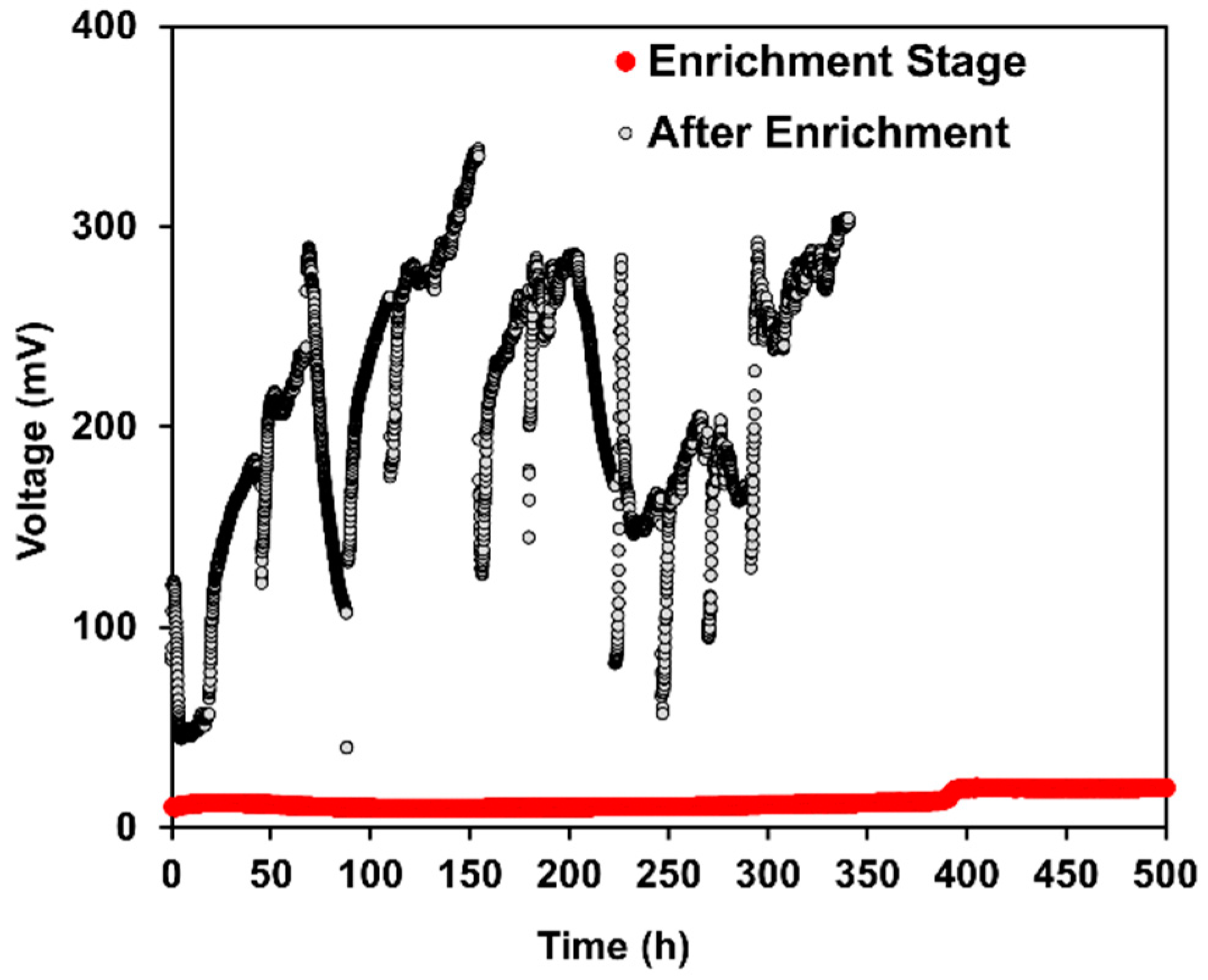
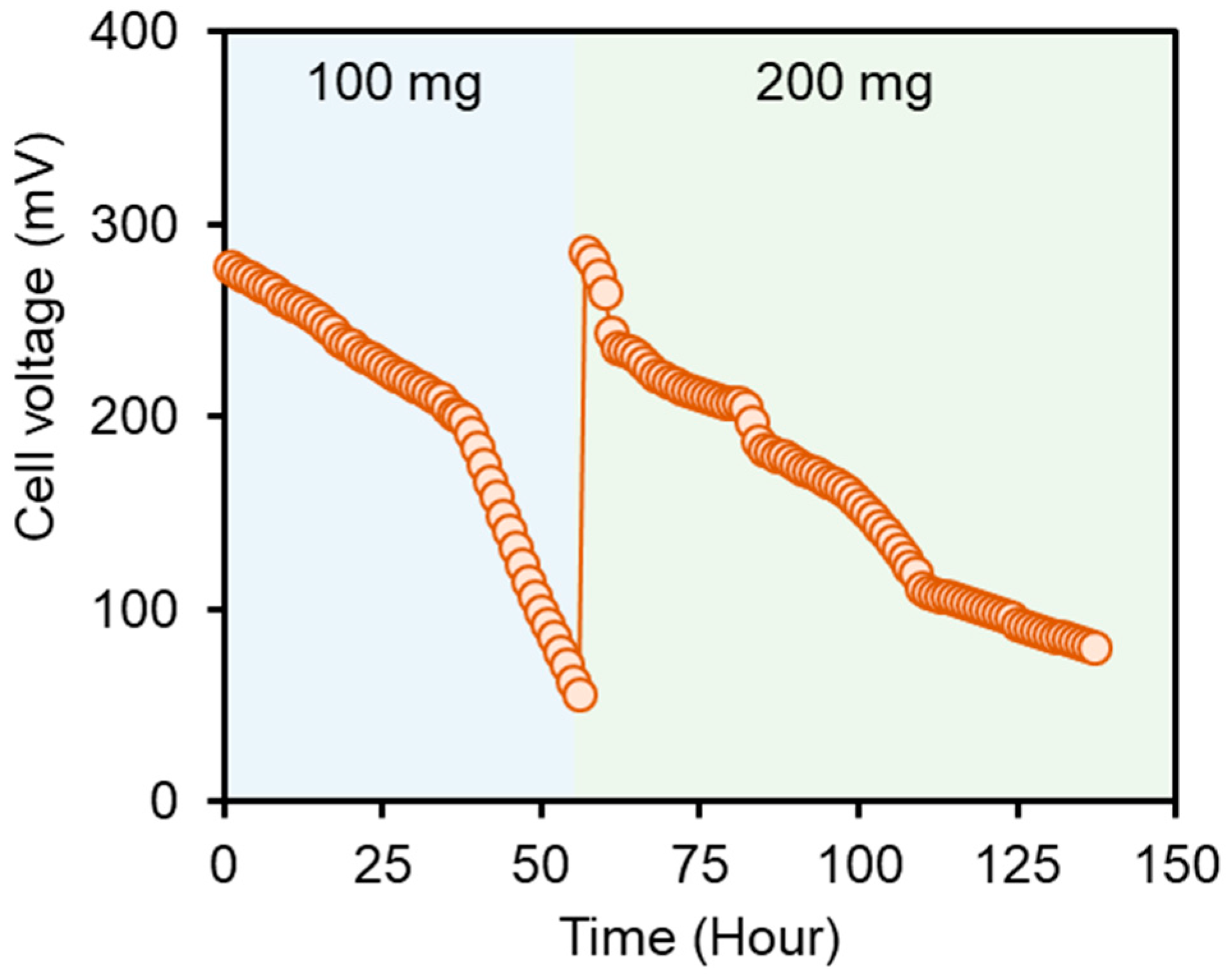
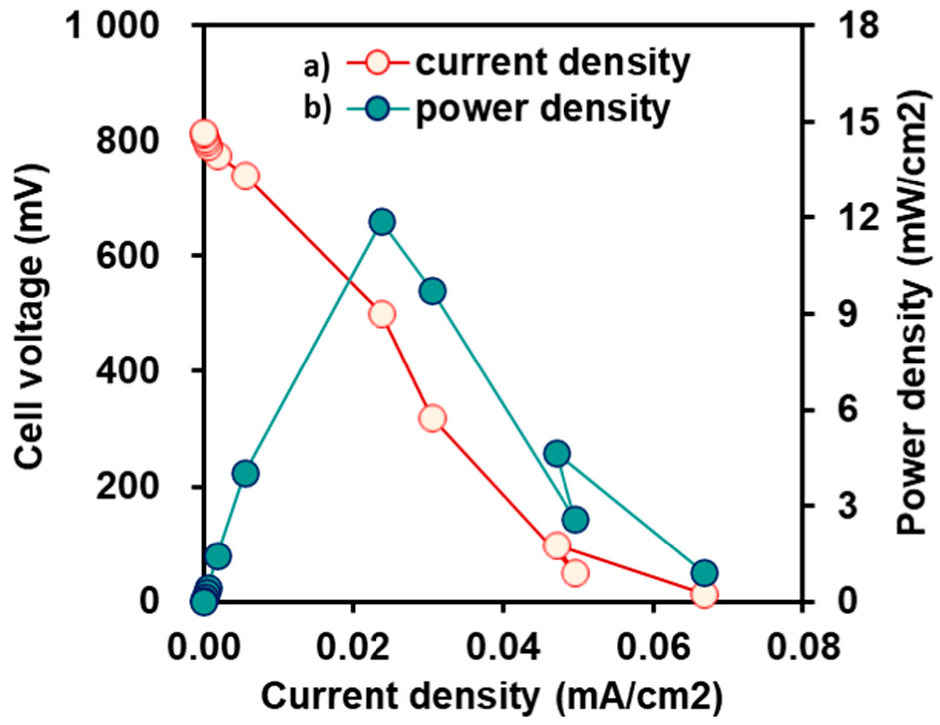
2.1. Bioelectrochemical Performance of DCMFCs

The DCMFC voltage outputs were observed from the start of operation, and are displayed in Figure 1. The adaptation of bacteria to the DCMFC environment is a major factor in the removal of dyes, and this may be enhanced by feeding cosubstrates and azo dyes concurrently and progressively increasing the influent azo dye concentration [15]. Figure 1 shows a lower output for the enrichment stage than for the enrichment stage. During the enrichment stage, the voltage was mostly less than 20 mV in this phase. This lower output during the enrichment stage can be attributed to the adaptation of electricigens to the new anodic environment containing Evans blue [16]. Following the enrichment stage shown in Figure 1, the reactor was stabilized for 3 days until the voltage decreased to 50 mV; at this point, the reactor was replenished with acetate and media. In the enriched stage, the output voltage was significantly greater than that in the initial enrichment stage, with a maximum voltage recorded at 337 mV when the initial dye concentration was 100 mg/L. However, increasing the dye concentration to 200 mg/L initially caused a decrease in voltage to an average of 180 mV for three cycles, but this gradually increased to 304 mV by the end of the experiment. This fluctuation in voltage suggests an inhibitory effect of the dye on bacteria, which then gradually adapt to the environment and regain maximum voltage output. This phenomenon aligns with previous studies by Sultana et al. and Khan et al., who observed similar trends with different dye concentrations [17]. Another explanation for our results is that a high concentration of EB may require more electrons to cleave the azo bond, which decreases the output voltage. This was also explained by Khan et al. [18]. Furthermore, Thung et al. observed that increasing the concentration of acid orange 7 resulted in a reduction in voltage from 167 mV to 148 mV, suggesting that electrons were diverted from the electrode to the azo dye, following acetate oxidation [19].
The biofilm and freshly acclimated bacteria in our study progressively adhered to the anode surface after being adjusted to the electrogenic environment. The concentrations of Evans blue were progressively increased from 100 to 200 mg/L over the course of the investigation. It is evident from Figure 2 that the voltage peak declined steadily as soon as the bacteria in the batch mode began to consume acetate; however, the highest voltage recovered upon the addition of new feed. The highest voltages for 100 mg/L and 200 mg/L EB azo dye were 280.2 and 287.34 mV, respectively, over a 500 Ω external resistance. Despite the high concentration of electrogens in the acclimatized anaerobic sludge, the DCMFC showed limited voltage output, suggesting that while the bioelectricity production mechanism is active, further optimization is needed to enhance the voltage performance.
At the end of the operational run, the electrochemical performance of the microbial fuel cells (MFCs) was characterized using polarization tests, and the results were encouraging (Figure 3). A maximum power density of 13.2 mW/cm2 and a maximum current density of 0.074 mA/cm2 were achieved, with an external load of 500 Ω. These results highlight how well the MFC bioreactor system works to simultaneously breakdown EB and produce bioelectricity. Interestingly, the current density decreased with increasing resistance, but the voltage increased with increasing resistance. This result is in line with the maximum power transfer theorem, which asserts that the best power output occurs when the external load is equal to the internal resistance of the power supply components. As a result, current plays a major role in MFC power generation, with Pmax occurring when Rext equals Rint. Our results are consistent with those of Rint, and support the observation of Pmax at 550 Ω made by Zafar et al. [2]. However, Raturi et al. [20] reported greater power densities when the exterior load moved from higher to lower resistances. The findings of the present study concur with the maximum power theorem, as stated by [21] and other previous research.

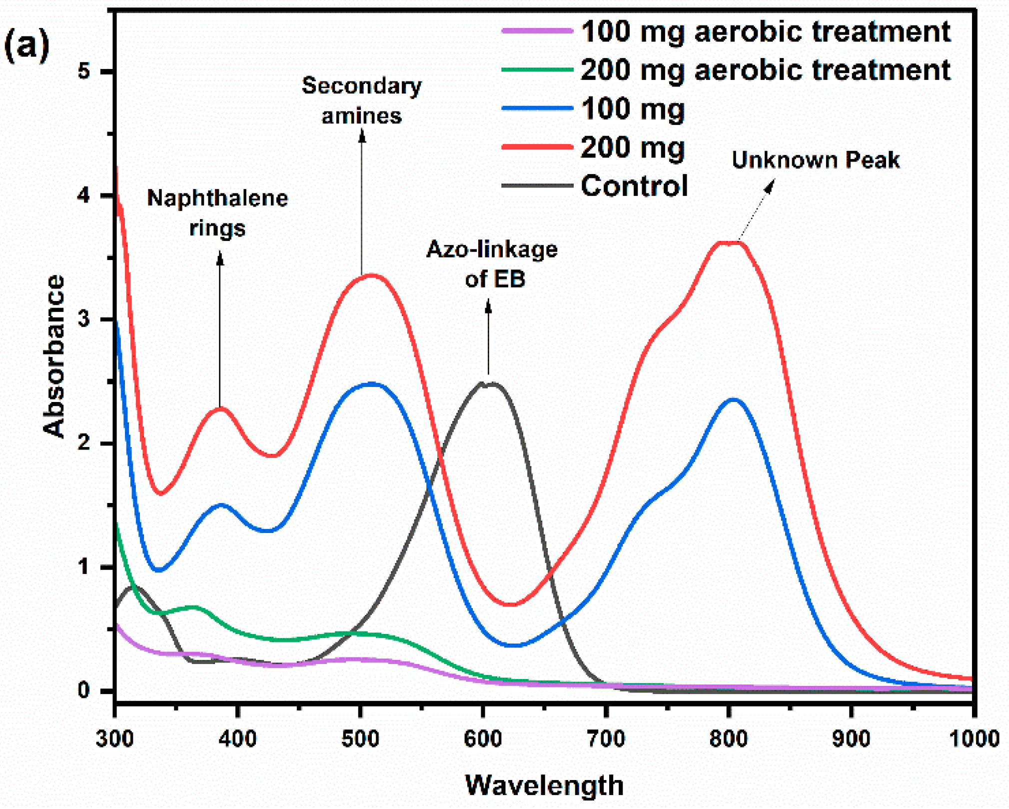
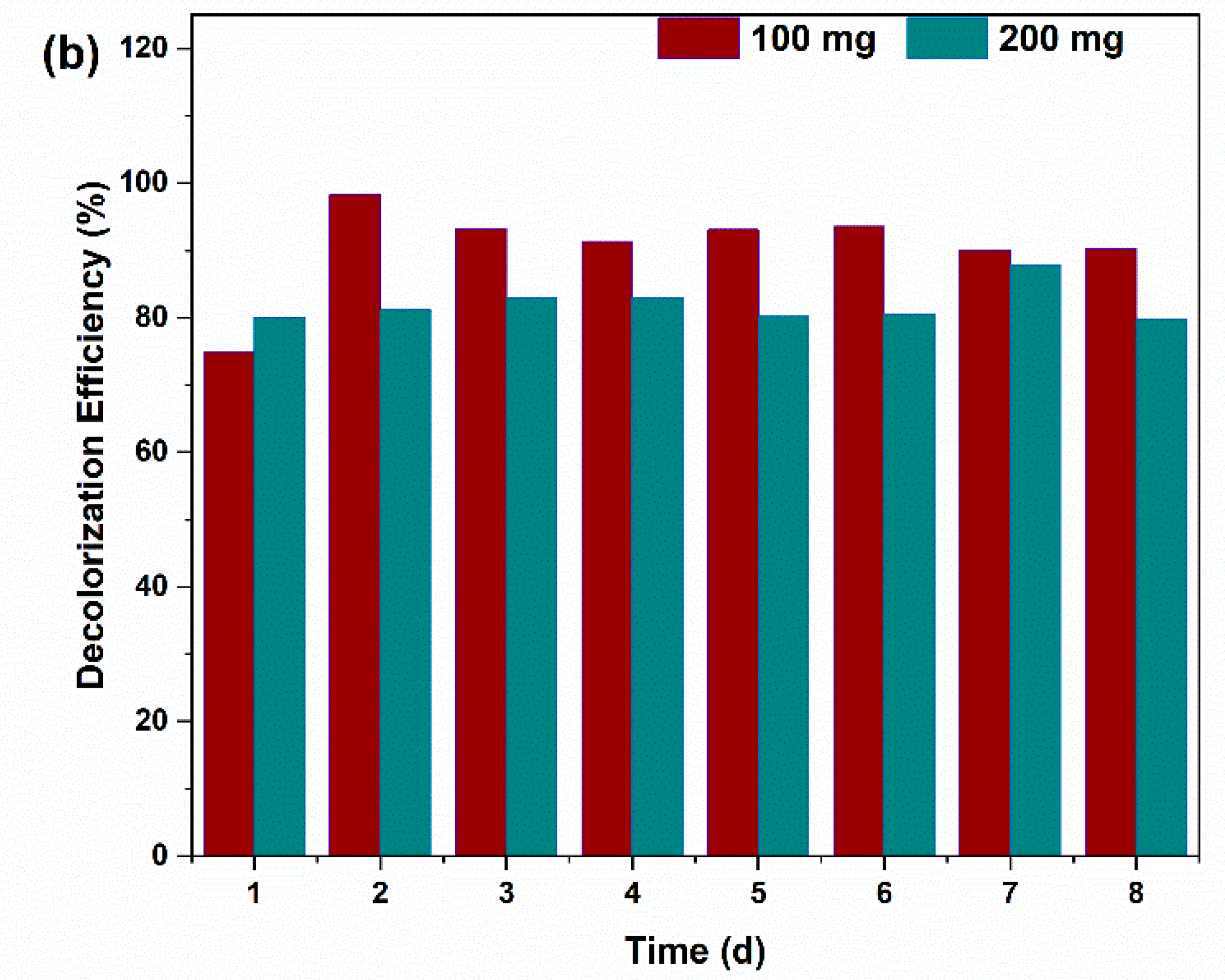
2.2. Decolorization Efficiency of the Dyes

Using a spectrophotometer, the UV–Vis spectra of the effluent treated with the dual-chamber microbial fuel cell (DCMFC) and the pure Evans blue sample were thoroughly scanned. The results are shown in Figure 4a. The spectrum of the pure Evans blue solution showed clear absorption peaks in the visible spectrum, most notably at 602 nm, which are associated with the distinctive azo linkage (–N=N–) that gives Evans blue its deep-blue color. Additionally, the presence of absorption peaks at 389 nm (naphthalene rings) [18] and 500 nm (secondary amines) [22] in the spectra of the anaerobically treated effluent indicated the occurrence of biodegradation processes that result in the generation of secondary compounds. The absorption peak at 800 nm remains unidentified, and will be the focus of further investigation in future studies.
The absorption peak observed at λmax = 602 nm for the pure EB dye is attributed to the presence of azo bonds. Thus, the reduction in absorbance at this wavelength was used to calculate the decolorization efficiency (DE%) of Evans blue in the effluent of the DCMFC. In the anaerobic treatment of EB, certain byproducts linked to aromatic amines were present in the effluent, as shown by the absorption peaks in Figure 4a. Overall, the results showed a notable decrease in color intensity, ascribed to the reductive breakage of the azo bond and conversion into aromatic amines in the DCMFC anaerobic environment. The effluent from DCMFC was then transferred to an aerobically treated chamber and showed no noticeable absorption peak, indicating that aromatic amines and other degraded products generated in an anaerobic environment were completely biodegraded in the integrated bioreactor (DCMFC–aerobic) treatment. To assess the different dye concentrations (100 and 200 mg/L), a comparative analysis was carried out. Before the double-chamber microbial fuel cell (DCMFC) experiment, a new carbon brush was subjected to an adsorption test, and the results were almost negligible. Interestingly, as Figure 4b illustrates, at an initial concentration of 100 mg/L, more than 90% of the color was removed, on average, but at 200 mg/L, approximately 80% of the color was effectively eliminated. The removal efficiency of Evans Blue observed in this study is comparable to the findings of Quan et al., where the anode, modified with palladium nanoparticles, achieved a 98.0% removal of Evans Blue. In contrast, the unmodified control anode demonstrated a 90% removal efficiency [23]. Bacterial inhibition has been found to cause the degradation time to increase with increasing dye concentration. Similar findings from earlier research indicate that higher dye concentrations are more biotoxic, which in turn suppresses microbial development and activity and decreases decolorization efficiency [24]. The observed decrease in decolorization efficiency could be explained by the decreased ability of the bacteria to withstand EB, which is essential for electron transfer and decolorization. This reduction probably limited electron transfer, acetate oxidation, and, as a result, decolorization efficiency. Consistent with our findings, Fang et al. [25] reported that high concentrations of active brilliant red X-3B inhibited microorganisms, which in turn led to a decrease in the number and activity of microorganisms because they formed toxic and resistant products that the bacteria were unable to use directly, which in turn caused the decolorization efficiency to decrease. Furthermore, it is important to keep in mind that complete decolorization in microbial fuel cells (DCMFCs) does not necessarily equate to complete mineralization, because of biotransformation into byproducts of aromatic amines, which are colorless but may not be biodegradable in anaerobic settings.

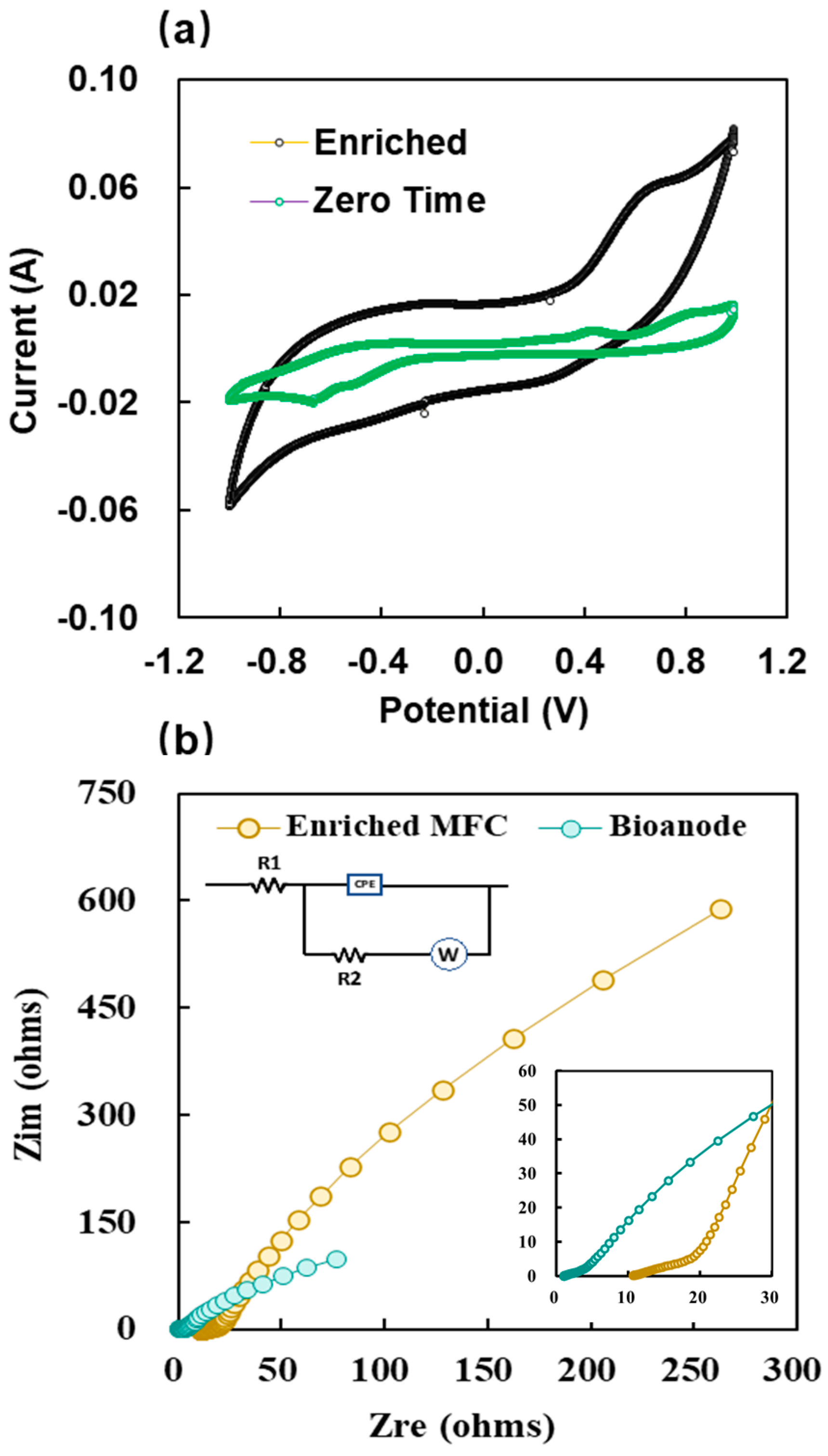
2.3. Electrochemical Characteristics of the Anode Biofilms

A typical method for examining the electroactive species in a medium is cyclic voltammetry (CV). CV was employed to analyze the degradation products of the anode dye, aiming to identify potential reduction end products. A comparison was made between the initial CV at zero time and the CV obtained after enriching the anode. The first peaks for oxidation and reduction were found at 0.99 V and −0.54 V, respectively, with currents of 0.016 A and −0.0001 A, respectively. Following the enrichment of the anode with deteriorated products, the subsequent CV curve showed reduction and oxidation peaks, referenced to Ag/AgCl, at 0.99 V and −0.73 V, respectively, with corresponding currents of 0.10 A and −0.0003 A. Figure 5a provides a graphic representation of these results. The results of cyclic voltammetry (CV) obtained in our studies are similar to those of Mani et al. in 2019 [10]. The results of the referenced study and our study are consistent with the presence of redox couples during the degradation of EB, as evidenced by the distinctive peak at 0.89 V/0.7 V. Based on these findings, we suggest that both studies involved the degradation of azo bonds (N=N). Furthermore, the results of the study by Khan et al., 2021 are consistent with our results, as both studies suggested redox couples of dye degradation, as both investigations demonstrated redox couples indicative of dye degradation, along with the detection of oxidation peaks in the degraded products [18].
The best technique for explaining the processes that occur in bioelectrochemical reactors is electrochemical impedance spectroscopy (EIS). This technique is widely employed to determine the ohmic resistance (Rs) or solution resistance and internal resistance (Rin). Additionally, EIS may be used to determine the Warburg resistance (W) and activation charge transfer (Rct). This resistance is important for understanding the kinetics of reactions [26,27]. A Nyquist plot is commonly used to construct complex impedance (Z) data (Figure 5b). Using the data from the EIS tests, a similar circuit with Rct, Rs, and W and a constant phase element (CPE) was constructed (Figure 5b). The DCMFC system’s total internal resistance, Rs = 11.19 Ω and Rct = 1.29 Ω, was found to be 12.48 Ω in our investigation. The considerable distance between the electrodes and the conductivity of the solution might cause the apparent internal resistance of the DCMFC system. Interestingly, the resistance of the bioanode (Rs + Rct) was found to be 1.4 Ω, indicating the presence of a fully developed biofilm on the anode surface and encouraging an increased rate of electron transfer at the interface between the electrolyte and bioanode. Higher electron transport to the cathode as a result of the anode’s higher biocatalytic activity increases the generation of bioelectricity.

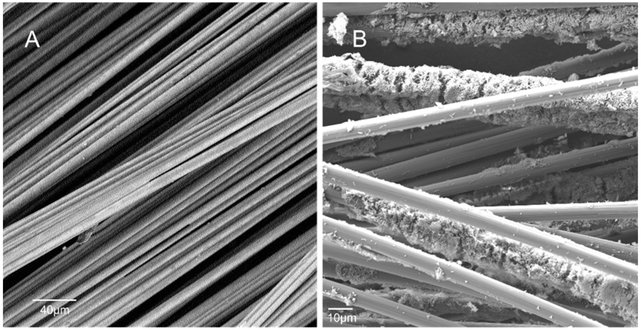
2.4. Surface Characterization and Microbial Biofilm Community Analysis

SEM analysis of the carbon brush anode was conducted to observe bacterial adhesion and biofilm formation. The bioanodes were removed from the DCMFC after the research studies were completed. SEM images of the plain carbon-brush anode surface at the start of the experiment are displayed in Figure 6A, while Figure 6B shows SEM images of the anode covered with a distinctive biofilm, demonstrating the presence of a compact microbial population on the surface of the anode. Electron micrographs of the plain carbon brush reveal a smooth, well-organized network and readily noticeable carbon strands that provide significant surface area for microbe adhesion. In line with earlier findings, the majority of the sticky structures (biofilms) are composed of cocci-shaped bacteria [28,29,30]. Despite the fact that electron transmission is enhanced by biofilm formation on the electrode surface, overly dense biofilms obstruct electron transmission and lower the DCMFC power output. Analysis of the microbial population may shed further light on the importance of morphology and the particular roles it plays.
Understanding the impact of DCMFCs on current generation and dye degradation requires analysis of the activated−sludge biomass microbial population in DCMFCs. Examining many microbial groups, including electricigens, dye-degrading organisms, exoelectrogens, fermentative bacteria, and others, is necessary to evaluate the overall performance of MFCs. These groups work together to attain maximum functioning. The microbial communities found in both the biofilm and inoculum were examined and categorized at the phylum and genus levels, using 16S rRNA gene sequencing. The main functional microorganisms that are present at both levels and have relative abundances of more than 2% are the subject of this section.
Figure 7a,b show changes at the phylum and genus levels in the inoculum and DCMFC anode biofilms. Pseudomonadota (45.55%), Bacteriodota (13.86%), Actinomycetota (10.87%), Chloroflexota (8.83%), Campylobacterota (6.85%), Bacillota (3.41%), Nitrospirota (2.9%), and others (3.3%) were initially the major phyla in the inoculum (Figure 7a). After acclimation, the percentage of Actinomycetota increased to 28.8%, and that of Pseudomonadota increased to 32% in dye wastewater containing a cosubstrate in DCMFCs. Thermodesulfobacteriota (4.77%), Bacteriodota (4.14%), Chloroflexota (9.94%), Bacillota (16.42%), and others (1.42%) were the other dominant phyla in the DCMFC (Figure 7a).
Thermodesulfobacteriota and Pseudomonodonotota, which we identified in our research, are considered important communities that can break down a variety of organic pollutants, including azo dyes, and immediately transfer electrons to the electrode [2,31]. Furthermore, Bacteroidota are described as the predominant phylum in anaerobic conditions that break down azo dyes, and potentially as exoelectrogens on the MFC bioanode [23]. Actinomycetota are typically found in MFCs, and have been shown to be effective at degrading organic pollutants [32,33]. However, their role in transporting electrons to the anode is not clear; rather, Actinomycetota contribute to the diversity and activity of electrogenic bacteria [34,35]. Bacillota may help in electron transport to the electrode and the production of bioelectricity [35]. A phylum of bacteria called Chloroflexota is important for the breakdown of azo dyes and the generation of electric current in microbial fuel cells [36]. The anode chamber of MFCs handling wastewater is enriched in a variety of electrochemically active bacteria from these six phyla [18,36,37].
The relative abundance of the microbial community at the genus level is shown in Figure 7b. The dominant species of the inoculum were Methylotenera (20.3%), Thermochromatium (6.9%), Actinomarinicola (4.5%), Caldilinea (3.0%), Acidothermus (2.5%), and others (3.3%). The main genera enriched in the anode biofilms were Actinomarinicola (13.7%), Methylotenera (9.4%), Thermochromatium (4.8%), Geobacter (4.5%), Acidothermus (4.04%), Soehngenia (3.6%), Propionivibrio (2.4%), Janibacter (2.4%), Proteiniclasticum (2.3%), Romboutsia (2.2%), Bellilinea (2.2) and others (1.42%). Figure 7b shows a significant prevalence of Actinomarinicola within the dye-containing chamber. This bacterium was initially discovered by He YQ, in 2020, is known to thrive in aerobic environments, and is often associated with sulfur and iron biomining processes [38]. Our findings are consistent with this characterization, especially given the high levels of sulfur derivatives found in the dye. Interestingly, while Actinomarinicola was previously identified as aerobic, our study detected it in an anaerobic chamber. This discrepancy may be due to the adaptability of the bacterium, which is potentially influenced by its natural habitat deep in the sea, where it may exhibit facultative anaerobic behavior.
Geobacter and Methylotenera play crucial roles in the degradation of azo dyes and producing electric current in microbial fuel cells. Geobacter-like species have been found to be involved in electricity generation in the presence of azo dyes [39] while Methylotenera has been implicated in the metabolism of EB dye degradation, and the secondary metabolite nitrobenzene is formed during Evans blue degradation [40]. This compound can be used as a substrate by Methylotenera to further degrade it and remove nitro groups from the benzene ring. This finding was also reported by Dai et al., who reported that the Methylotenera genera that developed on anodes were related to denitrification [41].
A range of studies have explored the potential of thermophilic microorganisms in microbial fuel cells; however, no direct studies have explored the role of Thermochromatium in MFCs. Choi et al. reported that Bacillus licheniformis and Bacillus thermoglucosidasius can generate significant amounts of electricity, and their performance is influenced by temperature and carbon sources [42]. Fu (2013) identified Caloramator-related bacteria as potential electricity generators in a thermophilic microbial fuel cell [43]. In 2006, Jong et al. further enriched and characterized the bacterial diversity in a thermophilic mediatorless microbial fuel cell, achieving high power density and electron recovery [44]. These studies collectively highlight the potential of thermophilic microorganisms, including Thermochromatium, in microbial fuel cells.
The presence of Acidothermus in our study may be linked to dye degradation, as these bacteria were found by Chao Li et al. [45] in their studies on the removal of dying textile water, because of the high COD removal in the high temperature range between 40 and 45 °C.
The role of Soehngenia in microbial fuel cells has not been directly addressed in the literature. However, Zhang et al. found this bacterial genus in their study using inoculum sludge from the anaerobic or anoxic tank of a wastewater treatment plant [46]. The bacteria in our study were also detected in the inoculum anaerobic sludge from the anoxic tank of the wastewater treatment plant. The possible role of these bacteria in biofilms could be as fermenters. Because Soehngenia is anaerobic or facultatively anaerobic, saccharolytic and proteolytic, it may ferment carbohydrates or proteins in the anode biofilm [47].
Many researchers have identified the genus Propionivibrio on anode biofilms in different studies, but none of them explained its role in the biofilm community [48,49,50]. However, Brune et al. [51] reported that the fermentative bacteria Propionivibrio specialize in breaking down hydroaromatic compounds.
In this study, Proteiniclasticum was enriched on anode biofilms from anaerobic sludge, and Liu et al. also isolated this bacterial genus from anaerobic acetochlor-degrading sludge [52]. Proteiniclasticum is thought to be involved in interspecies electron transfer mechanisms; this was also clarified by Perez et al., in systems that were amended with Fe3O4 and fed acetate. They link this to direct mechanisms of electron transfer between species, and strongly tie it to the role of Proteiniclasticum, which is predominant when the mineral (Fe3O4) is present on the anode of MFCs [53].
In the present study, Janibacter is involved in the degradation of the azo dye Evans blue under anaerobic conditions on anode biofilms. These results are in line with those of a study conducted by Ravadelli et al., who recognized Jiaibacter as an azo dye-degrading bacterium in an anoxic/oxic membrane bioreactor for treating azo dye-containing wastewater, and these bacteria were the most abundant (60%) of the classified sequences [54].
The genus Romboutsia may play a part in the production of bioelectricity and the transportation of electrons to the anode. It may also be important in the breakdown of azo dyes. The Romboutsia sp. abundance near the anode showed a significant positive correlation with bioelectricity generation, indicating that these results align with the findings of Li et al., who investigated the possibility that Romboutsia sp. are electricigens or similar bacteria. Additionally, he described how chlorinated pesticides degraded in the tests [55].
It has been shown that the genus Bellilinea is connected to the anaerobic breakdown of azo dyes and is electrochemically active, meaning that it can complete extracellular electron transfer [56].
The genera identified in this study exhibit diverse metabolic functions within the microbial community, categorizing them into three distinct groups. First, genera such as Geobacter, Thermochromatium, and Proteiniclasticum serve solely as exoelectron generators, contributing to the electron transfer process in microbial fuel cells by releasing electrons to the anode. Second, genera such as Methylotenera, Romboutsia, and Bellilinea, which function as both exoelectron generators and effective dye degraders, play dual roles. These adaptable genera demonstrate diverse metabolic capacities, because they are essential for both electron transport and dye degradation. Finally, certain genera, including Actinomarinicola, Acidothermus, Soehngenia, Propionivibrio, and Janibacter, are dedicated solely to dye degradation. These bacteria help in environmental remediation by degrading dye pollutants. This classification helps in understanding the function and role of these specialized genera within the microbial community inside microbial fuel cells and their efforts in environmental remediation.
The anode biofilm community developed differently from the inoculum community, which may be attributed to bacteria adapting to the DCMFC environment. A significant difference in the microbial community was observed between the beginning and end of the experiment. These findings, which are in line with earlier research, demonstrated that the presence of an azo dye and an anode in the anodic compartment of DCMFCs creates an environment that is conducive to the growth of both dye-degrading and exoelectrogenic bacteria [12,57].
The resulting phylogenetic tree exhibited a hierarchical structure with several well-supported clades or six different generations. However, every generation is represented by a different color. Subjected sequences SK2 and SK3 lie in the third generation of the tree.
By comparing the 16S rRNA gene sequences of two actinobacterial strains in an aerobic chamber with those of reference organisms found in GenBank data repositories, the strains were identified and named SK-2 and SK-3. According to sequence analysis, Sk-2 and Sk-3 were identified as Streptomyces sp. strain actinomycetes (KY235922.1) and Streptomyces sp. strain actinomycetes (KY236012.), respectively, whereas Sk-2 and Sk-3 were classified within Streptomyces sp. (Figure 7c).
These Streptomyces isolates were present in our investigation, which is consistent with the findings of other investigations. Evans blue (>97%) and other azo dyes, including Brilliant Green, triphenylmethane, Nigrosine, Amido black 10B, Congo red, and Methyl Red, are degraded by Streptomyces when shaken [58,59,60]. This confirms that Streptomyces spp. have a strong ability to degrade different types of azo dyes.

3. Materials and Methods

3.1. Materials and Chemicals

In accordance with the directions of Logan et al., the carbon brush electrode utilized in the anode and cathode was made from carbon fibers woven around two twisted titanium wire cores [61]. The RisingSun Membrane in Beijing, China, is a cation-exchange membrane (CEM). Acetate, sodium phosphate monobasic monohydrate (NaH2PO4·H2O), and sodium phosphate dibasic heptahydrate (Na2HPO4·7H2O) were acquired from Sigma Aldrich in Poland as analytical-grade chemicals for the experiment.

3.2. Anodic and Cathodic Media

A medium comprising nutrients, 100 and 200 mg/L Evans blue, 1 g/L acetate, and 50 mM phosphate buffer solution was fed to the anode, including vitamins and minerals, as previously described [12]. The cathode was supplied with an identical medium, except that NaHCO3 (0.3 g/L) was used instead of Evans blue and acetate.

3.3. Inoculum Preparation

For the anaerobic compartment of the double-chamber microbial fuel cell (DCMFC), mixed-culture anaerobic sludge from a municipal wastewater treatment plant in Gliwice, Poland, was used. Aerobic sludge from the same source was used for the biocathode. Two actinomycetes strains (SK-2 and SK-3) were isolated from garden compost in Gliwice, and biomass produced in a liquid Pochn medium was inoculated in a post-treatment aerobic reactor. The anaerobic and aerobic sludge had a mixed-liquor suspended solid (MLSS) concentration of 4 g/L and a starting optical density (OD600) of 1.5, while the actinomycetes had an OD600 of 0.5. The anaerobic chamber was acclimated with the mixed culture for one month, and the aerobic chamber with actinomycetes for seven days, to ensure the development of mature biofilms on both the anode and cathode before experimentation.

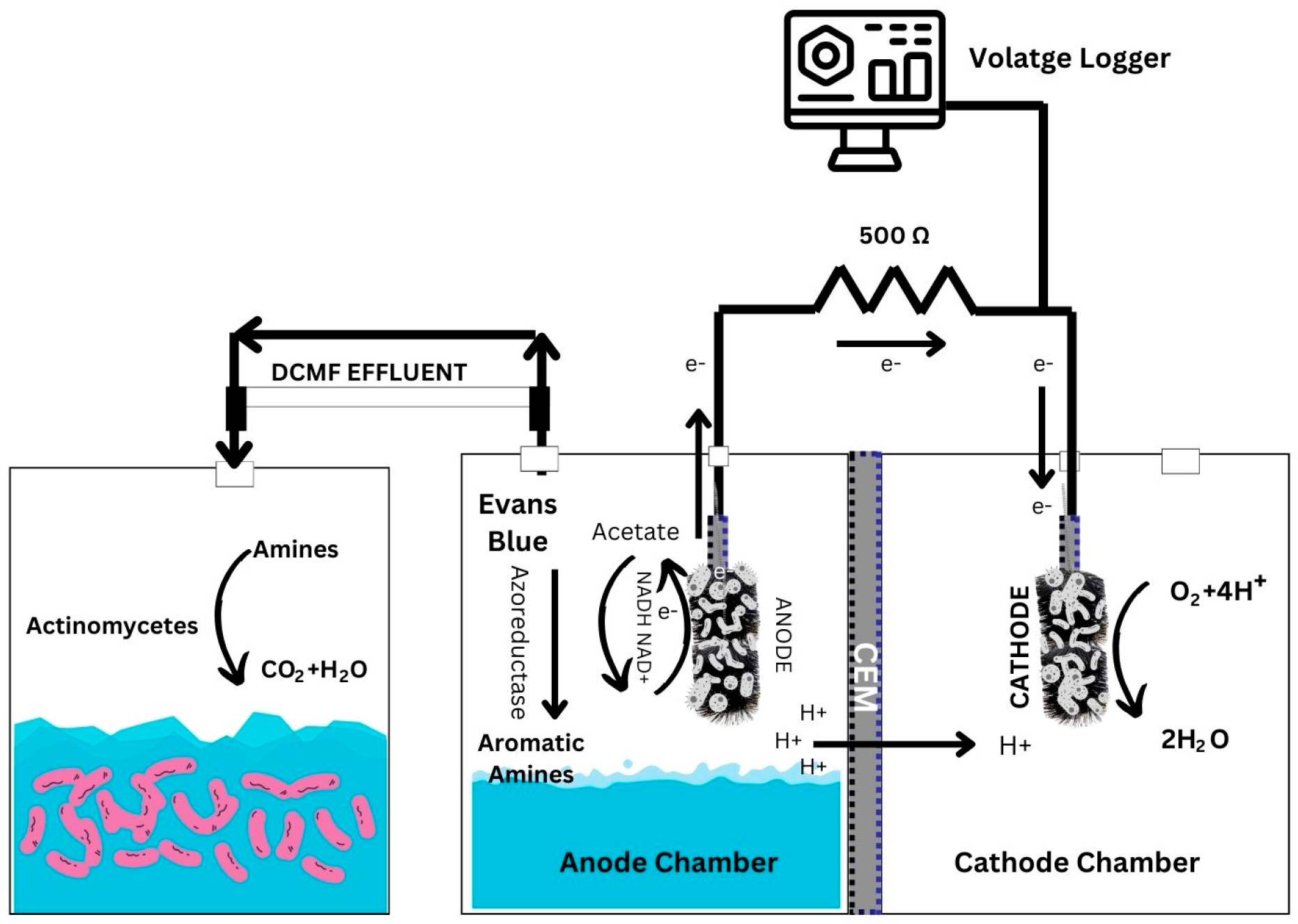
3.4. The Design and Functioning of the Integrated DCMFC–Aerobic Reactor System

The experimental DCMFC setup used in this work consisted of two 120 mL Plexiglass compartments, one for anode conditions that were controlled to be anaerobic and the other for cathode conditions, as illustrated in Figure 8. Both the cathode and anode electrodes were carbon brushes with a (1 cm × 2 cm) diameter and length, and were placed in the middle of the compartment. Copper wire with insulation was used to fix the connections on the electrodes, and nonconductive epoxy was cast off to insulate the connection interfaces.
A cation-exchange membrane (CMI-7000, RisingSun Membrane, Beijing, China) was used to divide the anode and cathode chambers. A voltage data logger (Picolog ADC-24, Pico Technology, Northamptonshire, UK) was connected to the external circuit of the MFCs with a 500 (ohms) resistor to collect voltage output data, with a data recording interval of 5 min.
Each chamber of the DCMFC had an effective working volume of 100 mL, and the ratio of sludge to dye wastewater was 4/1. The DCMFC bioreactor was injected with mixed-culture anaerobic sludge obtained from a municipal wastewater treatment plant in Gliwice, Poland, to acclimate the systems before initiating the experiment. Evans blue was gradually increased from 50 mg/Lto 100 mg/L in 50 mM phosphate buffer during the adaptation period (start-up phase). Anaerobic sludge and anodic medium were also fed into the DCMFC, along with this mixture, and the MFC was operated for almost a month. Nitrogen gas was used to span the anodic medium for fifteen minutes before inoculation, and nitrogen gas was also pumped into the DCMFC headspace.
Approximately half of the anode chamber media was replaced with a new dye-containing medium to complete one operational cycle when the acetate (electron donor) was nearly depleted and the cell’s potential fell below 50 mV. Following acclimation, DCMFCs were supplemented with acetate (1 g/L), two distinct concentrations of Evans blue (100 and 200 mg/L), minerals, and vitamins, to conduct feasibility experiments. To further treat the DCMFC effluent, an aerobic bioreactor with a working volume of 120 mL was built of plexiglass. To fully mineralize the intermediates created during anaerobic degradation, the effluent of the DCMFC anaerobic chamber was fed into an aerobic chamber.

3.5. Detection Method for Evans Blue Decolorization

Evans blue (EB)-treated samples were scanned in the full range of 300–900 nm by using a UV–visible spectrophotometer (Hitachi U-1900 UV–VIS Spectrophotometer Lab). The absorption peak at 602 nm for EB possibly corresponded to N=N. To convert the absorbance values to concentration values, a standard calibration curve was used (R2 = 0.998). The percentage of dye decolorization was calculated using the following formula:
D e c o l o r i z a t i o n   % = I n i t i a l   a b s o r b a n c e F i n a l   a b s o r b a n c e I n i t i a l   a b s o r b a n c e × 100

3.6. Monitoring of MFC Performance

The MFC reactor was run in a closed circuit with 500 (Ohms Ω) of external resistance. A Picolog data logging system was used to acquire voltage data from the reactor. Using Ohm’s law, the flow of current within the external circuit was determined as follows [61]:
R = I/V
where V is the potential difference (voltage) across the exterior load, R is the external resistor (load), and I is the current flow in the exterior circuit.
To calculate the power output of the MFC, the following formula is used [61]:
P = VI
where V is the potential difference (voltage) across the exterior load, I is the current, and P (watts) is the power.
Different external resistances, ranging from 10 ohms to 1 M ohm, were attached to exterior circuits at the completion of the MFC experimental setup. By applying the procedure outlined in the equation to normalize the resultant power and current values to the surface area (21 cm2) of the anode, polarization curves and power–current graphs were created [13].
In the last batch cycle of the acclimation period, DCMFC was given 100 mg/L of EB combined with 1 g/L of acetate, and it was able to maintain stability and reach its maximum cell potential. Electrochemical impedance spectroscopy (EIS) and cyclic voltammetry (CV) were performed by using CH Instruments, Inc., type 660C, Austin, TX, USA.
There were two different EIS measuring types used. First, the bioanode employed a three-electrode arrangement, including Ag/AgCl (reference), cathode (counter), and bioanode (working) electrodes, at potential amplitudes of 10 mV and frequencies ranging from 100 kHz to 10 mHz. For the entire cell (DCMFC), a two-electrode setup was used with cathode (working), anode (counter) and Ag/AgCl (reference) electrodes at an open-circuit voltage [18]. Cyclic voltammetry (CV) was carried out with a scan rate of 10 mV/s over a range of −1 V to 1 V, with degrading dye. To assess the electrochemical performance of the DCMFC, Ag/AgCl served as the reference electrode [62], while the anode served as the working electrode, and the cathode served as the counter electrode.

3.7. Scanning Electron Microscopy (SEM) of the Anode Surface

At the end of the experiment, the surface of the bioanode was evaluated by using SEM (Phenom ProX Desktop, Eindhoven, The Netherlands) to determine bacterial attachment and biofilm formation at the anode surface. Before being examined under an SEM, the anode fibers were cut into slices, fixed for one night with 2% glutaraldehyde, and thoroughly washed twice in a pH 7.2 phosphate-buffer solution. Next, 25, 50, 75, and 100% ethanol solutions were used to dehydrate the fragment samples [63]. For clear photos, the fragment samples needed to be completely dry. Consequently, a Baltec critical point drier CPD-030 (BAL-TEC, Balzers, Liechtenstein)was used to dry all the samples.

3.8. DNA Extraction, PCR Amplification and Illumina Sequencing

After the DCMFC experiment was completed, the anode carbon brush was removed, and the microbial population of the anode biofilm was investigated using the following methods. Genomic DNA was initially isolated from the biofilm sample using the DNA Isolation DNeasy PowerSoil Pro Kit (Qiagen, Hilden, Germany), as per the manufacturer’s instructions. The forward primer 338F (5′-ACTCCTACGGGAGGCAGCA-3′) and the reverse primer 806R (5′-GGACTACHVGGGTWTCTAAT-3′) were utilized to amplify the V3-V4 region of 16S rRNA. For Actinomycete strains in an integrated aerobic chamber, the actinobacterial primers 243 F (5′-GGATGAGCCCGCGGCCTA-3′) and 513 R (5’-CGGCCGCGGCTGCTGGCACGTA-3′) with 5% DMSO were used during PCR to avoid any metabolites. A GeneAmp® 9700 (Applied Biosystems, California, United States ) was used for PCR. The input gDNA sample (5 ng) was amplified through PCR using 5× reaction buffer, 1 mM dNTP, 500 nM each of the forward and reverse PCR primers, and DNA polymerase (Herculase II fusion, Agilent Technologies, Santa Clara, CA, USA). Heat activation at 95 °C for three minutes was used for the initial PCR cycle. After that, there were 25 cycles of 30 s at 95 °C, 55 °C, and 72 °C and a final 5 min extension at 72 °C. To create the final library with the index, 10 µL of the first PCR product was amplified using Nextera XT Indexed Primer(Illumina Inc., San Diego, CA, USA). The PCR products were purified using the AxyPrepTM DNA Gel Extraction Kit (Axygen Biosciences, AXYGEN Union City, CA, USA). After purification and quantification, the samples were delivered to a biotechnology company (Macrogen Europe, Amsterdam, The Netherlands) for 16S rRNA gene sequencing via the Illumina MiSeq platform. The genetic distance model (Tamura–Nei) was used to construct a phylogenetic tree and neighbor-joining tree method in Geneious Prime (version 9.8.1). The tree included 27 ITS sequences representing different strains of Actinomycetes, including 5 sequences, i.e., SK2 and SK3.

4. Conclusions

In conclusion, the integration of a double—chamber microbial fuel cell (DCMFC) with a biocathode and an aerobic bioreactor has shown promising potential for efficiently degrading the azo dye Evans blue and producing less-contaminated biodegraded products. High decolorization efficiencies were observed across various influent concentrations, ranging from 90 ± 2% to 98 ± 1.9% for 100 mg/L and from 79 ± 2% to 87 ± 1% for higher concentrations. The combined DCMFC–aerobic bioreactor treatment method effectively mitigated dye pollution, as evidenced by the lack of characteristic absorption peaks in the aerobically treated effluent, suggesting full biodegradation of Evans blue. Electrochemical impedance spectroscopy (EIS) analysis revealed a well-developed biofilm on the anode surface, facilitating enhanced electron-transfer rates. Microbial community analysis identified Pseudomonadota as the dominant phylum, with Actinomarinicola representing a significant portion (13.75%) at the genus level, a novel finding in MFC environments. This finding underscores the potential role of Actinomarinicola in dye degradation within MFC systems. Overall, this study emphasizes the importance of integrated double − chamber MFC–aerobic bioreactor setups for addressing dye pollution challenges while highlighting the contribution of Actinomarinicola to the field as a valuable discovery in MFC ecosystems.

Author Contributions

Conceptualization, K.A. and E.Z.-G.; methodology, K.A. and E.Z.-G.; software, K.A. and C.L.; validation, K.A., E.Z.-G. and C.L.; formal analysis, K.A. and C.L.; investigation, K.A.; resources, K.A. and E.Z.-G.; data curation, K.A. and C.L.; writing—original draft preparation, K.A.; writing—review and editing, K.A., E.Z.-G. and C.L.; visualization, K.A. and C.L.; supervision, E.Z.-G.; project administration, K.A. and E.Z.-G.; funding acquisition, K.A. All authors have read and agreed to the published version of the manuscript.

Funding

This work was funded by the Department of Air Protection, Silesian University of Technology (grant number BKM 632/RIE-2/2023 08/020/BKM_23/005) and Silesian University of Technology Groundbreaking Research (grant number 32/014/SDU/10-21-05).

Data Availability Statement

The raw data supporting the conclusions of this article will be made available by the authors on request.

Conflicts of Interest

The authors declare that they have no known competing financial interests or personal relationships that could have appeared to influence the work reported in this paper.

References

  1. Rabaey, K.; Verstraete, W. Microbial Fuel Cells: Novel Biotechnology for Energy Generation. Trends Biotechnol. 2005, 23, 291–298. [Google Scholar] [CrossRef] [PubMed]
  2. Zafar, Z.; Ayaz, K.; Nasir, M.H.; Yousaf, S.; Sharafat, I.; Ali, N. Electrochemical Performance of Biocathode Microbial Fuel Cells Using Petroleum-Contaminated Soil and Hot Water Spring. Int. J. Environ. Sci. Technol. 2019, 16, 1487–1500. [Google Scholar] [CrossRef]
  3. Kyazze, G. Taking the Waste out of Dye Wastewater Using Microbial Fuel Cells. In Proceedings of the 5th EU-ISMET Conference, Online, 13–15 September 2021. [Google Scholar]
  4. Rawat, D.; Mishra, V.; Sharma, R.S. Detoxification of Azo Dyes in the Context of Environmental Processes. Chemosphere 2016, 155, 591–605. [Google Scholar] [CrossRef] [PubMed]
  5. Kumar, R.; Ahmad, R. Biosorption of Hazardous Crystal Violet Dye from Aqueous Solution onto Treated Ginger Waste (TGW). Desalination 2011, 265, 112–118. [Google Scholar] [CrossRef]
  6. Popli, S.; Patel, U.D. Destruction of Azo Dyes by Anaerobic–Aerobic Sequential Biological Treatment: A Review. Int. J. Environ. Sci. Technol. 2015, 12, 405–420. [Google Scholar] [CrossRef]
  7. Ilamathi, R.; Jayapriya, J. Microbial Fuel Cells for Dye Decolorization. Environ. Chem. Lett. 2018, 16, 239–250. [Google Scholar] [CrossRef]
  8. Rahimnejad, M.; Adhami, A.; Darvari, S.; Zirepour, A.; Oh, S.E. Microbial Fuel Cell as New Technol Ogy for Bioelectricity Generation: A Review. Alex. Eng. J. 2015, 54, 745–756. [Google Scholar] [CrossRef]
  9. Slate, A.J.; Whitehead, K.A.; Brownson, D.A.C.; Banks, C.E. Microbial Fuel Cells: An Overview of Current Technology. Renew. Sustain. Energy Rev. 2019, 101, 60–81. [Google Scholar] [CrossRef]
  10. Mani, P.; Fidal, V.T.; Bowman, K.; Breheny, M.; Chandra, T.S.; Keshavarz, T.; Kyazze, G. Degradation of Azo Dye (Acid Orange 7) in a Microbial Fuel Cell: Comparison Between Anodic Microbial-Mediated Reduction and Cathodic Laccase-Mediated Oxidation. Front. Energy Res. 2019, 7, 101. [Google Scholar] [CrossRef]
  11. Yao, L.; Xue, X.; Yu, P.; Ni, Y.; Chen, F. Evans Blue Dye: A Revisit of Its Applications in Biomedicine. Contrast Media Mol. Imaging 2018, 2018, 7628037. [Google Scholar] [CrossRef]
  12. Sun, J.; Li, Y.; Hu, Y.; Hou, B.; Zhang, Y.; Li, S. Understanding the Degradation of Congo Red and Bacterial Diversity in an Air-Cathode Microbial Fuel Cell Being Evaluated for Simultaneous Azo Dye Removal from Wastewater and Bioelectricity Generation. Appl. Microbiol. Biotechnol. 2013, 97, 3711–3719. [Google Scholar] [CrossRef] [PubMed]
  13. Yadav, A.; Kumar, P.; Rawat, D.; Garg, S.; Mukherjee, P.; Farooqi, F.; Roy, A.; Sundaram, S.; Sharma, R.S.; Mishra, V. Microbial Fuel Cells for Mineralization and Decolorization of Azo Dyes: Recent Advances in Design and Materials. Sci. Total Environ. 2022, 826, 154038. [Google Scholar] [PubMed]
  14. Meenakshi, S.; Hiremath, J.; Meenakshi, M.H.; Shivaveerakumar, S. Actinomycetes: Isolation, Cultivation and Its Active Biomolecules. J. Pure Appl. Microbiol. 2024, 18, 118–143. [Google Scholar] [CrossRef]
  15. Kong, F.; Ren, H.Y.; Pavlostathis, S.G.; Wang, A.; Nan, J.; Ren, N.Q. Enhanced Azo Dye Decolorization and Microbial Community Analysis in a Stacked Bioelectrochemical System. Chem. Eng. J. 2018, 354, 351–362. [Google Scholar] [CrossRef]
  16. Sun, J.; Cai, B.; Zhang, Y.; Peng, Y.; Chang, K.; Ning, X.; Liu, G.; Yao, K.; Wang, Y.; Yang, Z.; et al. Regulation of Biocathode Microbial Fuel Cell Performance with Respect to Azo Dye Degradation and Electricity Generation via the Selection of Anodic Inoculum. Int. J. Hydrogen Energy 2016, 41, 5141–5150. [Google Scholar] [CrossRef]
  17. Sultana, S.; Khan, M.D.; Sabir, S.; Gani, K.M.; Oves, M.; Khan, M.Z. Bio-Electro Degradation of Azo-Dye in a Combined Anaerobic–Aerobic Process along with Energy Recovery. New J. Chem. 2015, 39, 9461–9470. [Google Scholar] [CrossRef]
  18. Khan, M.D.; Li, D.; Tabraiz, S.; Shamurad, B.; Scott, K.; Khan, M.Z.; Yu, E.H. Integrated Air Cathode Microbial Fuel Cell-Aerobic Bioreactor Set-up for Enhanced Bioelectrodegradation of Azo Dye Acid Blue 29. Sci. Total Environ. 2021, 756, 143752. [Google Scholar] [CrossRef]
  19. Thung, W.E.; Ong, S.A.; Ho, L.N.; Wong, Y.S.; Ridwan, F.; Oon, Y.L.; Oon, Y.S.; Lehl, H.K. A Highly Efficient Single Chambered Up-Flow Membrane-Less Microbial Fuel Cell for Treatment of Azo Dye Acid Orange 7-Containing Wastewater. Bioresour. Technol. 2015, 197, 284–288. [Google Scholar] [CrossRef]
  20. Katuri, K.P.; Scott, K.; Head, I.M.; Picioreanu, C.; Curtis, T.P. Microbial Fuel Cells Meet with External Resistance. Bioresour. Technol. 2011, 102, 2758–2766. [Google Scholar] [CrossRef]
  21. Potrykus, S.; León-Fernández, L.F.; Nieznański, J.; Karkosiński, D.; Fernandez-Morales, F.J. The Influence of External Load on the Performance of Microbial Fuel Cells. Energies 2021, 14, 612. [Google Scholar] [CrossRef]
  22. Fu, Y.; Gao, Y.; Chen, L.; He, Q.; Zhu, D.; Cao, H.; Cheng, J. Highly Efficient Single Fluorescent Probe for Multiple Amine Vapours via Reaction between Amine and Aldehyde/Dioxaborolane. RSC Adv. 2014, 4, 46631–46634. [Google Scholar] [CrossRef]
  23. Quan, X.; Xu, H.; Sun, B.; Xiao, Z. Anode Modification with Palladium Nanoparticles Enhanced Evans Blue Removal and Power Generation in Microbial Fuel Cells. Int. Biodeterior. Biodegrad. 2018, 132, 94–101. [Google Scholar] [CrossRef]
  24. Long, X.; Cao, X.; Liu, S.; Nishimura, O.; Li, X. The Azo Dye Degradation and Differences between the Two Anodes on the Microbial Community in a Double-Anode Microbial Fuel Cell. Water. Air. Soil Pollut. 2019, 230, 265. [Google Scholar] [CrossRef]
  25. Fang, Z.; Song, H.L.; Cang, N.; Li, X.N. Electricity Production from Azo Dye Wastewater Using a Microbial Fuel Cell Coupled Constructed Wetland Operating under Different Operating Conditions. Biosens. Bioelectron. 2015, 68, 135–141. [Google Scholar] [CrossRef]
  26. Sanchez-Herrera, D.; Pacheco-Catalan, D.; Valdez-Ojeda, R.; Canto-Canche, B.; Dominguez-Benetton, X.; Domínguez-Maldonado, J.; Alzate-Gaviria, L. Characterization of Anode and Anolyte Community Growth and the Impact of Impedance in a Microbial Fuel Cell. BMC Biotechnol. 2014, 14, 102. [Google Scholar] [CrossRef]
  27. Wang, H.; Long, X.; Sun, Y.; Wang, D.; Wang, Z.; Meng, H.; Jiang, C.; Dong, W.; Lu, N. Electrochemical Impedance Spectroscopy Applied to Microbial Fuel Cells: A Review. Front. Microbiol. 2022, 13, 973501. [Google Scholar] [CrossRef] [PubMed]
  28. Khan, N.; Khan, M.D.; Nizami, A.S.; Rehan, M.; Shaida, A.; Ahmad, A.; Khan, M.Z. Energy Generation through Bioelectrochemical Degradation of Pentachlorophenol in Microbial Fuel Cell. RSC Adv. 2018, 8, 20726–20736. [Google Scholar] [CrossRef] [PubMed]
  29. Offei, F.; Thygesen, A.; Mensah, M.; Tabbicca, K.; Fernando, D.; Petrushina, I.; Daniel, G. A Viable Electrode Material for Use in Microbial Fuel Cells for Tropical Regions. Energies 2016, 9, 35. [Google Scholar] [CrossRef]
  30. Yang, S.; Du, F.; Liu, H. Characterization of Mixed-Culture Biofilms Established in Microbial Fuel Cells. Biomass Bioenergy 2012, 46, 531–537. [Google Scholar] [CrossRef]
  31. Pham, T.H.; Boon, N.; Aelterman, P.; Clauwaert, P.; De Schamphelaire, L.; Vanhaecke, L.; De Maeyer, K.; Höfte, M.; Verstraete, W.; Rabaey, K. Metabolites Produced by Pseudomonas Sp. Enable a Gram-Positive Bacterium to Achieve Extracellular Electron Transfer. Appl. Microbiol. Biotechnol. 2008, 77, 1119–1129. [Google Scholar] [CrossRef]
  32. Hemdan, B.A.; El-Taweel, G.E.; Naha, S.; Goswami, P. Bacterial Community Structure of Electrogenic Biofilm Developed on Modified Graphite Anode in Microbial Fuel Cell. Sci. Rep. 2023, 13, 1255. [Google Scholar] [CrossRef] [PubMed]
  33. Arbour, T.J.; Gilbert, B.; Banfield, J.F. Diverse Microorganisms in Sediment and Groundwater Are Implicated in Extracellular Redox Processes Based on Genomic Analysis of Bioanode Communities. Front. Microbiol. 2020, 11, 508335. [Google Scholar] [CrossRef] [PubMed]
  34. Jimenez, L.; Kulko, M.; Kim, R.; Jashari, T.; Choe, T. 16S RRNA Analysis of Electrogenic Bacterial Communities in Microbial Fuel Cells Developed from Temperate Soils. Bios 2020, 91, 9–20. [Google Scholar] [CrossRef]
  35. De La Cruz-Noriega, M.; Benites, S.M.; Rojas-Flores, S.; Otiniano, N.M.; Sabogal Vargas, A.M.; Alfaro, R.; Cabanillas-Chirinos, L.; Rojas-Villacorta, W.; Nazario-Naveda, R.; Delfín-Narciso, D. Use of Wastewater and Electrogenic Bacteria to Generate Eco-Friendly Electricity through Microbial Fuel Cells. Sustainability 2023, 15, 10640. [Google Scholar] [CrossRef]
  36. Dai, Q.; Zhang, S.; Liu, H.; Huang, J.; Li, L. Sulfide-Mediated Azo Dye Degradation and Microbial Community Analysis in a Single-Chamber Air Cathode Microbial Fuel Cell. Bioelectrochemistry 2020, 131, 107349. [Google Scholar] [CrossRef]
  37. Rathour, R.; Patel, D.; Shaikh, S.; Desai, C. Eco-Electrogenic Treatment of Dyestuff Wastewater Using Constructed Wetland-Microbial Fuel Cell System with an Evaluation of Electrode-Enriched Microbial Community Structures. Bioresour. Technol. 2019, 285, 121349. [Google Scholar] [CrossRef]
  38. He, Y.Q.; Chen, R.W.; Li, C.; Shi, S.B.; Cui, L.Q.; Long, L.J.; Tian, X.P. Actinomarinicola tropica Gen. Nov. Sp. Nov., a New Marine Actinobacterium of the Family Iamiaceae, Isolated from South China Sea Sediment Environments. Int. J. Syst. Evol. Microbiol. 2020, 70, 3852–3858. [Google Scholar] [CrossRef] [PubMed]
  39. Sun, L.; Mo, Y.; Zhang, L. A Mini Review on Bio-Electrochemical Systems for the Treatment of Azo Dye Wastewater: State-of-the-Art and Future Prospects. Chemosphere 2022, 294, 133801. [Google Scholar] [CrossRef]
  40. Antonin, V.S.; Garcia-Segura, S.; Santos, M.C.; Brillas, E. Degradation of Evans Blue Diazo Dye by Electrochemical Processes Based on Fenton’s Reaction Chemistry. J. Electroanal. Chem. 2015, 747, 1–11. [Google Scholar] [CrossRef]
  41. Dai, M.; Zhang, Y.; Wu, Y.; Sun, R.; Zong, W.; Kong, Q. Mechanism Involved in the Treatment of Sulfamethoxazole in Wastewater Using a Constructed Wetland Microbial Fuel Cell System. J. Environ. Chem. Eng. 2021, 9, 106193. [Google Scholar] [CrossRef]
  42. Choi, Y.; Jung, E.; Park, H.; Paik, S.R.; Jung, S.; Kim, S. Construction of Microbial Fuel Cells Using Thermophilic Microorganisms, Bacillus Licheniformis and Bacillus Thermoglucosidasius. Bull. Korean Chem. Soc. 2004, 25, 813–818. [Google Scholar] [CrossRef]
  43. Fu, Q.; Kobayashi, H.; Kawaguchi, H.; Vilcaez, J.; Wakayama, T.; Maeda, H.; Sato, K. Electrochemical and Phylogenetic Analyses of Current-Generating Microorganisms in a Thermophilic Microbial Fuel Cell. J. Biosci. Bioeng. 2013, 115, 268–271. [Google Scholar] [CrossRef] [PubMed]
  44. Jong, B.C.; Kim, B.H.; Chang, I.S.; Liew, P.W.Y.; Choo, Y.F.; Kang, G.S. Enrichment, Performance, and Microbial Diversity of a Thermophilic Mediatorless Microbial Fuel Cell. Environ. Sci. Technol. 2006, 40, 6449–6454. [Google Scholar] [CrossRef]
  45. Li, C.; Zhang, Z.; Li, Y.; Cao, J. Study on Dyeing Wastewater Treatment at High Temperature by MBBR and the Thermotolerant Mechanism Based on Its Microbial Analysis. Process Biochem. 2015, 50, 1934–1941. [Google Scholar] [CrossRef]
  46. Zhang, G.; Feng, S.; Jiao, Y.; Lee, D.J.; Xin, Y.; Sun, H. Cathodic Reducing Bacteria of Dual-Chambered Microbial Fuel Cell. Int. J. Hydrogen Energy 2017, 42, 27607–27617. [Google Scholar] [CrossRef]
  47. Nazina, T.N.; Bidzhieva, S.K.; Grouzdev, D.S.; Sokolova, D.S.; Tourova, T.P.; Parshina, S.N.; Avtukh, A.N.; Poltaraus, A.B.; Talybly, A.K. Soehngenia longivitae sp. nov., a Fermenting Bacterium Isolated from a Petroleum Reservoir in Azerbaijan, and Emended Description of the Genus Soehngenia. Microorganisms 2020, 8, 1967. [Google Scholar] [CrossRef]
  48. Naz, I.; Hodgson, D.; Smith, A.; Marchesi, J.; Sehar, S.; Ahmed, S.; Lynch, J.; Avignone-Rossa, C.; Saroj, D.P. Investigation of the Active Biofilm Communities on Polypropylene Filter Media in a Fixed Biofilm Reactor for Wastewater Treatment. J. Chem. Technol. Biotechnol. 2018, 93, 3264–3275. [Google Scholar] [CrossRef]
  49. Wang, G.; Guo, Y.; Cai, J.; Wen, H.; Mao, Z.; Zhang, H.; Wang, X.; Ma, L.; Zhu, M. Electricity Production and the Analysis of the Anode Microbial Community in a Constructed Wetland-Microbial Fuel Cell. RSC Adv. 2019, 9, 21460. [Google Scholar] [CrossRef]
  50. Fischer, F.; Merino, N.; Sugnaux, M.; Huguenin, G.; Nealson, K.H. Microbial Community Diversity Changes during Voltage Reversal Repair in a 12-Unit Microbial Fuel Cell. Chem. Eng. J. 2022, 446, 137334. [Google Scholar] [CrossRef]
  51. Brune, A.; Ludwig, W.; Schink, B. Propionivibrio limicola Sp. Nov., a Fermentative Bacterium Specialized in the Degradation of Hydroaromatic Compounds, Reclassification of Propionibacter Pelophilus as Propionivibrio Pelophilus Comb. Nov. and Amended Description of the Genus Propionivibrio. Int. J. Syst. Evol. Microbiol. 2002, 52 Pt 2, 441–444. [Google Scholar] [CrossRef]
  52. Liu, J.; Bao, Y.; Zhang, X.; Zhang, K.; Chen, S.; Wu, H.; He, J. Proteiniclasticum sediminis Sp. Nov., an Obligate Anaerobic Bacterium Isolated from Anaerobic Sludge. Antonie Van Leeuwenhoek 2021, 114, 1541–1549. [Google Scholar] [CrossRef] [PubMed]
  53. Pérez-Díaz, M.I.; Zárate-Segura, P.; Bermeo-Fernández, L.A.; Nirmalkar, K.; Bastida-González, F.; García-Mena, J.; Jan-Roblero, J.; Guerrero-Barajas, C. Bacterial Consortium from Hydrothermal Vent Sediments Presents Electrogenic Activity Achieved under Sulfate Reducing Conditions in a Microbial Fuel Cell. J. Environ. Health Sci. Eng. 2020, 18, 1189. [Google Scholar] [CrossRef]
  54. Ravadelli, M.; Da Costa, R.E.; Lobo-Recio, M.A.; Akaboci, T.R.V.; Bassin, J.P.; Lapolli, F.R.; Belli, T.J. Anoxic/Oxic Membrane Bioreactor Assisted by Electrocoagulation for the Treatment of Azo-Dye Containing Wastewater. J. Environ. Chem. Eng. 2021, 9, 105286. [Google Scholar] [CrossRef]
  55. Li, Y.; Li, X.; Sun, Y.; Zhao, X.; Li, Y. Cathodic Microbial Community Adaptation to the Removal of Chlorinated Herbicide in Soil Microbial Fuel Cells. Environ. Sci. Pollut. Res. 2018, 25, 16900–16912. [Google Scholar] [CrossRef] [PubMed]
  56. Liu, S.; Xue, H.; Feng, X.; Pyo, S.H. Electrostimulation for Promoted Microbial Community and Enhanced Biodegradation of Refractory Azo Dyes. J. Environ. Chem. Eng. 2022, 10, 108626. [Google Scholar] [CrossRef]
  57. Zhong, D.; Liu, Y.; Liao, X.; Zhong, N.; Xu, Y. Facile Preparation of Binder-Free NiO/MnO2-Carbon Felt Anode to Enhance Electricity Generation and Dye Wastewater Degradation Performances of Microbial Fuel Cell. Int. J. Hydrogen Energy 2018, 43, 23014–23026. [Google Scholar] [CrossRef]
  58. Kameche, K.; Amrani, S.; Mouzaoui, S.; Aït-Amar, H. Biodegradation of Diazo Dye Evans Blue by Four Strains of Streptomyces Isolated from Soils of Algeria. Biocatal. Agric. Biotechnol. 2022, 46, 102529. [Google Scholar] [CrossRef]
  59. Ayaz, K.; Zabłocka-Godlewska, E. Screening of Actinomycetes Decolorizing the Synthetic Dyes from Rotten Poplar Wood and Garden Compost. In Contemporary Problems of Power Engineering and Environmental Protection; Pikoń, K., Bogacka, M., Eds.; Politechnika Śląska: Gliwice, Poland, 2022; pp. 119–131. [Google Scholar]
  60. Adenan, N.H.; Lim, Y.Y.; Ting, A.S.Y. Identification and Optimization of Triphenylmethane Dyes Removal by Streptomyces Sp. from Forest Soil. Sustain. Environ. Res. 2021, 31, 8. [Google Scholar] [CrossRef]
  61. Logan, B.E.; Hamelers, B.; Rozendal, R.; Schröder, U.; Keller, J.; Freguia, S.; Aelterman, P.; Verstraete, W.; Rabaey, K. Microbial Fuel Cells:  Methodology and Technology. Environ. Sci. Technol. 2006, 40, 5181–5192. [Google Scholar] [CrossRef]
  62. Yadav, R.S.; He, W.; Liang, D.; Li, C.; Yu, Y.; Feng, Y. Enrichment of Geobacter on Anode Biofilms from Domestic Wastewater without Posing Anode Potential in Microbial Electrochemical Cells. Microbiol. Res. 2024, 15, 1859–1869. [Google Scholar] [CrossRef]
  63. Yadav, R.S.; He, W.; Liang, D.; Li, C.; Yu, Y.; Ayaz, K.; Feng, Y. High-Efficiency Hydrogen Recovery from Corn Straw Hydrolysate Using Functional Bacteria and Negative Pressure with Microbial Electrolysis Cells. Water 2024, 16, 2423. [Google Scholar] [CrossRef]
Figure 1. Voltage output-time curves of MFCs with enrichment stage and after enrichment.
Energies 17 04896 g001
Figure 2. Impact of EB concentrations on the production of bioelectric power (500 Ω external load).
Energies 17 04896 g002
Figure 3. Polarization curves. a–b: power density as a function of current density in DCMFC.
Energies 17 04896 g003
Figure 4. (a) UV–visible absorption spectra of 100 mg and 200 mg of DCMFC effluent, pure dye (control), and aerobically treated effluent. (b) Decolorization of EB in DCMFC with time.
Energies 17 04896 g004aEnergies 17 04896 g004b
Figure 5. (a) Zero time and EB−enriched anode cyclic voltammetry in DCMFC, and (b) EIS Nyquist plots and fitted circuit drawings of the bioanode and DCMFC treated with EB.
Energies 17 04896 g005
Figure 6. (A) SEM Images plain carbon brush at 1000× magnification. (B) SEM Images of anodic biofilm in the DCMFC at 5000× magnification.
Energies 17 04896 g006
Figure 7. Microbial relative abundance at the (a) phylum and (b) genus level. (c) Phylogenetic tree of 16s RNA sequences of Streptomyces spp. (In = inoculum, E anode = enriched bioanode).
Energies 17 04896 g007
Figure 8. Schematic diagram of double—chamber microbial fuel cell.
Energies 17 04896 g008
Disclaimer/Publisher’s Note: The statements, opinions and data contained in all publications are solely those of the individual author(s) and contributor(s) and not of MDPI and/or the editor(s). MDPI and/or the editor(s) disclaim responsibility for any injury to people or property resulting from any ideas, methods, instructions or products referred to in the content.

