3.1. Structural Properties
The main structural differences between the Ga
FeN layers grown on GaN and those deposited on the Al
Ga
N buffers are summarized in
Figure 1, where the overall sample structure, including TEM cross-section and plan-view images for the reference sample (
= 0%) and for the sample with the highest Al concentration
= 41% are reported. A comparison between the overview cross-section images presented in
Figure 1b,e reveals a dislocation density in the Al
Ga
N buffer larger than the one in GaN, affecting the nanocrystal distribution in the Ga
FeN overlayer. As a consequence, the NCs are not all localized in one plane like those embedded in the layer grown on GaN, as demonstrated in the TEM micrographs reproduced in
Figure 1c,f. It is further observed that the majority of the NCs in the Ga
FeN/Al
Ga
N sample form at the end of dislocations propagating from the buffer, in contrast to the NCs in the layer grown on GaN, which are embedded in the Ga
FeN matrix volume. This is visualized in the plan-view images presented in
Figure 1d,g, where NCs with a round-shaped contour, distributed homogeneously in the plane with an average distance of (20–100) nm between nanocrystals, are observed. The NCs density increases from (5.0 ± 0.2)
NCs/cm
for the reference sample to (5.0 ± 0.3)
NCs/cm
for the sample grown on the Al
Ga
N buffer. Besides an increased NC density, there is a complex dislocation network connecting the NCs observed for the Ga
FeN layer grown on the Al
Ga
N buffer.
The nanocrystal phases are established from the HRXRD 2
-
scans collected along the
growth direction and reported in
Figure 2a for all samples. Besides the diffraction peaks from the Ga
FeN layer, from the Al
Ga
N (
%) buffer and from the Al
O
substrate, two additional diffraction peaks located around (41.28 ± 0.07)
and (47.72 ± 0.07)
are observed for all samples with Al in the buffer. The first diffraction peak is attributed to the (0002) plane of the hexagonal
-Fe
N phase, while the second one origins from the (200) plane of the fcc
’-Ga
Fe
N phase. The calculated lattice parameters for the two Fe
N phases are (0.437 ± 0.002) nm and (0.381 ± 0.002) nm, respectively. These values lie in the range of the reported literature values for both phases: the hexagonal
-Fe
N with
a = 0.469 nm and
c = 0.437 nm [
27], and the fcc
’-Ga
Fe
N with
a = 0.379 nm [
15]. For the reference sample, only the
’-Ga
Fe
N phase is observed.
A close-up of the region around the (0002) diffraction peak of the Ga
FeN overlayer and of the Al
Ga
N buffer is presented in
Figure 2b, showing the shift of the buffer peak to higher diffraction angles with increasing Al concentration, pointing at a reduction in the
c-lattice parameter. The position of the diffraction peak related to the Ga
FeN thin layer remains unchanged for the buffers with
10% and shifts to lower angles for increasing Al concentrations, i.e., larger
c-lattice parameter. This suggests that the Ga
FeN layer is compressively strained on the Al
Ga
N buffers.
To analyze the strain state and to obtain the in-plane
a-lattice parameter, reciprocal space maps at the (10
5) diffraction plane are acquired. The RSM for the samples with buffers containing 5% and 41% of Al are shown in
Figure 2c,d, demonstrating that while the Ga
FeN layer grows fully strained on the Al
Ga
N buffer, it is partially relaxed on the Al
Ga
N one. The in-plane percentage of relaxation
of the Ga
FeN thin layer with respect to the buffer is obtained directly from the respective in-plane
d-lattice spacings as [
30]:
where
refers to the in-plane lattice spacings
d. The values in the numerator are the measured ones and those in the denominator are the values for free-standing GaN and Al
Ga
N according to the Vegard’s law. The calculated
values for the samples considered here, are reported in
Table 1, showing that for
10%, the Ga
FeN layers grow fully strained on the buffers and the onset of relaxation occurs at
10%. This is also evident from the lattice parameters presented in
Figure 3a,b as a function of
, where the lattice parameter
a for the Ga
FeN layer is found to deviate from the one of the Al
Ga
N buffer with
10%. The dashed lines in
Figure 3a,b give the trend of the Vegard’s law and the dashed-dotted lines indicate the literature values for the lattice parameters for GaN [
31]. Although the
c-lattice parameter for the Ga
FeN layer is not significantly affected by increasing the Al concentration,
a matches the one of the buffer until
10% and then deviates significantly, confirming the relaxation of the Ga
FeN thin layer. Considering that the Ga
FeN thin layer has only biaxial in-plane strain, the strain
and stress
tensors are calculated employing a linear interpolation between the value of the Young modulus
E and the stiffness constants
of GaN (
E = 450 GPa, 2
= 0.509) and AlN (
E = 470 GPa, 2
/
= 0.579) [
31]. The values reported in
Table 1 show that independent of the Al concentration, the Ga
FeN layers are all under a comparable compressive strain.
The (0002) diffraction peak of the Al
Ga
N buffers presented in
Figure 2b broadens with increasing Al concentration, pointing at an increment of defects and dislocation density in the buffer layers. In
-oriented III-nitride films, three main types of threading dislocations are commonly observed: edge-, mixed- and screw-type. The analysis of the integral breadth of the diffraction peaks originating from the (000
l) planes allows estimating the density of screw dislocations, while the one in the (20
4) plane provides information on the density of edge and mixed type dislocations [
22]. According to Dunn and Koch, the density of dislocations
is given by [
32]:
where
is the integral breadth and
b is the Burgers vector. This equation was previously employed to estimate the dislocation density in GaN thin films [
33]. The dislocation densities obtained from HRXRD analysis for all buffer layers as a function of
are reported in
Figure 3c, where a linear increase is observed reaching values up to four times larger than those of the GaN buffer for both edge-mixed and screw dislocations in the buffer with the highest Al concentration. These results are consistent with the observations from the cross-section and plan-view TEM images shown in
Figure 1. The dislocation density is also estimated from TEM micrographs, yielding larger values for the Al
Ga
N buffers than those obtained from the XRD analysis, but following the same trend: the greater the concentration of Al in the buffer, the higher the dislocation density.
The increased dislocation density in the Al
Ga
N buffers with
10% leads to the relaxation of the Ga
FeN thin layers. As observed in
Figure 1f, a fraction of the dislocations from the Al
Ga
N buffer runs throughout the entire Ga
FeN layer, promoting the aggregation of Fe along the defects and, therefore, the preferential formation of nanocrystals. Interestingly, the nanocrystals stabilized at the dislocations are predominantly elongated along the
growth direction.
A more detailed analysis of the NCs sizes is performed on cross-section and plan-view TEM images. The size of the NCs is determined with an accuracy of ±0.5 nm by measuring the size of the areas where Moiré patterns are visible with the Fiji software [
34]. The results are presented in
Figure 4a–e, where the size distribution of 200 measured NCs per sample is reported. For this evaluation, the NCs are treated as ellipsoids according to the schematic representation in
Figure 4f with dimensions perpendicular (
A) and parallel (
C) to the
growth direction for the different
in the buffers. The solid line marks the aspect ratio (AR) equal to 1, i.e.,
. From the size distributions presented in
Figure 4, it is seen that the size of the NCs in the reference sample has a broader distribution and particularly a larger in-plane
A than in the samples grown on the Al
Ga
N buffers. Although the size of the NCs in the reference sample tends to lie on or below the solid line, indicating an AR≤1 and an oblate shape of the NCs—with their
y-axis elongated in the plane of the layer—the size of the NCs in the layers grown on the Al
Ga
N buffers lies above the solid line, i.e., with an AR>1, pointing at prolate NCs elongated along the
growth direction. From the measured dimensions of the NCs, the average sizes parallel and perpendicular to the growth direction
are estimated, confirming the decrease in the size perpendicular to the growth direction for the nanocrystals embedded in the Ga
FeN layers grown on the Al
Ga
N buffers.
Furthermore, it is found that in all samples the nanocrystals located at dislocation sites are predominantly prolate. This suggests that the increase in dislocation density for the layers grown on the Al
Ga
N buffers promotes the formation of prolate NCs, which are mostly arranged in pairs aligned along dislocations, as shown in
Figure 5a. In contrast, the oblate NCs are all located at the same depth in the layers.
In addition to providing the size and phase, the characterization of the Moiré patterns (MPs) observed in the HRTEM micrographs yields further relevant information about the embedded NCs. The origin of MPs in general is the result of the overlap of two lattices with equal spacings that are rotated with respect to each other, or of the superposition of lattices with slightly different spacings. This leads to a pattern with Moiré fringe spacings with either single periodicity (line pattern) or double periodicity (grid-like pattern). Exemplary NCs showing a double and a single MP are presented in
Figure 5b,c, respectively. The Moiré fringe spacings depend on the two underlying crystal structures, on their orientation relationship, and on the lattice strain. The fraction of nanocrystals displaying single MP (SMP) and double MPs (DMP) is shown in
Figure 5d. Up to 78% of the NCs exhibit single MPs and 22% produce double MPs in the reference Ga
FeN grown on GaN buffer, while for the films grown on the Al
Ga
N buffers this tendency is inverted. The double MP pattern is an indication of an in-plane misorientation of the NCs, which is related to the enhanced dislocation density in the underlying buffer layers and to the formation of the NCs along the dislocations, leading to slight distortions and strain within the GaN matrix.
The Fe
N phases identified in the HRXRD spectra depicted in
Figure 2a are confirmed by HRTEM analysis. In HRTEM micrographs showing NCs, the regions of interests are Fourier transformed by Fast Fourier Transformation (FFT) using the Gatan Digital Micrograph (Gatan Inc.) software. Micrographs of two NCs are shown in
Figure 6a,d along with the corresponding FFTs in
Figure 6b,e. The FFT images are used to determine the lattice parameters by measuring the spacings in the two directions of the diffraction pattern. To identify the NCs orientation with respect to the GaN matrix, a comparison with the diffraction patterns simulated by the JEMS software is performed [
35]. Employing this procedure, the investigated NC in
Figure 6a is identified as
-Fe
N oriented along the zone axis (ZA)
, which is parallel to the ZA
, and therefore corresponds to an epitaxial relation
‖
. A schematic representation of the epitaxial relation is sketched in
Figure 6c, showing that the NC is 30
rotated with respect to the crystallographic axis of GaN, but parallel to the one of the sapphire substrate, similarly to the fcc NCs studied in Ga
FeN/GaN layers [
13]. The above procedure is applied to the NCs found in the reference sample and reproduced in
Figure 6d, revealing the epitaxial relation
‖
presented in
Figure 6f and previously reported for
’-Ga
Fe
N NCs in Ga
FeN layers grown on GaN [
13]. The majority of the NCs found in the Ga
FeN layers grown on the Al
Ga
N buffers are identified as the hexagonal
-Fe
N phase, while those in the reference sample are associated with the cubic
’-Ga
Fe
N phase oriented preferentially as
‖
, in agreement with the results from the XRD spectra presented in
Figure 2a. From elemental composition analysis
via EDX line-scans, the presence of Al in the Ga
FeN layers is ruled out as shown in
Figure S1 (Supplementary Materials) of the Suplemental Material.
3.2. Magnetic Properties
In the previous section it has been demonstrated that the basic structural characteristics of the NCs change considerably with the incorporation of Al into the buffer layer. To shed light onto how the magnetic characteristics of the layers are modified by these structural changes, a comparative analysis of the magnetic properties of the reference Ga
FeN/GaN and the Ga
FeN/Al
Ga
N samples is performed. As indicated in
Table 1 and depicted in
Figure 2, the former contains mostly
’-Ga
Fe
N NCs, which are characterized by a balanced distribution of prolate and oblate shapes, whereas in the latter, prolate
-Fe
N NCs prevail over the
’-Ga
Fe
N ones.
The formation of the Fe-rich NCs in GaN is the direct consequence of the solubility limit of Fe in GaN being (
) cm
or 0.4% at the growth conditions considered here [
36,
37,
38]. Therefore, when the doping level exceeds this concentration, the Fe ions are found both in Ga substitutional sites as Fe
and in the phase-separated NCs. The Fe-rich NCs form disperse ensembles of large ferromagnetic macrospins with specific size and shape distributions. In the absence of mobile carriers, the randomly distributed Fe
ions, despite their high spin state (
), do not interact in the relevant temperature range between 2 K and 400 K and exhibit paramagnetic properties. Due to the high diffusivity of transition metal ions in GaN, these paramagnetic ions are found diffusing a few hundreds of nanometres below the Fe-
-doped layer [
39]. This substantially increases the total amount of the dilute Fe
, making the intensity of the paramagnetic signal at low temperatures comparable to the one of the ferromagnetic NCs. Therefore, a dedicated experimental approach is required to distinguish between the two contributions.
The isothermal magnetization curves with the magnetic moment as a function of the applied magnetic field
for the reference sample (
%) are plotted for selected temperatures (solid symbols) in
Figure 7. As mentioned, the bare magnetic signal consists of two distinct contributions. At temperatures above 50 K, the fast saturating response resembling a Langevin’s
function at weak fields is attributed to the ferromagnetic NCs. However, the lack of a systematic
T-dependency satisfying the
scaling [
40] and the presence of a weak magnetic hysteresis indicate that the majority of the NCs is not in thermal equilibrium and their magnetic response is affected by the presence of energy barriers and governed by their distribution. At temperatures below 50 K, the
gains in strength and a slowly saturating contribution originating from the non-interacting Fe
ions retaining their own magnetic moment dominates [
37,
38,
41,
42].
The paramagnetism of the Fe
ions is described by the Brillouin function
for
[
36,
43,
44], and the experimentally established difference
between
at, e.g., 2 K and 5 K permits the quantification of the ions’ contribution by fitting
to
with the procedure described in detail in Ref. [
38]. The open circles in
Figure 7 represent the experimental difference
between
at 2 K and 5 K, whereas the dotted line follows the magnitude of the expected change
corresponding to several ions
cm
. The dashed line indicates the magnitude of the paramagnetic contribution corresponding to
at 2 K.
Having established
in each of the investigated structures, the paramagnetic contribution
—where
g is the g-factor and
the Bohr magneton—is calculated and subtracted from the experimental data to obtain the magnitude
of the magnetization corresponding to the NCs. The results are indicated by solid lines in
Figure 7. It is worth noting that
saturates at all investigated temperatures for
kOe, confirming the ferromagnetic order within the NCs. The evolution of
and
as a function of the dislocation density is presented in
Figure 7b,c, respectively. The former decreases, whereas the latter increases with the dislocation density, suggesting that the dislocations originating at the sapphire/Al
Ga
N interface serve as preferential sites for the aggregation of the Fe ions. This is substantiated by the fact that the magnitude of
in the reference structure and related solely to the layer nominally containing Fe, i.e., (60–100) nm, corresponds to (
) cm
or
% of Fe ions, largely exceeding the Fe solubility limit in GaN. Thus, the Fe
ions are distributed across the entire depth in the structure of the reference sample, whereas in the layers grown on the Al
Ga
N buffers a significant fraction of the Fe ions migrates to the dislocations, where they aggregate into the hexagonal
-Fe
N NCs. Since the dislocation density is found to correlate with the Al content in the buffer, as presented in
Figure 3c, the Al content in the Al
Ga
N buffer is instrumental to control both the substitutional Fe atoms concentration and the strength of the ferromagnetic signatures related to the NCs.
The temperature dependence of the saturation magnetization
of the ferromagnetic signal specific to the NCs for the layer grown on the Al
Ga
N buffer (circles) and for the reference one (squares) is reproduced in
Figure 8. These dependencies are established upon performing a
analysis similar to the one exemplified in
Figure 7 (solid symbols), as well as from direct continuous sweeping of
T at
kOe (open symbols). This allows quantifying the temperature dependence of the saturation magnetization
of the
’-Ga
Fe
N and
-Fe
N present in the structures.
To quantify the magnetization of the NCs, their average volume is estimated from the size distribution shown in
Figure 4 and the average densities established from TEM by taking into account that (50-70)% of the prolate NCs in the Ga
FeN/Al
Ga
N structures grow in pairs along the dislocations, as shown in
Figure 5a. The estimated values of the NCs magnetization are
emu/cm
for the NCs in the reference sample containing
’-Ga
Fe
N NCs, and
emu/cm
for the NCs present in the Ga
FeN/Al
Ga
N structure, where about 80% of the NCs are
-Fe
N and 20% are
’-Ga
Fe
N. These values are consistent with those estimated from ferromagnetic resonance measurements [
17], shown in
Figure S2 of the Suplemental Material, and in good agreement with the respective ranges of
reported in the literature for these compounds. For
’-Fe
N, the
ranges between 1500 emu/cm
and 2000 emu/cm
[
2,
45,
46,
47], so that the values obtained for the
’-Ga
Fe
N NCs considered here point at high crystallinity and low dilution by Ga, i.e.,
. For the layer grown on the Al
Ga
N buffer the
established, taking into account a 20% contribution of
’-Ga
Fe
N NCs, yields a corrected value of
emu/cm
for the
-Fe
N NCs, consistent with previous studies [
2,
5,
48,
49,
50,
51,
52,
53,
54].
The resulting magnitudes of
for both compounds are represented as solid symbols in
Figure 8b. The experimental trends of
for both Fe
N compounds are compared with the spontaneous magnetization calculated as a function of
T based on the molecular field theory in the classical limit and with the Langevin function
, i.e., corresponding to a large magnetic moment of the NCs
(solid lines). It is observed that the low-
T fast drop of
starting at
K, is indeed well captured by
, and could not be reproduced by a Brillouin function. For comparison, the
functions are added to
Figure 8b as dashed lines. The
is then extrapolated to assess the
of the NCs in each sample.
In the reference sample containing mostly
’-Ga
Fe
N NCs a
K is found, i.e., about 100 K lower than the values reported for Ga-free
’-Fe
N of
K [
47] and 767 K [
6]. This is attributed to a partial replacement of the Fe ions by Ga, which leads to a magnetic dilution and randomization of spins breaking down the ferromagnetic order [
15,
55]. However, the Ga incorporation is minimal, since the ternary GaFe
N is weakly antiferromagnetic [
15]. The same extrapolation method yields
K for the layer grown on the Al
Ga
N buffer, which contains predominantly
-Fe
N NCs and a limited amount of
’-Ga
Fe
N. No quantitative conclusion about the
of
-Fe
N NCs can be made, nevertheless it can be stated that its value is significantly greater than the previously reported 575 K [
5] and (500–525) K [
53,
56]. This result is relevant, since despite the high potential of
-Fe
N for spintronics [
5], the technological development of this material has been limited by its high chemical reactivity and by challenges in obtaining the required stoichiometry [
57]. The magnitude reported here for
-Fe
N NCs points, on the other hand, to the possibility of stabilizing, in a controlled fashion, relevant Fe
N nanostructures in a GaN matrix.
The magnetothermal behavior of these ensembles of NCs traced for two orientations of
H, i.e.,
parallel (full symbols) and
perpendicular (open symbols) to the film plane is shown in
Figure 9a and follows a trend specific to ferromagnetic nanoparticle ensembles previously reported for Fe-rich NCs stabilized in GaN [
18,
37,
38]. These features indicate that independently of the orientation, a specific distribution of energy barriers
for the ferromagnetic moment reversal determines the response in the whole temperature range. Here
is the effective magnetic anisotropy energy density specific to a given NC with volume
. The effect is particularly significant in the Ga
FeN/Al
Ga
N layer for
. This finding demonstrates that the predominantly prolate character of the
-Fe
N NCs in the layers grown on the Al
Ga
N buffers dramatically affects the magnetic anisotropy (MA), which will be treated in detail later.
For an ensemble of non-interacting magnetic NCs the temperature derivative of the thermoremanence magnetization (TRM) provides qualitative information on the
distribution in the ensemble [
58]. From
, the
is calculated and displayed in
Figure 9b, with non-zero values in the whole
T-range and exhibiting a peak at around 50 K. From this, the magnitude of the superparamagnetic limit
in the layers is quantified. Here,
is the temperature above which a given magnetic NC or an ensemble of NCs is in thermal equilibrium and is defined by
[
59], where
is the Boltzmann constant and the numerical factor 25 corresponds to the typical magnetometry probing time of 100 s.
Due to the fact that all considered layers contain
’-Ga
Fe
N NCs, their size distribution is taken into account. For each NC, the individual
, where
erg/cm
is the magnitude of the cubic magnetocrystalline anisotropy parameter of
’-Fe
N [
60], is calculated. The positive sign indicates that the magnetic easy axes are directed along the
direction, which is parallel to the
c-axis of GaN. The shape contribution to the MA for each NC:
is determined by the difference
of the demagnetizing coefficients
N of the considered nanocrystals according to the ellipsoid with semi-axes
A and
C [
61]. The experimental magnitude of
emu/cm
established here is employed, considering that the main crystallographic axes of the NCs and their axes of revolution are aligned with those of the host lattice. The magnitudes of
and
can be added with the
that all NCs with negative values of
are discarded. This is because for
the easy plane of the magnetization
M rotates smoothly by 180
o to facilitate the reversal and the NCs are at thermal equilibrium at any
T, thus not contributing to TRM. Based on the data presented in
Figure 4a, as much as 50% of the NCs belong to this category, a decisive factor for understanding the magnetic softness of the ensembles of NCs [
18,
19,
24,
37,
38]. The large number of NCs in equilibrium explains also the low magnitude of
(and
), i.e., less than 20% of the total saturation value. Finally, for nearly spherical NCs (
), where the cubic
prevails,
is set, as expected for cubic anisotropy exhibiting magnetic easy axes oriented along the
family of directions (
) [
62]. The calculated
distribution as a function of the
is depicted in
Figure 9c and is in agreement with the experimental data in
Figure 9b. The calculated distribution peaks around 40 K, decreases at higher temperatures, and remains non-zero up to 400 K, as found experimentally.
The non-conventional behavior of
and
of the Ga
FeN/Al
Ga
N structure probed for
indicates that even at
K the field of 100 Oe is too weak to overcome the energy barriers. Therefore, direct TRM measurements to establish the actual magnitude of the low–
T are performed. To this end, the sample is cooled down at a saturating field of 10 kOe to
K, then the field is quenched and at
the TRM measurement is performed while warming up. For comparison, the same sequence is executed for
. The results and their
T-derivatives are presented in
Figure 9d,e, respectively. The magnitude of the irreversible response increases for the perpendicular orientation (empty symbols) to about 80% of the total magnetic saturation. Taking into account the significant MA of hexagonal
-Fe
N and the much weaker one of cubic
’-Ga
Fe
N, the 80% level is taken as a coarse estimate of the relative content of the
-Fe
N NCs in the layer grown on the Al
Ga
N buffer.
Both TRMs remain non-zero even at 400 K. By extrapolating the curves to zero, with the maximum value of located at 500 K. This procedure is valid because the derivatives increase as K. Interestingly, the T-derivative of for the in-plane configuration is featureless and larger than the one established at low fields in the ZFC and FC measurements, suggesting that in these two measurements two different subsets of NCs determine the response.
The normalized magnetization
of the layers as a function of the magnetic field is presented in
Figure 10a,b, where both
and
show the sensitivity of the magnetization to the orientation of
H for the reference structure and for the Ga
FeN/Al
Ga
N layer, respectively. The measured
saturates beyond
kOe and does not significantly depend on
H in the whole studied
T-range, as demonstrated earlier in
Figure 7a for the reference sample and in previous studies [
19,
24]. A similar behavior is observed for all the layers deposited on the Al
Ga
N buffers.
It is worth underlining that the main symmetry axes of the
-Fe
N NCs are fixed in the direction of the
c-axis of GaN, i.e., perpendicular to the sample plane, which is essential for modelling the results. The uniaxial magnetocrystalline anisotropy (UMA) of the hexagonal
-Fe
N NCs was found to be between (0.5–1
) emu/cm
[
54] with the easy axis along the
-direction. Due to preferential nucleation along the dislocations, the distribution of shapes of the
-Fe
N NCs is highly asymmetric, adding a sizeable shape contribution to the native crystalline UMA of
-Fe
N. The data presented in
Figure 4c yield the average elongation
for the prolate part of the distribution, what, according to Equation (
3) and
emu/cm
, points to
erg/cm
, which represents the most relevant contribution to the overall MA of this ensemble.
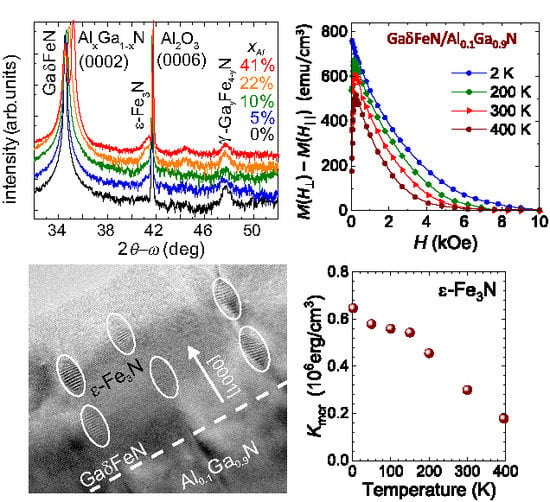
The large UMA along the growth direction is the origin of the pronounced squareness and the resemblance of the experimental
to the perpendicular magnetic anisotropy of bulk ferromagnets and layered structures. This is further demonstrated by the hard-axis-like shape of
. The magnitude of the UMA exerted by the considered ensemble of NCs is calculated by taking the experimental difference
, plotted for selected temperatures in
Figure 10c. By definition, the area under the
yields the magnitude of
. The established magnitudes are plotted against the corresponding magnitudes of
in
Figure 10d (diamonds). The nearly linear relationship
confirms the significant UMA in this ensemble, allowing the direct determination from Equation
(3) of
of
-Fe
N from the
T–dependence of
(
Figure 8). The resulting magnitudes of
established at all the measured temperatures, are shown in
Figure 10e (bullets). This is the first direct determination of the absolute magnitudes of
of
-Fe
N in such a broad and technologically relevant temperature range up to 400 K.
On the other hand, as indicated in
Figure 10b, the magnetization process in the Ga
FeN/Al
Ga
N structures is based on two rather independent switching processes. This is seen at the two temperatures exemplified in
Figure 10b. The
K case, where the thermal activation contribution to
can be neglected, is considered in detail. Here, about a third of the total magnetization of the NCs switches at very weak fields. This process completes at weak negative fields, where a kink is seen in
at about
kOe, marked by the arrows at
. Up to
about 30% of the total
M has switched or rotated to the new direction of
H. This is the result of a narrow band of weak switching fields brought about by the minority of the oblate NCs (which nominally reverse
M at
) and of several cubic
’-Ga
Fe
N NCs, which reverse
M at weak fields, as demonstrated in
Figure 10a. For the remaining 70% NCs, the switching process begins after
passes
and these are the prolate
-Fe
N NCs, which, due to their generally high
require larger magnitudes of
H to overcome the individual anisotropy fields
. Since the majority of the NCs is in the single domain state, the different magnitudes of
contribute to a broad distribution of switching (coercive) fields
, resulting in the wide
for
. From the magnitude of
,
kOe at low temperatures is obtained and it is also extrapolated directly from the
curve in
Figure 10b. Since the reversal process of
M of the prolate fraction of the NCs ensemble in the Ga
FeN/Al
Ga
N structures starts after the magnetically soft part of the ensemble has reversed, the
cannot be determined at
. The
after
is assigned to the prolate
-Fe
N, marked by the arrows in
Figure 10b, from where the corresponding
can be obtained. It is worth noting that the difference in
between the two branches of
corresponds to the magnitude of the soft part of
M which switches within
,
i.e. the magnetically hard part of
corresponding to the prolate NCs is broken up by the magnetically soft component of the distribution.

 ,
, 


