1. Introduction
Textiles are some of the most used materials in the world with a wide range of applications. Their diversified use in domains such as medicine [
1], clothing [
2], and electronics [
3] have transformed the textile industry. Classic methods of production are insufficient to satisfy the demands, so new techniques and apparatuses are needed to comply to the increased needs. The interest in developing new textiles with “smart properties” is very high [
4,
5] due to, among others, the evolution of antibiotic-resistant bacteria. Thus, cotton gauzes, medical gowns, and hospital bed sheets are just some examples of cotton textiles that could favor bacterial growth due to their moisture absorbing property.
The ionic state of Ag
+ has an antimicrobial effect and is used as a common microbial inhibitor in various commercial and medical products [
6]. Furthermore, silver nanoparticles showed antibacterial activity against
E. coli,
P. aeruginosa, and methicillin-resistant
S. aureus [
7]. A synergistic antibacterial effect was observed even when silver nanoparticles were combined with other natural and synthetic compounds [
8]. The method of obtaining stable dispersions of Ag and ZnO nanoparticles relied on using PVP (polyvinylpyrrolidone) and chitosan as dispersing matrices. Gamma irradiation offers an alternative to the classical approach of chemical reduction of silver ions in PVP and surfactants. Nonetheless, gamma irradiation is one of the most promising methods to produce Ag NPs at room temperature, using fewer reducing agents and byproducts. Increasing the γ-irradiation doses from 1 to 5 kGy enhanced the concentration of Ag NPs, indicated by UV–vis spectroscopy [
9].
Gamma irradiation is still gaining a lot of popularity in medicine [
10], the clothing industry [
11], grafting [
12], and even heritage artefact conservation [
13]. In the work of Geba et al. [
14] it was concluded that gamma irradiation at 10 kGy of accelerated aged silk and wool fabrics did not cause significant changes of their physical and chemical features.
Nanoparticles have a worldwide use, with an increasing trend in production every year [
15]. These materials are used in medicine, from devices [
16] to drug delivery [
17], electronics [
18], and textiles [
19]. Because of their heavy usage around the world, research is being conducted to reduce production cost, modify physical or chemical properties, or create new material types. Antibacterial fabrics were obtained by Truong et al. [
20] by gamma irradiation of cotton materials in an AgNO
3 chitosan solution.
The study’s aim was to propose replacing the high-energy consumption, classical textile finishing methods, Ref. [
21] used to fix the enhancing substance on the textile with the more ecological gamma technologies. For this objective, we chose to develop a basic set of methods for creating antibacterial textiles with gamma irradiation and nanoparticles combined treatment. The novelty of the study consists of preparing new antibacterial cotton textile materials by various nanoparticles and gamma irradiation treatments.
2. Materials and Methods
2.1. Silver and ZnO Nanoparticles Preparation
2.1.1. Ag Nanoparticles Were Prepared by Two Different Methods, Using Chitosan and PVP, Respectively
The Ag/chitosan (Ag/C) nanoparticles were prepared with a chitosan aqueous solution (concentration 0.2%), which was prepared by dissolving the polymer powder in 1% acetic acid under magnetic stirring (300 rpm) at room temperature for 12 h. The chitosan used was a product with average viscosity, molecular mass = 300 kDa, and an 82% degree of deacetylation (Sigma-Aldrich, St. Louis, MO, USA). In the chitosan solution, different volumes of AgNO
3 (0.1 M) were added as was 0.1 mL of NaOH 1 M for adjusting the pH and speeding up the reduction reaction. The mixture was left to react for 8 h at 90 °C. The verification of the nanoparticle obtaining was done by UV-Vis spectrophotometry; data are available in the
Supplementary Material.
Ag/PVP nanoparticles were obtained by the hydrothermal method by reducing silver salts. An AgNO
3 solution with a 0.1 M concentration was prepared, and 1 mL of NaBH4 0.5 M was added under strong stirring (300 rpm) for the reduction of Ag
+ ions. Afterwards, the Ag nanoparticles were dispersed in a 2% polyvinylpyrrolidone solution. The schematic chemical structure of the composites is given in the
Supplementary Materials along with their characterization.
2.1.2. Zinc Oxide Nanoparticles Preparation
The ZnO nanoparticles (ZnO/PVP) were obtained by hydrothermal synthesis at a normal pressure and moderate temperature. A total of 100 mL of Zn(NO
3)
2·6H
2O with a 0.5 M concentration was introduced into a 3 pits balloon, and 50 mL of NaOH was added under vigorous stirring (300 rpm) at 65 °C. The mixture became opalescent immediately, and stirring was continued for 8 h. Subsequently, the resulting suspension was filtered and washed 2–3 times with distilled water and alcohol. To increase the crystallinity factor, the ZnO nanopowder was calcined in the oven at 150 °C for 4 h. Then, the ZnO nanoparticles were dispersed in a polyvinylpyrrolidone 2% solution. Characterization of the nanoparticles is given in the
Supplementary Materials.
2.2. Textile Materials Treatment
Bleached, plain weave 100% cotton fabric with a weight per unit area of 168 g/m2 was used for the functionalization process. Sixteen samples were cut; one was kept as a reference (R), and 15 samples, i.e., 5 for each type of prepared composites, were impregnated using ultrasonic bath. One of the 5 impregnated samples was kept as an impregnated control (M), 2 were irradiated while still wet (W) at 5 kGy and 20 kGy (5W and 20W), respectively, and another 2 were irradiated at 5 kGy and 20 kGy in a dry state after drying at room temperature (5D and 20D).
Gamma Irradiation
The irradiation treatments were conducted at the IRASM Centre of Technological Irradiations of the Horia Hulubei National Institute for Physics and Nuclear Engineering. The cotton textiles were irradiated in a Gamma Cell 5000 research unit that uses Co-60 sources at a dose rate of 3.3 kGy/h. The doses were measured using the ECB (ethanol-chlorobenzene) method using ISO/ASTM 51538:2009. The uncertainty of the absorbed dose measurement was 5%, and the dose uniformity ratio (ratio between delivered maximum and minimum absorbed dose) was 1.1.
2.3. Materials Characterization
The cotton samples were characterized before and after irradiation and nanoparticles treatment by FTIR spectroscopy and thermal analysis. A Netzsch STA 409 PC Luxx Simultaneous Thermal Analyzer with a TG/DSC sample carrier was used for testing under an inert atmosphere (50 mL/min pure nitrogen). The tests were performed from 40 °C to 600 °C at 10 K/min in aluminum crucibles with pierced caps. The sample mass was about 10 mg. Each measurement was performed in triplicate. For thermal analysis, the cotton samples were cut in discs of about 5.5 mm (close to the size of the Al crucibles).
The FTIR spectra were obtained with a Vertex 70 equipment (Bruker Optics) as an average of 5 measuring points in the ATR mode using a diamond crystal accessory. The spectra were acquired with OPUS software for the range of 400–4000 cm−1 with 64 scans and were adjusted by vector normalization and baseline correction, available in the OPUS software package.
The antimicrobial activity of the treated textiles was tested against Pseudomonas aeruginosa (ATCC 9027) and Staphylococcus aureus (ATCC 6538) by the disc diffusion test. The textile samples were placed on Mueller–Hinton sterilized agar, and the agar plate was inoculated uniformly with 108 UFC/mL; lastly, the plates were incubated at 36 °C for 24 h. The bacteria developed and enveloped the medium, except the resulting sterile area named “inhibition zone”, generated by the antimicrobial textiles. After 24 h, the inhibition zone diameter was measured.
3. Results and Discussion
3.1. FTIR-ATR Analysis
FTIR spectra of reference cotton, impregnated, and gamma-irradiated textiles are shown in
Figure 1a–c.
No major modification in the cellulose textile FTIR spectra was induced by the presence of the nanocomposites or gamma irradiation as may be seen from
Figure 1a–c. In
Table 1, the main peaks of cotton cellulose and some of its impurities are presented based on the references from Gilbert et al. [
22] and Chung et al. [
23].
The broad peak of 3550–3100 cm−1 is attributed to the OH stretch, and the one at 2980–2800 cm−1 is the CH stretch. A first observation is the presence of the 2850 cm−1 peak correlated to the symmetric CH2 stretch and of a small shoulder at around 2916 cm−1 corresponding to the asymmetric CH2 stretch, related to the presence of waxes in the cotton. There are also two other peaks that appear at 1577 cm−1 and 1542 cm−1 that are attributed to impurities, and due to amide II, aromatic C=C stretching vibrations, or C-O vibrations from the COOR group. Due to their small intensity, all the above peaks of impurities were no more visible in the FTIR spectra of cotton materials after the nanocomposites’ application and irradiation.
Because of the low concentration of chitosan and PVP and the fact that many of their peaks overlap with the cellulose peaks, their presence could not be detected directly but indirectly as a decrease in the intensity of cellulose peaks. Differences in the case of peak intensities are small, around ±0.005 ATR units for the Ag/PVP and ZnO/PVP samples in all their variants, but for the Ag/C samples, there is a marked reduction in intensity for the 1054, 1030, and 1001 cm−1 peaks. All newly obtained materials have a similar tendency of a higher 3550–3100 peak intensity that may have been caused by increased hydrophilicity and a lower intensity for the 1054, 1030, and 1001 cm−1 peaks. Peak shifts are around ±0.5 cm−1, which shows that there are no major changes in the chemical structure for the cotton textile caused by the applied treatments.
The main spectral differences can be observed for the analyzed textile materials, especially for the C-O-C vibration mode of cellulose at about 1004 cm−1. The most important spectral changes, i.e., decrease of cellulose band intensity, is observed amid the control and the material impregnated and irradiated with Ag/chitosan nanoparticles, showing the adhesion of the nanoparticles on the material surface.
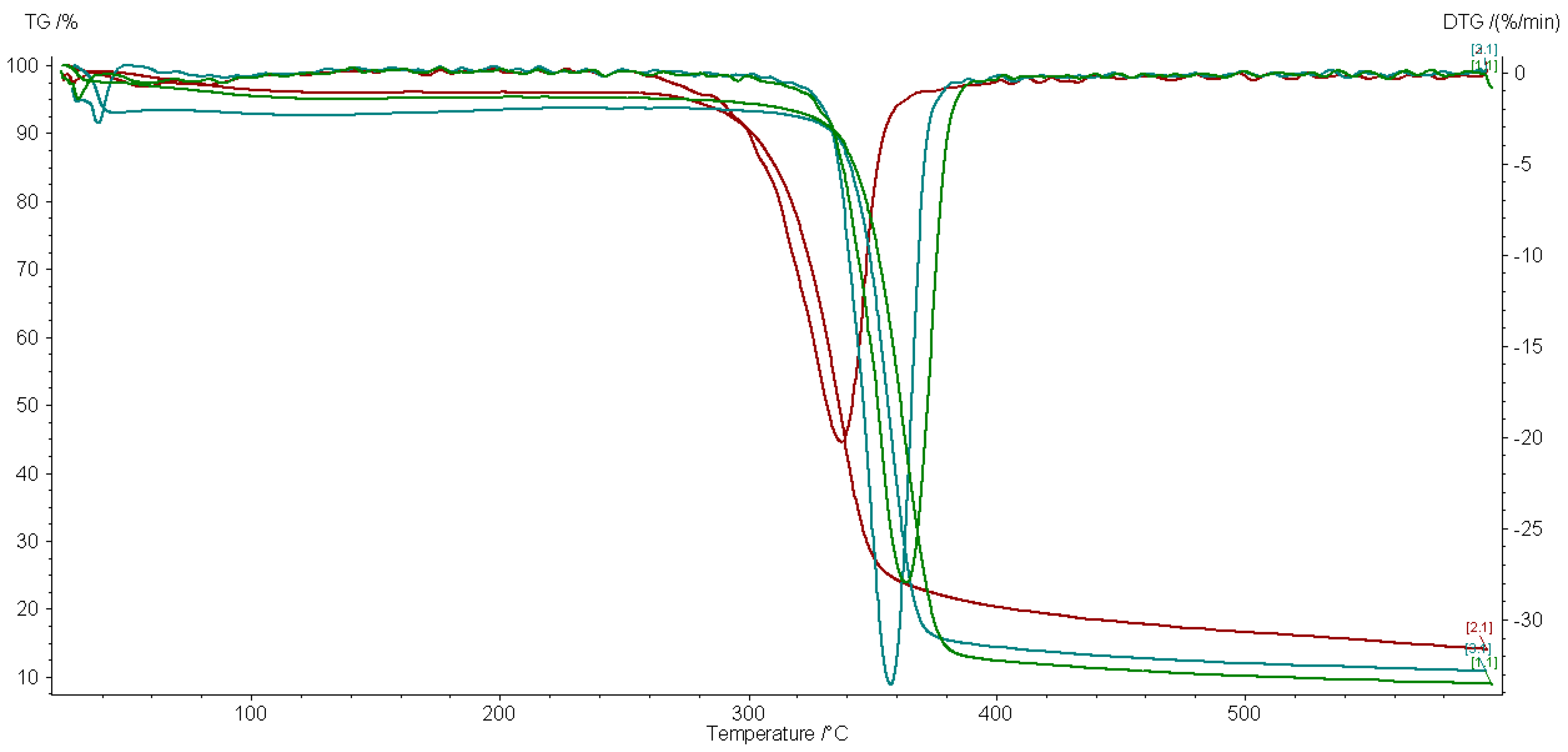
3.2. TG/DTG Analysis
Selected TG/DTG curves of impregnated and 5 kGy gamma-irradiated textiles are shown in
Figure 2 and
Figure 3.
The TG/DTG tests were done to observe the effect of our ecological gamma alternative treatment on the properties of the material. Each nanocomposite affects the thermal behavior of the cotton textile to a different extent.
Table 2 displays that the irradiation does not produce the same effect on each material in a dry or wet state. Ag/C-treated samples have the lowest residual mass and Ag/PVP the biggest residual mass in correlation with a lower thermal stability as revealed by the decomposition temperature.
The TG/DTG curves shown in
Figure 2 and
Figure 3 have only one decomposition peak corresponding to the decomposition of cellulose materials [
24]. The control sample has a maximum decomposition temperature at 365.2 °C, which is close to the decomposition temperature from the literature [
24,
25].
Both dry- and wet-state-treated samples have similar TG and DTG curves but with small differences: the wet-state-irradiated samples have a lower maximum decomposition temperature of around 2–3 °C while almost all the dry-state-treated samples have a lower thermal stability compared to the control group. The Ag/C-treated material is the one with the closest thermal parameters to the original materials. In a wet state, the radiolysis processes produce more reactive oxygen than in a dry state. These short life reactive species may further interact with the cotton support material. The thermal parameters of samples irradiated at 20 kGy have an even smaller variation toward the reference sample, as expected, due to the predominance of crosslinking effects against bond scission, which prevails at lower doses.
3.3. Antimicrobial Testing
The antimicrobial activity of tested samples using the disk diffusion method are represented in the
Figure 4, where nanoparticles impregnated textile disks were inoculated on Muller–Hinton agar plates with a
Staphylococcus aureus and
Pseudomonas aeruginosa 24 h fresh suspension.
Two potent antibiotics, gentamicin 10 mg and chloramphenicol 30 mg, were used as positive controls to compare the antimicrobial activity of textile samples. While chloramphenicol 30 mg managed to inhibit only the growth of
Staphylococcus aureus, gentamicin 10 mg showed a good inhibition zone for both tested microorganisms (see
Figure 5).
Both Ag/C and Ag/PVP, in all tested forms (M, 5D, 5W, 20D, 20W), exhibit an antimicrobial effect against Staphylococcus aureus and Pseudomonas aeruginosa, comparable with the control.
All variants of Ag/C display a strong effect on Staphylococcus aureus, with the inhibitory zones ranging from 9–10 mm. Pseudomonas aeruginosa displays slightly less sensitivity to Ag/C nanoparticles, with inhibitory areas of 7–8 mm.
The sample Ag/PVP, in all tested forms, displays similar antimicrobial inhibitory capacity for both tested microorganisms, with inhibition zones around 9–10 mm.
Both samples did not exhibit significant antimicrobial inhibitory differences between tested variances. Similar results were obtained by Truong et al. [
20].
The fact that the samples (textiles) impregnated with ZnO/PVP nanoparticles had no antimicrobial activity on tested microorganisms is surprising. The experiments will be continued using higher nanoparticles concentrations and other microorganisms.
4. Conclusions
While the nanocomposites’ presence has been identified indirectly by FTIR-ATR spectroscopy, thermal analysis proved the presence of the nanoparticles on the material along with the antimicrobial testing that emphasized that there is an important antimicrobial effect produced by Ag/C and Ag/PVP nanoparticles for the two selected studied microorganisms.
Compared to the standardized antibiotics disk used as a positive control, our materials show promising results, their antimicrobial capacity being around 25–30% of the tested standardized antibiotics (gentamicin 10 mg and chloramphenicol 10 mg). Further investigations should be performed on various concentrations and other pathogens, such as E. coli, Salmonella sp., and Klebsiella.
Small differences in the IR peak intensities prove that the selected doses of gamma irradiation do not have a major impact on the physical-chemical properties of the cotton textile material. The small decrease in thermal stability of the samples at 5 kGy gamma irradiation dose is observed in correlation with the known predominance effects of gamma irradiation of bond breaking at low doses and crosslinking at higher doses.
From a preliminary perspective, the experiment proved to be a success as we obtained antibacterial textiles while also providing some additional information about the interaction of gamma irradiation with the nanoparticles impregnated cotton material.
These findings support a starting point for further research in utilizing gamma technologies as a more ecological method to the classical textile finishing methods and possibly even as an alternative for the other textile processing steps.
Supplementary Materials
The following supporting information can be downloaded at:
https://www.mdpi.com/article/10.3390/ma15082734/s1, Figure S1: Chemical structure of the Ag/chitosan, Ag/PVP and ZnO/PVP composites, Figure S2: UV-Vis spectrum of dispersion of Ag NPs in 0.2% Chitosan polymer solution(right) the image of dispersion of Ag NPs prepared in Chitosan solution 0.2% after 48 h (left), Figure S3: SEM image of the ZnO nanopowder prepared by the hydrothermal method (a) and the size and size distribution of the nanoparticles for the sample obtained at 65 °C, before calcination.
Author Contributions
Conceptualization, L.C., L.O.C. and I.S.; methodology, L.C., L.O.C. and I.S.; validation, O.-A.C., B.L., M.V., M.C. (Mihai Constantin) and M.C. (Mihalis Cutrubinis); formal analysis, O.-A.C., B.L., M.V., M.C. (Mihai Constantin), M.C. (Mihalis Cutrubinis), L.C., L.O.C. and I.S.; investigation, L.C., L.O.C. and I.S.; resources, M.V.; data curation, O.-A.C.; writing—original draft preparation, O.-A.C.; writing—review and edit, O.-A.C., B.L., L.C., L.O.C. and I.S.; supervision, I.S.; project administration, L.C., L.O.C. and I.S.; funding acquisition, L.C., L.O.C. and I.S. All authors have read and agreed to the published version of the manuscript.
Funding
This research was funded by UEFISCDI, grant number 44 PCCDI. APC fee was paid by the University of Bucharest.
Institutional Review Board Statement
Not applicable.
Informed Consent Statement
Not applicable.
Data Availability Statement
Data are available on request from the authors.
Conflicts of Interest
The authors declare no conflict of interest.
References
- Mostafalu, P.; Kiaee, G.; Giatsidis, G.; Khalilpour, A.; Nabavinia, M.; Dokmeci, M.R.; Sonkusale, S.; Orgill, D.P.; Tamayol, A.; Khademhosseini, A. A textile dressing for temporal and dosage-controlled drug delivery. Adv. Funct. Mater. 2017, 27, 1702399. [Google Scholar] [CrossRef]
- Schmidt, A.; Paul, R.; Classen, E.; Morlock, S.; Beringer, J. 2 comfort testing and fit analysis of military textiles. In Performance Testing of Textiles, Woodhead Publishing Series in Textiles; Wang, L., Ed.; Woodhead Publishing: Amsterdam, The Netherlands, 2016; pp. 25–37. [Google Scholar] [CrossRef]
- Carey, T.; Cacovich, S.; Divitini, G.; Ren, J.; Mansouri, A.; Kim, J.M.; Wang, C.; Ducati, C.; Sordan, R.; Torrisi, F. Fully inkjet-printed two-dimensional material field-effect heterojunctions for wearable and textile electronics. Nat. Commun. 2017, 8, 1–11. [Google Scholar] [CrossRef] [PubMed] [Green Version]
- Mahmud, R.; Nabi, F. Application of nanotechnology in the field of textile. IOSR J. Polym. Text. Eng. 2017, 4, 2181p–2348p. [Google Scholar] [CrossRef]
- Mashitah, M.D.; Chan, Y.S.; Jaison, J. Antimicrobial properties of nanobiomaterials and the mechanism. In Nanobiomaterials in Antimicrobial Therapy, Applications of Nanobiomaterials; William Andrew Publishing, Elsevier: Amsterdam, The Netherlands, 2016; pp. 261–312. [Google Scholar]
- Zheng, K.; Setyawati, M.I.; Leong, D.T.; Xie, J. Antimicrobial silver nanomaterials. Coord. Chem. Rev. 2018, 357, 1–17. [Google Scholar] [CrossRef]
- Guzmán, M.G.; Dille, J.; Godet, S. Synthesis of silver nanoparticles by chemical reduction method and their antibacterial activity. Int. J. Chem. Biomol. Eng. 2009, 2, 104–111. [Google Scholar]
- Durán, N.; Nakazato, G.; Seabra, A.B. Antimicrobial activity of bio-genic silver nanoparticles, and silver chloride nanoparticles: An over-view and comments. Appl. Microbiol. Biotechnol. 2016, 100, 6555–6570. [Google Scholar] [CrossRef]
- Sheikh, N.; Akhavan, A.; Kassaee, M.Z. Synthesis of antibacterial silver nanoparticles by γ-irradiation. Radiat. Phys. Chem. 2009, 42, 132–135. [Google Scholar] [CrossRef]
- Landolt, A.M.; Lomax, N. Gamma knife radiosurgery for prolactinomas. J. Neurosurg. 2000, 93 (Suppl. 3), 14–18. [Google Scholar] [CrossRef]
- Chirila, L.; Popescu, A.; Cutrubinis, M.; Stanculescu, I.; Moise, V.I. The influence of gamma irradiation on natural dyeing properties of cotton and flax fabrics. Radiat. Phys. Chem. 2018, 145, 97–103. [Google Scholar] [CrossRef]
- Wang, J.-P.; Chen, Y.-Z.; Zhang, S.-J.; Yu, H.-Q. A chitosan-based flocculant prepared with gamma-irradiation-induced grafting. Bioresour. Technol. 2008, 99, 3397–3402. [Google Scholar] [CrossRef]
- Moise, V.; Stanculescu, I.; Vasilca, S.; Cutrubinis, M.; Pincu, E.; Oancea, P.; Raducan, A.; Meltzer, V. Consolidation of very degraded cultural heritage wood artefacts using radiation curing of polyester 180 resins. Radiat. Phys. Chem. 2019, 156, 314–319. [Google Scholar] [CrossRef]
- Geba, M.; Lisa, G.; Ursescu, C.M.; Olaru, A.; Spiridon, I.; Leon, A.L.; Stanculescu, I. Gamma irradiation of protein-based textiles for historical collections decontamination. J. Therm. Anal. Calorim. 2014, 118, 977–985. [Google Scholar] [CrossRef]
- Inshakova, E.; Inshakov, O. World market for nanomaterials: Structure and 185 trends. MATEC Web Conf. 2017, 129, 02013. [Google Scholar] [CrossRef]
- Eby, D.M.; Luckarift, H.R.; Johnson, G.R. Hybrid antimicrobial enzyme and silver nanoparticle coatings for medical instruments. ACS Appl. Mater. Interfaces 2009, 1, 1553–1560. [Google Scholar] [CrossRef]
- Kalishwaralal, K.; Luboshits, G.; Firer, M.A. Synthesis of Gold Nanoparticle: Peptide–Drug Conjugates for Targeted Drug Delivery; Springer: New York, NY, USA, 2020; pp. 145–154. [Google Scholar] [CrossRef]
- Huang, D.; Liao, F.; Molesa, S.; Redinger, D.; Subramanian, V. Plasticcompatible low resistance printable gold nanoparticle conductors for flexible electronics. J. Electrochem. Soc. 2003, 150, G412. [Google Scholar] [CrossRef] [Green Version]
- Chirila, L.; Radulescu, D.E.; Cinteza, L.O.; Radulescu, D.M.; Tanase, M.; Stanculescu, I.R. Hybrid materials based on ZnO and SiO2 nanoparticles as hydrophobic coatings for textiles. Ind. Text. 2020, 71, 297–301. [Google Scholar] [CrossRef]
- Hanh, T.T.; Van Phu, D.; Thu, N.T.; Hien, N.Q. Gamma irradiation of cotton fabrics in AgNO3 solution for preparation of antibacterial fabrics. Carb. Polym. 2014, 101, 1243–1248. [Google Scholar] [CrossRef]
- Available online: https://www.sciencedirect.com/topics/engineering/textile-finishing (accessed on 23 February 2022).
- Gilbert, C.; Kokot, S.; Meyer, U. Application of DRIFT Spectroscopy and Chemometrics for the Comparison of Cotton Fabrics. Appl. Spectrosc. 1993, 47, 741–748. [Google Scholar] [CrossRef]
- Chung, C.; Lee, M.; Choe, E.K. Characterization of cotton fabric scouring by FT-IR ATR spectroscopy. Carbohydr. Polym. 2004, 58, 417–420. [Google Scholar] [CrossRef]
- Siriviriyanun, A.; O’Rear, E.; Yanumet, N. Self-Extinguishing Cotton Fabric with Minimal phosphorous deposition. Cellulose 2008, 15, 731–737. [Google Scholar] [CrossRef]
- Alongi, J.; Colleoni, C.; Rosace, G.; Malucelli, G. Phosphorus- and nitrogen-doped silica coatings for enhancing the flame retardancy of cotton: Synergisms or additive effects? Polym. Degrad. Stab. 2013, 98, 579–589. [Google Scholar] [CrossRef]
| Publisher’s Note: MDPI stays neutral with regard to jurisdictional claims in published maps and institutional affiliations. |
© 2022 by the authors. Licensee MDPI, Basel, Switzerland. This article is an open access article distributed under the terms and conditions of the Creative Commons Attribution (CC BY) license (https://creativecommons.org/licenses/by/4.0/).


 ,
, 


