Article

Effect of Photoluminophore Light-Correcting Coatings and Bacterization by Associative Microorganisms on the Growth and Productivity of Brassica juncea L. Plants

by
Natalia S. Zakharchenko
1,
Elena B. Rukavtsova
1,
Ilia V. Yampolsky
2,
Dmitry O. Balakirev
3,
Ivan V. Dyadishchev
3,
Sergey A. Ponomarenko
3,
Yuriy N. Luponosov
3,
Andrey E. Filonov
4,
Pavel A. Mikhailov
4,
Anton N. Zvonarev
4,
Lenar I. Akhmetov
4,
Vasily V. Terentyev
5,
Alexandra Yu. Khudyakova
5,
Lubov V. Zalomova
6,
Sergey V. Tarlachkov
1,4,
Alexander V. Aripovsky
7,
Irina F. Puntus
4,* and
Robert N. Khramov
8
1
Branch of Shemyakin-Ovchinnikov Institute of Bioorganic Chemistry of the Russian Academy of Scences, Science Avenue 6, Pushchino, Moscow 142290, Russia
2
Shemyakin-Ovchinnikov Institute of Bioorganic Chemistry of the Russian Academy of Sciences, Miklukho-Maklaya, 16/10, Moscow 117997, Russia
3
Enikolopov Institute of Synthetic Polymeric Materials of the Russian Academy of Sciences, Profsoyuznaja ul. 70, Moscow 117373, Russia
4
Pushchino Scientific Center for Biological Research of the Russian Academy of Sciences, G.K. Skryabin Institute of Biochemistry and Physiology of Microorganisms of the Russian Academy of Sciences, Science Avenue 5, Pushchino, Moscow 142290, Russia
5
Pushchino Scientific Center for Biological Research of the Russian Academy of Sciences, Institute of Basic Biological Problems of the Russian Academy of Sciences, Institutskaya Str. 2, Pushchino, Moscow 142290, Russia
6
Pushchino Scientific Center for Biological Research of the Russian Academy of Sciences, Institute of Cell Biophysics of the Russian Academy of Sciences, Institutskaya Str. 3, Pushchino, Moscow 142290, Russia
7
R&D and Manufacturing Company ‘A-BIO’, Institutskaya ul. 4, Pushchino, Moscow 142290, Russia
8
Institute of Theoretical and Experimental Biophysics of the Russian Academy of Sciences, Institutskaya Str. 3, Pushchino, Moscow 142290, Russia
*
Author to whom correspondence should be addressed.
Microbiol. Res. 2024, 15(4), 1957-1972; https://doi.org/10.3390/microbiolres15040131 (registering DOI)
Submission received: 12 September 2024 / Revised: 23 September 2024 / Accepted: 24 September 2024 / Published: 25 September 2024

Abstract

:
The effect of a coating material containing organic photoluminophore (PL) on the growth and development of mustard Brassica juncea L. plants colonized with beneficial associative bacteria Pseudomonas putida KT2442 and Rhodococcus erythropolis X5 was studied in vitro and in vivo. Plants grown with the use of microbial bacterization in combination with a photoluminophore coating (PLC) had significantly faster growth rates in vitro (2.1 times faster, P. putida; 1.8 times faster, R. erythropolis) than those grown using PLC alone (1.2 times faster). The leaves of plants grown with PLC had higher contents of glucose and fructose (28.4 ± 0.3% more glucose and 60.4 ± 0.3% more fructose accumulated compared to plants grown without PLC). It was found that seed weights and seed number increased 1.9-fold and 1.6-fold, respectively, for plants grown with PLC and colonized with beneficial P. putida KT2442 bacteria. The stimulatory effect of PLC on photosynthetic parameters of Photosystem II (PSII) was observed in colonized plants grown in vitro. For the first time, it was shown that providing plants with a PLC for only 4 weeks may make it possible to support further plant growth without PLC to obtain higher yields in the future. Thus, PLCs that convert shorter-wavelength radiation into red light may induce enhancement of biochemical processes not only in plants but also in microorganisms that supply plants with growth regulators and other active compounds. The results indicate the need for further research to understand the mechanisms of photobiological and photoregulatory systems in the interaction of microbes and plants.

1. Introduction

The use of light-correcting coatings (films) as covering materials containing photoluminophores is a promising method that can be used to increase crop yield [1,2,3,4]. The efficiency of the use of this material for plant cultivation is due to the ability of light-correcting coatings to absorb the short-wavelength component of the light spectrum and transmit it into the red region of the spectrum (Figure 1) [2,5]. This process is also called the down-shifting of a single photon and involves the transformation of one absorbed high-energy photon into one lower-energy photon. It obeys Stokes law of wavelength change, known as Stokes shift [6]. Excessive exposure to UV radiation is known to induce damage to various cellular systems, which may lead to increased repair expenses and affect plant growth and photosynthesis. In addition to these, green light is less efficient for photosynthesis than red and blue light. The effects of different light spectral compositions on the photosynthetic apparatus and many other plant systems are mainly due to the activity of numerous plant photoreceptors [7].
With the application of light-correcting polymeric coating materials, it is possible to change the compositions of spectral light, controlling photosynthetic activity, respiration, and other processes that determine plant growth and development. New-generation polymeric coating materials can not only maintain higher temperatures when cold but also protect plants against UV radiation, which is harmful to plants at high doses, converting UV light to more useful red light. This range includes the absorption maximum of phytochromes, the key photoreceptors that stimulate plant growth [8].
It is worth noting that exposure to red light changes the structure of membranes; their permeability increases, thus contributing to an increase in the effectiveness of cytokinins and gibberellins, leading to an enhanced cell division rate. Along with it, plants grow and develop faster, and their productivity increases. At the same time, no changes in the content of photosynthetic pigments are observed [9,10,11,12,13].
Currently, there are many photoluminophores that are more or less suitable for creating light-correcting coatings. Photoluminophores based on organic dyes have an undeniable advantage due to their low cost and ease of use and are characterized by high luminescence yield [14,15,16,17,18]. The use of biodegradable light-correcting polymer coatings is an attractive approach to solving environmental pollution problems because, unlike plastic films, they can gradually decompose in the soil without releasing toxic residues into the environment [19,20]. The use of biodegradable light-correcting polymeric coatings can be an effective method of reducing soil pathogens and improving crop productivity. This approach can have a positive impact on beneficial soil microbiota [21,22,23]. Biodegradable polymeric films have already been tested on several crops, such as tomato, pepper, eggplant, melon, sweet corn, strawberry, zucchini, and lettuce [24,25,26,27,28,29].
Under natural conditions, plants exist in close association with a complex of endo- and exo-microorganisms, which have a stimulating effect on plant growth and development. This process stems from the ability of microorganisms to fix nitrogen, mobilize nutrients from soil, produce physiologically active substances, suppress phytopathogen growth, and detoxify foreign chemical compounds [30,31,32].
Previously, using the example of bacterial strains of the genus Pseudomonas, it was shown that light-correcting PL coating transforms sunlight in such a way that it stimulates the growth processes, population dynamics and oxygenase activity of local soil microflora in the process of crude oil degradation [33,34,35]. It is obvious that the use of light-correcting coatings promotes the effect of transformed light not only on the plants themselves but also on associative microorganisms (e.g., methylobacteria, pseudomonads, and rhodococci). The role of associative microorganisms in the stimulation of plant growth under PLC has not yet been practically investigated.
The aim of our work was to study the effect of PLC with luminescence maximum (660 nm) on plant growth and bioproductivity of Sarepta mustard Brassica juncea L. colonized with beneficial associative microorganisms P. putida KT2442 and R. erythropolis X5 under in vitro (climatic chamber) and in vivo (greenhouse “Biotron”) conditions. The choice of these bacteria was determined by the prospects for their use in agriculture and in soil bioremediation. The development of methods for stimulating and optimizing the growth of beneficial associative bacteria by means of PL is an important task of microbiology and biotechnology. The study of plant growth associated with beneficial bacteria under PLC is important for the development of methods to improve plant productivity and phytoremediation technologies. It is known that Sarepta mustard seed oil is used in food, pharmaceutical and cosmetic industries and also helps to strengthen immunity and improves the quality of vision and has bactericidal, wound healing, and analgesic properties.
Pseudomonas spp. is a promising group of PGPR (plant growth-promoting rhizobacteria) with a set of useful properties (growth stimulation, mobilization of minerals from the soil, protection of plants from pathogens, etc.), which makes them successful symbionts of various plant species. The Pseudomonas putida KT2442 strain also has the ability to utilize salicylate, a key intermediate in the biodegradation pathways of naphthalene, phenanthrene, anthracene, and other toxic and carcinogenic compounds that pollute the environment [36,37]. The accumulation of salicylates in the environment suppresses the growth of beneficial microorganisms, but bacteria such as P. putida KT2442, which utilize these aromatic compounds as a source of carbon and energy, neutralize this negative impact.
The Rhodococcus erythropolis X5 strain isolated from oil-contaminated soil is a psychrotrophic (cold-adapted) bacterium that degrades hydrocarbons (petroleum products) and releases biosurfactants; therefore, it has potential applications in bioremediation [38,39]. The ability to associate with plants makes R. erythropolis X5 a more versatile microorganism for plant and soil protection against abiotic stresses.
Previously, we obtained the first results of studies of the effect of photoluminescent coatings (PLCs) on plant growth in the presence of associative microorganisms [40]. These results served as a starting point for further studies. In a series of new experiments described in the presented article, we tried to evaluate the stimulating effect of PLCs in combination with bacterization on the photosynthetic parameters of photosystem II (PSII) of plants, on the content of hydroxy acids and sugars in plant leaves, and on plant productivity.

2. Materials and Methods

2.1. Objects of Study and Cultivation Conditions

Plants of the variety ‘Russian’ Sarepta mustard B. juncea L. were used in this work. Seeds were sterilized for 1 min in 70% ethanol (Okabiolab, Pushchino, Russia) and then 10 min in 2% sodium hypochlorite (Kaustik, Volgograd, Russia) and washed three times for 10 min each in sterile water. The seeds were germinated in vitro on hormone-free Murashige–Skoog medium [41] containing 9 g/L agar (Difco Lab., Detroit, MI, USA). Then, the plants were grown for 60 days under sterile conditions in a climate-controlled chamber (in vitro) at 22–24 °C in the daylight (under 16 h photoperiod) at a light intensity of 190 μmol m−2 s−1 (Osram L 36W/765 G13 Tubular fluorescent lamp (Smolensk, Russia)) and 65% relative humidity.
Associative bacterial strains P. putida KT2442 Kmr Gfp+ [36] and R. erythropolis X5 Hgmr Gfp+ [38] both containing the gfp gene were used for plant bacterization. Microorganisms were obtained from the Laboratory of Plasmid Biology, IBPM RAS, Pushchino, Russia. Bacterial cultures were grown in LB liquid medium [42] containing 10 g/L bacto-tryptone (Difco Lab., Detroit, MI, USA), 5 g/L yeast extract (Difco Lab., Detroit, MI, USA), and 10 g/L NaCl, on an orbital shaker (IBP RAS, Pushchino, Russia) at 28 °C, 160 rpm.
To determine the effect of associative bacteria on plant growth, maturation and productivity, colonized plants cultivated in vitro under PLC were transferred into 5 L pots containing a sterile peat–sand mixture (1:1) (Gera, Moscow, Russia) and were then grown indoors at the artificial climate station “Biotron” (BIBCh RAS, Pushchino, Russia). Plants were grown at 22–24 °C under a 16 h photoperiod and a 65% relative humidity. The artificial lighting environment (Sodium lamp with clear tubular outer bulb MASTER SON-T 400W (Philips, Moscow, Russia) at a light intensity (photosynthetically active radiation) of 190 μmol m−2 s−1 was additionally used in the evening. At the end of the growing season, seed weight and the number of seeds per plant were determined.
Light-correcting PLCs were used: polypropylene non-woven spunbond (20 ± 3 g/m2), coated with polylactide varnish with the organic photoluminophore (PL), with a resulting concentration for PL of 0.25% (w/w), which mainly absorbs in the 460–560 nm range and re-emits with a spectral maximum of 660 nm and a half-width of 610–730 nm. As PL, the luminescent molecule of the donor–acceptor type based on triphenylamine with effective radiation in the red range was used [43,44]. More detailed information on preparing the PLCs and PL characterization was described in [3].

2.2. Analysis of Auxin Content in the Culture Broth of P. putida KT2442 and R. erythropolis X5 Bacteria

The production of indole-3-acetic acid (IAA) by bacteria was assessed using the method presented in [45] with modifications. The bacteria were grown for 24 h in LB liquid medium at 28 °C and 160 rpm on an orbital shaker. For analysis, 1 mL of culture was centrifuged at 10,000× g for 6 min. Two parts of Salkowski’s reagent (0.1 g NH4Fe(SO4)2 × 12H2O in 100 mL of 50% H2SO4) were added to one part of the supernatant, incubated in the dark for 30 min, and the optical density was measured on a spectrophotometer Shimadzu UV-1800, Japan at 530 nm. A standard solution of IAA (Sigma-Aldrich, St. Louis, MO, USA) was used to plot a calibration curve for the determination of IAA concentration.

2.3. Bacterization of Plants and Analysis of Stability of Their Association with Plants

Sterile one-month-old mustard plants B. juncea L., grown from seeds, were colonized once by spraying a cell suspension (103–104 CFU/mL) of P. putida KT2442 or R. erythropolis X5 bacteria (20 plants for each variant). The bacteria were applied to the leaves using sterile brushes. Then, the glass test tubes containing plants were covered with PL and PL-free coating materials. Different plant explants (leaves, roots) were examined to determine whether they contained microorganisms in 7, 14, and 21 days after bacterization according to the method described in [46]. For this purpose, extracts obtained from colonized plants (100 mg of plant tissue) grown under PL and PL-free coating materials were plated on the Petri dishes with rich nutrient medium (LB) with selective antibiotics (10 µg/L hygromycin for R. erythropolis X5 strain and 50 µg/L kanamycin for P. putida KT2442) and incubated for 2 days at 22–24 °C. After two days, the CFU number per g of plant fresh weight was counted.

2.4. Microscopic Analysis of B. juncea L. Colonized Plants

A Zeiss Axio Imager A1 fluorescence microscope (Zeiss, Germany) was applied to detect the localization of microorganisms in leaves and roots of plants colonized with P. putida KT2442 and R. erythropolis X5 bacteria (after 3-week bacterization). An Axiocam 506C camera was used to take pictures.

2.5. Analysis of Sugars and Hydroxy Acids in Plants

2.5.1. Samples Preparation

Leaves of 8-week-old Sarepta mustard plants (0.5 g) were ground in a mortar with quartz sand and centrifuged at 10,000× g (Mini-Spin centrifuge, Eppendorf, Germany). The supernatant was used for analysis. To 50 µL of the tested plant juice, 50 µL of an aqueous solution of D-mannitol with a concentration of 1.0 mg/mL was added, and the solution was evaporated to dryness in a vacuum on a Speedvac rotary dryer (Savant Instrument, Midland, MI, USA). To the dry residue, 50 μL of a 2% solution of methoxyamine hydrochloride in pyridine was added and heated at 80 °C for 15 min, and then 120 μL of pure N,O-Bis(trimethylsilyl)trifluoroacetamide was added and further heated at 80 °C for 30 min. The composition of the obtained trimethylsilyl ethers mixture was analyzed via gas chromatography.

2.5.2. Chromatography

The analysis was performed on an analytical gas chromatograph HP5890 (Hewlett-Packard, Poway, CA, USA) with the following process parameters: quartz capillary column SPB-1, size 20 m × 0.2 mm × 0.2 µm, carrier gas helium (1.8 mL/min), with carrier gas flow split (1:65), 1 µL liquid sample was introduced, temperature analysis ranged from 80 °C (0.5 min) to 310 °C at a rate of 10 °C/min, and the temperatures of the evaporator and flame ionization detector (FID) were 280 and 320 °C, respectively. The HP3396A peak integrator was used for signal registration. The internal standard (D-mannitol) method was used for quantitative processing of the results [47].

2.6. Analysis of Photosynthetic Activity of Plants

Eight-week-old plants were used to analyze photosynthetic activity. Chlorophyll fluorescence was measured in vitro using a MULTI-COLOR PAM fluorometer (Waltz, Germany) on leaves of plants adapted to darkness for at least 30 min. The maximum quantum yield of PSII was calculated as Fm/Fv, where Fv = Fm − F0, where F0 is background chlorophyll fluorescence, and Fm is maximum fluorescence output induced by saturating flash (duration of 500 msec, λ = 625 nm, ~12,000 µmol photons m−2 s−1). The effective fluorescence quantum yield (Y(II)), quantum yields of regulated (Y(NPQ), and unregulated (Y(NO)) non-photochemical quenching of chlorophyll were calculated using the formulas given in the instrument manual (as follows [48]):
Y(II) = (Fm′ − F)/Fm′,
Y(NPQ) = F/Fm′ − F/Fm,
Y(NO) = F/Fm,
where Fm′ is the fluorescence maximum induced by a saturating flash, and F is the steady-state level of chlorophyll fluorescence in leaves adapted to light with an intensity of 212 µmol photons m−2 s−1. In this case, Y(II) + Y(NPQ) + Y(NO) = 1.

2.7. Statistical Analysis

Statistica 6.0 software and the MS Excel 2007 built-in tool were used for statistical data processing. Three analytical and three biological replicates were conducted to obtain the measurements. The graphs show mean values and their standard deviations. The Mann–Whitney test, a nonparametric statistical test, was used to assess whether differences were significant for the results obtained.

3. Results

3.1. The Effect of PLC on IAA Content in Bacterial Culture Medium

One of the valuable properties of associative microorganisms is the synthesis of auxin phytohormones, in particular, IAA. Auxins are known to influence cell division and differentiation as well as seed germination and promote the formation of lateral and adventitious roots. Root elongation increases the root surface area, thereby enhancing nutrient uptake, which provides plants with essential nutrients [49]. The treatment of plants with auxin-producing bacteria stimulates the development of their root system [50,51].
In our study, it was shown that the content of IAA varied depending on the type of coating. When bacteria were grown in culture medium in flasks coated without and with PL, the IAA contents in cells of P. putida strain KT2442 were 1.7 ± 0.1 and 2 ± 0.1 µg/mL, respectively (Table S1 of Supplementary Materials). For R. erythropolis X5 strain, the IAA contents were 1.6 ± 0.21 and 1.8 ± 0.15 µg/mL, respectively. Most likely, the greater number of the studied bacteria on plants under PLC was due to enhanced auxin synthesis, which led to the stimulation of plant root growth.

3.2. Bacterization of Plants by Associative Microorganisms

After the 3-week growth of mustard plants colonized with bacteria covered with the different coating materials (material with or without PL), the number of bacteria on plant roots was determined. It was found that the presence of PL in the covering material stimulated the growth of bacteria on the plant roots. It was shown that in plants under PLC, the number of P. putida KT2442 bacteria increased by 1.4 times and R. erythropolis X5 by 1.8 times, as compared to that on roots of plants grown and covered with material without PL. Our results indicate that 660 nm red light exerts a stimulating effect on bacterial growth during plant bacterization.
Plants colonized with strains P. putida KT2442 and R. erythropolis X5 exhibited increased growth rate and weight as compared to the control, non-colonized plants (Figure 2 and Figure 3).
Presumably, the intensification of metabolic processes in bacteria irradiated with PL- transformed light promotes a greater accumulation of physiologically active substances as well as increased mobilization of nutrients from the environment or soil, thereby having a favorable effect on plant growth. Thus, the average height of three-week-old colonized plants with P. putida (6.11 ± 0.27 cm) and R. erythropolis (5.4 ± 0.23 cm) under PLC was almost 2.1 and 1.7 times higher than that of control plants (non-colonized) under the agrotextile conditions without PL (2.8 ± 0.22 cm, respectively) (Figure 3a). The weight of colonized plants with P. putida (0.764 ± 0.078 g) and R. erythropolis (0.599 ± 0.028 g) under PLC was almost by 3.1 and 2.4 times higher than that of control plants (non-colonized) under agrotextile conditions without PL (0.245 ± 0.023 respectively) (Figure 3b). Red light is known to stimulate bacterial growth by increasing nucleic acid synthesis and cell respiration rate, which may induce certain changes in the metabolic regulatory program [10].
It is likely that plants grew better when they were covered with PLC because of the effect of light transformed by PL on the metabolism of bacteria. After the 60-day cultivation of plants in vitro, the plants were transferred to the greenhouse “Biotron”. All of the plants grew in soil without the use of PLC.

3.3. Microscopic Study of Colonized Plants

Microscopic analysis confirmed the presence of colonizing bacteria P. putida KT2442 and R. erythropolis X5 on plants grown in vitro (Figure 4). It was shown that the bacterial cells are unevenly distributed. This is especially noticeable on the leaves. Microbial cells are mainly concentrated in the intercellular space; perhaps, they are attracted by plant cellular metabolites. On the leaf surface and on the roots of Sarepta mustard plants grown under PLC, the number of bacteria was visually greater than that observed in plants grown without PLC, which was consistent with the CFU number per 1 g of fresh weight. In subsequent cycles of micropropagation of plants, the content of bacteria in leaves and roots remained unchanged throughout the entire vegetation period, indicating their strong association with plants.

3.4. Analysis of the Content of Hydroxy Acids and Sugars in Plant Leaves

The contents of hydroxy acids (malic and citric acids), as well as fructose and glucose, were analyzed in the leaf juice of mustard plants grown within 8 weeks (Figure 5, Table S2 of Supplementary Materials). Differences in the amount of sugars were revealed in plants grown under PLC: the content of glucose and fructose in leaves was higher by 28.4 ± 0.3% and 60.4 ± 0.3%, respectively, compared to plants grown without PLC. Practically no differences in the amounts of hydroxy acids were observed. The bacterization of plants by microorganisms R. putida KT2442 and R. erythropolis X5 had no effect on the content of sugars and hydroxy acids).

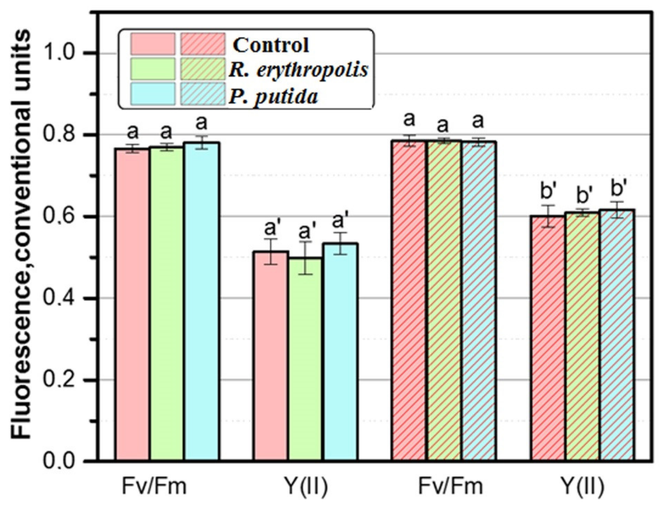
3.5. Analysis of Photosynthetic Apparatus Efficiency in Leaves of B. juncea L. Plants Grown In Vitro

The photosynthetic apparatus activity was analyzed in the leaves of Sarepta mustard plants grown within 8 weeks in vitro. The maximum chlorophyll fluorescence quantum yield (Fv/Fm), indicating the maximum capacity of PSII for photochemistry in dark-adapted samples, was ~0.77 in the leaves of the control plants (without bacterization and in the absence of PLC action) and was not significantly different in colonized plants alone or in plants grown under PLC (Figure 6).
However, when the effective chlorophyll fluorescence quantum yield (Y(II), which indicates the photochemistry capability of PSII in light-adapted samples) was measured, statistically significant differences were found in plants under PLC as opposed to PLC-free grown plants; Y(II) value for plants under PLC was higher by ~17% than that for plants without PLC. For plants colonized with P. putida KT2442 and R. erythropolis X5, these values were higher by ~22% and ~17%, respectively. Thus, the data obtained reveal a stimulating effect of the PLC on the photosynthetic parameters of PSII in plants in vitro.
The mechanism of the stimulatory effect of PLCs on photosynthesis is not fully understood. One possible mechanism is the activation of the phytochrome system in plants by fluorescent red light emitted by PL [2] in the spectrum of solar radiation: PL increases the red light/far-red light (RL/FRL) ratio and, consequently, the content of the active form of phytochrome. This can promote an increase in photosynthetic rate, stress tolerance, and plant growth [4,52,53]. Different plant responses are known to require a diverse set of photoreceptors to control them. These regulatory photoreceptors consist of a light-absorbing pigment (chromophore) bound to an effector protein molecule (apoprotein) and represented by phytochromes PHYA–E (RL/FRL red light/far-red light), cryptochromes CRY1, CRY2 and CRY3 (blue light/UV-A), photopropins PHOT1 and PHOT2, Zeitlupe photoreceptors, and underexplored photoreceptors UVR8 (UV-B) in the green spectrum range [54,55,56,57]. Under indoor conditions, the effect of PLC on PSII parameters is much more pronounced [3]. This is probably due to the peculiarities of plant growth in vitro, as the ability to photoautotrophy in this case depends on the type of nutrient medium, vessel coverage, and light conditions [58]. When transplanted from in vitro to soil conditions, plants gradually adapt to full photoautotrophy; the presence of microorganisms in the soil may be beneficial [59].

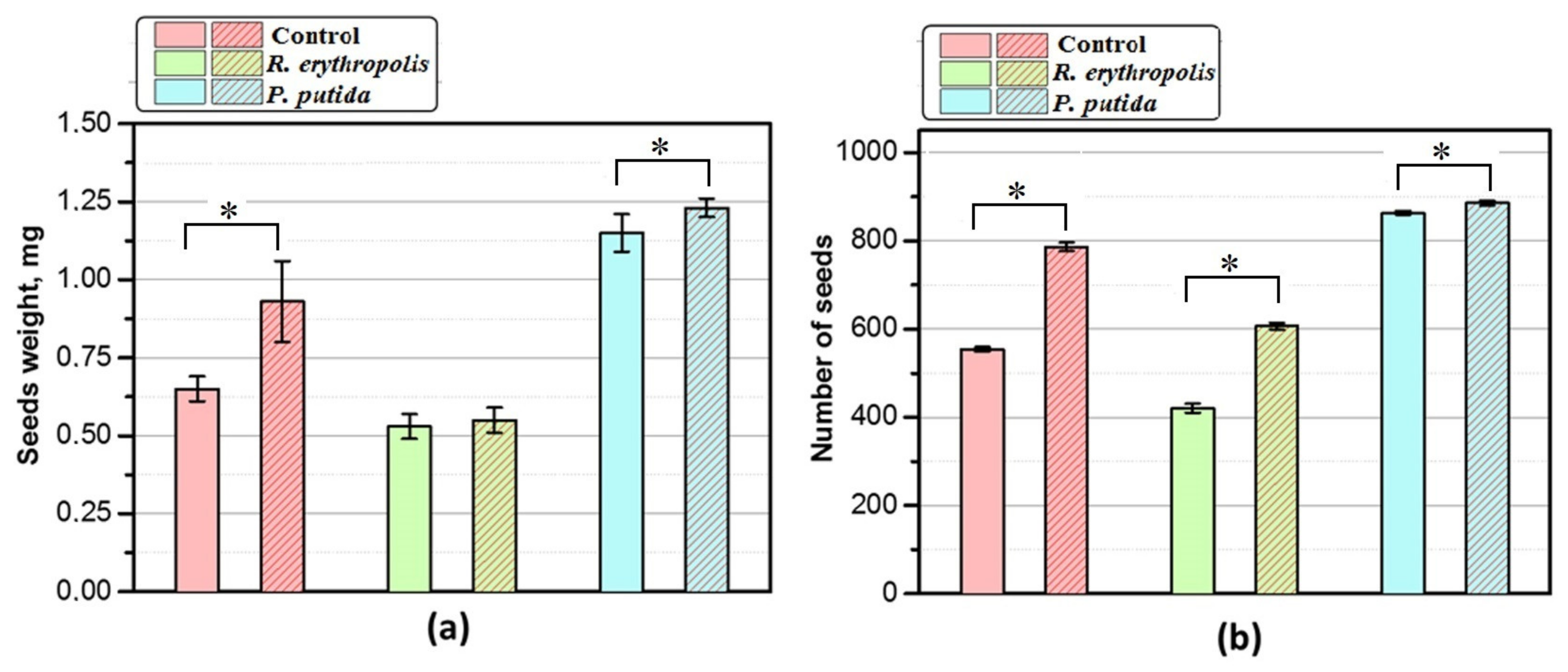
3.6. Plants Productivity Research

B. juncea L. plants colonized with P. putida strains KT2442 and R. erythropolis X5, grown in vitro under PLC for 8 weeks, were planted without coating in a greenhouse, where they grew for about 5 months until seed maturation. The productivity of all plants was higher when grown with PLC, compared to plants without PLC.
The weight (0.93 ± 0.13) and number (786.36 ± 9.47) of seeds were higher in control plants under PLC by 43% and 41.8%, respectively, as compared to plants grown without PLC (Figure 7). At the same time, the productivity of plants colonized with P. putida KT2442 was significantly higher than that of control plants. Thus, the seed weight of plants grown in vitro with PLC (1.23 ± 0.03) was higher (by 89%) compared to plants without bacterization and without PLC (0.65 ± 0.04) (control). The number of seeds of P. putida KT2442 colonized plants (885.6 ± 5.85) grown with PLC was higher (by 59.7%) than that in the control (554.2 ± 5.06) (Figure 7). Despite the fact that the growth and weight of colonized plants by R. erythropolis X5 in vitro were higher than those in the control, the productivity in the greenhouse was lower (Figure 7), which indicates the ambiguity of these bacteria. Such plants tolerated adaptation to in vivo conditions worse since the bacteria R. erythropolis X5 are characterized mainly as producers of biosurfactants for bioremediation of soils contaminated with oil products, and they do not have such antibiotic properties compared to Pseudomonas strains. Our data indicate a positive effect of associative bacteria P. putida KT2442 in combination with light-correcting agrotextile (PLC) on plant growth and productivity.

4. Discussion

Our studies showed that light passed through the light-correcting film and transformed by photoluminophores emitting with a maximum at 660 nm and a half-width of the spectrum of 610–730 nm stimulated plant and microbial growth processes. The combined application of plant bacterization and PLC resulted in significantly stronger plant growth stimulation compared to the use of these factors separately. An increase in the weight and number of seeds collected from plants colonized with beneficial bacteria P. putida KT2442 and grown under PLC was shown. P. putida KT2442 bacteria have a number of useful properties, such as the synthesis of physiologically active substances, the mobilization of nutrients from the soil, the inhibition of the growth of phytopathogens, and the detoxification of foreign chemical compounds. All of these contribute to the good adaptation of plants when transplanted from sterile conditions to the greenhouse. We have shown that the use of bacterization in combination with PLC, which converts shorter wavelength radiation into red light, can increase plant productivity by enhancing biochemical processes not only in plants but also in microorganisms that supply plants with growth regulators and other active compounds.
The potential of R. erythropolis X5 [38,39] is just beginning to be investigated; we were the first to study their ability for associative bacterization of plants and to show the ability of these bacteria to stimulate plant growth in vitro due to the expression of the gene for the synthesis of IAA and the use of PLC (Table S1 of Supplementary Materials). After transplanting the plants covered with PLC and colonized with R. erythropolis X5 in the greenhouse, they retained an increased growth rate compared to plants without PLC, but due to insufficient adaptation to in vivo conditions, the yields were lower compared to plants colonized with P. putida KT2442.
It turned out that the use of PLC, converting shorter-wavelength radiation into red light, may induce enhancement of biochemical processes not only in plants but also in microorganisms that supply plants with growth regulators and other active compounds. In this study, we found a stimulatory effect of PLC on PSII in colonized plants growing in vitro. Several mechanisms for the positive effect of PL used in light-correcting coatings are possible. For example, there may be an activation of the phytochrome system by the fluorescent red light emitted by PL [2]. Red light in the spectrum of solar radiation incident on plant leaves can increase the RL/FRL ratio and, consequently, the content of the active form of phytochrome. As a consequence, this can lead to increased photosynthetic rate, stress tolerance, and plant growth rate [4,52,53]. Previously, a marked increase in photosynthesis rate and biomass accumulation in cabbage and lettuce seedlings was shown when PL-containing coatings were used, which is associated with an increase in the proportion of orange-red light in the solar radiation spectrum [3].
Some researchers have shown that the incorporation of the organic dye Lumogen F-Red 300 into an agricultural polyethylene film, which converts part of the light in the green-yellow region of the spectrum to red, can be successfully used in tomato and rose cultivation [13]. The use of these materials as greenhouse covers increased tomato fruit yield by 19.6% and the number of flowering branches on rose bushes by 26.7% compared to covers without fluorescent dye. The positive effect of fluorescence in the blue region of the spectrum on the productivity of strawberry plants has previously been demonstrated by incorporating fluorescent pigments into coatings [14]. Because photosynthesis is sensitive to different components of the spectrum, the light of different wavelengths has different effects on the light and dark stages of photosynthesis and, ultimately, on biomass accumulation in plants [60,61]. The researchers found that photoconversion films accelerated the growth of eggplant, cucumber and tomato plants. The highest growth rate was observed in tomato plants, and the lowest was observed in cucumber. The leaf area of tomato plants growing under photoconversion coating was significantly larger (up to 50%) compared to control plants [62]. Eu3+-based films have been shown to absorb UV light and exhibit strong red luminescence when exposed to sunlight. Such coatings provide significant growth acceleration, size increase, and biomass production for crops (the Swiss chard) and trees (Japanese larch) in the northern region. Plants grown using Eu3+-based films had a 1.2-fold increase in height and a 1.4-fold increase in total biomass compared to those cultures without Eu3-luminophores [4].
Laboratory studies of bacterization and PLC under in vitro conditions can be a convenient model for studying the mechanisms of photoregulatory systems. Previously, photoregulatory systems in plants and microorganisms under the action of PL were studied separately [33,34]. We have shown for the first time the possibility of their joint action since plant growth stimulation by PL was improved in combination with bacterization by associative microorganisms. Previously, we showed that Sarepta mustard plants grown in the laboratory in vitro and transferred to the experimental field with PLC in the open ground had a significantly higher weight (1.5 times) than non-colonized plants, which confirms the stimulating effect of PLC in combination with bacterization with beneficial microflora [40]. There are few studies in which it has been demonstrated that when light-correcting coatings are used, soil microbiota, as well as changes in phytohormonal balance in plants, along with an increase in photosynthesis, can make a significant contribution to the stimulation of plant growth and development [9,63].
The strains of associative microorganisms that we selected for the bacterization of mustard plants (P. putida KT2442) are promising both for use in agriculture and for cleaning soils from oil pollution. It has been previously shown that sunlight passed through the light-correcting film and transformed by photoluminophores can stimulate growth processes and oxygenase activity of soil microflora during oil biodegradation [33,34,35].
In addition, it is very important that the organic PL used in this work does not contain toxic components. This is an advantage compared to previously used organometallic PLs containing moderately toxic rare earth metals [64] and colloidal nanocrystals based on highly toxic cadmium [65]. The use of a large variety of sheltering materials (polymer films, glasses, varnishes, textiles) containing organic PLs may be promising in agrobiotechnology not only for vegetable crops but also for various tree crops, which requires further study. A promising direction for agricultural practice may be the creation of polymeric covering film and agrotextiles containing organic photoluminophores, which we successfully tested for the first time in work [3] and used in this work. Photoluminophores of this type turned out to be quite photostable and effective, and by modifying their chemical composition, it is possible in the future to select light-converting properties (absorption and emission of sunlight) of these materials for different plants. An even more interesting direction for the future is the development of biodegradable materials [66]. The polylactide varnish used in this work to coat polypropylene spunbond can be successfully used in the future to create woven and non-woven biodegradable mulch covers with light-converting properties.
Currently, container pot-in-pot technology is a promising method of growing and propagating plants [67]. It is implemented in nurseries in the USA, Canada, and some EU countries. This method solves problems relating to overwintering, watering, feeding, drying out, and other region-specific issues. Our bacterization method with beneficial microflora combined with PLC can be used in this technology to increase the growth rate and yield and protect plants from stress factors (increased UV radiation).
Thus, there are preconditions for the practical use of PLC based on biodegradable polylactide materials containing organic PL in combination with the bacterization of plants with beneficial associative microorganisms.

5. Conclusions

The combined use of microbial bacterization and PLC resulted in significantly faster plant growth than the use of photoluminophore coating or bacterization alone. It was shown for the first time that even rather short-term in vitro cultivation of non-colonized and colonized plants (for 4 weeks) under PLC provides subsequent stimulation of productivity during plant cultivation without coatings in the greenhouse. The weight and number of seeds of plants of Sarepta mustard grown in vitro under PLC and colonized with P. putida KT2442 were higher than those of non-colonized plants grown both with and without PLC. The stimulating effect of PLC on photosynthetic parameters of Photosystem II (PSII) in colonized plants grown in vitro was revealed. For a deeper understanding of the mechanism of the stimulating effect of transformed light on the growth and biochemical activity of microflora, further research is needed. The study of the regulatory action of light and molecular–physiological processes of interactions between plants and associative microorganisms under the influence of red light is promising for both fundamental biological science and agrotechnological practice.

Supplementary Materials

The following supporting information can be downloaded at https://www.mdpi.com/article/10.3390/microbiolres15040131/s1, Table S1. Content of indole-3-acetic acid (µg/mL) in bacterial cultures of Pseudomonas putida KT 2442 and Rhodococcus erythropolis X5. Table S2. The content of hydroxy acids and sugars (mg·mL−1) in leaves of control, non-colonized Sarepta mustard plants (growth within eight weeks, in vitro).

Author Contributions

Investigation, N.S.Z., E.B.R., A.N.Z., V.V.T., A.Y.K., L.V.Z., A.V.A. and P.A.M.; methodology and writing—review and editing, A.E.F.; writing—original draft software, L.I.A. and E.B.R.; validation, S.V.T.; supervision, R.N.K. and I.V.Y.; conceptualization, formal analysis, I.F.P.; visualization, D.O.B. and I.V.D.; resources, S.A.P. and Y.N.L. All authors have read and agreed to the published version of the manuscript.

Funding

This research was supported by the grant of the Ministry of Science and Higher Education of the Russian Federation № 075-15-2023-610 “Herbicide screening platform based on self-sustained bioluminescence”.

Institutional Review Board Statement

Not applicable.

Informed Consent Statement

Not applicable.

Data Availability Statement

The data presented in this study are contained within this article and Supplementary Materials.

Conflicts of Interest

The authors declare no conflicts of interest.

Abbreviations

CFU—colony-forming units; GFP—green fluorescence protein; Hgm—hygromycin; Km—kanamycin; IAA—indole-3-acetic acid; PL—photoluminophore; PLC—photoluminophore coating; PLA—polylactic acid; PP—polypropylene; PSII—Photosystem II, RL/FRL—red light/far-red light.

References

  1. Golovatskaya, I.F.; Raida, V.S.; Leshchuk, R.I.; Karnachuk, R.A.; Minich, A.S.; Bolshakova, S.A.; Prikhodko, S.A.; Usacheva, E.Y.; Burkovskaya, V.T.; Tolstikov, G.A. Physiological and biochemical features of growth and productivity of vegetable crops when growing under light-correctingfilms. Sel’skokhoz. Biol. 2002, 37, 47–51. [Google Scholar]
  2. Khramov, R.N.; Kreslavski, V.D.; Svidchenko, E.A.; Surin, N.M.; Kosobryukhov, A.A. Influence of photoluminophore-modified agro textile spunbond on growth and photosynthesis of cabbage and lettuce plants. Opt. Express 2019, 28, 31967. [Google Scholar] [CrossRef] [PubMed]
  3. Khramov, R.; Kosobryukhov, A.; Kreslavski, V.; Balakirev, D.; Khudyakova, A.; Svidchenko, E.; Surin, N.; Ponomarenko, S.; Luponosov, Y. Luminescence of agrotextiles based on red-light-emitting organic luminophore and polypropylene spunbond enhances the growth and photosynthesis of vegetable plants. Front. Plant Sci. 2022, 13, 827679. [Google Scholar] [CrossRef] [PubMed]
  4. Shoji, S.; Saito, H.; Jitsuyama, Y.; Tomita, K.; Haoyang, O.; Sakurai, Y.; Okazaki, Y.; Aikawa, K.; Sasaki, K.; Fushimi, K.; et al. Plant growth acceleration using a transparent Eu3+-painted UV-to-red conversion film. Sci. Rep. 2022, 12, 17155. [Google Scholar] [CrossRef]
  5. Raida, V.S.; Ivanitskii, A.S.; Bushkov, A.V.; Andrienko, O.S.; Tolstikov, G.A. Investigation of peculiarities in conversion of the UV and visible sunlight by light transforming films with europium luminophores. Atmos. Ocean. Opt. 2003, 16, 1029–1034. [Google Scholar]
  6. Huang, X.; Han, S.; Huang, W.; Liu, X. Enhancing solar cell efficiency: The search for luminescent materials as spectral converters. Chem. Soc. Rev. 2013, 42, 173–201. [Google Scholar] [CrossRef] [PubMed]
  7. Pashkovskiy, P.; Ivanov, Y.; Ivanova, A.; Kreslavski, V.D.; Vereshchagin, M.; Tatarkina, P.; Kuznetsov, V.V.; Allakhverdiev, S.I. Influence of light of different spectral compositions on growth parameters, photosynthetic pigment contents and gene expression in scots pine plantlets. Int. J. Mol. Sci. 2023, 24, 2063. [Google Scholar] [CrossRef]
  8. Kreslavski, V.D.; Los, D.A.; Schmitt, F.J.; Zharmukhamedov, S.K.; Kuznetsov, V.V.; Allakhverdiev, S.I. The impact of the phytochromes on photosynthetic processes. Biochim. Biophys. Acta Bioenerg. 2018, 1859, 400–408. [Google Scholar] [CrossRef]
  9. Minich, A.S.; Minich, I.B.; Zelen’chukova, N.S.; Karnachuk, R.A.; Golovatskaya, I.F.; Efimova, M.V.; Raida, V.S. The role of low intensity red luminescent radiation in the control of Arabidopsis thaliana morphogenesis and hormonal balance. Russ. J. Plant Physiol. 2006, 53, 762–767. [Google Scholar] [CrossRef]
  10. He, W.; Chen, S.; Ling, D.; Gan, L.; Xu, Z.G. Effects of red and blue light on potato leaf senescence and tuber-seed yield. Russ. J. Plant Physiol. 2022, 69, 118. [Google Scholar] [CrossRef]
  11. Xu, J.M.; Liu, Y.; Liu, M.X.; Xu, Z.G. Proteomic, physiological, and anatomical analyses reveal the effects of red, blue, and white light on the growth of potato plantlets under in vitro culture. Rus. J. Plant Physiol. 2022, 69, 139. [Google Scholar] [CrossRef]
  12. Lu, C.H.; Liu, Y.C.; Hsu, Y.T.; Wang, H.L.; Der Chung, J. Enhancing flower stalk emergence in Phalaenopsis by red light supplementation. Am. J. Plant Sci. 2016, 7, 639–648. [Google Scholar] [CrossRef]
  13. Novoplansky, A.; Sachs, T.; Cohen, D.; Bar, R.; Bodenheimer, J.; Reisfeld, R. Increasing plant productivity by changing the solar spectrum. Sol. Energy Mater. 1990, 21, 17–23. [Google Scholar] [CrossRef]
  14. Hemming, S.; van Os, E.A.; Hemming, J.; Dieleman, J.A. The effect of new developed fluorescent greenhouse films on the growth of Fragaria × ananassa “Elsanta”. Eur. J. Hortic. Sci. 2006, 71, 145–154. [Google Scholar]
  15. Schettini, E.; de Salvador, F.R.; Scarascia-Mugnozza, G.; Vox, G. Radiometric properties of photoselective and photoluminescent greenhouse plastic films and their effects on peach and cherry tree growth. J. Hortic. Sci. Biotechnol. 2011, 86, 79–83. [Google Scholar] [CrossRef]
  16. Yalçın, R.A.; Ertürk, H. Improving crop production in solar illuminated vertical farms using fluorescence coatings. Biosyst. Eng. 2020, 193, 25–36. [Google Scholar] [CrossRef]
  17. Kang, J.H.; Yoon, H.I.; Lee, J.M.; Kim, J.P.; Son, J.E. Electron transport and photosynthetic performance in Fragaria × ananassa Duch. acclimated to the solar spectrum modified by a spectrum conversion film. Photosynth. Res. 2021, 151, 31–46. [Google Scholar] [CrossRef]
  18. Sánchez-Lanuza, M.B.; Menéndez-Velázquez, A.; Peñas-Sanjuan, A.; Navas-Martos, F.J.; Lillo-Bravo, I.; Delgado-Sánchez, J.M. Advanced photonic thin films for solar irradiation tuneability oriented to greenhouse applications. Materials 2021, 14, 2357. [Google Scholar] [CrossRef]
  19. Castronuovo, D.; Candido, V.; Margiotta, S.; Manera, C.; Miccolis, V.; Basile, M.; D’Addabbo, T. Potential of a corn-starch based biodegradable plastic film for soil solarization. Acta Hortic. 2005, 698, 201–205. [Google Scholar] [CrossRef]
  20. Tsia, H.Y.; Tsen, W.C.; Shu, Y.C.; Chuang, F.S.; Chen, C.C. Compatibility and characteristics of poly (butylene succinate) and propylene-coethylene copolymer blend. Polym. Test. 2009, 28, 875–885. [Google Scholar] [CrossRef]
  21. Balakrishna, A.N.; Lakshmipathy, R.; Bagyaraj, D.J.; Ashwin, R. Effect of soil solarization on native AM fungi and microbial biomass. Agric. Res. 2015, 4, 196–201. [Google Scholar] [CrossRef]
  22. Fernández-Bayo, J.D.; Achmona, Y.; Harrold, D.R.; Claypool, J.T.; Simmons, B.A.; Singer, S.W.; Dahlquist-Willard, R.M.; Stapleton, J.J.; Vander Gheynst, J.S.; Simmons, C.W. Comparison of soil biosolarization with mesophilic and thermophilic solid digestates on soil microbial quantity and diversity. Appl. Soil Ecol. 2017, 119, 183–191. [Google Scholar] [CrossRef]
  23. Di Mola, I.; Ventorino, V.; Cozzolino, E.; Ottaiano, L.; Romano, I.; Duri, L.G.; Pepe, O.; Mori, M. Biodegradable mulching vs traditional polyethylene film for sustainable solarization: Chemical properties and microbial community response to soil management. Appl. Soil Ecol. 2021, 163, 103921. [Google Scholar] [CrossRef]
  24. Moreno, M.M.; Moreno, A.; Mancebo, I. Comparison of different mulch materials in a tomato (Solanum lycopersicum L.) crop. Span. J. Agric. Res. 2009, 7, 454–464. [Google Scholar] [CrossRef]
  25. Waterer, D. Evaluation of biodegradable mulches for production of warm-season vegetable crops. Can. J. Plant Sci. 2010, 90, 737–743. [Google Scholar] [CrossRef]
  26. Costa, R.; Saraiva, A.; Carvalho, L.; Duarte, E. The use of biodegradable mulch films on strawberry crop in Portugal. Sci. Hortic. 2014, 173, 65–70. [Google Scholar] [CrossRef]
  27. Di Mola, I.; Cozzolino, E.; Ottaiano, L.; Duri, L.G.; Riccardi, R.; Spigno, P.; Leone, V.; Mori, M. The effect of novel biodegradable films on agronomic performance of zucchini squash grown under open-field and greenhouse conditions. Aust. J. Crop Sci. 2019, 13, 1810. [Google Scholar] [CrossRef]
  28. Cozzolino, E.; Giordano, M.; Fiorentino, N.; El-Nakhel, C.; Pannico, A.; Di Mola, I.; Mori, M.; Kyriacou, M.C.; Cola, G.; Rouphael, Y. Appraisal of biodegradable mulching films and vegetal-derived biostimulant application as eco-sustainable practices for enhancing lettuce crop performance and nutritive value. Agronomy 2020, 10, 427. [Google Scholar] [CrossRef]
  29. Zhao, Z.; Wu, H.; Jin, T.; Liu, H.; Men, J.; Cai, G.; Cernava, T.; Duan, G.; Jin, D. Biodegradable mulch films significantly affected rhizosphere microbial communities and increased peanut yield. Sci. Total Environ. 2023, 871, 162034. [Google Scholar] [CrossRef]
  30. Kloepper, J.W.; Schroth, M.N.; Miller, T.D. Effects of rhizosphere colonization by plant growth promoting rhizobacteria on potato plant development and yield. Phytopathology 1980, 70, 1078–1082. [Google Scholar] [CrossRef]
  31. Welbaum, G.; Sturz, A.V.; Dong, Z.; Nowak, J. Managing soil microorganisms to improve productivity of agroecosystems. Crit. Rev. Plant Sci. 2004, 23, 175–193. [Google Scholar] [CrossRef]
  32. Patten, C.L.; Glick, B.R. The role of bacterial indoleacetic acid in development of the host plant root system. Appl. Environ. Microbiol. 2002, 68, 3795–3801. [Google Scholar] [CrossRef] [PubMed]
  33. Svarovskaya, L.I.; Altunina, L.K.; Filatov, D.A. Biodegradation of petroleum hydrocarbons by soil microflora activated with photoluminescent films. Pet. Chem. 2007, 47, 219–224. [Google Scholar] [CrossRef]
  34. Svarovskaya, L.I.; Altunina, L.K.; Filatov, D.A. Activation of indigenous micorflora in oil-contaminated soils using photoluminescent films. Appl. Biochem. Microbiol. 2008, 44, 585–589. [Google Scholar] [CrossRef]
  35. Filatov, D.A.; Svarovskaya, L.I.; Ovsyannikova, V.S.; Altunina, L.K. The effect of film-corrected light on oxygenase activity of microorganisms of the genus Pseudomonas. Microbiology 2011, 80, 158–163. [Google Scholar] [CrossRef]
  36. Smalla, K.; Heuer, H.; Götz, A.; Niemeyer, D.; Krögerrecklenfort, E.; Tiedze, E. Exogenous isolation of antibiotic resistance plasmids from piggery manure slurries reveals a high prevalence and diversity of IncQ-like plasmids. Appl. Environ. Microbiol. 2000, 66, 4854–4862. [Google Scholar] [CrossRef]
  37. Habe, H.; Omori, T. Genetics of polycyclic aromatic hydrocarbon metabolism in diverse aerobic bacteria. Biosci. Biotechnol. Biochem. 2003, 67, 225–243. [Google Scholar] [CrossRef]
  38. Delegan, Y.; Valentovich, L.; Petrikov, K.; Vetrova, A.; Akhremchuk, A.; Akimov, V. Complete genome sequence of Rhodococcus erythropolis X5, a psychrotrophic hydrocarbon-degrading biosurfactant-producing bacterium. Microbiol. Resour. Announc. 2019, 8, e01234-19. [Google Scholar] [CrossRef]
  39. Abashcheva, M.A.; Horavidi, E.X.; Akatova, E.V.; Nechaeva, I.A.; Babkina, E.E.; Ponamoreva, O.N. Influence of physico-chemical environmental factors on the surface properties of biosurfactant produced by the strain Rhodococcus erythropolis X5. Izv. TulGU 2023, 4, 28–37. [Google Scholar] [CrossRef]
  40. Zakharchenko, N.S.; Anisimov, S.; Dyadishchev, I.V.; Ponomarenko, S.A.; Robert Khramov, R.N. Effect of light-converting coatings on the growth of Sarepta mustard (Brassica juncea L.) plants colonized by associative microorganisms. BIO Web Conf. 2022, 42, 01024. [Google Scholar] [CrossRef]
  41. Murashige, T.; Skoog, F.A. Revised medium for rapid growth and bioassays with tobacco cultures. Physiol. Plant. 1962, 15, 473–497. [Google Scholar] [CrossRef]
  42. Sambrook, J.; Fritsch, E.E.; Maniatis, T. Molecular Cloning: A Laboratory Manual; Cold Spring Harbor Lab. Press: Cold Spring Harbor, NY, USA, 1989; 1626p. [Google Scholar]
  43. Luponosov, Y.N.; Solodukhin, A.N.; Balakirev, D.O.; Surin, N.M.; Svidchenko, E.A.; Pisarev, S.A.; Fedorov, Y.V.; Ponomarenko, S.A. Triphenylamine-based luminophores with different side and central aromatic blocks: Synthesis, thermal, photophysical and photochemical properties. Dyes Pigments 2020, 179, 108397. [Google Scholar] [CrossRef]
  44. Balakirev, D.O.; Solodukhin, A.N.; Peregudova, S.M.; Svidchenko, E.A.; Surin, N.M.; Fedorov, Y.V.; Ponomarenko, S.A.; Luponosov, Y.N. Luminescent push-pull triphenylamine-based molecules end-capped with various electron-withdrawing groups: Synthesis and properties. Dyes Pigments 2023, 208, 110777. [Google Scholar] [CrossRef]
  45. Gordon, S.A.; Weber, R.P. Colorimetric estimation of indoleacetic acid. Plant Physiol. 1951, 26, 192. [Google Scholar] [CrossRef] [PubMed]
  46. Kandel, S.L.; Herschberger, N.; Kim, S.H.; Doty, S.L. Diazotrophic endophytes of poplar and willow for growth promotion of rice plants in nitrogen-limited conditions. Crop Sci. 2015, 55, 1765–1772. [Google Scholar] [CrossRef]
  47. Knapp, D.R. Handbook of Analytical Derivatization Reactions; Wiley-InterScience Publication: Hoboken, NJ, USA, 1979; 768p. [Google Scholar]
  48. Genty, B.; Harbinson, J.; Cailly, A.L.; Rizza, F. Fate of excitation at PS II in leaves: The non-photochemical side. In Proceedings of the Third BBSRC Robert Hill Symposium on Photosynthesis, University of Sheffield, Department of Molecular Biology and Biotechnology, Western Bank, Sheffield, UK, 31 March–3 April 1996. Abstract no. P28 31. [Google Scholar]
  49. Tsavkelova, E.A.; Cherdyntseva, T.A.; Botina, S.G.; Netrusov, A.I. Bacteria associated with orchid roots and microbial production of auxin. Microbiol. Res. 2007, 162, 69–76. [Google Scholar] [CrossRef]
  50. Kudoyarova, G.; Arkhipova, T.; Korshunova, T.; Bakaeva, M.; Loginov, O.; Dodd, I.C. Phytohormone mediation of interactions between plants and non-symbiotic growth promoting bacteria under edaphic stresses. Front. Plant Sci. 2019, 10, 1368. [Google Scholar] [CrossRef]
  51. Arkhipova, T.N.; Evseeva, N.V.; Tkachenko, O.V.; Burygin, G.L.; Vysotskaya, L.B.; Akhtyamova, Z.A.; Kudoyarova, G.R. Rhizobacteria inoculation effects on phytohormone status of potato microclones cultivated in vitro under osmotic stress. Biomolecules 2020, 10, 1231. [Google Scholar] [CrossRef]
  52. Kasperbauer, M.J. Phytochrome involvement in regulation of the photosynthetic apparatus and plant adaptation. Plant Physiol. Biochem. 1988, 26, 519–524. [Google Scholar]
  53. Cao, K.; Yu, J.; Xu, D.; Ai, K.; Bao, E.; Zou, Z. Exposure to lower red to far-red light ratios improve tomato tolerance to salt stress. BMC Plant Biol. 2018, 18, 92. [Google Scholar] [CrossRef]
  54. Senger, H.; Schmidt, W. Diversity of Photoreceptors. In Photomorphogenesis in Plants, 2nd ed.; Kendrick, R.E., Kronenberg, G.H.M., Eds.; Springer: Dordrecht, The Netherlands, 1994; pp. 137–158. [Google Scholar] [CrossRef]
  55. Neff, M.M.; Fankhauser, C.; Chory, J. Light: An indicator of time and place. Genes Dev. 2000, 14, 257–271. [Google Scholar] [CrossRef] [PubMed]
  56. Devlin, P.F.; Christie, J.M.; Terry, M.J. Many hands make light work. J. Exp. Bot. 2007, 58, 3071–3077. [Google Scholar] [CrossRef] [PubMed]
  57. Kami, C.; Lorrain, S.; Hornitschek, P.; Fankhauser, C. Light-regulated plant growth and development. Curr. Top. Dev. Biol. 2010, 91, 29–66. [Google Scholar] [CrossRef]
  58. Demenko, V.I.; Lebedev, V.I. Adaptation of plants obtained in vitro to non-sterile conditions. RSAU–MTAA Bull. 2011, 1, 60–70. [Google Scholar]
  59. Zakharchenko, N.S.; Furs, O.V.; Rukavtsova, E.B.; Tarlachkov, S.V.; Kosobryukhov, A.A.; Kreslavski, V.D.; Dyachenko, O.V.; Buryanov, Y.I.; Shevchuk, T.V. The Brassica napus L. plants expressing antimicrobial peptide cecropin P1 are safe for colonization by beneficial associative microorganisms. J. Plant Interact. 2021, 16, 432–442. [Google Scholar] [CrossRef]
  60. Bian, Z.H.; Yang, Q.C.; Liu, W.K. Effects of light quality on the accumulation of phytochemicals in vegetables produced in controlled environments: A review. J. Sci. Food Agric. 2015, 95, 869–877. [Google Scholar] [CrossRef]
  61. Martirosyan, Y.T.; Martirosyan, L.Y.; Kosobryukhov, A.A. Dynamic regulation of photosynthetic processes under variable spectral led irradiation of plants. Agric. Biol. 2019, 54, 130–139. [Google Scholar] [CrossRef]
  62. Gudkov, S.V.; Simakin, A.V.; Bunkin, N.F.; Shafeev, G.A.; Astashev, M.E.; Glinushkin, A.P.; Grinberg, M.A.; Vodeneev, V.A. Development and application of photoconversion fluoropolymer films for greenhouses located at high or polar latitudes. J. Photochem. Photobiol. B 2020, 213, 112056. [Google Scholar] [CrossRef]
  63. Minich, A.S.; Minich, I.B.; Shaitarova, O.V.; Permyakova, N.L.; Zelenchukova, N.S.; Ivanitsky, A.E.; Filatov, D.A.; Ivlev, G.A. Vital activity of Lactuca sativa and soil microorganisms under fluorescent films. Vestn. TGPU 2011, 8, 78–84. [Google Scholar]
  64. Kusnetsov, S.I.; Leplianin, G.V.; Murinov, Y.I.; Schelokov, R.N.; Karasev, V.E.; Tolstikov, G.A. “Polisvetan”, a high-performance material for cladding greenhouses. Plasticulture 1989, 3, 13–20. [Google Scholar]
  65. Simakin, A.V.; Ivanyuk, V.V.; Dorokhov, A.S.; Gudkov, S.V. Photoconversion fluoropolymer films for the cultivation of agricultural plants under conditions of insufficient insolation. Appl. Sci. 2020, 10, 8025. [Google Scholar] [CrossRef]
  66. Kopitar, D.; Marasovic, P.; Jugov, N.; Schwarz, I. Biodegradable nonwoven agrotextile and films. Polymers 2022, 2, 2272. [Google Scholar] [CrossRef]
  67. Available online: http://www.tnstate.edu/faculty/ablalock/documents/Pot-N-Pot.pdf (accessed on 1 December 2009).
Figure 1. The ability of light-correcting coatings to absorb short-wavelength component of the light spectrum and transmit it into the red region of the spectrum: (a) normalized absorption (black) and luminescence (red) spectra of polylactic acid (PLA) films (thickness is ca. 40 μm) containing (w/w 0.25%) luminophore PL; (b) on a black backing polypropylene (PP) agrotextiles (pieces 2 × 5 cm in size) in the daylight (solar radiation) and UV-irradiation only (365 nm maximum): I—with PLA + PL (w/w 0.25%); II—with PLA only; (c) changes in the absorption spectrum of artificial light (Solar Irradiance) after passing through the reference PP + PLA textile and agrotextiles with addition (w/w 0.25%) of the PL (PP + PLA + PL ); (d) changes in the absorption spectrum of light (lamp irradiance) after passing through the reference PP + PLA textile and agrotextiles with addition (w/w 0.25%) of PL (PP + PLA + PL). Arrows indicate the fit of the graphs to the ordinate axis used.
Microbiolres 15 00131 g001
Figure 2. Brassica juncea L. plants (growth within 3 weeks, in vitro) without PL and under PL. 1–2—control, non-colonized plants; 3–4—colonized by R. erythropolis X5; 5–6—colonized by P. putida KT2442.
Microbiolres 15 00131 g002
Figure 3. Development of Sarepta mustard plants (growth within 3 weeks, in vitro) depending on bacterization by R. erythropolis X5 (green columns), and P. putida KT2442 (light blue columns) and photoluminophore coating. Pink columns represent control plants without bacterization; unshaded columns, plants with coating without PL; shaded columns, plants under PLC. (a) height of plants (cm); (b) weight of plants (g). * Statistically significant at Mann–Whitney test (p < 0.05).
Microbiolres 15 00131 g003
Figure 4. Microscopic analysis of Sarepta mustard plant tissues colonized with R. erythropolis X5 (growth within 3 weeks, in vitro): (a) leaves without PL; (b) leaves under PL, (c) roots without PL, (d) roots under PL. Arrows indicate individual bacterial cells. Bar = 10 µm.
Microbiolres 15 00131 g004
Figure 5. The content of hydroxy acids (malic and citric acids) and sugars (fructose, glucose) in leaves of control, non-colonized Sarepta mustard plants (growth within eight weeks, in vitro). Unshaded columns—coating without PL; shaded ones—under PLC. * Statistically significant at Mann–Whitney test (p < 0.05).
Microbiolres 15 00131 g005
Figure 6. Maximum quantum yield (Fv/Fm) and efficient one (YII) of PSII in Sarepta mustard plants without (control, pink) and with bacterization by R. erythropolis X5 (green), and P. putida KT2442 (light blue) depending on the application of PLC: unshaded columns—no PLC applied; shaded ones—under PLC. Data are presented as a mean ± SD (n = 6–8 Letters indicate statistically significant difference in the results at p < 0.05).
Microbiolres 15 00131 g006
Figure 7. Productivity of Sarepta mustard plants depending on bacterization and photoluminophore coating (without PL or under PL), growth within 5 months, in vivo: (a) seed weight; (b) number of seeds. Pink columns—control, without bacterization; green—bacterization by R. erythropolis X5; light blue—P. putida KT2442. Unshaded columns—coating without PL; shaded ones—under PLC. * Statistically significant at Mann–Whitney test (p < 0.05).
Microbiolres 15 00131 g007
Disclaimer/Publisher’s Note: The statements, opinions and data contained in all publications are solely those of the individual author(s) and contributor(s) and not of MDPI and/or the editor(s). MDPI and/or the editor(s) disclaim responsibility for any injury to people or property resulting from any ideas, methods, instructions or products referred to in the content.

