Article

Genome-Wide Identification of the Rehmannia glutinosa miRNA Family and Exploration of Their Expression Characteristics Caused by the Replant Disease Formation-Related Principal Factor

1
College of Bee Science and Biomedicine, Fujian Agriculture and Forestry University, Fuzhou 350002, China
2
Key Laboratory of Ministry of Education for Genetics, Breeding and Multiple Utilization of Crops, Fujian Agriculture and Forestry University, Fuzhou 350002, China
3
College of Bioengineering, Henan University of Technology, Zhengzhou 450001, China
4
School of Pharmacy, Henan University of Chinese Medicine, Zhengzhou 450046, China
*
Author to whom correspondence should be addressed.
Genes 2024, 15(9), 1239; https://doi.org/10.3390/genes15091239
Submission received: 17 August 2024 / Revised: 15 September 2024 / Accepted: 19 September 2024 / Published: 23 September 2024
(This article belongs to the Section Plant Genetics and Genomics)

Abstract

:
Background/Objectives: Rehmannia glutinosa, a highly valuable medicinal plant in China, is encountering severe replant disease. Replant disease represents a complex stress driven by multiple principal factors (RDFs), including allelochemicals, microbes, and their interactions. miRNAs are recognized as key regulators of plant response to stresses; however, their specific roles within RDFs are not entirely clear. Methods: This study builds six RDF treatments, comprising R. glutinosa continuously planted (SP), normally planted (NP), and NP treated with ferulic acid (FA), Fusarium oxysporum (FO), and a combination of FA with FO (FAFO). sRNA-seq technology was used to identify crucial miRNAs in response to diverse RDFs. Results: In total, 30 sRNA datasets were generated from the SP, NP, FA, FO, and FAFO samples. A total of 160 known and 41 novel miRNAs (RgmiRNAs) were identified in the R. glutinosa genome based on the sRNA database. Abundance analysis revealed that RgmiRNAs in SP exhibited a distinct expression profile in comparison with others. Of these, 124, 86, 86, and 90 RgmiRNAs were differentially expressed in SP, FA, FO, and FAFO compared with NP. Target analysis indicated that RgmiRNAs downregulated in both SP and RDFs impede the organism growth of R. glutinosa. RgmiRNAs upregulated in SP can disrupt root formation and nutrient metabolism, in which, two RgmiR398 were uniquely expressed in SP. It was confirmed to target RgCSD genes. The expression patterns of RgmiR398 and RgCSD indicated that replant disease induces the oxidative damage of R. glutinosa through RgmiR398. Conclusions: RgmiRNA profiling under RDFs provides a theoretical basis for the further clarification of RgmiRNA function in replant disease.

1. Introduction

Rehmannia glutinosa (Gaert.) Libosch. (R. glutinosa) is a perennial herb belonging to the Rehmannia genus of the Scrophulariaceae family [1], which comprises six species. Of these, only R. glutinosa is widely cultivated in China, with its tuberous roots employed as raw materials for traditional Chinese herbal medicine, Rehmanniae radix in Chinese Pharmacopoeia [1,2]. At present, a number of significant active ingredients have been identified in the tuberous roots, fibrous roots, and leaves of R. glutinosa, including monosaccharides, polysaccharides, phenylethanoid, iridoid glycosides, catalpol, and trichitosans. The in vitro and in vivo experiments have demonstrated that these ingredients exhibit a range of pharmacological activities, such as antitumour, immunomodulatory, cardiovascular, cardioprotective, cerebrovascular, and antioxidant effects [1,2,3]. In recent years, with the increase in market demand for R. glutinosa and the reduction in cultivated areas, R. glutinosa has begun to be monoculturally cultivated in a large area in production. However, the continuous planting of R. glutinosa in the same field will inevitably give rise to the emergence of consecutive monoculture problems (CMP), also referred to as replant disease (RD) [4,5]. The practice of replanting has resulted in a range of adverse effects on the growth and development of R. glutinosa, including stunted growth, the arrested formation of tuberous roots, an increased prevalence of plant diseases and insect pests, and higher death rates [4,5,6]. Currently, no effective method or technology is available to control replant disease.
The formation of replant disease cannot be attributed to a single factor; rather, it results from the interaction of multiple factors within the “plant-soil-microbe” system [7,8]. An increasing number of studies indicate that the continuous accumulation of autotoxic allelochemicals in the rhizosphere of plants is a key factor in the onset of replant disease [9,10,11]. The proliferation of rhizosphere pathogenic microbes has been observed to be promoted by the presence of autotoxic allelochemicals in R. glutinosa [12]. Further studies have demonstrated that the representative autotoxic allelochemical, ferulic acid, either when present alone or in combination with R. glutinosa-specific microbes, Fusarium oxysporum, has the potential to exert a detrimental effect on the growth of R. glutinosa. It is currently hypothesized that autotoxic allelochemicals, harmful microbes, and their interactions within the rhizosphere are the principal factors influencing replant disease formation (RDFs). Despite the identification of numerous RDFs in plants, the precise mechanisms by which they cause damage to replanted plants, particularly at the molecular level, remain unclear.
The early growth stage of R. glutinosa has been identified as a critical period for the development of replant disease, as evidenced by field studies [4]. At this stage, multiple omics has revealed that several key cellular processes, including reactive oxygen species (ROS) generation, ethylene signalling, calcium signalling, and chromatin modification, exhibit abnormal alterations in replant R. glutinosa [13,14,15]. Furthermore, a significant number of non-coding RNAs, particularly microRNAs, have been demonstrated to regulate these cellular processes [15]. Furthermore, miRNAs have been identified as key regulators of essential cellular processes in plants exhibiting symptoms of replant disease, as evidenced by studies on Pogostemon cablin [16], Polygonatum odoratum [17], and Salvia miltiorrhiza [18]. Consequently, recent advances have demonstrated that miRNAs facilitate the establishment of replant disease by regulating specific response genes. Replant disease is characterized by the presence of multiple factors, which form a complex stress environment in rhizospheres [8,9,10]. The evidence indicates that miRNAs play a crucial role in a wide range of biotic and abiotic stressors [19]. Accordingly, it can be postulated that the number of miRNAs should be greater in replant disease than in single stress factors. In a previous study, a preliminary screening of a list of R. glutinosa miRNAs related to the formation of replant disease was conducted in the field [15]. The precise role of these miRNAs in allelochemicals, autotoxins, specific microbes, and their interactions remains ambiguous, and their specific role in the formation of replant disease cannot be ascertained. It is therefore of the utmost importance to analyze the expression variances of miRNA in single factors, multi-factors, and compound factors, as well as in real replant soils, with the aim of identifying the specific miRNA that responds to actual replant disease.
A fundamental prerequisite for the study of miRNAs lies in the comprehensive identification of the mature and precursor sequence of miRNA families. Previous studies have encountered considerable challenges in identifying R. glutinosa miRNA precursors due to the lack of genomic data available for R. glutinosa. In 2021, Ma et al. [20] published the genome sequence of R. glutinosa, thereby providing substantial support for the more comprehensive identification of miRNA coding sites on a whole-genome scale. Moreover, this offered vital insight into the function of miRNAs in replant disease. This study, based on the principal factors influencing replant disease formation (RDFs) identified in previous studies, established different treatments under controlled conditions, including NP, SP, FA, FO, and FAFO, and constructed corresponding sRNA libraries. The encoded site, precursor, and mature sequences of the R. glutinosa miRNA (RgmiRNA) were meticulously extracted from the R. glutinosa genome. A further analysis was conducted to investigate the cellular processes regulated by RgmiRNAs. On this basis, the expression profiling of RgmiRNAs was identified under different RDFs. From this, specific miRNAs associated with replant disease were identified. This study provides fundamental data for the in-depth analysis of the roles of miRNAs in R. glutinosa growth and development, stress response, and the biosynthesis of active components. Furthermore, it establishes a foundation for elucidating the function of non-coding RNAs in replant disease and proposing alleviated technology.

2. Materials and Methods

2.1. Plant Materials and Each Treatment

Two distinct soil types were extracted from the 25 cm layer of soil in two separate isolated pools: one that had never been planted with R. glutinosa and one that had only been planted with R. glutinosa once previously. These isolation plots were located at the Wen Agricultural Institute, Jiaozuo City, Henan Province, China. No specific permissions were required for this field experiment or the related activities. The methodology employed in the construction of the isolation plots has been previously described [4]. For the sake of convenience, the former soil was designated NP soil, and the latter was designated SP soil. Subsequently, both soils were transferred into plastic pots with a diameter of 25 cm and a height of 22 cm. A total of five treatments were conducted in pot experiments, comprising NP soils, SP soils, NP soils irrigated by FA, NP soils irrigated by FO [identified as Fusarium oxysporum f. sp. R. glutinosa] [12], and NP soils irrigated by a mixed application of FA with FO. Each treatment was assessed in 20 pots. The pot experiments were conducted in a controlled environment (28 °C, 10,000 lx, photoperiod of 14 h light/10 h dark) at the Institute of GAP for Chinese Medicinal Materials, Fujian Agriculture and Forestry University, China.
R. glutinosa cultivars ‘Wen 85-5’, which exhibited the characteristic symptoms of replant disease, were used as the experimental materials. The R. glutinosa seedlings were initially cultivated in nutrient-rich soils using their tuberous roots as the propagating material. In summary, the tuberous roots of R. glutinosa were divided into several sections, each containing one or two bud eyes. The tuberous root-derived sections were planted in a nutrient-rich medium under controlled conditions (28 °C, 10,000 lx, photoperiod of 14 h light/10 h dark) until the development of four-leaf seedlings. Subsequently, the seedlings were transferred from the nutrient soil to the plastic pots containing different soil types. In accordance with the RDFs employed in previous studies [12], along with the quantity of soil present in the plastic pots and the characteristics of the soil, each treatment was conducted as follows. The pots designated for the FA treatment were irrigated using 1 mmol L−1 FA solution until a negative appearance, analogous to that induced by replant disease, was observed. The final concentration of FA in the soil was maintained at a maximum of 10 mmol L−1. The FO treatment was conducted using F. oxysporum, which was initially cultivated in 100 mL of potato dextrose broth (PDB) for a period of 4–5 days at 24 °C. Thereafter, the mycelia were extracted from the PDB solution cultivated F. oxysporum using four layers of gauze, and the resulting solution was diluted to 1 × 10⁶ conidia/mL with sterilized water. The FO suspension solution was applied to R. glutinosa until the disease symptoms became apparent. In the case of FAFO treatments, the aforementioned FA and FO solutions were applied simultaneously to R. glutinosa until the disease symptoms became apparent. The NP and SP treatments were irrigated with an identical volume of sterilized water. At 10, 20, 30, 40, and 50 days following each treatment, the roots of the different treatments were sampled, rapidly frozen in liquid nitrogen and stored at −80 °C for subsequent analysis. The samples after treatment exhibiting clear symptoms of replant disease were used to construct sRNA library.

2.2. Construction and Sequencing of the R. glutinosa sRNA Library

The total RNA was isolated from root samples subjected to the various treatments using TRIZOL methods. The quality and purity of the total RNA from each sample were evaluated using a NanoDrop 2000 spectrophotometer. The 18–35 nt small RNA (sRNA) was separated from the total RNA through 15% denaturing polyacrylamide gel electrophoresis (PAGE). The isolated small RNAs were subsequently ligated to the 5′ and 3′ ends of oligonucleotide adapters. The linked products were then reverse-transcribed with specific primers into cDNA, which was subsequently amplified by PCR. cDNA production was further screened on 15% denaturing PAGE, followed by sequencing on the Illumina HiSeq™ 2000 platform. In order to obtain clean reads of sRNA from the different samples, the raw reads from the sequencing platform were filtered to remove low-quality sequences, adapters, and polyN sequences. This was accomplished through the utilization of the Cutadapt v3.2 software [21].

2.3. Annotation of Clean Reads of R. glutinosa sRNA

The sRNA clean reads were collapsed to obtain unique reads through the col-lapse_reads_md.pl scripts of the miRdeep2 v0.1.3 software [22]. The genome of R. glutinosa (NCBI Assembly accession number: ASM1608111v2 [20]) was obtained from the NCBI database. The clean reads were subsequently aligned to the R. glutinosa genome to identify sRNA reads derived from the R. glutinosa genome using the Bowtie 1.0 software (https://github.com/BenLangmead/bowtie, accessed on 20 May 2022) with no mismatches. In order to ascertain the category of small RNA (sRNA) to which the clean reads in different samples belong, the various sRNA datasets were downloaded from public databases. The snRNA and snoRNA sequences were obtained from the RNAcentral database (https://rnacentral.org/, accessed on 15 October 2023), the tRNA sequences from the GtRNAdb 2.0 database (http://lowelab.ucsc.edu/GtRNAdb/, accessed on 15 May 2022), and the repetitive DNA sequence from the Repbase database (V20.05, https://www.girinst.org/repbase/, accessed on 26 May 2022). The clean reads were then aligned in sequence to the aforementioned datasets, comprising rRNA, sRNA, snoRNA, tRNA, and Repbase, with the objective of verifying the types of sRNA. This was conducted using the Bowtie 1.0 software, with one mismatch permitted. Following the alignment of the clean reads in the various sRNA datasets and subsequent filtering, the remaining sequences were employed in the identification of miRNAs within the R. glutinosa genome.

2.4. Identification of miRNA Coding Sites in the R. glutinosa Genome

The sRNAminer v1.1.2 software [23] was used to identify potential miRNA family in the R. glutinosa genome, employing 30 sRNA libraries with the default parameters. The identified candidate R. glutinosa miRNAs, including their respective precursors, mature sequences, and star sequences (miRNA*), were collated. The quantifier.pl script, as presented in mirdeep2 [22], was then employed to ascertain the abundance of miRNA and miRNA* in the 30 samples. To ensure the reliability of the identified R. glutinosa miRNAs, the candidate miRNAs were validated following their expression levels across different treatments. Only those miRNAs or miRNAs* with a read count exceeding 100 in at least three samples were retained. Subsequently, these miRNAs were subjected to manual checking for validating the final R. glutinosa miRNAs. The DESeq2 v1.45.3 software was used to identify significantly differentially expressed miRNAs between SP or RDFs and NP that satisfied the following criteria: log2 (fold change)> or <−1, p-value < 0.05. The heatmap for miRNA expression and differentially expressed miRNAs was generated using the MeV 4.9 software [24].

2.5. Target Identification of R. gluitnosa miRNAs

The mRNA of selected samples including FA and FAFO was captured from total RNA through the use of beads attached to oligo(dT). The enriched mRNA fragments were then ligated with a 5′ RNA adapter. Subsequently, the linked products were subjected to reverse transcription using biotinylated random primers, followed by amplification through the use of PCR methods. The PCR products were subjected to further purification to obtain degradome libraries, which were then sequenced on an Illumina HiSeq 2500. The raw data obtained from sequencing was filtered to remove reads with adapters, low-quality sequences and uncertain base sequences, thus obtaining clean reads. Additionally, two degradome libraries [15] in a previous study were selected to identif the target genes of R. glutinosa miRNAs. The transcriptome constructed in the preceding study [4], which was revised according to R. glutinosa genome information, was employed to identify the target genes of the miRNAs. The target transcripts of the miRNAs were identified using CleaveLand 4.0 software [25] based on the four degradome libraries and the R. glutinosa transcriptome. Target sites with a p-value of less than 0.05 were considered reliable cleavage sites caused by the miRNA. The function of the target genes was subsequently investigated through the utilization of Nr annotation and GO annotation information.

2.6. Verification of the Relationship between Key miRNAs and Candidate Target Genes

The dual-luciferase reporter system is employed to substantiate the cleavage functions of pivotal R. glutinosa miRNAs in response to replant disease against their respective targets. Two vectors, pGreen-3′UTR-sensor and pGreen-GUS-competitor [26], were selected for the expression of the miRNA precursor and the sites of cleavage by miRNA in vivo, respectively. The precursor of the key miRNAs was cloned from the roots of R. glutinosa using primers with Xho I and EcoR I (Table S1 in the Supplementary File), and then inserted into the pEASY vector (Beijing TransGen Biotech Co., Ltd., Beijing, China) to form the pEASY-miRNA-Pre vector. The precursor sequences of the miRNAs were validated through Sanger sequencing. The pGreen-competitor plasmid was linearized using the restriction enzymes Xho I and EcoR I. The miRNA-predigested pEASY-miRNA-Pre was ligated into the pGreen-competitor vector, thereby forming the pGreen-competitor-RgmiRNA-Pre construct. The binding regions of the miRNAs on their respective targets were identified. Subsequently, the Age I and Avr II sites were incorporated into both ends of the binding regions, which were then biosynthesized in vitro. The biosynthesized fragments were subsequently introduced into the pGreen-3′UTR-sensor vector, which had been previously digested with Age I and Avr II. This process yielded the pGreen-3′UTR-RgTarget-sensor construct. A 20-nucleotide sequence of random sequence was introduced into the sites between Age I and Avr II in the pGreen-dualluc-Space-sensor vector as a negative control.
The pGreen-RgmiRNA-Pre-competitor, pGreen-3′UTR-RgTarget-sensor, and pGreen-Space-sensor vectors were introduced into the Agrobacterium tumefaciens (A. tumefaciens) GV3101 (pSoup) strain, and cultivated in LB plates with kanamycin (Kan). The positive clones were fully proliferated in liquid LB medium and subsequently collected by centrifugation at 4000 rpm for five minutes. The pellets of A. tumefaciens were resuspended in a mixture composed of 10 mM MES, pH 5.6, 10 mM MgCl₂, and 200 µM acetosyringone, respectively. The A. tumefaciens with pGreen-RgmiRNA-Pre were immediately mixed with the A. tumefaciens with pGreen-3′UTR-RgTarget-sensor, thereby forming the experimental group. The pGreen-RgmiRNA-Pre were then mixed with the pGreen-Space-sensor, thereby forming the control group. The prepared mixture was cultivated at 28 °C in the dark for a period of three hours. The experiment was conducted on Nicotiana benthamiana (N. benthamiana) with four leaves to ascertain the cleavage activity of the miRNAs on their target sites. In practice, the abaxial surfaces of the leaves were infiltrated using a 1-mL needle-free syringe with the A. tumefaciens mixture suspension. Each N. benthamiana plant with two leaves infected by A. tumefaciens was considered a single biological replicate, with three biological replicates for both the treatment and control groups. Following a 72 h cultivation period of the infected plant, the infiltrated leaves were harvested and processed using the Dual Luciferase Reporter Assay Kit DL101-01 (Vazyme Biotech Co., Ltd., Nanjing, China). Luciferase (LUC) and Renilla (REN) activities were quantified using a Promega GloMax 20/20 luminometer (Promega Biotech Co., Ltd., Fitchburg, WI, USA). The ratio of luciferase (LUC) to Renilla (REN) activity is employed as a means of characterizing the inhibitory effect of miRNAs on their respective targets.

2.7. The Expression Pattern Analysis of Key microRNAs and Their Target Genes

The step-loop method was employed for the quantification of miRNAs, and the step-loop primers were designed through CE Design v1.04 (Vazyme Biotech Co., Ltd., Nanjing, China). The total RNA from the various samples was employed as a template for reverse transcription using the miRNA 1st cDNA Synthesis Kit (Vazyme Biotech Co., Ltd., Nanjing, China). The 20 μL qPCR mixtures were prepared with 1 μL cDNA, 0.4 μL miRNA-specific primer (10 μM) (Table S1 in the Supplementary File), 0.4 μL mQ Primer R, 10 μL SYBR mixture (Vazyme Biotech Co., Ltd., Nanjing, China), and 8.2 μL deionized water. The following parameters were employed for the qPCR programme: the reaction was initially heated to 95 °C for 30 s, followed by 40 cycles of 95 °C for 10 s, and 59°C for 10 s. The abundance of miRNAs was normalized using R. glutinosa 18S as the reference gene. The relative expression of miRNAs was calculated using the 2−ΔΔCT method [27], with R. glutinosa seedlings, before their transplantation into designated soils, which served as the control. The value of the control was set to 1.
The qPCR primers for target genes of R. glutinosa miRNA were manually designed. First-strand cDNA was synthesized from the total RNA by employing the HiScript II 1st Strand cDNA Synthesis Kit (Vazyme Biotech Co., Ltd., Nanjing, China). qPCR was then performed using a total reaction mixture of 20 μL, comprising 1 μL cDNA, 0.4 μL each of a pair of primers (10 μM) (Table S1 in the Supplementary File), 10 μL SYBR mixture (Vazyme Biotech Co., Ltd., Nanjing, China), and 8.2 μL deionized water. The qPCR programme was conducted in accordance with the aforementioned miRNA qPCR. Furthermore, the 18S gene of R. glutinosa was used to normalize the expression levels of the target genes across various samples. Subsequently, the relative expression of the target genes was calculated using the same methodology employed for miRNA expression analysis.

3. Results

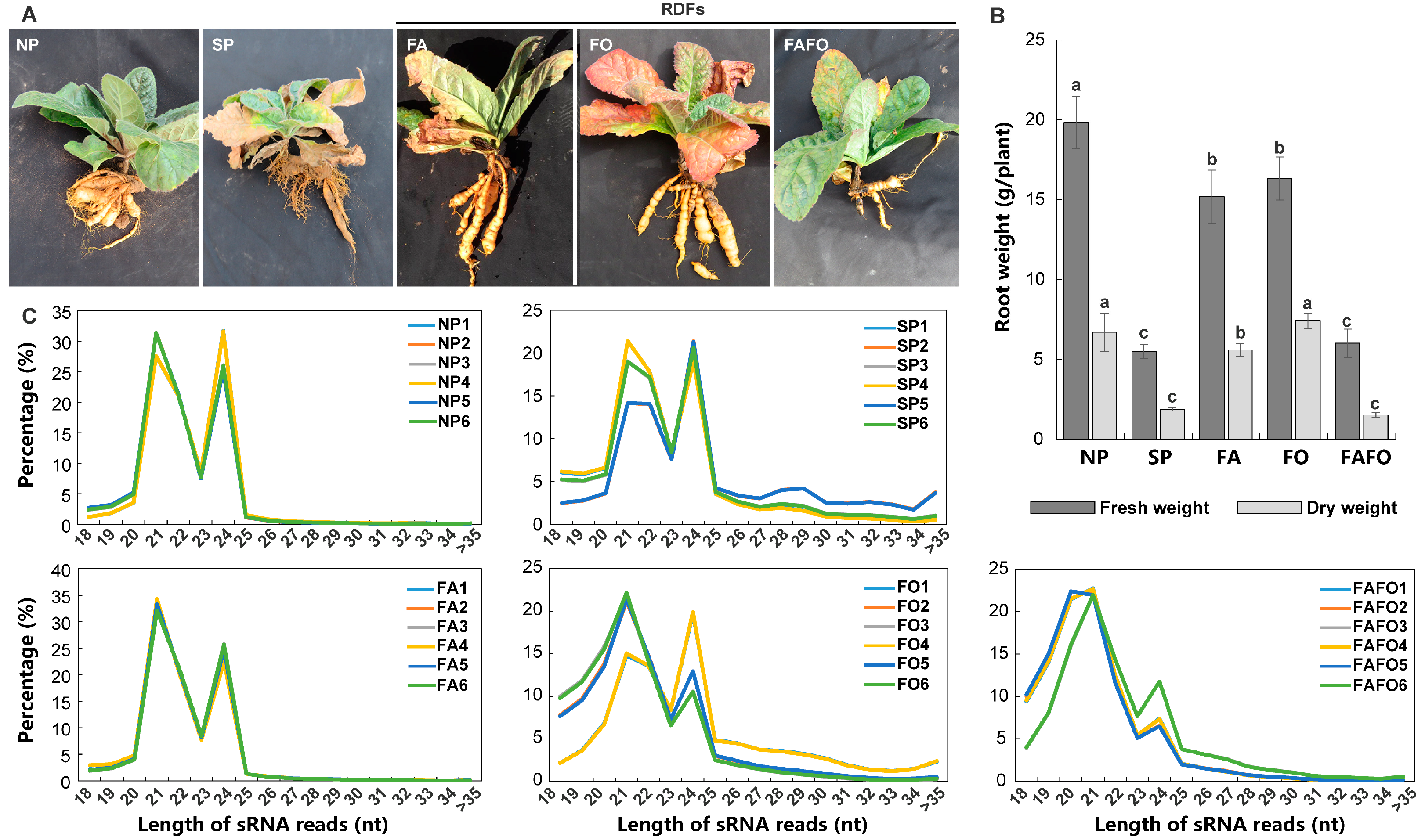
3.1. Construction of sRNA Libraries of R. glutinosa in Relation to RDFs

To accurately identify the specific miRNAs that respond to replant disease and to minimize the interference of miRNAs elicited by non-replant disease-related environmental factors, five treatments were established under controlled environments. The experimental design comprised three RDFs (FA, FO, and FAFO), one SP, and NP. The NP soils was employed as a control for the other treatments. The results demonstrated that the diverse treatments exerted a significant influence on the growth and development of R. glutinosa, when compared with NP (Figure 1A). At 30 days after transplantation (DATs), distinctive features were identified in R. glutinosa for the various treatments (Figure 1A). Nevertheless, the severity of the observed symptoms in the R. glutinosa plants varied depending on the specific treatment employed. The root formation of the SP R. glutinosa was most severely affected, exhibiting the most prominent fibrous roots (Figure 1A). The symptoms of FAFO are similar to those of SP; however, there are still notable differences between the two. The primary effect of FAFO is the elongation of tuberous roots, with relatively minimal damage to the aboveground portions. Furthermore, FA and FO demonstrated a diminished effect on root growth, while exhibiting pronounced damage to the leaves (Figure 1A). A subsequent analysis of the changes in root biomass of R. glutinosa under different treatments revealed that in comparison with the NP, the biomass of R. glutinosa treated with SP and FAFO was significantly reduced, while FA and FO had relatively minor effects on the root biomass (Figure 1B). R. glutinosa samples at 40 DATs were collected for the establishment of sRNA libraries. The number of raw reads generated in six replicates of each sample is presented in the following table: the number of raw reads ranged from 7.74 to 12.76 million in the NP, 8.91 to 13.35 million in the SP, 7.96 to 13.59 million in the FA, 9.82 to 17.53 million in the FO, and 8.58 to 13.61 million in the FAFO (Table S2 in the Supplementary File). Following quality control, the number of clean reads obtained from each sample ranged from 6.65 to 11.21 million for NP, 6.72 to 10.42 million for SP, 6.37 to 11.11 million for FA, 6.59 to 9.95 million for FO, and 6.14 to 10.14 million for FAFO (Table S2 in Supplementary File).
The length of the sRNA present in the aforementioned 30 sRNA libraries was determined. The results demonstrated that the clean reads in the various sRNA libraries exhibited lengths spanning from 21 to 25 nt (Figure 1C). The most prevalent sRNAs were those with lengths of 21 nt and 24 nt (Figure 1C). This distribution pattern is largely in accordance with the typical range of plant sRNA lengths (Figure 1C). In order to obtain clean reads originating from the R. glutinosa genome, the clean reads of all samples were initially aligned to the R. glutinosa genome. The results demonstrated that there were 4.79–7.65 million clean reads in NP, 3.13–4.61 million in SP, 4.47–7.84 million in FA, 1.98–3.53 million in FO, and 1.42–2.89 million in FAFO that were mapped to the R. glutinosa genome (Table S3 in Supplementary File). The mapped clean reads were subsequently annotated using non-coding sRNA datasets, including rRNA, snRNA, snoRNA, and tRNA, extracted from the public database. The proportion of clean reads that were successfully annotated ranged from 11.23% to 18.24% in NP, 12.83% to 17.18% in SP, 9.73% to 27.18% in FA, 10.58% to 17.39% in FO, and 7.12% to 18.52% in FOFA (Table S3 in Supplementary File). In order to mitigate the influence of these reads on the identification of miRNAs, they were eliminated. The remaining clean reads were employed for the identification of the miRNA family in R. glutinosa.

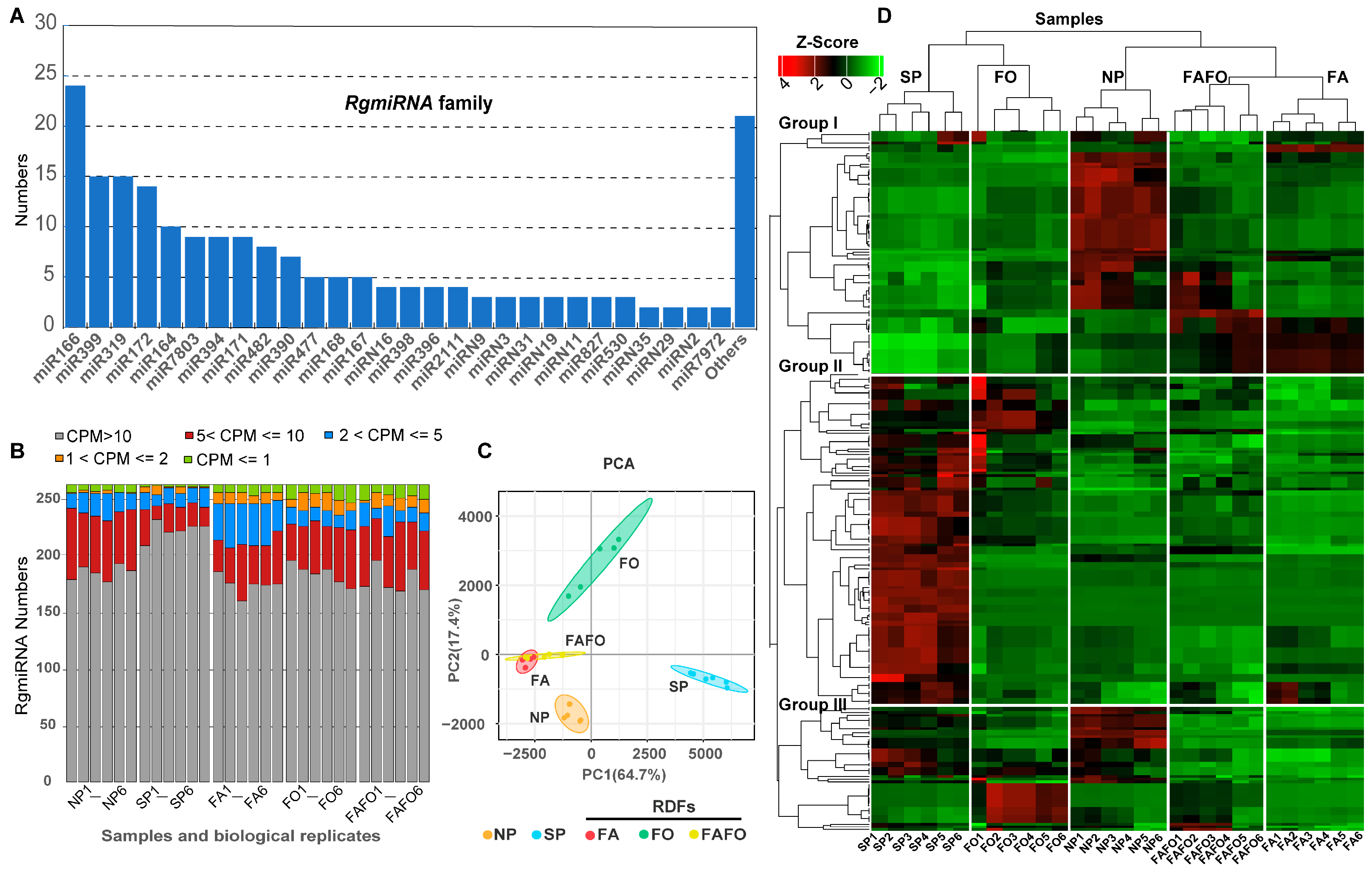
3.2. Identification of the miRNA Family Existing in the Genome of R. glutinosa

In order to comprehensively identify the miRNAs of R. glutinosa, 30 sRNA libraries were utilized to explore the miRNA sites within the R. glutinosa genome by means of the sRNA-miner v1.1.2 software [23]. As a result, 343 miRNA loci were identified within the R. glutinosa genome. Subsequently, the expression abundance of all miRNAs was calculated using mirdeep2 [22]. To obtain a more accurate representation of the R. glutinosa miRNAs, only those miRNAs or miRNA* that had mapped clean reads of more than 100 in at least three samples out of thirty samples were retained as candidate R. glutinosa miRNAs. Subsequently, 201 miRNAs were identified as eligible for classification as R. glutinosa miRNAs (RgmiRNAs) (Table S4 in the Supplementary File). Of these, 160 were identified as conserved miRNAs, while 41 were not identified in other plants and are therefore regarded as novel or specific miRNAs in R. glutinosa (Table S4 in the Supplementary File). These RgmiRNAs were classified into 49 distinct miRNA families, as detailed in Table S4 in the Supplementary File. The RgmiR166, RgmiR399, RgmiR319, and RgmiR172 family was the most abundant, comprising more than 14 members (Figure 2A and Table S4 in the Supplementary File). Subsequently, 10, 9, 9, 9, 8, and 7 members were identified, respectively, for the RgmiR164, RgmiR7803, RgmiR394, RgmiR171, RgmiR482, and RgmiR390 families (Figure 2A and Table S4 in Supplementary File). It is noteworthy that the RgmiR477, RgmiR168, RgmiR167 family comprises five members (Figure 2A and Table S4 in Supplementary File). The remaining 36 miRNA families comprised between one and four individual members (Figure 2A and Table S4 in the Supplementary File).
In order to gain a comprehensive understanding of the expression levels and patterns of all RgmiRNAs, the abundance of each mature RgmiRNA and its star (RgmiRNA*) sequence was calculated based on the number of clean reads per million mapped reads (CPM) (Table S5 in Supplementary File). A comprehensive analysis of RgmiRNA expression across diverse samples demonstrates that the expression of RgmiRNAs is relatively uniform among biological replicates within a single sample. However, conspicuous differences in expression patterns emerge between samples (Figure 2B; Table S5 in Supplementary File). A principal component analysis (PCA) of the RgmiRNA abundance from the various samples revealed that SP was distinctly separated from the other samples. FAFO and FA demonstrated crossover and aggregation (Figure 2C). To demonstrate the expression profile of RgmiRNAs across different samples, a heatmap was constructed using the expression matrix of all RgmiRNAs, with Z-score normalization and hierarchical clustering (Figure 2D). The clustering of the five samples indicates that the SP samples were conspicuously grouped into a single category, while the FA, FO, and FAFO samples were grouped into another category (Figure 2D). This also implies that the effects of SP and RDFs on the expression of miRNAs are markedly dissimilar. Furthermore, SP is considerably more complex, encompassing all of the replant disease-related stress factors and their interactions.
The expression levels of RgmiRNAs in different samples were employed to classify the RgmiRNAs expression pattern into five groups (Figure 2D). Group I comprise 91 RgmiRNAs, some of which exhibited higher expression levels in NP, FA, and FAFO than in the other samples (Figure 2D). Group II comprises 125 RgmiRNAs, majority of which exhibited higher expression levels in SP in comparison with the other groups (Figure 2D). Furthermore, some RgmiRNAs displayed elevated expression levels in FO relative to other groups. It is noteworthy that the abundance of this group of RgmiRNAs in SP was significantly higher than in other treatments, indicating that these miRNAs may be specifically expressed in SP samples (Figure 2D). Furthermore, group III contains 47 RgmiRNAs, some of which were predominantly expressed in SP, FO, and NP in comparison with other treatments (Figure 2D). Furthermore, 72 RgmiRNAs* were identified as having higher abundance in various samples (Table S5 in the Supplementary File), indicating that both mature miRNAs and their star sequences with higher abundance may also exert a significant role in R. glutinosa. The miRNA star sequence will be the focus of further analysis in subsequent studies.

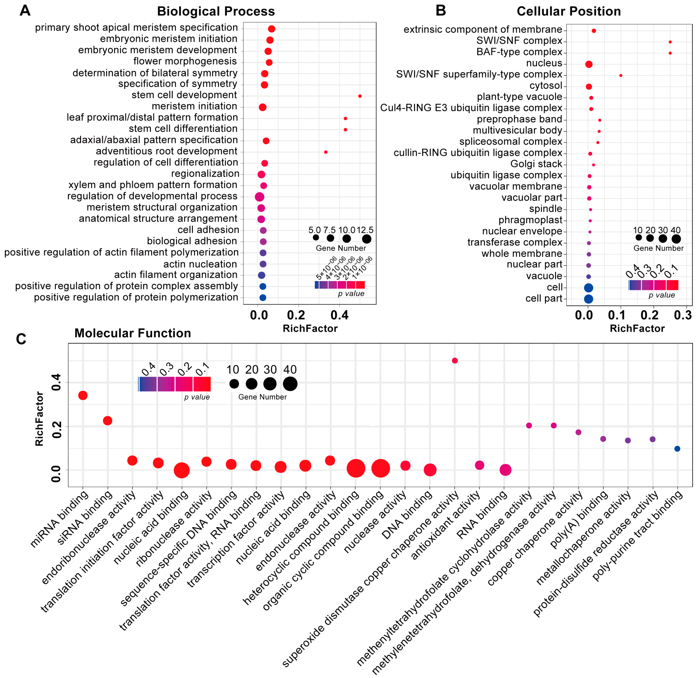
3.3. Identification and Functional Analysis of R. glutinosa RgmiRNAs Target Genes

In order to investigate the function of RgmiRNAs in R. glutinosa, NP and FAFO samples were selected for the establishment of a degradome library. The results revealed that 16.20 million and 11.58 million raw reads were obtained from the two samples, respectively. Following quality filtering, 15.55 million and 10.18 million clean reads were obtained from each sample. Furthermore, two degradome libraries from a previous study [15] were selected to identify the targets of RgmiRNAs. Concurrently, a transcriptome established in a previous study was further revised following the genome information of R. glutinosa, to identify targets of RgmiRNA. The candidate targets of RgmiRNAs were primarily obtained through Cleaveland 4.0 [25], based on the four degradomes and the transcriptome of R. glutinosa. Consequently, 107 RgmiRNAs or RgmiRNAs* were identified to target 189 transcripts (Table S6 in the Supplementary File). To gain further insight into the cellular pathways associated with RgmiRNAs, the function of the transcripts targeted by RgmiRNAs was investigated through gene ontology (GO) analysis. The GO enrichment analysis revealed that the 189 transcripts were involved in three functional groups (Figure 3). The predominant biological processes were found to be the ‘regulation of developmental process’, ‘xylem and phloem pattern formation’, ‘meristem structure organisation’, and another category (Figure 3A). In the category of cellular position, the most significantly enriched groups were ‘cell part’, ‘nucleus’, ‘cytosol’, and others (Figure 3B). With regard to molecular function, the most highly enriched groups were ‘organic cyclic compound binding’, ‘heterocyclic compound binding’, ‘nucleic acid binding’, and others (Figure 3C). These findings indicate that miRNAs are involved in the regulation of diverse cellular pathways and exert pivotal functions during the growth and development of R. glutinosa.

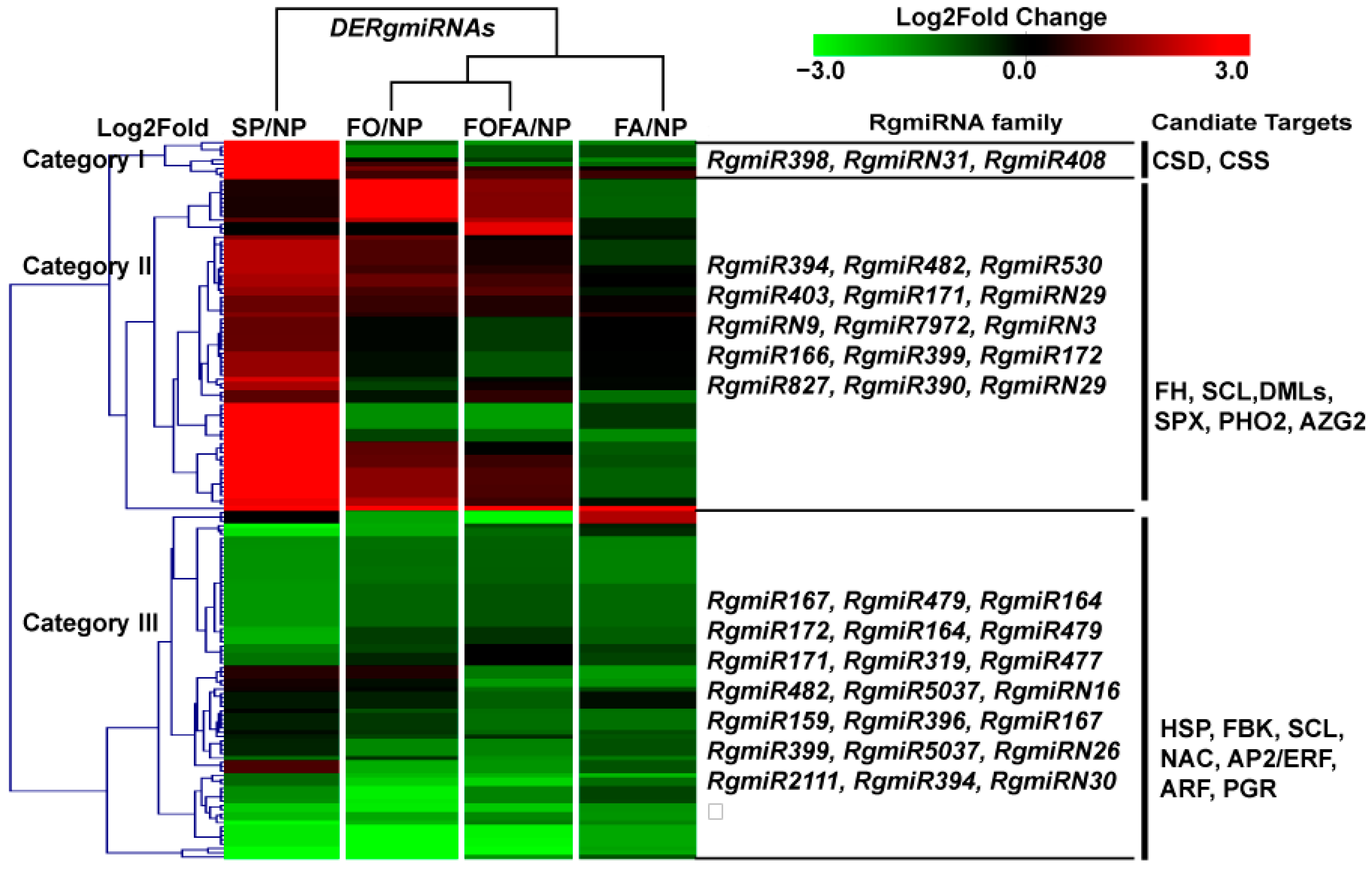
3.4. Analysis of Differentially Expressed miRNAs in RDFs

In order to gain insight into the role of RgmiRNAs in the formation of replant disease, differentially expressed RgmiRNAs (DeRgmiRNAs) were identified in various SP and RDFs in comparison with NP. A total of 124 DeRgmiRNAs were identified in SP, with 70 exhibiting increased expression and 54 displaying decreased expression (Table S7 in the Supplementary File). In FA, a total of 86 DeRgmiRNAs were identified, comprising 4 upregulated and 82 downregulated (Table S8 in the Supplementary File). Similarly, FO exhibited 86 DeRgmiRNAs, comprising 25 upregulated and 61 downregulated sequences (Table S9 in the Supplementary File). Finally, a total of 90 DeRgmiRNAs were identified in FAFO, comprising 14 upregulated and 76 downregulated (Table S10 in the Supplementary File). A heatmap and cluster analysis were conducted to illustrate the distribution of DeRgmiRNAs among different samples (Figure 4). The results demonstrated that the clustering pattern of DeRgmiRNAs across samples was essentially identical to that of RgmiRNAs.
It is worthy of note that RgmiRNAs in category 1 exhibited overexpression exclusively in SP, as exemplified by RgmiR398c, RgmiR398b, RgmiR398d, and RgmiR408, with negligible or no expression in the other treatments (Figure 4). In particular, the expression of RgmiR398c exceeded 3000 CPM in the SP samples, while it was less than 10 CPM in NP and RDFs (Figure 4, Table S5 and S7–S10 in the Supplementary File). This indicates that RgmiR398 is abnormally concentrated in SP and is associated with the development of replant disease. These miRNAs have been observed to primarily target genes encoding copper/zinc-superoxide dismutase (CSD) and copper/zinc-superoxide dismutase copper chaperone precursor (CSS). The category 2 RgmiRNAs were found to be upregulated in SP, FO, and FAFO, including RgmiR394, RgmiR171, RgmiR166, RgmiR399, RgmiR827, RgmiR7972, and so forth. This category of RgmiRNAs was observed to target genes including scarecrow-like protein (SCL), SPX domain-containing membrane protein (SPX), transcriptional activator DEMETER (DMLs), and so forth (Figure 4). The miRNAs in category 3 exhibited a notable decline in expression levels in SP and different RDFs. This group primarily comprises RgmiR172, RgmiR2111, RgmiR164, RgmiR319, and so forth. These miRNAs have been observed to target NAC transcription factors (NAC), AP2/ERF transcription factors (AP2/ERF), and F-box/kelch-repeat proteins (FBK) (Figure 4). In general, category 1 comprises a relatively limited number of DERgmiRNAs. However, RgmiR398, which is specifically expressed in SP, mainly targets genes related to the oxidation balance. It can thus be postulated that RgmiR398 may be intimately associated with the regulation of the oxidation balance of replant disease of R. glutinosa.

3.5. Expression Changes of RgmiR398/RgCSD in Replant Disease and RDFs

To elucidate the regulatory impact of RgmiR398 on the oxidative balance of replant R. glutinosa, the RgmiR398c precursor was cloned following the predicted RgmiR398c precursor (Figure 5A). The results demonstrated that the RgmiR398c precursor cloned in R. glutinosa exhibited near-perfect identity with the theoretical prediction, with only two mismatch bases. The RgmiR398c precursor exhibits all the properties expected of an miRNA precursor. Concurrently, RgmiR398c was found target R. glutinosa CSD genes (RgCSD) in the degradome (Figure 5B). The two RgCSD genes were successfully cloned from R. glutinosa. The lengths of the two RgCSDs, designated RgCSD1 and RgCSD2, was 880 and 767 base pairs (bp), respectively, and both exhibited complete coding regions (Figure 5C). To validate the targeting relationship between RgmiR398c and RgCSD, a dual luciferase assay was employed to assess the cleavage ability of RgmiR398c on RgCSD. The results demonstrated that RgmiR398c was capable of effective cleavage within the 5′ UTR regions of RgCSD1 and RgCSD2 (Figure 5D). These findings indicate that RgmiR398 plays a role in regulating the oxidation system of replanted R. glutinosa.
The expression characteristics of RgmiR398c and two RgCSDs were subjected to further in-depth analysis under NP, SP, and RDFs. The findings revealed that the RgmiR398c and RgCSD genes exhibited distinguishable expression profiles in response to the diverse treatments. RgmiR398c in R. glutinosa plants grown in NP soils displayed high expression levels at 10 DATs, followed by a gradual decline. While the targeted RgSOD1 and RgSOD1 demonstrated relatively lower expression at 10–40 days after transplanting (DATs), as illustrated in Figure 5E. The expression of RgmiR398c in R. glutinosa treated with SP soils demonstrated a gradual increase at 10 DATs, whereas its two targeted RgCSDs exhibited an inverse expression trend. In the context of FA stress, RgmiR398c exhibited lower expression levels at 10–40 DATs, followed by a rapid increase. In contrast, the two target genes exhibited higher expression levels at 10–40 DATs (Figure 5E), indicating that allelochemical autotoxicity had a limited impact on RgmiR398c during the initial stages of R. glutinosa development. In response to FO stress, RgmiR398c demonstrated a notable elevation in expression after 10–20 DATs, which subsequently declined. The targeted RgCSD displayed an inverse trend (Figure 5E). Furthermore, the expression of RgmiR398c displayed a comparable trend to that of the FO treatment under FAFO conditions. RgmiR398c displayed higher expression levels at the initial stages of the treatment, followed by a decline at subsequent stages (Figure 5E). This suggests that FO and FAFO treatments significantly induce the expression of RgmiR398c at an earlier stage.

4. Discussion

In response to a multitude of stress factors, plants meticulously regulate the expression of a series of miRNAs. These particular response miRNAs are present in multiple forms and undergo significant alterations in expression levels, thereby functioning as pivotal regulators of plants’ responses to a wide variety of stress factors [28,29,30]. The development of replant disease is a complex process that involves a range of principal factors, including allelochemicals, microorganisms, and their interactions. Consequently, replant disease exhibits both common biotic and abiotic stress characteristics, while also displaying distinctive properties. It is therefore of the utmost importance to elucidate the formation mechanism of replant disease through a detailed interpretation of the common and specific miRNAs in response to replant disease. This study constructed 30 sRNA libraries associated with SP and RDFs in a controlled environment. In conclusion, a total of 201 miRNA loci were accurately identified within the R. glutinosa genome. Moreover, a number of crucial DeRgmiRNAs were identified in SP and RDFs when compared with NP. The expression pattern analysis of RgmiRNAs and DeRgmiRNAs revealed that the impact of replant soils on R. glutinosa miRNAs was markedly distinct from that of single RDFs. The analysis of RgmiRNA target genes indicated that replant soils and RDFs exerted a specific influence on the normal growth and development of R. glutinosa. Moreover, replant disease led to a significant disturbance in the ROS equilibrium in R. glutinosa, which was mediated by RgmiR398. The results of the miRNA analysis under replant disease and RDFs provided crucial data that further elucidated the formation mechanism of replant disease in R. glutinosa.
In response to various types of stress, plants have been observed to modulate the expression of specific miRNAs [30]. In this study, a notable decline in the expression of RgmiRNAs was observed in response to SP and RDFs. This indicates that SP and RDFs may impede essential cellular processes in R. glutinosa by inhibiting miRNAs. Among the RgmiRNAs that were inhibited by SP and RDFs, miR172 plays a pivotal role in regulating the size of plant roots, stems, leaves, and fruit, as well as the development of floral organs [31,32,33]. The overexpression of Arabidopsis miR172 has been demonstrated to result in a significant increase in root length [34]. In potato, the overexpression of miR172 has been observed to facilitate tuber expansion [35]. This suggests that there is a positive correlation between miR172 expression and the formation of plant organs. miR2111b is closely associated with the processes of root morphology and structure [36,37]. The overexpression of legume miR2111b has been demonstrated to result in a significant increase in root density and root diameter [38]. The miR164-NAC regulatory module is involved in the development of roots, the uptake of nutrients, and the adaptation to stress. In Arabidopsis and maize, miR164 reduces the sensitivity of auxin and inhibits lateral root development by degrading NAC [39]. The upregulation of wheat miR164 has been demonstrated to suppress NAC1, which subsequently inhibits the formation of new adventitious roots [40]. The downregulation of RgmiR164 in a variety of treatments indicates that SP and RDFs promote the development of adventitious roots in R. glutinosa. These findings suggest that SP and RDFs may potentially inhibit the root development of replanted R. glutinosa through the reduction of RgmiR2111b, RgmiR172, and RgmiR164 levels. The preceding studies have demonstrated that both replant disease and RDFs exert an influence on the root growth of R. glutinosa [12,41]. Furthermore, an increase in fibrous roots is a primary consequence of replant disease [4]. It is hypothesized that these repressed miRNAs may be closely related to the manifestation of R. glutinosa symptoms in response to diverse stress treatments.
Specific stress factors not only inhibit the expression of specific miRNAs but also enrich key miRNAs. It was observed that SP and RDFs significantly promoted the accumulation of specific RgmiRNAs. It is noteworthy that the number of significantly upregulated miRNAs in SP was found to be higher than in single RDFs. miR827 has been demonstrated to target two SPX-containing region proteins (SPX), thereby confirming its role in the absorption and storage of phosphorus (P) [42]. The findings of this study demonstrate that RgmiR827 is markedly upregulated in SP. R. glutinosa. This indicates that the SP soil disordered the absorption and metabolism of nutrients in R. glutinosa via RgmiR827. Furthermore, another upregulated miR399 in replant R. glutinosa was identified as a regulator of P metabolism, exerting its influence on the expression of the UBC gene [43,44]. The overexpression of miR399 has been observed to result in root P poisoning [43]. The present study demonstrated that RgmiR399 exhibited a specific upregulation in response to SP, whereas it displayed a downregulation or no significant change in other RDFs. This suggests that replant disease may have a significant impact on the nutritional metabolism of R. glutinosa through specific miRNAs. miR7972 could indirectly determine the regulation of gene expression by targeting DEMETER-like DNA glycosylase (DML) [45]. DML, a DNA damage repair enzyme, plays a pivotal role in the dynamic regulation of DNA methylation levels [46,47]. The expression of RgmiR7972 in replant R. glutinosa indicates its involvement in the regulation of gene expression in replant R. glutinosa. miR171 primarily targets SCL genes and is extensively implicated in diverse cellular processes, including the maintenance of the shoot apical meristem, the transition between growth phases, and stress response [48,49]. The overexpression of Arabidopsis miR171 has been observed to result in a reduction in branch number, root length, and leaf shape [50]. The expression of RgmiR171 was observed to be repressed in R. glutinosa replant, indicating that replant disease may affect the development of R. glutinosa tissues and organs by upregulating specific miRNAs.
It is noteworthy that RgmiR398 was significantly upregulated in R. glutinosa plants subjected to replant disease, while no notable changes were observed in RDFs. Moreover, the expression of RgmiR398 in R. glutinosa subjected to replant disease exhibited aberrant accumulation in comparison with other RDFs. RgmiR398 was identified as a target of two RgCSDs, suggesting that replant disease may potentially disrupt the expression of core members of the antioxidant enzyme system through RgmiR398. As a result, this has an impact on the functionality of the antioxidant enzyme system of R. glutinosa. A study of the changes in ROS and antioxidant enzyme activities during the formation of replant disease in R. glutinosa revealed a gradual increase in ROS content and a corresponding decrease in superoxide dismutase (SOD) activity [12,51]. The expression changes of RgmiR398 and RgCSD in response to RDFs were analysed. It was demonstrated that RDFs could also exert a significant influence on the alteration of RgmiR398. The FA treatment was conducted in the later stage, while the FA and FAFO treatments were conducted in the early stage. Concurrently, the impact of FA, FO, and FAFO on RgmiR398 was markedly inferior to that of replant disease. This is consistent with previous studies, indicating that different RDFs significantly impact the oxidation balance of R. glutinosa, although the extent of this alteration varies [12]. These findings indicate that SP exerts a more pronounced effect on the oxidative damage of R. glutinosa than RDFs. Previous studies on replant R. glutinosa have demonstrated a correlation between oxidative balance and the development of replant disease [4,12]. Several studies on various plants with replant disease characteristics, including Solanum melongena, Lilium davidii, Angelica sinensis, Codonopsis tangshen, have provided evidence to support this conclusion [52,53,54,55]. It has been demonstrated that oxidative imbalance plays a significant role in the decline of plants subjected to replant disease. The precise mechanism by which oxidative imbalance occurs in replant disease remains uncertain. The present study has identified RgmiR398, which is specifically upregulated in R. glutinosa subjected to replant disease, as a regulator of antioxidant enzyme activity. This finding establishes a crucial point of entry for elucidating the mechanisms of replant disease-induced oxidative imbalance. It provides a foundation for further insights into the death processes in replant plants.
Moreover, this study demonstrated that a considerable number of RgmiRNAs displayed a significant upregulation in replant disease and a notable downregulation in RDFs. However, the target genes of these RgmiRNAs remained unidentified within the degradome. These RgmiRNAs may play a pivotal role in replant disease, and further investigation is required to elucidate their precise functions. A comparison of RgmiRNAs expression in response to different treatments indicates that SP soils and single RDFs both influence RgmiRNAs expression. However, notable discrepancies in the expression patterns across treatments have been observed. Although the combined treatment of FA and FO (FAFO) involved two RDFs, this is still quite different from actual replant disease, which involves a large number of RDFs interacting with one another. Furthermore, the impact of these RDFs on replant disease is not merely additive; rather, there is a sophisticated interplay between them. Consequently, replant disease represents a highly distinctive stressor, differing from general stressors. A specific set of response RgmiRNAs is thus activated during the process of replant disease formation.

5. Conclusions

In conclusion, in this study, the RgmiRNA family was first identified on a whole-genome scale, along with the determination of their candidate target genes. The identification of these miRNAs in R. glutinosa provides a valuable data resource for the in-depth clarification of the roles of RgmiRNAs during replant disease formation, growth and development, stress response, and the biosynthesis of active components in R. glutinosa. Furthermore, this study elucidates the expression profiles of RgmiRNAs under NP, SP, and RDFs, identifying specific RgmiRNAs in response to replant disease. Additionally, the potential roles of RgmiR398/RgCSD in the formation of oxidative stress of R. glutinosa subjected to replant disease were proposed. These findings significantly enhance our comprehension of the functions of miRNAs in intricate multi-factor stress.

Supplementary Materials

The following supporting information can be downloaded at: https://www.mdpi.com/article/10.3390/genes15091239/s1, Table S1. Primer lists utilized in this study; Table S2. Statistics of sRNA raw reads, filtered reads, and clean reads generated from different R. glutinosa sRNA libraries; Table S3. The annotation of clean reads from R. glutinosa sRNA libraries under different treatments in non-coding sRNA database; Table S4. RgmiRNAs identified in the genome of R. glutinosa; Table S5. Expression abundance (CPM) of RgmiRNAs in R. glutinosa under different treatments; Table S6. Candidate transcripts targeted by RgmiRNAs identified in the degradome; Table S7. DeRgmiRNAs in SP compared with NP R. glutinosa; Table S8. DeRgmiRNAs in FA compared with NP R. glutinosa; Table S9. DeRgmiRNAs in FO compared with NP R. glutinosa; Table S10. DeRgmiRNAs in FAFO compared with NP R. glutinosa.

Author Contributions

Data curation, L.G., Y.L., G.Z. and B.Z.; Formal analysis, Y.Y. and M.L.; Funding acquisition, M.L.; Investigation, J.W.; Methodology, Y.Y. and Z.Z.; Project administration, L.G. and M.L.; Resources, Z.Z. and J.W.; Software, Y.L., G.Z. and B.Z.; Writing—original draft, Y.L. and M.L.; Writing—review and editing, G.Z. and M.L. All authors have read and agreed to the published version of the manuscript.

Funding

This work was supported by grants from the National Natural Science Foundation of China (Grant No. 82073965; 82073962; 82274049; 82104331).

Institutional Review Board Statement

Not applicable.

Informed Consent Statement

Not applicable.

Data Availability Statement

The original contributions presented in the study are included in the article and Supplementary Material, further inquiries can be directed to the corresponding author.

Conflicts of Interest

The authors declare no conflicts of interest.

References

  1. Zhang, R.X.; Li, M.X.; Jia, Z.P. Rehmannia glutinosa: Review of botany, chemistry and pharmacology. J. Ethnopharmacol. 2008, 117, 199–214. [Google Scholar] [CrossRef] [PubMed]
  2. Bian, Z.Y.; Zhang, R.; Zhang, X.; Zhang, J.Y.; Xu, L.C.; Zhu, L.H.; Ma, Y.; Liu, Y.H. Extraction, structure and bioactivities of polysaccharides from Rehmannia glutinosa: A review. J. Ethnopharmacol. 2023, 305, 116132. [Google Scholar] [CrossRef] [PubMed]
  3. Piątczak, E.; Kuźma, Ł.; Kozłowska, W.; Lisiecki, P.; Szemraj, M.; Płachno, B.J.; Gonciarz, W.; Chmiela, M.; Matkowski, A.; Zielińska, S. Phenylethanoid and iridoid glycosides production in Rehmannia elata N.E.Brown ex Prein. in vitro shoot cultures and their biological activity. Ind. Crop. Prod. 2020, 158, 113050. [Google Scholar] [CrossRef]
  4. Li, M.J.; Yang, Y.H.; Feng, F.J.; Zhang, B.; Chen, S.Q.; Yang, C.Y.; Gu, L.; Wang, F.Q.; Zhang, J.Y.; Chen, A.G.; et al. Differential proteomic analysis of replanted Rehmannia glutinosa roots by iTRAQ reveals molecular mechanisms for formation of replant disease. BMC Plant Biol. 2017, 17, 116. [Google Scholar] [CrossRef]
  5. Liu, Y.Z.; Liu, Y.; Zeng, C.L.; Wang, J.Y.; Nyimbo, W.J.; Jiao, Y.Y.; Wu, L.K.; Chen, T.; Fang, C.X.; Lin, W.X. Intercropping with Achyranthes bidentata alleviates Rehmannia glutinosa consecutive monoculture problem by reestablishing rhizosphere microenvironment. Front. Plant Sci. 2022, 13, 1041561. [Google Scholar] [CrossRef]
  6. Yang, Q.X.; Wang, R.F.; Xu, Y.Y.; Kang, C.X.; Miao, Y.; Li, M.J. Dynamic change of the rhizosphere microbial community in response to growth stages of consecutively monocultured Rehmanniae glutinosa. Biologia 2016, 71, 1320–1329. [Google Scholar] [CrossRef]
  7. Gruber, K. Deep influence of soil microbes. Nat. Plants. 2015, 1, 15194. [Google Scholar] [CrossRef]
  8. Kong, C.H.; Zhang, S.Z.; Li, Y.H.; Xia, Z.C.; Yang, X.F.; Meiners, S.J.; Wang, P. Plant neighbor detection and allelochemical response are driven by root-secreted signaling chemicals. Nat. Commun. 2018, 9, 3867. [Google Scholar] [CrossRef]
  9. Liao, J.M.; Xia, P.G. Continuous cropping obstacles of medicinal plants: Focus on the plant-soil-microbe interaction system in the rhizosphere. Sci. Hortic. 2024, 328, 112927. [Google Scholar] [CrossRef]
  10. Zhou, Q.; Wang, Y.; Yue, L.; Ye, A.L.; Xie, X.F.; Zhang, M.L.; Tian, Y.; Liu, Y.; Turatsinze, A.N.; Uwaremwe, C.; et al. Impacts of continuous cropping on the rhizospheric and endospheric microbial communities and root exudates of Astragalus mongholicus. BMC Plant Biol. 2024, 24, 340. [Google Scholar] [CrossRef]
  11. Jin, X.; Jia, H.T.; Ran, L.Y.; Wu, F.Z.; Liu, J.J.; Schlaeppi, K.; Dini-Andreote, F.; Wei, Z.; Zhou, X.G. Fusaric acid mediates the assembly of disease-suppressive rhizosphere microbiota via induced shifts in plant root exudates. Nat. Commun. 2024, 15, 5125. [Google Scholar] [CrossRef] [PubMed]
  12. Li, Z.F.; He, C.L.; Wang, Y.; Li, M.J.; Dai, Y.J.; Wang, T.; Lin, W.X. Enhancement of trichothecene mycotoxins of Fusarium oxysporum by ferulic acid aggravates oxidative damage in Rehmannia glutinosa Libosch. Sci. Rep. 2016, 6, 33962. [Google Scholar] [CrossRef] [PubMed]
  13. Yang, Y.H.; Li, M.J.; Chen, X.J.; Wang, P.F.; Wang, F.Q.; Lin, W.X.; Yi, Y.J.; Zhang, Z.W.; Zhang, Z.Y. De novo characterization of the Rehmannia glutinosa leaf transcriptome and analysis of gene expression associated with replanting disease. Mol. Breeding. 2014, 34, 905–915. [Google Scholar] [CrossRef]
  14. Yang, Y.H.; Li, M.J.; Li, X.Y.; Chen, X.J.; Lin, W.X.; Zhang, Z.Y. Transcriptome-wide identification of the genes responding to replanting disease in Rehmannia glutinosa L. roots. Mol. Biol. Rep. 2015, 42, 881–892. [Google Scholar] [CrossRef] [PubMed]
  15. Li, M.J.; Yang, Y.H.; Chen, X.J.; Wang, F.Q.; Lin, W.X.; Yi, Y.J.; Zeng, L.; Yang, S.Y.; Zhang, Z.Y. Transcriptome/degradome-wide identification of R. glutinosa miRNAs and their targets: The role of miRNA activity in the replanting disease. PLoS ONE 2013, 8, e68531. [Google Scholar] [CrossRef]
  16. Yan, W.P.; Liu, X.F.; Cao, S.J.; Yu, J.; Zhang, J.F.; Yao, G.L.; Yang, H.G.; Yang, D.M.; Wu, Y.G. Molecular basis of Pogostemon cablin responding to continuous cropping obstacles revealed by integrated transcriptomic, miRNA and metabolomic analyses. Ind. Crops Prod. 2023, 200, 116862. [Google Scholar] [CrossRef]
  17. Wang, Y.; Liu, K.T.; Zhou, Y.Y.; Chen, Y.; Jin, C.Z.; Hu, Y.H. Integrated analysis of microRNA and RNA-Seq reveals phenolic acid secretion metabolism in continuous cropping of Polygonatum odoratum. Plants 2023, 12, 943. [Google Scholar] [CrossRef]
  18. Zhang, H.H.; Jin, W.B.; Zhu, X.L.; Liu, L.; He, Z.G.; Yang, S.S.; Liang, Z.S.; Yan, X.J.; He, Y.F.; Liu, Y. Identification and characterization of Salvia miltiorrhizain miRNAs in response to replanting disease. PLoS ONE 2016, 11, e0159905. [Google Scholar] [CrossRef]
  19. Shang, R.; Lee, S.; Senavirathne, G.; Lai, E.C. microRNAs in action: Biogenesis, function and regulation. Nat. Rev. Genet. 2023, 24, 816–833. [Google Scholar] [CrossRef]
  20. Ma, L.G.; Dong, C.M.; Song, C.; Wang, X.L.; Zheng, X.K.; Niu, Y.; Chen, S.L.; Feng, W.S. De novo genome assembly of the potent medicinal plant Rehmannia glutinosa using nanopore technology. Comput. Struct. Biotechnol. J. 2021, 19, 3954–3963. [Google Scholar] [CrossRef]
  21. Martin, M. Cutadapt removes adapter sequences from high-throughput sequencing reads. EMBnet. J. 2011, 17, 10–12. [Google Scholar] [CrossRef]
  22. Friedländer, M.R.; Mackowiak, S.D.; Li, N.; Chen, W.; Rajewsky, N. miRDeep2 accurately identifies known and hundreds of novel microRNA genes in seven animal clades. Nucleic Acids Res. 2012, 40, 37–52. [Google Scholar] [CrossRef] [PubMed]
  23. Li, G.L.; Chen, C.J.; Chen, P.K.; Meyers, B.C.; Xia, R. sRNAminer: A multifunctional toolkit for next-generation sequencing small RNA data mining in plants. Sci. Bull. 2024, 69, 784–791. [Google Scholar] [CrossRef] [PubMed]
  24. Saeed, A.I.; Sharov, V.; White, J.; Li, J.; Liang, W.; Bhagabati, N.; Braisted, J.; Klapa, M.; Currier, T.; Thiagarajan, M.; et al. TM4: A free, open-source system for microarray data management and analysis. Biotechniques 2003, 34, 374–378. [Google Scholar] [CrossRef]
  25. Addo-Quaye, C.; Miller, W.; Axtell, M.J. CleaveLand: A pipeline for using degradome data to find cleaved small RNA targets. Bioinformatics 2009, 25, 130–131. [Google Scholar] [CrossRef]
  26. Liu, Q.K.; Wang, F.; Axtell, M.J. Analysis of complementarity requirements for plant microRNA targeting using a Nicotiana benthamiana quantitative transient assay. Plant Cell 2014, 26, 741–753. [Google Scholar] [CrossRef]
  27. Livak, K.J.; Schmittgen, T.D. Analysis of relative gene expression data using real-time quantitative PCR and the and the 2−ΔΔCT Method. Methods 2001, 25, 402–408. [Google Scholar] [CrossRef]
  28. Šečić, E.; Kogel, K.H.; Ladera-Carmona, M.J. Biotic stress-associated microRNA families in plants. J. Plant Physiol. 2021, 263, 153451. [Google Scholar] [CrossRef]
  29. Zhang, H.M.; Zhu, J.H.; Gong, Z.Z.; Zhu, J.K. Abiotic stress responses in plants. Nat. Rev. Genet. 2022, 23, 104–119. [Google Scholar] [CrossRef]
  30. Yadav, A.; Mathan, J.; Dubey, A.K.; Singh, A. The emerging role of non-coding RNAs (ncRNAs) in plant growth, development, and stress response signaling. Non-Coding RNA 2024, 10, 13. [Google Scholar] [CrossRef]
  31. Sang, Q.; Vayssières, A.; Ó’Maoiléidigh, D.S.; Yang, X.; Vincent, C.; Bertran Garcia de Olalla, E.; Cerise, M.; Franzen, R.; Coupland, G. MicroRNA172 controls inflorescence meristem size through regulation of APETALA2 in Arabidopsis. New Phytol. 2022, 235, 356–371. [Google Scholar] [CrossRef] [PubMed]
  32. Zhang, B.L.; Chen, X.M. Secrets of the MIR172 family in plant development and flowering unveiled. PLoS Biol. 2021, 19, e3001099. [Google Scholar] [CrossRef] [PubMed]
  33. Zhou, Z.; Zhu, Y.M.; Zhang, H.T.; Zhang, R.P.; Gao, Q.M.; Ding, T.Y.; Wang, H.; Yan, Z.L.; Yao, J.L. Transcriptome analysis of transgenic apple fruit overexpressing microRNA172 reveals candidate transcription factors regulating apple fruit development at early stages. PeerJ 2021, 9, e12675. [Google Scholar] [CrossRef] [PubMed]
  34. Kim, B.H.; Kwon, Y.; Lee, B.H.; Nam, K.H. Overexpression of miR172 suppresses the brassinosteroid signaling defects of bak1 in Arabidopsis. Biochem. Biophys. Res. Commun. 2014, 447, 479–484. [Google Scholar] [CrossRef]
  35. Martin, A.; Adam, H.; Díaz-Mendoza, M.; Zurczak, M.; González-Schain, N.D.; Suárez-López, P. Graft-transmissible induction of potato tuberization by the microRNA miR172. Development 2009, 136, 2873–2881. [Google Scholar] [CrossRef]
  36. Weng, S.T.; Kuo, Y.W.; King, Y.C.; Lin, H.H.; Tu, P.Y.; Tung, K.S.; Jeng, S.T. Regulation of microRNA2111 and its target IbFBK in sweet potato on wounding. Plant Sci. 2020, 292, 110391. [Google Scholar] [CrossRef]
  37. Sexauer, M.; Bhasin, H.; Schön, M.; Roitsch, E.; Wall, C.; Herzog, U.; Markmann, K. A microRNA mediates shoot control of root branching. Nat. Commun. 2023, 14, 8083. [Google Scholar] [CrossRef]
  38. Zhang, M.B.; Su, H.N.; Gresshoff, P.M.; Ferguson, B.J. Shoot-derived miR2111 controls legume root and nodule development. Plant Cell Environ. 2021, 44, 1627–1641. [Google Scholar] [CrossRef]
  39. Guo, H.S.; Xie, Q.; Fei, J.F.; Chua, N.H. MicroRNA directs mRNA cleavage of the transcription factor NAC1 to downregulate auxin signals for Arabidopsis lateral root development. Plant Cell 2005, 17, 1376–1386. [Google Scholar] [CrossRef]
  40. Li, J.F.; Zhang, H.H.; Zhu, J.H.; Shen, Y.; Zeng, N.D.; Liu, S.Q.; Wang, H.Q.; Wang, J.; Zhan, X.H. Role of miR164 in the growth of wheat new adventitious roots exposed to phenanthrene. Environ. Pollut. 2021, 284, 117204. [Google Scholar] [CrossRef]
  41. Chen, A.G.; Gu, L.; Xu, N.; Feng, F.J.; Chen, D.X.; Yang, C.Y.; Zhang, B.; Li, M.J.; Zhang, Z.Y. NB-LRRs not responding consecutively to Fusarium oxysporum proliferation caused replant disease formation of Rehmannia glutinosa. Int. J. Mol. Sci. 2019, 20, 3203. [Google Scholar] [CrossRef] [PubMed]
  42. Lin, W.Y.; Lin, Y.Y.; Chiang, S.F.; Syu, C.H.; Hsieh, L.C.; Chiou, T.J. Evolution of microRNA827 targeting in the plant kingdom. New Phytol. 2018, 217, 1712–1725. [Google Scholar] [CrossRef]
  43. Aung, K.; Lin, S.I.; Wu, C.C.; Huang, Y.T.; Su, C.L.; Chiou, T.J. pho2, a phosphate overaccumulator, is caused by a nonsense mutation in a microRNA399 target gene. Plant Physiol. 2006, 141, 1000–1011. [Google Scholar] [CrossRef] [PubMed]
  44. Wang, R.; Fang, Y.N.; Wu, X.M.; Qing, M.; Li, C.C.; Xie, K.D.; Deng, X.X.; Guo, W.W. The miR399-CsUBC24 module regulates reproductive development and male fertility in Citrus. Plant Physiol. 2020, 183, 1681–1695. [Google Scholar] [CrossRef] [PubMed]
  45. Li, J.; Li, C.L.; Lu, S.F. Systematic analysis of DEMETER-like DNA glycosylase genes shows lineage-specific Smi-miR7972 involved in SmDML1 regulation in Salvia miltiorrhiza. Sci. Rep. 2018, 8, 7143. [Google Scholar] [CrossRef]
  46. Bharti, P.; Mahajan, M.; Vishwakarma, A.K.; Bhardwaj, J.; Yadav, S.K. AtROS1 overexpression provides evidence for epigenetic regulation of genes encoding enzymes of flavonoid biosynthesis and antioxidant pathways during salt stress in transgenic tobacco. J. Exp. Bot. 2015, 66, 5959–5969. [Google Scholar] [CrossRef]
  47. Yu, A.; Lepère, G.; Jay, F.; Wang, J.Y.; Bapaume, L.; Wang, Y.; Abraham, A.L.; Penterman, J.; Fischer, R.L.; Voinnet, O.; et al. Dynamics and biological relevance of DNA demethylation in Arabidopsis antibacterial defense. Proc. Natl. Acad. Sci. USA 2013, 110, 2389–2394. [Google Scholar] [CrossRef]
  48. Fan, T.; Li, X.M.; Yang, W.; Xia, K.F.; Ouyang, J.; Zhang, M.Y. Rice osa-miR171c mediates phase change from vegetative to reproductive development and shoot apical meristem maintenance by repressing four OsHAM transcription factors. PLoS ONE 2015, 10, e0125833. [Google Scholar] [CrossRef]
  49. Curaba, J.; Talbot, M.; Li, Z.Y.; Helliwell, C. Over-expression of microRNA171 affects phase transitions and floral meristem determinacy in barley. BMC Plant Biol. 2013, 13, 6. [Google Scholar] [CrossRef]
  50. Huang, W.; Peng, S.Y.; Xian, Z.Q.; Lin, D.B.; Hu, G.J.; Yang, L.; Ren, M.Z.; Li, Z.G. Overexpression of a tomato miR171 target gene SlGRAS24 impacts multiple agronomical traits via regulating gibberellin and auxin homeostasis. Plant Biotechnol. J. 2017, 15, 472–488. [Google Scholar] [CrossRef]
  51. Zhang, Z.Y.; Yin, W.J.; Li, J.; Du, J.F.; Yang, Y.H.; Chen, X.J.; Lin, W.X. Physio-ecological properties of continuous cropping Rehmannia glutinosa. Chin. J. Plant Ecol. 2010, 34, 547–554. [Google Scholar] [CrossRef]
  52. Wang, M.Y.; Wu, C.N.; Cheng, Z.H.; Meng, H.W. Growth and physiological changes in continuously cropped eggplant (Solanum melongena L.) upon relay intercropping with garlic (Allium sativum L.). Front. Plant Sci. 2015, 6, 262. [Google Scholar] [CrossRef] [PubMed]
  53. Hua, C.P.; Xie, Z.K.; Wu, Z.J.; Zhang, Y.B.; Guo, Z.H.; Qiu, Y.; Wang, L.; Wang, Y.J. The physiological and biochemical effects of phthalic acids and the changes of rhizosphere fungi diversity under continuous cropping of Lanzhou Lily (Lilium davidii var. unicolor). Hortscience 2019, 54, 253–261. [Google Scholar] [CrossRef]
  54. Zhang, X.H.; Zhang, E.H.; Wang, H.Z.; Lang, D.Y. Effects of continuous cropping obstacle on growth of Angelica sinensis and its mechanism. China J. Chin. Mater. Medica 2010, 35, 1231–1234. [Google Scholar] [CrossRef]
  55. He, Y.S.; Zhang, M.D.; Zhou, W.X.; Ai, L.Q.; You, J.W.; Liu, H.H.; You, J.M.; Wang, H.; Wassie, M.; Wang, M.; et al. Transcriptome analysis reveals novel insights into the continuous cropping induced response in Codonopsis tangshen, a medicinal herb. Plant Physiol. Biochem. 2019, 141, 279–290. [Google Scholar] [CrossRef]
Figure 1. Construction of sRNA libraries of R. glutinosa treated with NP, SP, and RDFs (FA, FO, and FAFO). (A,B) Phenotypic characteristics and biomass changes of R. glutinosa roots under different treatments. (C) The length distribution of clean reads from sRNA libraries under different treatments and their replicates. The use of lower-case letters indicates that the observed differences are statistically significant (p < 0.05; LSD). The data are presented as the mean ± standard deviation (n = 3).
Genes 15 01239 g001
Figure 2. RgmiRNAs identified in the genome of R. glutinosa and their expression profiling under NP, SP, and RDFs. (A) RgmiRNA family and their members within the genome of R. glutinosa. (B) The overall distribution of the abundance of RgmiRNA in different samples. (C) PCA analysis of RgmiRNA abundance derived from different sRNA libraries. (D) The expression levels of RgmiRNAs in the various samples. The expression levels of all miRNAs in the heatmap were transformed using the Z-Score method.
Genes 15 01239 g002
Figure 3. Analysis of GO categories encompassing biological process (A), cellular position (B), and molecular function (C) for candidate target genes of RgmiRNAs.
Genes 15 01239 g003
Figure 4. Identification of differentially expressed RgmiRNAs (DeRgmiRNAs) in R. glutinosa treated with SP and RDFs in comparison with those treated with NP. In the identification of DeRgmiRNAs, NP was employed as the control, while SP and RDFs were subjected to treatment. The heat map provides a visual representation of the distribution of DeRgmiRNAs across the various treatments. The DERgmiRNA family and their target genes identified in the degradome are displayed on the right side of the heat map.
Genes 15 01239 g004
Figure 5. Verification of RgmiR398 and its target genes, as well as analysis of their expression characteristics during the treatment of various stress. (A) Structural representation of the RgmiR398c precursor. (B) The cleavage of RgmiR398c against RgCSD was identified through degradome analysis. (C) The cleavage sites of RgmiR398 on RgCSD were illustrated in a diagrammatic representation. (D) The target site of RgmiR398 in RgCSD was further validated through a dual luciferase reporter gene assay. (E) The expression pattern of RgmiR398c and the target RgCSD during different treatments. R. glutinosa seedlings prior to transplantation into the experimental pots for treatment were employed as controls, and the expression level was set at 1, which was not depicted in the figure. The use of lower-case letters indicates that the observed differences are statistically significant (p < 0.05; LSD). *** indicates a significant difference at the 0.001 level of confidence. The data are presented as the mean ± standard deviation (n = 3).
Genes 15 01239 g005
Disclaimer/Publisher’s Note: The statements, opinions and data contained in all publications are solely those of the individual author(s) and contributor(s) and not of MDPI and/or the editor(s). MDPI and/or the editor(s) disclaim responsibility for any injury to people or property resulting from any ideas, methods, instructions or products referred to in the content.

