Article

Does Mobility of the Ankle Joint Depends on Length of the Free Part of the Achilles Tendon?

by
Tomasz Wawrzyński
1,
Bernadeta Angelika Pietrzak
2 and
Anna Mika
1,*
1
Institute of Clinical Rehabilitation, University of Physical Education in Kraków, al. Jana Pawła II 78, 31-571 Kraków, Poland
2
Laboratory of Neuropharmacology and Epigenetics, Department of Pharmacology, Maj Institute of Pharmacology, Polish Academy of Sciences, ul. Smetna 12, 31-343 Kraków, Poland
*
Author to whom correspondence should be addressed.
Symmetry 2022, 14(11), 2313; https://doi.org/10.3390/sym14112313
Submission received: 3 October 2022 / Revised: 28 October 2022 / Accepted: 2 November 2022 / Published: 4 November 2022

Abstract

:
The aim of our study was to evaluate whether the length of the free part of the Achilles tendon affects the mobility of the ankle joint in active motion without a load, as well as in functional motion with a body-weight load. We examined 36 healthy people, aged 21 to 30 years, and divided them into two groups: 1 (n = 15)—participants with a normal range of dorsiflexion in the ankle joint (20° or more), and 2 (n = 21)—participants with a reduced range of dorsiflexion in the ankle joint (below 20°). The length of the free part of the Achilles tendon was measured using ultrasonography. Ankle joint range of dorsiflexion was assessed, and a weight-bearing lunge test (WBLT) was conducted. Group 1 performed the WBLT better and demonstrated significantly greater Achilles tendon length compared to Group 2. A moderate, significant correlation was observed between ankle joint range of dorsiflexion and Achilles tendon length (r = 0.53, p < 0.05); between WBLT and Achilles tendon length (r = 0.61, p < 0.05); as well as between ankle joint range of dorsiflexion and WBLT (r = 0.63, p < 0.05). Thus, we can suggest that both the length of the tendon (measured by USG) and the ankle range of motion under a body-weight load (measured by WBLT) are good indicators regarding the range of foot dorsiflexion, but only up to specific values (6 cm of tendon length and 11 cm of WBLT reach). Therefore, Achilles tendon length, e.g., after injury and during tendon healing, may be monitored using the method of ultrasound imaging presented in this study.

1. Introduction

The Achilles tendon is the thickest and strongest tendon in the human body. It begins with the tendon cord, which starts at the descent of the medial and the lateral head of the gastrocnemius muscle and the soleus tendon, and ends with an attachment on the calcaneus. Its average length is 15 cm (11–26 cm), the average width in the proximal part is 6.8 cm (4.5–8.6 cm), the average width in the middle part is 1.8 cm (1.2–2.6 cm), while the average width on the distal trailer is 3.4 cm (2–4.8 cm). According to Chan’s classification, the Achilles tendon is divided into three parts—the heel (from attachment on the calcaneus to the upper part of the calcaneus), the free part (from the upper part of the calcaneus to the beginning of the soleus muscle fibers), and the inter-muscular section [1].
The Achilles tendon is an especially important structure and crucial for proper functioning of the entire body. This tendon, along with the calf triceps muscle, are a part of the superficial posterior anatomical tape, which includes the plantar flexors of the ankle joint, the knee joint flexors, and some of the hip extensors, as well as the spine extensors. According to the model of the Myers anatomical tapes, a disturbance in any of its parts affects the functioning of the whole tape; thus, it may be concluded that the length of the Achilles tendon and its free part may affect the biomechanics of the entire lower limb and cause disturbances in maintaining correct body posture [2]. Achilles tendon length has an influence on the range of motion in the ankle joint, depending on the position of the knee [3]. It has been demonstrated that, along with the change in the length of the Achilles tendon, the resting angle of the ankle changes, which may affect the stability of the entire lower limb, leading to disturbances in gait pattern [4]. A shortened Achilles tendon, on the other hand, may adversely affect the posture of the whole body [5]. Therefore, it seems essential to investigate whether Achilles tendon length may be related to ankle joint dorsiflexion, which may have important clinical implications during the treatment of ankle joint dysfunctions. It has also been reported that the stiffness and elasticity of the Achilles tendon may change with age [6]. Stenroth et al. [6] compared several parameters related to Achilles tendon function in young (mean age 24 ± 2 years) and older (mean age 75 ± 3 years) subjects. They noted a decrease in the stiffness of the Achilles tendon with a simultaneous increase in its cross-section area with age.
Restrictions in ankle motion can arise due to various factors; i.e., any deviations from correct anatomical as well as proper functional positioning of the entire foot and ankle joint. A limitation of dorsal flexion can be affected by the length of the Achilles tendon and the abdominal muscles of the posterior shin group: the gastrocnemius and soleus (too short a tendon or muscles inhibit the flexion movement in the ankle joint). The second important aspect regards changes in soft tissues (mainly spurs), articular limitations (contracted joint capsule, blocked sliding or rolling movement between articular surfaces), and injuries within the ankle joint [7,8,9,10]. It was suggested, that the crural fascia may play an important role in locomotor mechanics and, moreover, that an increase of fascia thickness may reduce the ankle joint flexibility in patients with chronic pain, e.g., in basketball players with a history of recurrent ankle sprains [11,12]. Some authors demonstrated that ultrasound imaging may be a good tool for assessing the fascial layers of the leg and any fascial abnormalities present due to previous injury. It was also reported that Achilles tendinopathy may strongly restrict the ankle joint dorsiflexion, especially via limited gliding of the crural fascia [11]. Pirri et al. [13] observed that ultrasound evaluation of the Achilles tendon was very useful in monitoring the effectiveness of tendinopathy treatment.
Considering the fact that the length of the whole Achilles tendon determines the extent of foot dorsiflexion, the question arises whether a similar relationship exists between the free part of the Achilles tendon and the mobility in the ankle joint. According to del Buono et al. [1], different parts of the Achilles tendon have distinct properties, i.e., they have different patterns of vascularization, and they differ structurally and mechanistically. In addition, the free part of the Achilles tendon is not restrained by muscles or calcaneus insertion. Therefore, it may be possible that ultrasonographic assessment of only the free part of the tendon length may be clinically more informative than whole Achilles tendon evaluation. The examination of this relationship may be useful in clinical assessment of post-traumatic changes, assessing treatment progress, and in differentiating an injured tendon from a healthy one [12]. We suggest that if this relationship between the length of the tendon free part and the ankle range of motion exists, this may be helpful in post-traumatic treatment, especially in patients with a short Achilles tendon free part visible in USG. In these patients, the increase of ankle dorsiflexion may be limited, not only due to trauma, but also due to anatomical properties. In addition, the length of the Achilles tendon may be significant from the point of limb length and joint mobility symmetry, which is essential for the proper functioning of movement patterns. Asymmetry was reported as an overuse injury risk factor [14]; therefore, sensitive methods of musculoskeletal system assessment are of interest.
Measurement of Achilles tendon length can be performed using indirect methods (such as measuring resting angle of the ankle joint in the Achilles tendon length measure (ATLM) and Achilles tendon resting angle (ATRA) tests), and by direct methods, such as magnetic resonance imaging (MRI), ultrasonography (USG), or calculating the length of the tendon isolated post mortem [15,16,17]. The methods of musculoskeletal system evaluation must be sensitive enough and allow for the detection of even minor abnormalities (e.g., asymmetries). One such method is ultrasonographic imaging of the musculoskeletal system. It has been reported that USG is an effective and reproducible method for measuring the length of the Achilles tendon, as well as its free part [15,16,17]. The great advantage of ultrasound is its general availability, low cost, and the possibility of carrying out mobile examinations. Barfod et al. [16], in a group of 19, non-injured patients, examined both lower limbs using MRI (magnetic resonance imaging) and ultrasound. They reported that the results obtained by three independent examiners using ultrasound were reliable and comparable with those achieved using MRI. They further underlined that using ultrasound examination among the non-traumatized patients made it possible to detect differences between right and left tendons greater than 4 mm [16]. It was also suggested that ultrasound imaging is a useful method for examining the length of the free part of the Achilles tendon [15,16,17,18]. The range of ankle joint motion may be determined with a goniometer or a WBLT. Both of these methods are widely used and considered reliable [19,20,21]. A goniometer is a basic tool for examining the range of motion in the joints [19], while WBLT is the preferred method applied for measuring functional dorsiflexion of the ankle [20]. It is also one of the basic functional tests used for dorsiflexion assessment after ankle and Achilles tendon injuries [21].
Therefore, the aim of our study was to evaluate whether the length of the free part of the Achilles tendon affects the mobility of the ankle joint in active motion without a load, as well as in functional motion with a body-weight load. We hypothesized that the length of the free part of the Achilles tendon may influence ankle joint dorsiflexion.

2. Methods

2.1. Study Group

A group of 36 healthy people (14 men and 22 women) aged 21 to 30 years participated in the study (Table 1). They had to meet the following inclusion criteria: (1) age between 20 and 30 years, (2) no trauma history of at least one ankle joint, (3) no oncological diseases, (4) no diseases of the nervous system. Professional athletes were also excluded. All participants provided their written informed consent to participate in the study. Before beginning the study, the participants were informed about all measurements in detail and were familiarized with all tests.
The study group was then divided into two subgroups [22]. The purpose of this division was to check whether participants with a normal range of dorsiflexion also had a greater length of the free Achilles tendon and a higher WBLT result.
Based on a cut-off value of 20° ankle joint dorsiflexion (which is considered the norm) [22], the following groups were distinguished:
-
Group 1 (n = 15)—participants with a normal range of dorsiflexion in the ankle joint (20° or more)
-
Group 2 (n = 21)—participants with a reduced range of dorsiflexion in the ankle joint (below 20°)

2.2. Sample Size Estimation

An independent t-test power analysis determined that at least 23 subjects were required to obtain a power of 0.8 at a two-sided level of 0.05 with an effect size d = 0.8 [23]. This analysis was based on data derived from previous literature [24,25].

2.3. Length Calculation of the Free Part of the Achilles Tendon

During the measurements, the subject lied in the prone position with the ankle joint in a neutral position. Ultrasound imaging was used to visualize the Achilles tendon in two axes: long (Figure 1A) and short (Figure 1B). To examine the distal attachments, the long axis was visualized (with longitudinal probe placement), and then the place where the soleus muscle releases its fibers to the Achilles tendon was visualized in the short axis (with transverse probe placement), due to the possibility of a more precise determination [26]. By rotating the probe head by 90°and slowly moving it proximally, we located the place where the free part of the Achilles tendon ends (Figure 1B). At this point, on the medial side, the beginning of the soleus muscle fibers can be noticed, being the end of the free part of the Achilles tendon [26].
Length measurements regarding the free part of the Achilles tendon were performed using a Mindray DP-50 (Shenzhen Mindray BioMedical Electronics Co., Ltd., Shenzhen, China) ultrasound apparatus. The first step was to mark the end of the free part on the side of the calcaneus, and then to mark the beginning at the level where the soleus muscle begins to release its fibers into the Achilles tendon. Based on the obtained image, appropriate points were marked on the skin, followed by measuring the distance between them using an anthropometric tape measure (Figure 2).

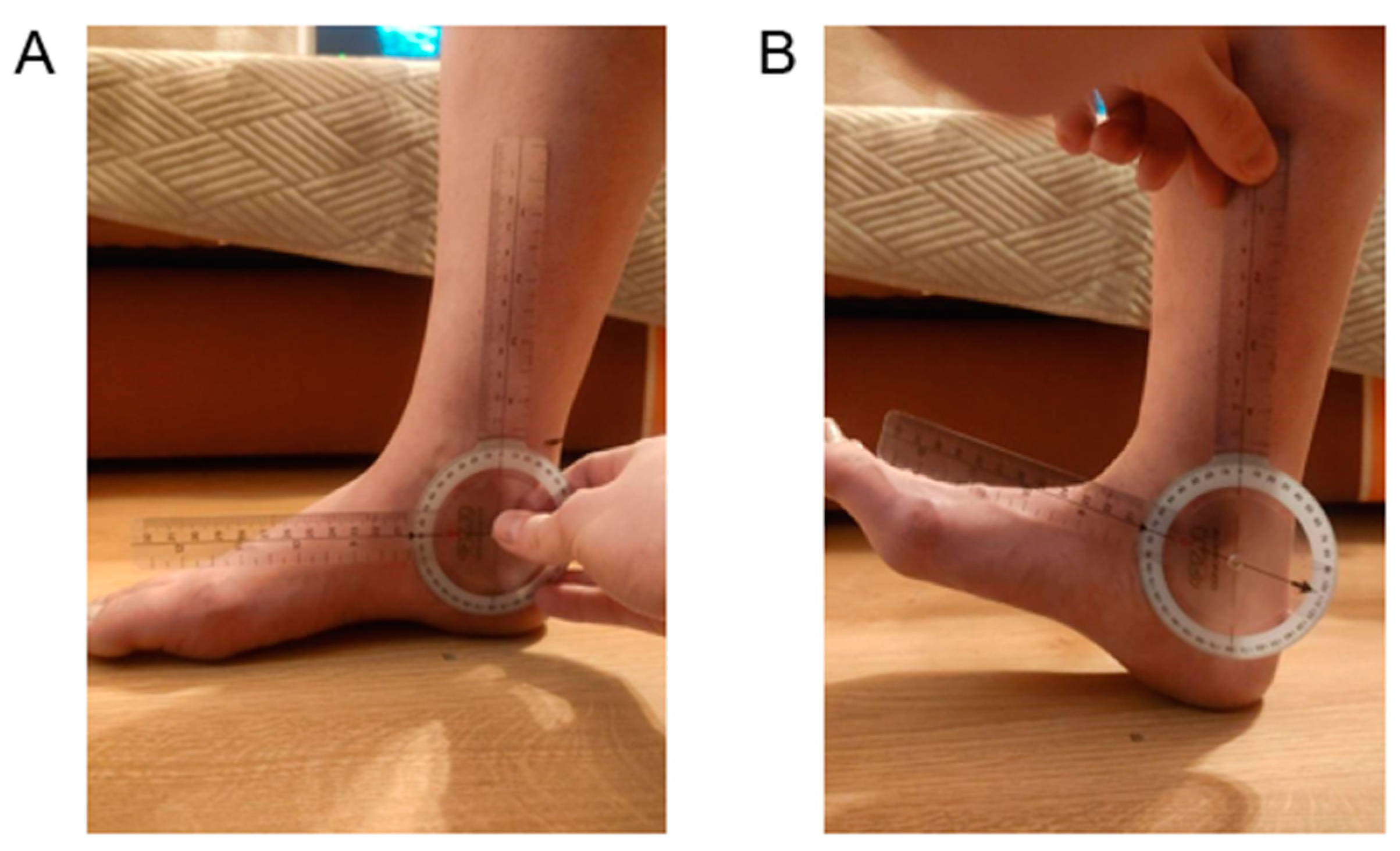
2.4. Dorsiflexion Measurements of the Ankle Joint Using a Goniometer

In order to measure the range of ankle joint dorsiflexion, the subject assumed a seated position with the legs lowered, and the examined joint was in an intermediate position, with the plantar surface of the foot resting on the floor. The axis of the goniometer was placed on the lateral ankle, in line with the transverse axis of the joint. The stationary arm of the goniometer was placed along the long axis of the shin, with the movable arm parallel along the edge of the foot. The subject performed a dorsiflexion movement of the foot, and the movable arm of the goniometer moved with the forefoot (Figure 3) [22]. According to Zembaty, a normal range of the ankle joint dorsiflexion is 20° [22].

2.5. Measurement of Functional Mobility Regarding Dorsiflexion of the Ankle Joint Using the Weight-Bearing Lunge Test (WBLT)

During the WBLT, the subject was positioned on their front lower limb with 10 cm from the wall to the tip of their toes. The back lower limb was positioned by the subject in such a way that they could stand in a stable and comfortable position. In this position, by bending the knee along the axis of the lower limb, they tried to touch the wall without taking their heel off the ground. If the heel lifted, the distance to the wall was shortened until the heel did not lift. If the heel did not rise, the distance was gradually increased until the heel rose and the final distance at which the heel did not lift was marked (Figure 4) [21]. The final distance between the tip of the toes and the wall was measured in cm; therefore, a longer distance indicated a higher ankle joint dorsiflexion. Two repetitions of WBLT were performed and the higher score was analyzed.

2.6. Statistical Analyses

Statistical analysis was performed using STATISTICA 13.0 Pl software. The data distribution was evaluated with the Shapiro–Wilk test. The t-test for independent samples was applied to determine differences between groups. Additionally, Pearson’s linear correlation coefficient (r) was calculated (below 0.50—“poor”; between 0.50 and 0.75—“moderate”; between 0.75 and 0.90—“good”; above 0.90—“excellent”) [27]. For all variables, sensitivity, specificity, ROC (receiver–operator characteristics) curve, and cut-off values were also calculated [28]. The variability within each data set was described using the arithmetic mean and standard deviation (SD) with 95% confidence intervals (95%CI), coefficients of variation (CV), and standard error of the mean (SEM). The effect size (ES) was calculated using Cohen’s d and interpreted as small (0.2–0.3), medium (0.5), or large (>0.8). Differences were considered statistically significant at a level of (p < 0.05).

3. Results

3.1. Between-Group Comparison

A significantly higher value of WBLT was observed in Group 1 compared to Group 2. The subjects with a normal ankle joint range of motion performed the WBLT better. In addition, the subjects in Group 1 demonstrated significantly longer Achilles tendons compared to Group 2 (Table 2).

3.2. Correlations

A moderate, significant correlation was observed between ankle joint range of dorsiflexion and Achilles tendon length (r = 0.53, p < 0.05). Additionally, a moderate, significant correlation was noted between WBLT and Achilles tendon length (r = 0.61, p < 0.05). Furthermore, ankle joint range of dorsiflexion and WBLT were significantly correlated with each other (r = 0.63, p < 0.05).

3.3. Diagnostic Value of Data

The sensitivity (0.800) and specificity (0.762) of the WBLT indicated a cut-off value at 11 cm on the ROC (receiver–operator characteristics) curve. This means that WBLT results below 11 cm were strongly related to a higher range of dorsiflexion in the ankle joint. WBLT results above 11 cm demonstrated a higher specificity (close to 1); therefore, they were not indicative enough for a higher range of ankle dorsiflexion (Figure 5. The sensitivity (0.733) and specificity (0.857) of Achilles tendon length for a cut-off value of 6 cm on the ROC curve suggested that a tendon length below 6 cm was strongly related to a higher range of ankle dorsiflexion, but a tendon length above 6 cm did not indicate a higher range of ankle dorsiflexion (Figure 6).

4. Discussion

In our research, a correlation between the length of the free part of the Achilles tendon and the mobility of the ankle joint has been demonstrated for the first time. The objective of our study was to investigate if all subjects with a normal range of ankle joint dorsiflexion also demonstrated a longer free part of the Achilles tendon, as well as a higher value of the WBLT. The ROC curve indicated the cut-off point for the tendon length at 6 cm and at 11 cm for the WBLT. This means that a free part length of the tendon below 6 cm clearly allowed for higher range of ankle joint dorsiflexion, but if the length was above 6 cm, the compliance was lower (sensitivity decreased, while specificity increased). A WBLT with a value of 11 cm indicated a greater degree of ankle joint range of motion, but above 11 cm, the compliance decreased. Our data indicated that the shorter the length of the free part of the Achilles tendon, the smaller the range of ankle joint dorsiflexion and the smaller functional mobility of the this joint.
There are no studies in the literature in which the relationship between the length of the free part of the Achilles tendon and active, as well as functional, ankle joint dorsiflexion is demonstrated. In our study, we have considered this issue for the first time. However, there are some existing studies in which an indirect evaluation of this relationship was conducted. Costa et al. [29] reported a linear relationship between Achilles tendon length and the extent of ankle joint dorsiflexion, in the case of cadavers. The separation of the Achilles tendon from other structures did not increase the ankle joint dorsiflexion, while lengthening the tendon by 1 cm significantly increased ankle joint mobility. The authors observed that the extent of dorsiflexion appears to be useful in assessing the clinical length of the tendon [29]. If an appropriate range of foot dorsiflexion is a crucial condition for correct execution of many functional movements, and if the limitations of dorsiflexion are one of the risk factors for lower limb injuries, there is a need to determine how the anatomical conditions in the structure of the tendon may influence the functional mobility of the ankle joint. It should be determined whether people with a shorter free part of the Achilles tendon also have smaller range of dorsiflexion, regardless of other factors that limit mobility. The results obtained in this study indicated the existence of a relationship (r = 0.53, p < 0.05) between the length of the free part of the Achilles tendon and the mobility of the ankle joint. It has been shown that the shorter the length of the free part of the Achilles tendon, the smaller the range of ankle joint dorsiflexion. Moreover, we observed that an Achilles tendon length below 6 cm is strongly related to a higher ankle range of dorsiflexion, but a tendon length above 6 cm did not indicate a higher ankle range of dorsiflexion.
Examination of Achilles tendon length is also an important aspect in the prevention of injuries and disease, and for control of the healing process after tearing or surgical anastomosis. Unfortunately, researchers most often focus on assessing the entire Achilles tendon, not just the free part. This is probably due to the different anatomical structure of the free part, indicating a different flexibility than the whole Achilles tendon; therefore, intensive stretching can lead to excessive stress accumulation and micro-tearing of some fibers. Thus, from a clinical point of view, it is important to know how the length of the tendon free part is related to ankle joint dorsiflexion. We suggest that our results may be useful, especially in diagnostics and treatment of post-traumatic restriction of ankle joint dorsiflexion, and especially when the applied treatment methods are not effective. Then, ultrasonographic measurement of the Achilles tendon may reveal a too short free Achilles tendon part and, therefore, may indicate the potential cause of the restricted ankle dorsiflexion. The significant correlation between the length of the free part of the tendon and ankle joint dorsiflexion observed in our study may explain why sometimes clinically we cannot increase the ankle range of motion, despite many methods being applied. As was reported by del Buono et al. [1], different parts of the Achilles tendon differ structurally and mechanistically; therefore, the measurement of only the tendon’s free part length may be clinically more informative than whole Achilles tendon assessment.
The occurrence of previous injuries is also a significant aspect in the examination of Achilles tendon functionality. A rupture or tear to this tendon may reduce its functional activity, although this may cause it to lengthen [30]. Carmont et al. [30] examined 26 patients following injury, post-surgery as well as during rehabilitation (6 weeks after surgery; 3, 6, 9 and 12 months), and reported increased tendon lengths after injury, which decreased after surgery and then increased again during rehabilitation. In this study, the importance of precise and sensitive measurements of the Achilles tendon length at each step of the treatment following injury was illustrated. The ultrasound imaging used in our study seems to be an appropriate method to be implemented for this purpose. This relationship may be also of significance during the selection of proper and effective methods of training or physiotherapy in people with a limited range of ankle joint dorsiflexion. However, if the tendon length is a potential factor limiting mobility, the same methods for increasing the range of motion may not be effective for everyone. Therefore, it is necessary to assess the length of the free part of the Achilles tendon in people undergoing therapy for dorsiflexion restriction, in order to select appropriate treatment methods.
Another significant aspect is the evaluation and comparison between healthy and post-traumatic tendons. In this context, researchers typically consider changes in muscle strength following an injury [31,32]. However, this approach rarely allows for the observation of differences between the injured and non-injured sides, which indicates a great need to develop new measures that will more accurately track traumatic changes and treatment progress. The relative symmetry of the Achilles tendons in both limbs is one of the factors determining gait pattern, the presence of compensation, predisposition to injuries, and the quality of movement patterns in the lower limbs. The formation of asymmetries within the structures of the limb may occur; for example, as a result of injuries or practicing asymmetric sports [33], or more frequently, by overloading a given limb during the performance of everyday activities [34].
Changes in the Achilles tendon length occurring after injury and during tendon healing may be monitored using the method of ultrasound imaging presented in this study. The differentiation of changes in length and the assessment of symmetry between an injured and uninjured limb could be very useful in selecting appropriate methods for therapy. In this study, it was demonstrated that people with a short free part of the Achilles tendon (up to 6 cm) also have a smaller range of ankle dorsiflexion, suggesting that this should be taken into account by therapists when selecting methods aimed at increasing ankle joint mobility.
Silbernagel et al. [35] used the functional heel-rise test to compare non-traumatic and post-traumatic Achilles tendons in patients after an Achilles tendon rupture. They evaluated them 6 and 12 months post-injury and demonstrated that the applied test clearly indicated differences in the tested tendons. The limb symmetry index was also calculated. The authors suggested that post-traumatic changes within a tendon may be diagnosed with functional tests, which are sensitive to detecting differences the between the healthy and injured side. The use of functional tests to assess ankle joint motion restrictions related to Achilles tendon dysfunctions has been reported by many authors [35,36,37,38,39]. In our study, significant correlations between the WBLT and Achilles tendon length (r = 0.61, p < 0.05), as well as between the ankle joint range of dorsiflexion and WBLT (r = 0.63, p < 0.05), indicated the usefulness of functional tests in Achilles tendon assessment. Moreover, in our study, the ROC curve cut-off point for WBLT was at 11 cm, which means that WBLT results below 11 cm are strongly related to a higher range of dorsiflexion in the ankle joint. WBLT results above 11 cm were not sufficiently indicative of a higher ankle joint range of dorsiflexion.
One limitation of this study is the fact that the study group consisted of young subjects, without any Achilles tendon dysfunctions. Therefore, the results of this study should be considered as a reference only for non-dysfunctional conditions. There is a need to carry out future research including different age groups, as well as groups of patients suffering from, e.g., overuse Achilles tendon ailments or traumatic injury.

5. Conclusions

The results obtained in this study clearly indicate the existence of a relationship between the length of the Achilles tendon free part and mobility of the ankle joint. It has been shown that the shorter the length of the free part of the Achilles tendon, the smaller the range of ankle joint dorsiflexion and the smaller functional mobility of this joint. Thus, it may be suggested that both the length of the tendon (measured via USG) and the ankle range of motion under a body-weight load (measured by WBLT) are good indicators of range of foot dorsiflexion, but only up to specific values (6 cm of tendon length and 11 cm of WBLT reach). However, WBLT results above 11 cm and tendon lengths higher than 6 cm demonstrated higher specificity (close to 1); therefore, these values are not sufficiently indicative of a higher ankle joint range of dorsiflexion.

Author Contributions

Conceptualization T.W. and A.M.; methodology T.W. and A.M.; formal analysis A.M.; investigation T.W.; resources T.W. and A.M.; data curation A.M.; writing—original draft preparation T.W., B.A.P. and A.M.; writing—review and editing T.W., B.A.P. and A.M. All authors have read and agreed to the published version of the manuscript.

Funding

This research received no external funding.

Institutional Review Board Statement

This study was approved by Ethical Committee of the Regional Medical Chamber in Krakow (No. 56/KBL/OIL/2019, 10 September 2019). All procedures were performed in accordance with the 1964 Declaration of Helsinki and its later amendments.

Informed Consent Statement

Informed consent was obtained from all subjects involved in the study.

Data Availability Statement

All data generated or analyzed during this study are included in the published article.

Conflicts of Interest

The authors declare no conflict of interest.

References

  1. Del Buono, A.; Chan, O.; Maffulli, N. Achilles tendon: Functional anatomy and novel emerging models of imaging classification. Int. Orthop. 2013, 37, 715–721. [Google Scholar] [CrossRef] [Green Version]
  2. Myers, T. Anatomy Trains: Myofascial Meridians for Manual and Movement Therapists, 2nd ed.; Churchill Livingstone: Edinburgh, UK, 2009. [Google Scholar]
  3. Hug, F.; Lacourpaille, L.; Maïsetti, O.; Nordez, A. Slack length of gastrocnemius medialis and Achilles tendon occurs at different ankle angles. J. Biomech. 2013, 46, 2534–2538. [Google Scholar] [CrossRef]
  4. Zellers, J.A.; Carmont, M.R.; Silbernagel, K.G. Achilles Tendon Resting Angle Relates to Tendon Length and Function. Foot Ankle Int. 2017, 39, 343–348. [Google Scholar] [CrossRef]
  5. Munteanu, S.E.; Barton, C.J. Lower limb biomechanics during running in individuals with Achilles tendinopathy: A systematic review. J. Foot Ankle Res. 2011, 4, 15. [Google Scholar] [CrossRef] [Green Version]
  6. Stenroth, L.; Peltonen, J.; Cronin, N.J.; Sipilä, S.; Finni, T. Age-related differences in Achilles tendon properties and triceps surae muscle architecture in vivo. J. Appl. Physiol. 2012, 113, 1537–1544. [Google Scholar] [CrossRef] [Green Version]
  7. Akeson, W.H.; Amiel, D.; Abel, M.F.; Garfin, S.R.; Woo, S.L. Effects of immobilization on joints. Clin. Orthop. Relat. Res. 1987, 219, 28–37. [Google Scholar] [CrossRef]
  8. Ghanem, I.; Massaad, A.; Assi, A.; Rizkallah, M.; Bizdikian, A.J.; El Abiad, R.; Seringe, R.; Mosca, V.; Wicart, P. Understanding the foot's functional anatomy in physiological and pathological conditions: The calcaneopedal unit concept. J. Child. Orthop. 2019, 13, 134–146. [Google Scholar] [CrossRef]
  9. Kaltenborn, F.; Evjenth, O. Manuelle Therapie Teil I—Extrernitäten [Eng. Manual Therapy Part 1—The Extremities], 10A; Olaf Norlis Bokhandel: Oslo, Norway, 1999. [Google Scholar]
  10. Schünke, M.; Schulte, E.; Schumacher, U. Prometheus Learn Atlas of Anatomy. General Anatomy and Musculoskeletal System 5. Auflage; Georg Thieme Verlag KG: Stuttgart, Germany, 2018. [Google Scholar]
  11. Pirri, C.; Fede, C.; Stecco, A.; Guidolin, D.; Fan, C.; De Caro, R.; Stecco, C. Ultrasound Imaging of Crural Fascia and Epimysial Fascia Thicknesses in Basketball Players with Previous Ankle Sprains Versus Healthy Subjects. Diagnostics 2021, 11, 177. [Google Scholar] [CrossRef]
  12. McAuliffe, S.; McCreesh, K.; Culloty, F.; Purtill, H.; O’Sullivan, K. Can ultrasound imaging predict the development of Achilles and patellar tendinopathy? A systematic review and meta-analysis. Br. J. Sports Med. 2016, 50, 1516–1523. [Google Scholar] [CrossRef]
  13. Pirri, C.; Stecco, C.; Fede, C.; De Caro, R.; Özçakar, L. Dynamic Ultrasound Examination of the Paratenon and Fascia in Chronic Achilles Tendinopathy. Am. J. Phys. Med. Rehabil. 2021, 100, e75. [Google Scholar] [CrossRef]
  14. Bredeweg, S.W.; Buist, I.; Kluitenberg, B. Differences in kinetic asymmetry between injured and non-injured novice runners: A prospective cohort study. Gait Posture 2013, 38, 847–852. [Google Scholar] [CrossRef]
  15. Barfod, K.W.; Riecke, A.F.; Boesen, A.; Hansen, P.; Maier, J.F.; Doessing, S.; Troelsen, A. Validity and reliability of an ultrasound measurement of the free length of the Achilles tendon. Dan Med. J. 2018, 65, A5453. [Google Scholar]
  16. Barfod, K.W.; Riecke, A.F.; Boesen, A.; Hansen, P.; Maier, J.F.; Døssing, S.; Troelsen, A. Validation of a novel ultrasound measurement of Achilles tendon length. Knee Surg. Sports Traumatol. Arthrosc. 2015, 23, 3398–3406. [Google Scholar] [CrossRef]
  17. Silbernagel, K.G.; Shelley, K.; Powell, S.; Varrecchia, S. Extended field of view ultrasound imaging to evaluate Achilles tendon length and thickness: A reliability and validity study. Muscles Ligaments Tendons. J. 2016, 6, 104–110. [Google Scholar] [CrossRef]
  18. Hansen, M.S.; Kristensen, M.T.; Hölmich, P.; Barfod, K.W. The Achilles Tendon Length Measure and the Achilles Tendon Resting Angle show acceptable construct validity using the Copenhagen Achilles Length Measure as gold standard. Foot Ankle Surg. 2021, 27, 655–659. [Google Scholar] [CrossRef]
  19. Krause, D.A.; Cloud, B.A.; Forster, L.A.; Schrank, J.A.; Hollman, J.H. Measurement of ankle dorsiflexion: A comparison of active and passive techniques in multiple positions. J. Sport Rehabil. 2011, 20, 333–344. [Google Scholar] [CrossRef]
  20. Langarika-Rocafort, A.; Emparanza, J.I.; Aramendi, J.F.; Castellano, J.; Calleja-González, J. Intra-rater reliability and agreement of various methods of measurement to assess dorsiflexion in the Weight Bearing Dorsiflexion Lunge Test (WBLT) among female athletes. Phys. Ther. Sport. 2017, 23, 37–44. [Google Scholar] [CrossRef]
  21. Powden, C.J.; Hoch, J.M.; Hoch, M.C. Reliability and minimal detectable change of the weight-bearing lunge test: A systematic review. Man Ther. 2015, 20, 524–532. [Google Scholar] [CrossRef]
  22. Zembaty, A. (Ed.) Kinesitherapy. Vol. I. Outline of Theoretical Foundations and Kinesitherapy Diagnostics; “Kasper” Sp. z.o.o.: Kraków, Poland, 2002. [Google Scholar]
  23. Schmidt, S.A.J.; Lo, S.; Hollestein, L.M. Research Techniques Made Simple: Sample Size Estimation and Power Calculation. J. Investig. Dermatol. 2018, 138, 1678–1682. [Google Scholar] [CrossRef] [Green Version]
  24. Zhang, X.; Deng, L.; Xiao, S.; Li, L.; Fu, W. Sex Differences in the Morphological and Mechanical Properties of the Achilles Tendon. Int. J. Environ. Res. Public Health 2021, 18, 8974. [Google Scholar] [CrossRef]
  25. Intziegianni, K.; Cassel, M.; Hain, G.; Mayer, F. Gender Differences of Achilles tendon Cross-sectional Area during Loading. Sports Med. Int. Open. 2017, 1, E135–E140. [Google Scholar] [CrossRef]
  26. McNally, E. Practical Musculoskeletal Ultrasound, 2nd ed.; Churchill Livingstone Elsevier: London, UK, 2014. [Google Scholar]
  27. Schober, P.; Boer, C.; Schwarte, L.A. Correlation Coefficients: Appropriate Use and Interpretation. Anesth. Analg. 2018, 126, 1763–1768. [Google Scholar] [CrossRef]
  28. Hajian-Tilaki, K. Receiver Operating Characteristic (ROC) Curve Analysis for Medical Diagnostic Test Evaluation. Caspian J. Intern. Med. 2013, 4, 627–635. [Google Scholar]
  29. Costa, M.L.; Logan, K.; Heylings, D.; Donell, S.T.; Tucker, K. The effect of Achilles tendon lengthening on ankle dorsiflexion: A cadaver study. Foot Ankle Int. 2006, 27, 414–417. [Google Scholar] [CrossRef]
  30. Carmont, M.R.; Grävare Silbernagel, K.; Brorsson, A.; Olsson, N.; Maffulli, N.; Karlsson, J. The Achilles tendon resting angle as an indirect measure of Achilles tendon length following rupture, repair, and rehabilitation. Asia Pac. J. Sports Med. Arthrosc. Rehabil. Technol. 2015, 2, 49–55. [Google Scholar] [CrossRef] [Green Version]
  31. Barfod, K.W. Achilles tendon rupture; assessment of non-operative treatment. Dan Med. J. 2014, 61, B4837. [Google Scholar]
  32. Glazebrook, M.; Rubinger, D. Functional Rehabilitation for Nonsurgical Treatment of Acute Achilles Tendon Rupture. Foot Ankle Clin. 2019, 24, 387–398. [Google Scholar] [CrossRef]
  33. Bravo-Sánchez, A.; Abián, P.; Jiménez, F.; Abián-Vicén, J. Myotendinous asymmetries derived from the prolonged practice of badminton in professional players. PLoS ONE 2019, 14, e0222190. [Google Scholar] [CrossRef] [Green Version]
  34. Bohm, S.; Mersmann, F.; Marzilger, R.; Schroll, A.; Arampatzis, A. Asymmetry of Achilles tendon mechanical and morphological properties between both legs. Scand. J. Med. Sci.Sports 2015, 25, e124–e132. [Google Scholar] [CrossRef]
  35. Silbernagel, K.G.; Nilsson-Helander, K.; Thomeé, R.; Eriksson, B.I.; Karlsson, J. A new measurement of heel-rise endurance with the ability to detect functional deficits in patients with Achilles tendon rupture. Knee Surg. Sports Traumatol. Arthrosc. 2010, 18, 258–264. [Google Scholar] [CrossRef]
  36. Steinfeld, Y.; Akian, R.; Rovitsky, A.; Puchkov, N.; Keren, Y. Assessment of Functional Conservative Treatment of Acute Complete Achilles Tendon Rupture. Isr Med. Assoc. J. 2021, 23, 510–515. [Google Scholar] [PubMed]
  37. Barfod, K.W.; Nielsen, E.G.; Olsen, B.H.; Vinicoff, P.G.; Troelsen, A.; Holmich, P. Risk of Deep Vein Thrombosis After Acute Achilles Tendon Rupture: A Secondary Analysis of a Randomized Controlled Trial Comparing Early Controlled Motion of the Ankle Versus Immobilization. Orthop. J. Sports Med. 2020, 8, 2325967120915909. [Google Scholar] [CrossRef] [PubMed]
  38. Sikorski, Ł.; Czamara, A. Assessment of Effectiveness of 15 Weeks of Physical Therapy on Biplanar Ankle Mobility, Gait and Pain Level in Patients Following Operative Repair of the Achilles Tendon. Ortop. Traumatol. Rehabil. 2021, 23, 33–44. [Google Scholar] [CrossRef] [PubMed]
  39. Ferreira, V.M.L.M.; Oliveira, R.R.; Nazareno, T.S.; Freitas, L.V.; Mendonça, L.D. Interaction of foot and hip factors identifies Achilles tendinopathy occurrence in recreational runners. Phys. Ther. Sport. 2020, 45, 111–119. [Google Scholar] [CrossRef] [PubMed]
Figure 1. Method of placing the USG probe on the long (A) and short axis (B), together with ultrasound images.
Symmetry 14 02313 g001
Figure 2. Examining the length of the free part of the Achilles tendon, marking points on the skin (A), and measuring the distance between them (B).
Symmetry 14 02313 g002
Figure 3. Measurement of ankle joint dorsiflexion with a goniometer, baseline position (A), and final position (B).
Symmetry 14 02313 g003
Figure 4. Measurement of ankle dorsiflexion functional mobility using the weight-bearing lunge test.
Symmetry 14 02313 g004
Figure 5. Receiver–operator characteristics (ROC) curve for weight-bearing lunge test (WBLT).
Symmetry 14 02313 g005
Figure 6. Receiver–operator characteristics (ROC) curve for Achilles tendon length.
Symmetry 14 02313 g006
Table 1. Study group characteristics.
Outcome MeasureGroup 1Group 2p
Number of subjects1521
Age (years)25.2 ± 2.224.0 ± 1.90.09
Body weight (kg)73.5 ± 14.168.0 ± 18.80.34
Body height (cm)177.0 ± 10.7170.8 ± 10.60.09
BMI23.3 ± 3.122.9 ± 3.60.74
Sex (women/men)7/814/70.31
BMI—body mass index; p—p value.
Table 2. Between-group comparison of Achilles tendon length, WBLT, and ankle joint ROM.
Outcome Measure Mean ± SDCI 95%SEMCV pES
Achilles tendon length (cm)Group 16.5 ± 1.85.5–7.60.4828.60.0021.10
Group 24.7 ± 1.44.0–5.30.3131.0
WBLT (cm)Group 112.4 ± 2.710.8–13.90.7222.50.00011.37
Group 29.0 ± 2.08.0–9.90.4523.3
Ankle joint ROM (degrees)Group 119.9 ± 0.219.7–20.10.061.290.00003.68
Group 214.4 ± 2.113.5–15.40.4614.7
The standard error of the mean (SEM); coefficient of variation (CV); 95% confidence interval (CI 95%); standard deviation (SD); p value; effect size (ES); weight-bearing lunge test (WBLT); range of motion (ROM).
Publisher’s Note: MDPI stays neutral with regard to jurisdictional claims in published maps and institutional affiliations.

