Association of Radiation Doses and Cancer Risks from CT Pulmonary Angiography Examinations in Relation to Body Diameter


 ,
,  ,
,
Abstract
:1. Introduction
2. Materials and Methods
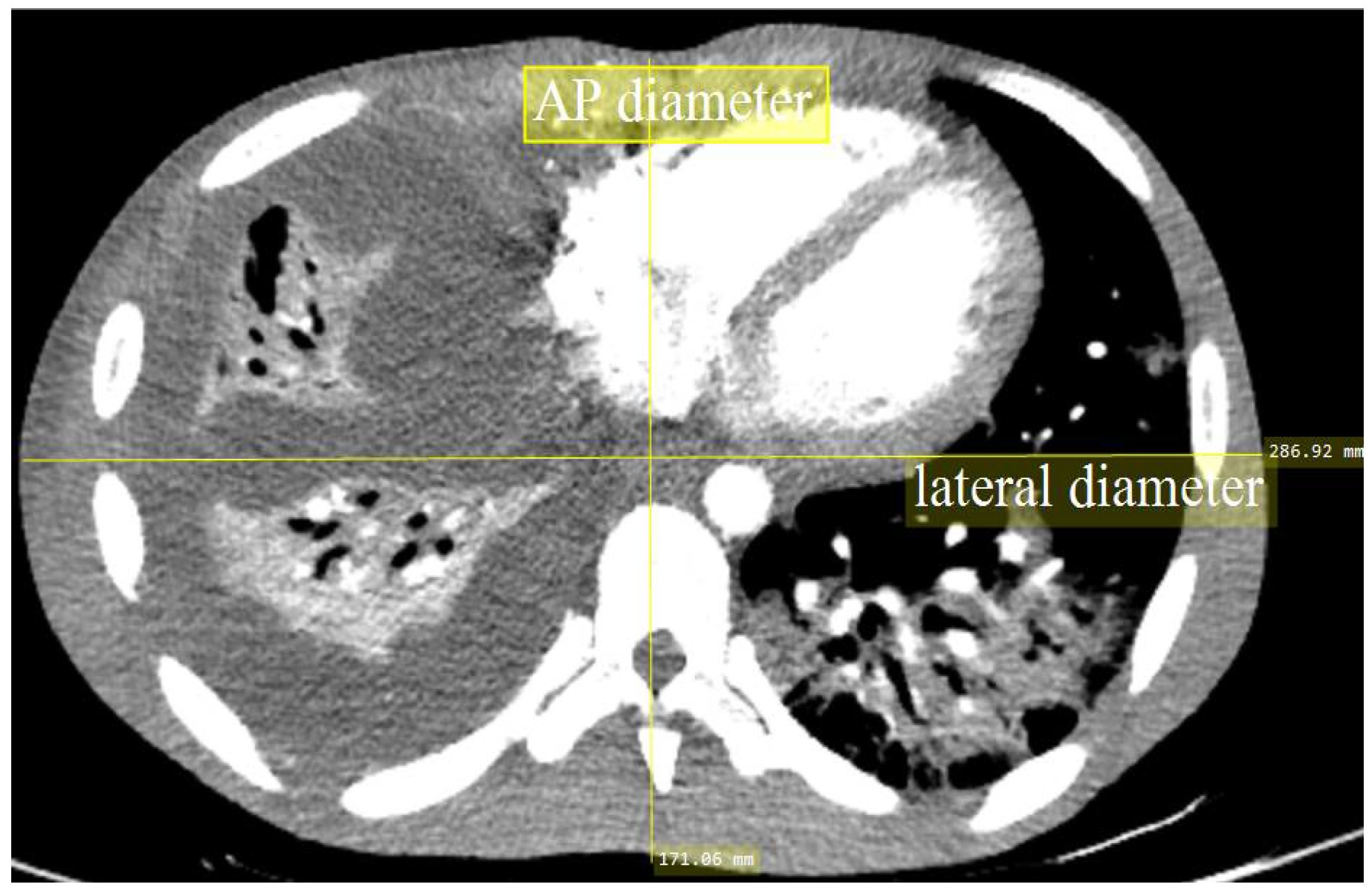
2.1. CT Parameter Measurements
2.2. Radiation Dose
2.3. Risk Assessments
2.4. Statistical Analysis
3. Results
4. Discussion
5. Conclusions
Author Contributions
Funding
Acknowledgments
Conflicts of Interest
References
- International Commission on Radiological Protection. The 2007 Recommendations of the International Commission on Radiological Protection. ICRP Publication 103. Ann. ICRP 2007, 37, 2–4. [Google Scholar]
- Sauter, A.; Koehler, T.; Brendel, B.; Aichele, J.; Neumann, J.; Noël, P.B.; Rummeny, E.J.; Muenzel, D. CT pulmonary angiography: Dose reduction via a next generation iterative reconstruction algorithm. Acta Radiol. 2018, 60, 1–10. [Google Scholar] [CrossRef]
- Halid, B.; Karim, M.K.A.; Sabarudin, A.; Bakar, K.A.; Shariff, N.D. Assessment of lifetime attributable risk of stomach and colon cancer during abdominal CT examinations based on Monte Carlo simulation. In IFMBE Proceedings; Springer: Cham, Switzerland, 2018; Volume 63, pp. 455–459. [Google Scholar]
- Daudelin, A.; Medich, D.; Andrabi, S.Y.; Martel, C. Comparison of methods to estimate water-equivalent diameter for calculation of patient dose. J. Appl. Clin. Med. Phys. 2018, 19, 718–723. [Google Scholar] [CrossRef]
- Burton, C.S.; Szczykutowicz, T.P. Evaluation of AAPM Reports 204 and 220: Estimation of effective diameter, water-equivalent diameter, and ellipticity ratios for chest, abdomen, pelvis, and head CT scans. J. Appl. Clin. Med. Phys. 2018, 19, 228–238. [Google Scholar] [CrossRef] [Green Version]
- Pourjabbar, S.; Singh, S.; Padole, A.; Saini, A.; Blake, M.A.; Kalra, M.K. Size-Specific dose estimates: Localizer or transverse abdominal computed tomography images? World J. Radiol. 2014, 6, 210–217. [Google Scholar] [CrossRef] [Green Version]
- Anam, C.; Haryanto, F.; Widita, R.; Arif, I.; Dougherty, G.; McLean, D. The impact of patient table on size-specific dose estimate (SSDE). Australas. Phys. Eng. Sci. Med. 2017, 40, 153–158. [Google Scholar] [CrossRef]
- O’Daniel, J.C.; Stevens, D.M.; Cody, D.D. Reducing radiation exposure from survey CT scans. AJR Am. J. Roentgenol. 2005, 185, 509–515. [Google Scholar] [CrossRef]
- Matsubara, K.; Koshida, K.; Noto, K.; Takata, T. Reduction of breast dose in abdominal CT examinations: Effectiveness of automatic exposure control system. Radiat. Meas. 2011, 46, 2056–2059. [Google Scholar] [CrossRef]
- Muhammad, N.A.; Karim, M.K.A.; Hassan, H.A.; Kamarudin, M.A.; Wong, J.H.D.; Ibahim, M.J. Estimation of effective dose and organ cancer risk from paediatric computed tomography thorax—Abdomen—Pelvis examinations. Radiat. Phys. Chem. 2019, 165, 108438. [Google Scholar] [CrossRef]
- Karim, M.K.A.; Hashim, S.; Bakar, K.A.; Bradley, D.A.; Ang, W.C.; Bahrudin, N.A.; Mhareb, M.H.A. Estimation of radiation cancer risk in CT-KUB. Radiat. Phys. Chem. 2017, 137, 130–134. [Google Scholar] [CrossRef]
- Brenner, D.J.; Hall, E.J. Computed tomography—An increasing source of radiation exposure. N. Engl. J. Med. 2007, 357, 2277–2284. [Google Scholar] [CrossRef] [PubMed] [Green Version]
- American Association of Physicists in Medicine. AAPM TG 220: Use of Water Equivalent Diameter for Calculating Patient Size and Size-Specific Dose Estimates (SSDE) in CT (AAPM Rep. 220); American Association of Physicists in Medicine: Alexandria, VA, USA, 2014; pp. 1–23. [Google Scholar]
- Sabel, B.O.; Buric, K.; Karara, N.; Thierfelder, K.M.; Dinkel, J.; Sommer, W.H.; Meinel, F.G. High-Pitch CT pulmonary angiography in third generation dual-source CT: Image quality in an unselected patient population. PLoS ONE 2016, 11, e0146949. [Google Scholar] [CrossRef] [Green Version]
- Kim, E.Y.; Kim, T.J.; Goo, J.M.; Kim, H.Y.; Lee, J.W.; Lee, S.; Lim, J.T.; Kim, Y. Size-Specific dose estimation in the Korean lung cancer screening project: Does a 32-cm diameter phantom represent a standard-sized patient in korean population? Korean J. Radiol. 2018, 19, 1179–1186. [Google Scholar] [CrossRef]
- Samei, E.; Richard, S. Assessment of the dose reduction potential of a model-based iterative reconstruction algorithm using a task-based performance metrology. Med. Phys. 2015, 42, 314–323. [Google Scholar] [CrossRef]
- Joemai, R.M.S.; Veldkamp, W.J.H.; Kroft, L.J.M.; Hernandez-Giron, I.; Geleijns, J. Adaptive iterative dose reduction 3d versus filtered back projection in CT: Evaluation of image quality. Am. J. Roentgenol. 2013, 201, 1291–1297. [Google Scholar] [CrossRef]
- Laqmani, A.; Regier, M.; Veldhoen, S.; Backhaus, A.; Wassenberg, F.; Sehner, S.; Groth, M.; Nagel, H.-D.D.; Adam, G.; Henes, F.O. Improved image quality and low radiation dose with hybrid iterative reconstruction with 80 kV CT pulmonary angiography. Eur. J. Radiol. 2014, 83, 1962–1969. [Google Scholar] [CrossRef]
- Dane, B.; Patel, H.; O’Donnell, T.; Girvin, F.; Brusca-Augello, G.; Alpert, J.B.; Niu, B.; Attia, M.; Babb, J.; Ko, J.P. Image quality on dual-energy CTPA virtual monoenergetic images: Quantitative and qualitative assessment. Acad. Radiol. 2018, 25, 1075–1086. [Google Scholar] [CrossRef]
- Sookpeng, S.; Martin, C.J.; Gentle, D.J. Comparison of different phantom designs for CT scanner automatic tube current modulation system tests. J. Radiol. Prot. 2013, 33, 735–761. [Google Scholar] [CrossRef]
- Kalra, M.K.M.; Maher, M.M.; Toth, T.T.L.; Schmidt, B.; Westerman, B.L.; Morgan, H.T.; Saini, S. Techniques and applications of automatic tube current modulation for CT. Radiology 2004, 233, 649–657. [Google Scholar] [CrossRef]
- Smith-Bindman, R.; Lipson, J.; Marcus, R.; Kim, K.-P.; Mahesh, M.; Gould, R.; Berrington de González, A.; Miglioretti, D.L. Radiation dose associated with common computed tomography examinations and the associated lifetime attributable risk of cancer. Arch. Intern. Med. 2009, 169, 2078–2086. [Google Scholar] [CrossRef]
- Isa, I.N.; Rahmat, S.M.S.; Dom, S.M.; Kayun, Z.; Karim, M.K. The effects of mis-centering on radiation dose during CT head examination: A phantom study. J. X Ray Sci. Technol. 2019, 27, 631–639. [Google Scholar] [CrossRef] [PubMed]
- Fukuda, A.; Ichikawa, N.; Fujita, Y.; Lin, P.J.P.; Matsubara, K.; Miyati, T. Does gantry rotation time influence accuracy of volume computed tomography dose index (CTDIvol) in modern CT? Phys. Med. 2017, 37, 43–48. [Google Scholar] [CrossRef] [PubMed]
- Bashier, E.H.; Suliman, I.I. Multi-Slice CT examinations of adult patients at Sudanese hospitals: Radiation exposure based on size-specific dose estimates (SSDE). Radiol. Med. 2018, 123, 424–431. [Google Scholar] [CrossRef] [PubMed]
- Karim, M.K.A.; Rahim, N.A.A.; Matsubara, K.; Hashim, S.; Mhareb, M.H.A.H.A.; Musa, Y. The effectiveness of bismuth breast shielding with protocol optimization in CT Thorax examination. J. X-Ray Sci. Technol. 2019, 27, 139–147. [Google Scholar] [CrossRef]


| Baseline Characteristic | Values | ||
|---|---|---|---|
| Male | Female | Total | |
| Age (years/old) * | 49.26 ± 14.57 | 48.60 ± 19.12 | 48.88 ± 17.28 |
| Anteroposterior (AP) (cm) * | 21.68 ± 3.68 | 21.88 ± 2.71 | 21.80 ± 3.14 |
| Lateral (LAT) (cm) * | 33.46 ± 4.17 | 32.85 ± 3.53 | 33.10 ± 3.80 |
| DW (cm) | Dose Descriptors * | Organ Equivalent Dose (mSv) * | ||||
|---|---|---|---|---|---|---|
| CTDIvol (mGy) | DLP (mGy cm) | E (mSv) | Breast | Lung | Liver | |
| Group 1 (19–25) | 6.44 ± 2.63 | 239.59 ± 97.36 | 5.19 ± 2.50 | 10.94 ± 4.62 | 10.62 ± 4.12 | 9.15 ± 4.23 |
| Group 2 (25–28) | 9.86 ± 6.46 | 351.85 ± 231.85 | 7.47 ± 4.11 | 15.48 ± 7.93 | 15.55 ± 8.39 | 13.48 ± 6.99 |
| Group 3 (>28) | 17.42 ± 6.90 | 631.46 ± 274.43 | 13.90 ± 5.66 | 23.81 ± 12.17 | 27.39 ± 11.87 | 23.19 ± 11.76 |
| p-value | <0.05 | <0.05 | <0.05 | <0.05 | <0.05 | <0.05 |
| TOTAL | 11.06 ± 7.17 | 400.38 ± 259.10 | 8.68 ± 5.47 | 17.05 ± 10.40 | 17.55 ± 10.86 | 15.04 ± 9.75 |
| DW (cm) | Dose Descriptors | |||
|---|---|---|---|---|
| SSDE a (mGy) | SSDE b (mGy) | Ratio a SSDE/CTDIvol | Ratio b SSDE/CTDIvol | |
| Group 1 (19–25) | 9.93 ± 3.89 | 9.01 ± 3.78 | 1.54 | 1.30 |
| Group 2 (25–28) | 13.70 ± 9.04 | 13.41 ± 7.74 | 1.42 | 1.34 |
| Group 3 (>28) | 22.29 ± 7.35 | 23.98 ± 9.63 | 1.28 | 1.31 |
| TOTAL | 14.62 ± 8.41 | 15.37 ± 9.67 | 1.41 | 1.32 |
| Cancer Risk (Per Million Procedures) | DW (cm) | p-Value | |||
|---|---|---|---|---|---|
| Group 1 (19–25) | Group 2 (25–28) | Group 3 (>28) | Total | ||
| Breast | 46.34 | 91.76 | 136.34 | 93.82 | <0.05 |
| Lung | 24.54 | 66.93 | 107.45 | 66.39 | <0.05 |
| Liver | 16.44 | 49.10 | 70.87 | 45.94 | <0.05 |
| Variable | Sex | p-Value | ||
|---|---|---|---|---|
| Male | Female | |||
| Total E (mSv) * | 7.47 ± 4.11 | 9.53 ± 5.68 | <0.05 | |
| Organ Dose (mSv) * | Breast | n/a | 17.05 ± 10.40 | n/a |
| Lung | 15.55 ± 8.39 | 17.63 ± 10.41 | NS | |
| Liver | 13.48 ± 6.99 | 14.58 ± 9.28 | NS | |
| Cancer Risk (per million procedures) | Breast | n/a | 93.81 | n/a |
| Lung | 50.31 | 78.03 | NS | |
| Liver | 38.86 | 51.07 | NS | |
© 2020 by the authors. Licensee MDPI, Basel, Switzerland. This article is an open access article distributed under the terms and conditions of the Creative Commons Attribution (CC BY) license (http://creativecommons.org/licenses/by/4.0/).
Share and Cite
Harun, H.H.; Abdul Karim, M.K.; Abbas, Z.; Abdul Rahman, M.A.; Sabarudin, A.; Ng, K.H. Association of Radiation Doses and Cancer Risks from CT Pulmonary Angiography Examinations in Relation to Body Diameter. Diagnostics 2020, 10, 681. https://doi.org/10.3390/diagnostics10090681
Harun HH, Abdul Karim MK, Abbas Z, Abdul Rahman MA, Sabarudin A, Ng KH. Association of Radiation Doses and Cancer Risks from CT Pulmonary Angiography Examinations in Relation to Body Diameter. Diagnostics. 2020; 10(9):681. https://doi.org/10.3390/diagnostics10090681
Chicago/Turabian StyleHarun, Hanif Haspi, Muhammad Khalis Abdul Karim, Zulkifly Abbas, Mohd Amir Abdul Rahman, Akmal Sabarudin, and Kwan Hoong Ng. 2020. "Association of Radiation Doses and Cancer Risks from CT Pulmonary Angiography Examinations in Relation to Body Diameter" Diagnostics 10, no. 9: 681. https://doi.org/10.3390/diagnostics10090681