8 pages, 226 KiB  
Article
Differences in Clinical Characteristics and Chest Images between Coronavirus Disease 2019 and Influenza-Associated Pneumonia
by Si-Ho Kim, Yu Mi Wi, Sujin Lim, Kil-Tae Han and In-Gyu Bae
Diagnostics 2021, 11(2), 261; https://doi.org/10.3390/diagnostics11020261 - 8 Feb 2021
Cited by 5 | Viewed by 2546
Abstract
Background: Concerns are arising about the simultaneous occurrence of the coronavirus disease 2019 (COVID-19) pandemic and the influenza epidemic, the so-called “twindemic”. In this study, we compared clinical characteristics and chest images from patients with COVID-19 and influenza. Methods: We conducted a case-control [...] Read more.
Background: Concerns are arising about the simultaneous occurrence of the coronavirus disease 2019 (COVID-19) pandemic and the influenza epidemic, the so-called “twindemic”. In this study, we compared clinical characteristics and chest images from patients with COVID-19 and influenza. Methods: We conducted a case-control study of COVID-19 and age- and sex-matched influenza patients. Clinical characteristics and chest imaging findings between patients with COVID-19 and matched influenza patient controls were compared. Results: A total of 47 patients were enrolled in each group. Anosmia (14.9%) and ageusia (21.3%) were only observed in COVID-19 patients. There were 31 (66%) and 23 (48.9%) patients with COVID-19 and influenza who had pulmonary lesions confirmed by chest computed tomography (CT), respectively. The interval between symptom onset and pneumonia was significantly longer in patients with COVID-19. Round opacities were more common in images from COVID-19 patients (41.9% vs. 8.7%, p = 0.007), whereas pure consolidation (0% vs. 34.9%, p < 0.001) and pleural effusion (0% vs. 17.4%, p = 0.028) were more common in images from influenza patients. Notably, the difference in the number of involved pulmonary lobes observed on CT and pulmonary fields observed on radiographic images was significantly higher in COVID-19-associated pneumonia than that in influenza-associated pneumonia (2.32 ± 1.14 vs. 1.48 ± 0.99, p = 0.010). Conclusions: Chest images and thorough review of clinical findings could provide value for proper differential diagnoses of COVID-19 patients, but they are not sufficiently sensitive for initial diagnoses. In addition, chest radiography could underestimate COVID-19 lung involvement because of the lesion characteristics of COVID-19-associated pneumonia. Full article
(This article belongs to the Special Issue Imaging of Infections and Inflammatory Diseases)
14 pages, 2870 KiB  
Article
Autofluorescence Imaging of the Skin Is an Objective Non-Invasive Technique for Diagnosing Pseudoxanthoma Elasticum
by Klára Farkas, Szabolcs Bozsányi, Dóra Plázár, András Bánvölgyi, Luca Fésűs, Pálma Anker, Sára Zakariás, Ilze Lihacova, Alexey Lihachev, Marta Lange, Tamás Arányi, Norbert M. Wikonkál, Márta Medvecz and Norbert Kiss
Diagnostics 2021, 11(2), 260; https://doi.org/10.3390/diagnostics11020260 - 8 Feb 2021
Cited by 9 | Viewed by 3568
Abstract
Pseudoxanthoma elasticum (PXE) is a rare multisystemic autosomal recessive connective tissue disease. In most cases, skin manifestations of PXE are the first to develop, followed later by severe ocular and cardiovascular complications. In our present study, in addition to dermoscopy, we introduced novel [...] Read more.
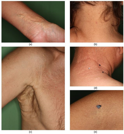
Pseudoxanthoma elasticum (PXE) is a rare multisystemic autosomal recessive connective tissue disease. In most cases, skin manifestations of PXE are the first to develop, followed later by severe ocular and cardiovascular complications. In our present study, in addition to dermoscopy, we introduced novel techniques, autofluorescence (AF) and diffuse reflectance (DR) imaging for the assessment of affected skin sites of five PXE patients. PXE-affected skin areas in most skin sites showed a previously observed pattern upon dermoscopic examination. With the novel imaging, PXE-affected skin lesions displayed high AF intensity. During our measurements, significantly higher mean, minimum and maximum AF intensity values were found in areas of PXE-affected skin when compared to uninvolved skin. Conversely, images acquired with the use of 660 and 940 nm illumination showed no mentionable difference. Our results demonstrate that AF imaging may be used in the in vivo diagnostics and quantification of the severity of the skin lesions of PXE patients. In addition, it is a safe, fast and cost-effective diagnostic method. AF imaging may be also used to objectively monitor the efficacy of the possible novel therapeutic approaches of PXE in the future. Full article
Show Figures

Figure 1

15 pages, 3524 KiB  
Article
Passive Microwave Radiometry for the Diagnosis of Coronavirus Disease 2019 Lung Complications in Kyrgyzstan
by Batyr Osmonov, Lev Ovchinnikov, Christopher Galazis, Berik Emilov, Mustafa Karaibragimov, Meder Seitov, Sergey Vesnin, Alexander Losev, Vladislav Levshinskii, Illarion Popov, Chingiz Mustafin, Turat Kasymbekov and Igor Goryanin
Diagnostics 2021, 11(2), 259; https://doi.org/10.3390/diagnostics11020259 - 7 Feb 2021
Cited by 18 | Viewed by 3569
Abstract
The global spread of severe acute respiratory syndrome coronavirus 2, which causes coronavirus disease 2019 (COVID-19), could be due to limited access to diagnostic tests and equipment. Currently, most diagnoses use the reverse transcription polymerase chain reaction (RT-PCR) and chest computed tomography (CT). [...] Read more.
The global spread of severe acute respiratory syndrome coronavirus 2, which causes coronavirus disease 2019 (COVID-19), could be due to limited access to diagnostic tests and equipment. Currently, most diagnoses use the reverse transcription polymerase chain reaction (RT-PCR) and chest computed tomography (CT). However, challenges exist with CT use due to infection control, lack of CT availability in low- and middle-income countries, and low RT-PCR sensitivity. Passive microwave radiometry (MWR), a cheap, non-radioactive, and portable technology, has been used for cancer and other diseases’ diagnoses. Here, we tested MWR use first time for the early diagnosis of pulmonary COVID-19 complications in a cross-sectional controlled trial in order to evaluate MWR use in hospitalized patients with COVID-19 pneumonia and healthy individuals. We measured the skin and internal temperature using 30 points identified on the body, for both lungs. Pneumonia and lung damage were diagnosed by both CT scan and doctors’ diagnoses (pneumonia+/pneumonia−). COVID-19 was determined by RT-PCR (covid+/covid−). The best MWR results were obtained for the pneumonia−/covid− and pneumonia+/covid+ groups. The study suggests that MWR could be used for diagnosing pneumonia in COVID-19 patients. Since MWR is inexpensive, its use will ease the financial burden for both patients and countries. Clinical Trial Number: NCT04568525. Full article
(This article belongs to the Special Issue Electromagnetic Imaging for a Novel Generation of Medical Devices)
Show Figures

Figure 1

14 pages, 1125 KiB  
Article
Significant Changes in Serum MicroRNAs after High Tibial Osteotomy in Medial Compartmental Knee Osteoarthritis: Potential Prognostic Biomarkers
by Yoon Hae Kwak, Dae-Kyung Kwak, Hyun-Soo Moon, Nan Young Kim, Jae-Sung Yee and Je-Hyun Yoo
Diagnostics 2021, 11(2), 258; https://doi.org/10.3390/diagnostics11020258 - 7 Feb 2021
Cited by 6 | Viewed by 2152
Abstract
High tibial osteotomy (HTO) is an effective alternative for medial compartmental knee osteoarthritis (OA). Circulating microRNAs (miRNAs) are known to serve as OA-related biomarkers. The present study investigated the differential expression of serum miRNAs before and after HTO to identify potential miRNAs as [...] Read more.
High tibial osteotomy (HTO) is an effective alternative for medial compartmental knee osteoarthritis (OA). Circulating microRNAs (miRNAs) are known to serve as OA-related biomarkers. The present study investigated the differential expression of serum miRNAs before and after HTO to identify potential miRNAs as prognostic biomarkers. miRNA-polymerase chain reaction (PCR) arrays were used to screen for miRNAs in the serum at preoperative and 6-month postoperative time points from six patients, and the differentially expressed miRNAs identified in the profiling stage were validated using real-time PCR at post-operative months 6 and 18 in 27 other HTO-treated patients. Among 84 miRNAs involved in the inflammatory process, three (miR-19b-3p, miR-29c-3p, and miR-424-5p) showed differential expression patterns in the profiling stage (p = 0.011, 0.015, and 0.021, respectively). Levels of these three and four other miRNAs (miR-140-3p, miR-454-3p, miR-let-7e-5p, and miR-885-5p) known to be related to OA progression were evaluated in the serum of 27 patients. Only four miRNAs (miR-19b-3p, miR-140-3p, miR-454-3p, and miR-let-7e-5p) were significantly upregulated at postoperative month 6 (p = 0.003, 0.005, 0.004, and 0.004, respectively), and only miR-140-3p was significantly upregulated up to 18 months after operation (p = 0.003). Together, this study reveals the significantly upregulated serum miRNAs after HTO as potential prognostic biomarkers; however, further studies are warranted to elucidate their clinical implications. Full article
(This article belongs to the Special Issue Biomarkers in Osteoarthritis)
Show Figures

Figure 1

10 pages, 1238 KiB  
Article
Recurrence and Coniglobus Volumetric Resolution of Subacute and Chronic Subdural Hematoma Post-Middle Meningeal Artery Embolization
by Ambooj Tiwari, Adam A. Dmytriw, Ryan Bo, Nathan Farkas, Phillip Ye, David S. Gordon, Karthikeyan M. Arcot, David Turkel-Parrella and Jeffrey Farkas
Diagnostics 2021, 11(2), 257; https://doi.org/10.3390/diagnostics11020257 - 7 Feb 2021
Cited by 22 | Viewed by 2990
Abstract
Objective: To study the efficacy of middle meningeal artery (MMA) embolization for the treatment of chronic subdural hematoma (SDH) and characterize its post-embolization volumetric resolution. Methods: Ten patients diagnosed with 13 cSDH underwent MMA embolization. SDH volumes were measured from time of initial [...] Read more.
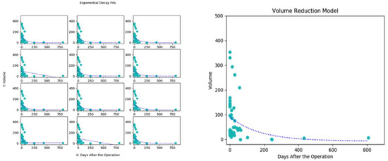
Objective: To study the efficacy of middle meningeal artery (MMA) embolization for the treatment of chronic subdural hematoma (SDH) and characterize its post-embolization volumetric resolution. Methods: Ten patients diagnosed with 13 cSDH underwent MMA embolization. SDH volumes were measured from time of initial discovery on imaging to pre-operative, post-operative, short-term and long-term follow-up. Time between procedure to obliteration was also measured. Volumetric analysis was done using the coniglobus formula, and recurrence rate as well as resolution timeline was defined using best-fit models. Results: Out of 10 patients, five were recurrent lesions, three were bilateral and seven unilateral cSDH. Average and median pre-operative volumes were 105.3 cc and 97.4 cc, respectively. Embolization on average was performed 21 days after discovery. Sixty percent of patients had concurrent antiplatelets or anticoagulation use. Forty percent underwent embolization treatment as the primary therapy. Recurrence was not seen in any patients treated with embolization. There were no peri- or post-operative complications. Five patients experienced complete or near-complete obliteration, while those with partial resolution showed a composite average of 75% volumetric reduction in 45 days. Post-embolization, the volumetric resolution followed an exponential decay curve over time and was independent of initial volume. Conclusion: MMA embolization contributed to a marked reduction in SDH volume post-operatively and can be used as a curative therapy for primary or recurrent chronic SDH. Full article
(This article belongs to the Special Issue Diagnosis and Management of Ischemic and Hemorrhagic Stroke)
Show Figures

Figure 1

24 pages, 4344 KiB  
Review
The Importance of Multimodality Imaging in the Diagnosis and Management of Patients with Infiltrative Cardiomyopathies: An Update
by Radu Sascău, Larisa Anghel, Alexandra Clement, Mădălina Bostan, Rodica Radu and Cristian Stătescu
Diagnostics 2021, 11(2), 256; https://doi.org/10.3390/diagnostics11020256 - 7 Feb 2021
Cited by 5 | Viewed by 4285
Abstract
Infiltrative cardiomyopathies (ICMs) comprise a broad spectrum of inherited and acquired conditions (mainly amyloidosis, sarcoidosis, and hemochromatosis), where the progressive buildup of abnormal substances within the myocardium results in left ventricular hypertrophy and manifests as restrictive physiology. Noninvasive multimodality imaging has gradually eliminated [...] Read more.
Infiltrative cardiomyopathies (ICMs) comprise a broad spectrum of inherited and acquired conditions (mainly amyloidosis, sarcoidosis, and hemochromatosis), where the progressive buildup of abnormal substances within the myocardium results in left ventricular hypertrophy and manifests as restrictive physiology. Noninvasive multimodality imaging has gradually eliminated endomyocardial biopsy from the diagnostic workup of infiltrative cardiac deposition diseases. However, even with modern imaging techniques’ widespread availability, these pathologies persist in being largely under- or misdiagnosed. Considering the advent of novel, revolutionary pharmacotherapies for cardiac amyloidosis, the archetypal example of ICM, a standardized diagnostic approach is warranted. Therefore, this review aims to emphasize the importance of contemporary cardiac imaging in identifying specific ICM and improving outcomes via the prompt initiation of a targeted treatment. Full article
Show Figures

Figure 1

9 pages, 702 KiB  
Article
Seeking Medical Assistance for Dysphonia Is Associated with an Improved Survival Rate in Laryngeal Cancer: Real-World Evidence
by Yi-An Lu, Ming-Shao Tsai, Li-Ang Lee, Shu-Ru Lee, Li-Yun Lin, Chain-Fen Chang, Wan-Ni Lin, Li-Jen Hsin, Chun-Ta Liao, Hsueh-Yu Li, Yu-Wen Wen and Tuan-Jen Fang
Diagnostics 2021, 11(2), 255; https://doi.org/10.3390/diagnostics11020255 - 7 Feb 2021
Cited by 1 | Viewed by 2186
Abstract
(1) Background: Patients with laryngeal cancer usually present with dysphonia. However, some studies reported that the duration from dysphonia to cancer diagnosis has been prolonged significantly in recent years. This study aimed to evaluate that in the initial dysphonia-related diagnosis and the interval [...] Read more.
(1) Background: Patients with laryngeal cancer usually present with dysphonia. However, some studies reported that the duration from dysphonia to cancer diagnosis has been prolonged significantly in recent years. This study aimed to evaluate that in the initial dysphonia-related diagnosis and the interval between the diagnosis of laryngeal cancer may affect the overall survival (OS). (2) Methods: The 1997–2013 Longitudinal Health Insurance Database was used in this study. A propensity score with 1-to-1 matching was applied to balance the baseline characteristics. The OS was examined by the Kaplan-Meier method and log-rank test. (3) Results: A total of 2753 patients with a first primary laryngeal cancer diagnosis were identified. The patients without prior dysphonia-related diagnosis (PD−) group did have a significantly worse five-year survival (p = 0.015) comparing with those with a prior dysphonia-related diagnosis (PD+) group among glottic cancer patients. The group with a shorter dysphonia-to-diagnosis interval had a better five-year OS than the prolonged group (p = 0.007) in laryngeal cancer. (4) Conclusions: Looking for medical assistance before a diagnosis of glottic cancer is associated with a better overall survival, while a diagnostic delay of more than 30 days from the first medical examination for dysphonia is associated with a worse outcome among in patients with laryngeal cancer. Full article
(This article belongs to the Section Pathology and Molecular Diagnostics)
Show Figures

Figure 1

11 pages, 1860 KiB  
Case Report
Medulloblastoma Associated with Down Syndrome: From a Rare Event Leading to a Pathogenic Hypothesis
by Alessandra Boni, Marco Ranalli, Giada Del Baldo, Roberto Carta, Mariachiara Lodi, Emanuele Agolini, Martina Rinelli, Diletta Valentini, Sabrina Rossi, Viola Alesi, Antonella Cacchione, Evelina Miele, Iside Alessi, Anna Maria Caroleo, Giovanna Stefania Colafati, Maria Antonietta De Ioris, Luigi Boccuto, Mario Balducci, Andrea Carai and Angela Mastronuzzi
Diagnostics 2021, 11(2), 254; https://doi.org/10.3390/diagnostics11020254 - 7 Feb 2021
Cited by 4 | Viewed by 3033
Abstract
Down syndrome (DS) is the most common chromosome abnormality with a unique cancer predisposition syndrome pattern: a higher risk to develop acute leukemia and a lower incidence of solid tumors. In particular, brain tumors are rarely reported in the DS population, and biological [...] Read more.
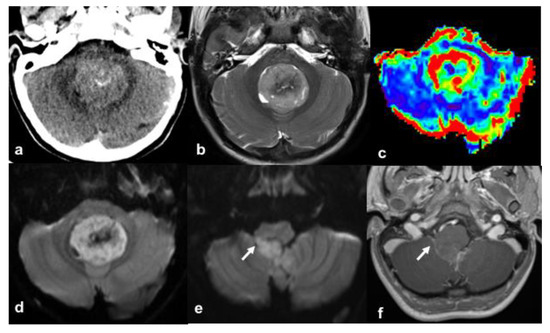
Down syndrome (DS) is the most common chromosome abnormality with a unique cancer predisposition syndrome pattern: a higher risk to develop acute leukemia and a lower incidence of solid tumors. In particular, brain tumors are rarely reported in the DS population, and biological behavior and natural history are not well described and identified. We report a case of a 10-year-old child with DS who presented with a medulloblastoma (MB). Histological examination revealed a classic MB with focal anaplasia and the molecular profile showed the presence of a CTNNB1 variant associated with the wingless (WNT) molecular subgroup with a good prognosis in contrast to our case report that has shown an early metastatic relapse. The nearly seven-fold decreased risk of MB in children with DS suggests the presence of protective biological mechanisms. The cerebellum hypoplasia and the reduced volume of cerebellar granule neuron progenitor cells seem to be a possible favorable condition to prevent MB development via inhibition of neuroectodermal differentiation. Moreover, the NOTCH/WNT dysregulation in DS, which is probably associated with an increased risk of leukemia, suggests a pivotal role of this pathway alteration in the pathogenesis of MB; therefore, this condition should be further investigated in future studies by molecular characterizations. Full article
(This article belongs to the Special Issue Advances in Pediatric Neuro-Oncology)
Show Figures

Figure 1

13 pages, 2820 KiB  
Article
Increased Tenascin C, Osteopontin and HSP90 Levels in Plasmatic Small Extracellular Vesicles of Pediatric ALK-Positive Anaplastic Large Cell Lymphoma: New Prognostic Biomarkers?
by Federica Lovisa, Anna Garbin, Sara Crotti, Piero Di Battista, Ilaria Gallingani, Carlotta Caterina Damanti, Anna Tosato, Elisa Carraro, Marta Pillon, Erfan Mafakheri, Filippo Romanato, Enrico Gaffo, Alessandra Biffi, Stefania Bortoluzzi, Marco Agostini and Lara Mussolin
Diagnostics 2021, 11(2), 253; https://doi.org/10.3390/diagnostics11020253 - 6 Feb 2021
Cited by 7 | Viewed by 3266
Abstract
Over the past 15 years, several biological and pathological characteristics proved their significance in pediatric anaplastic lymphoma kinase (ALK)-positive anaplastic large-cell lymphoma (ALCL) prognostic stratification. However, the identification of new non-invasive disease biomarkers, relying on the most important disease mechanisms, is still necessary. [...] Read more.
Over the past 15 years, several biological and pathological characteristics proved their significance in pediatric anaplastic lymphoma kinase (ALK)-positive anaplastic large-cell lymphoma (ALCL) prognostic stratification. However, the identification of new non-invasive disease biomarkers, relying on the most important disease mechanisms, is still necessary. In recent years, plasmatic circulating small extracellular vesicles (S-EVs) gathered great importance both as stable biomarker carriers and active players in tumorigenesis. In the present work, we performed a comprehensive study on the proteomic composition of plasmatic S-EVs of pediatric ALCL patients compared to healthy donors (HDs). By using a mass spectrometry-based proteomics approach, we identified 50 proteins significantly overrepresented in S-EVs of ALCL patients. Gene Ontology enrichment analysis disclosed cellular components and molecular functions connected with S-EV origin and vesicular trafficking, whereas cell adhesion, glycosaminoglycan metabolic process, extracellular matrix organization, collagen fibril organization and acute phase response were the most enriched biological processes. Of importance, consistently with the presence of nucleophosmin (NPM)-ALK fusion protein in ALCL cells, a topological enrichment analysis based on Reactome- and Kyoto Encyclopedia of Genes and Genomes (KEGG)-derived networks highlighted a dramatic increase in proteins of the phosphatidylinositol 3-kinase (PI3K)/AKT pathway in ALCL S-EVs, which included heat shock protein 90-kDa isoform alpha 1 (HSP90AA1), osteopontin (SPP1/OPN) and tenascin C (TNC). These results were validated by Western blotting analysis on a panel of ALCL and HD cases. Further research is warranted to better define the role of these S-EV proteins as diagnostic and, possibly, prognostic parameters at diagnosis and for ALCL disease monitoring. Full article
Show Figures

Figure 1

14 pages, 321 KiB  
Review
Tumour-Agnostic Therapy for Pancreatic Cancer and Biliary Tract Cancer
by Shunsuke Kato
Diagnostics 2021, 11(2), 252; https://doi.org/10.3390/diagnostics11020252 - 6 Feb 2021
Cited by 3 | Viewed by 2732
Abstract
The prognosis of patients with solid tumours has remarkably improved with the development of molecular-targeted drugs and immune checkpoint inhibitors. However, the improvements in the prognosis of pancreatic cancer and biliary tract cancer is delayed compared to other carcinomas, and the 5-year survival [...] Read more.
The prognosis of patients with solid tumours has remarkably improved with the development of molecular-targeted drugs and immune checkpoint inhibitors. However, the improvements in the prognosis of pancreatic cancer and biliary tract cancer is delayed compared to other carcinomas, and the 5-year survival rates of distal-stage disease are approximately 10 and 20%, respectively. However, a comprehensive analysis of tumour cells using The Cancer Genome Atlas (TCGA) project has led to the identification of various driver mutations. Evidently, few mutations exist across organs, and basket trials targeting driver mutations regardless of the primary organ are being actively conducted. Such basket trials not only focus on the gate keeper-type oncogene mutations, such as HER2 and BRAF, but also focus on the caretaker-type tumour suppressor genes, such as BRCA1/2, mismatch repair-related genes, which cause hereditary cancer syndrome. As oncogene panel testing is a vital approach in routine practice, clinicians should devise a strategy for improved understanding of the cancer genome. Here, the gene mutation profiles of pancreatic cancer and biliary tract cancer have been outlined and the current status of tumour-agnostic therapy in these cancers has been reported. Full article
8 pages, 1967 KiB  
Case Report
Native Aortic Valve Endocarditis Complicated by Splenic Infarction and Giant Mitral-Aortic Intervalvular Fibrosa Pseudoaneurysm—A Case Report and Brief Review of the Literature
by Andreea Varga, Ioan Tilea, Cristina Maria Tatar, Dragos Gabriel Iancu, Maria Andrada Jiga, Robert Adrian Dumbrava, Marian Pop and Horatiu Suciu
Diagnostics 2021, 11(2), 251; https://doi.org/10.3390/diagnostics11020251 - 6 Feb 2021
Cited by 3 | Viewed by 2789
Abstract
Background: Pseudoaneurysm of the mitral-aortic intervalvular fibrosa (P-MAIVF) is an unusual complication related to various injuries or conditions which involve the mitro-aortic region; it communicates with the left ventricular outflow tract and is associated with a high-risk of redoubtable complications or sudden death. [...] Read more.
Background: Pseudoaneurysm of the mitral-aortic intervalvular fibrosa (P-MAIVF) is an unusual complication related to various injuries or conditions which involve the mitro-aortic region; it communicates with the left ventricular outflow tract and is associated with a high-risk of redoubtable complications or sudden death. The cerebral and splenic localizations are frequently seen as manifestations of systemic embolism in infective endocarditis. Currently, there are no specific recommendations related to the diagnosis, management, treatment, or further evolution of patients with P-MAIVF and concomitant splenic infarction. This paper presents the case of a 43-year-old Caucasian woman with a late diagnosis of mixed bicuspid aortic valve disease, affected by an under-detected and undertreated episode of infective endocarditis leading to asymptomatic P-MAIVF. Prime clinical and imagistic diagnosis of splenic infarction indicated further extended investigations were required to clarify the source of embolism. Methods: Integrated multimodality imaging techniques confirmed the unexpected diagnosis of P-MAIVF. Results: The case had a fatal outcome following an uncomplicated yet laborious cardiac surgery. Patient death was attributed to a malignant ventricular arrhythmia. Conclusion: The present case raises awareness by highlighting an unexplained and unexpected splenic infarction association with P-MAIVF as a result of infective endocarditis related to mixed bicuspid aortic valve disease. Full article
(This article belongs to the Section Medical Imaging and Theranostics)
Show Figures

Figure 1

13 pages, 3250 KiB  
Article
Deep Learning for Diagnosis of Paranasal Sinusitis Using Multi-View Radiographs
by Yejin Jeon, Kyeorye Lee, Leonard Sunwoo, Dongjun Choi, Dong Yul Oh, Kyong Joon Lee, Youngjune Kim, Jeong-Whun Kim, Se Jin Cho, Sung Hyun Baik, Roh-eul Yoo, Yun Jung Bae, Byung Se Choi, Cheolkyu Jung and Jae Hyoung Kim
Diagnostics 2021, 11(2), 250; https://doi.org/10.3390/diagnostics11020250 - 5 Feb 2021
Cited by 30 | Viewed by 10362
Abstract
Accurate image interpretation of Waters’ and Caldwell view radiographs used for sinusitis screening is challenging. Therefore, we developed a deep learning algorithm for diagnosing frontal, ethmoid, and maxillary sinusitis on both Waters’ and Caldwell views. The datasets were selected for the training and [...] Read more.
Accurate image interpretation of Waters’ and Caldwell view radiographs used for sinusitis screening is challenging. Therefore, we developed a deep learning algorithm for diagnosing frontal, ethmoid, and maxillary sinusitis on both Waters’ and Caldwell views. The datasets were selected for the training and validation set (n = 1403, sinusitis% = 34.3%) and the test set (n = 132, sinusitis% = 29.5%) by temporal separation. The algorithm can simultaneously detect and classify each paranasal sinus using both Waters’ and Caldwell views without manual cropping. Single- and multi-view models were compared. Our proposed algorithm satisfactorily diagnosed frontal, ethmoid, and maxillary sinusitis on both Waters’ and Caldwell views (area under the curve (AUC), 0.71 (95% confidence interval, 0.62–0.80), 0.78 (0.72–0.85), and 0.88 (0.84–0.92), respectively). The one-sided DeLong’s test was used to compare the AUCs, and the Obuchowski–Rockette model was used to pool the AUCs of the radiologists. The algorithm yielded a higher AUC than radiologists for ethmoid and maxillary sinusitis (p = 0.012 and 0.013, respectively). The multi-view model also exhibited a higher AUC than the single Waters’ view model for maxillary sinusitis (p = 0.038). Therefore, our algorithm showed diagnostic performances comparable to radiologists and enhanced the value of radiography as a first-line imaging modality in assessing multiple sinusitis. Full article
(This article belongs to the Special Issue Deep Learning for Computer-Aided Diagnosis in Biomedical Imaging)
Show Figures

Figure 1

11 pages, 1775 KiB  
Article
Methods for Testing the Subjective Visual Vertical during the Chronic Phase of Menière’s Disease
by Samira Ira Zabaneh, Linda Josephine Voss, Agnieszka J. Szczepek, Heidi Olze and Katharina Stölzel
Diagnostics 2021, 11(2), 249; https://doi.org/10.3390/diagnostics11020249 - 5 Feb 2021
Cited by 8 | Viewed by 3348
Abstract
The subjective visual vertical (SVV) evaluates the function of the utricle, which, in patients with Menière’s disease (MD), can be affected by endolymphatic hydrops. This study aimed to determine the SVV in MD patients during the chronic phase of illness compared to healthy [...] Read more.
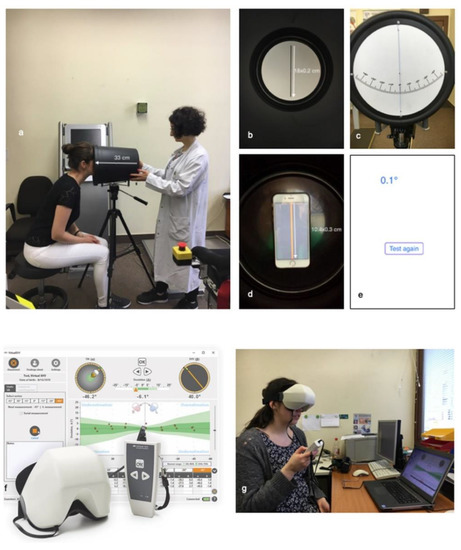
The subjective visual vertical (SVV) evaluates the function of the utricle, which, in patients with Menière’s disease (MD), can be affected by endolymphatic hydrops. This study aimed to determine the SVV in MD patients during the chronic phase of illness compared to healthy participants. The second aim was to compare the SVV measurement tools: the analog bucket test, digital bucket test, and C-SVV© goggles. The SVV scores differed significantly between MD patients and the control group for the analog bucket test (p < 0.001) and the C-SVV® goggles (p = 0.028), but no significance was shown when using the digital bucket test (p = 0.062). When comparing the analog bucket test and the C-SVV® goggles applying the calculated threshold (1.125° in analog bucket test, 2.5° in C-SVV® goggles), the bucket test showed higher accuracy (bucket test 73.84%, C-SVV® goggles 69.23%). When examining the influence of betahistine on SVV scores, there were no statistically significant differences in both the analog bucket test and C-SVV© goggles. We conclude that SVV test can be used as an additional tool to evaluate utricle function during the chronic phase of MD and that the analog bucket test produces the most reliable results. The intake of betahistine does not influence the perception of SVV. Full article
Show Figures

Figure 1

10 pages, 1954 KiB  
Article
Shear-Wave Elastography—Diagnostic Value in Children with Chronic Autoimmune Thyroiditis
by Cristina Mihaela Cepeha, Corina Paul, Andreea Borlea, Renata Fofiu, Florin Borcan, Cristina Adriana Dehelean, Viviana Ivan and Dana Stoian
Diagnostics 2021, 11(2), 248; https://doi.org/10.3390/diagnostics11020248 - 5 Feb 2021
Cited by 15 | Viewed by 2474
Abstract
Chronic autoimmune thyroiditis (CAT) is the most common thyroid disorder in the pediatric population. Ultrasound evaluation may suggest the diagnosis. Additionally, shear-wave elastography (SWE) proved to be a valuable additional diagnosis tool in adults with CAT by assessing thyroid stiffness (TS). This study [...] Read more.
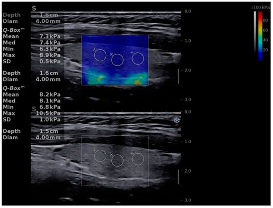
Chronic autoimmune thyroiditis (CAT) is the most common thyroid disorder in the pediatric population. Ultrasound evaluation may suggest the diagnosis. Additionally, shear-wave elastography (SWE) proved to be a valuable additional diagnosis tool in adults with CAT by assessing thyroid stiffness (TS). This study aims to assess its use also in detecting children with CAT. The study group consisted of 50 children with confirmed diagnosis of CAT, who were compared to the control group, consisting of 50 children with no thyroid pathology and with an adult group of 50 subjects with CAT. The evaluation included, besides bioimmunochemical evaluation, also thyroid ultrasound evaluation and elastography measurements in the same session (Aixplorer Mach 30, Supersonic imagine, France). The mean TS values were significantly lower for children in the CAT group compared to adults with CAT (15.51 ± 4.76 kPa vs. 20.96 ± 6.31 kPa; p < 0.0001) and higher compared to the healthy aged matched controls (15.51 ± 4.76 kPa vs. 10.41 ± 2.01 kPa; p < 0.0001). SWE elastography definitely seems a promising technique in the evaluation of children with autoimmune thyroid pathology. Full article
(This article belongs to the Special Issue Elastography 2.0)
Show Figures

Figure 1

3 pages, 175 KiB  
Editorial
Anatomical Variation and Clinical Diagnosis
by Heather F. Smith
Diagnostics 2021, 11(2), 247; https://doi.org/10.3390/diagnostics11020247 - 5 Feb 2021
Cited by 5 | Viewed by 2622
Abstract
In the anatomical sciences, it has long been recognized that the human body displays a range of morphological patterns and arrangements, often termed “anatomical variation” [...] Full article
(This article belongs to the Special Issue Anatomical Variation and Clinical Diagnosis)